
Mesothelin Expression in Human Tumors: A Tissue Microarray Study on 12,679 Tumors

 , , , ,
  add
Show full author list
add
Show full author list
Abstract
:1. Introduction
2. Material and Methods
2.1. Tissue Microarrays (TMAs)
2.2. Immunohistochemistry (IHC)
2.3. Statistics
3. Results
3.1. Technical Issues
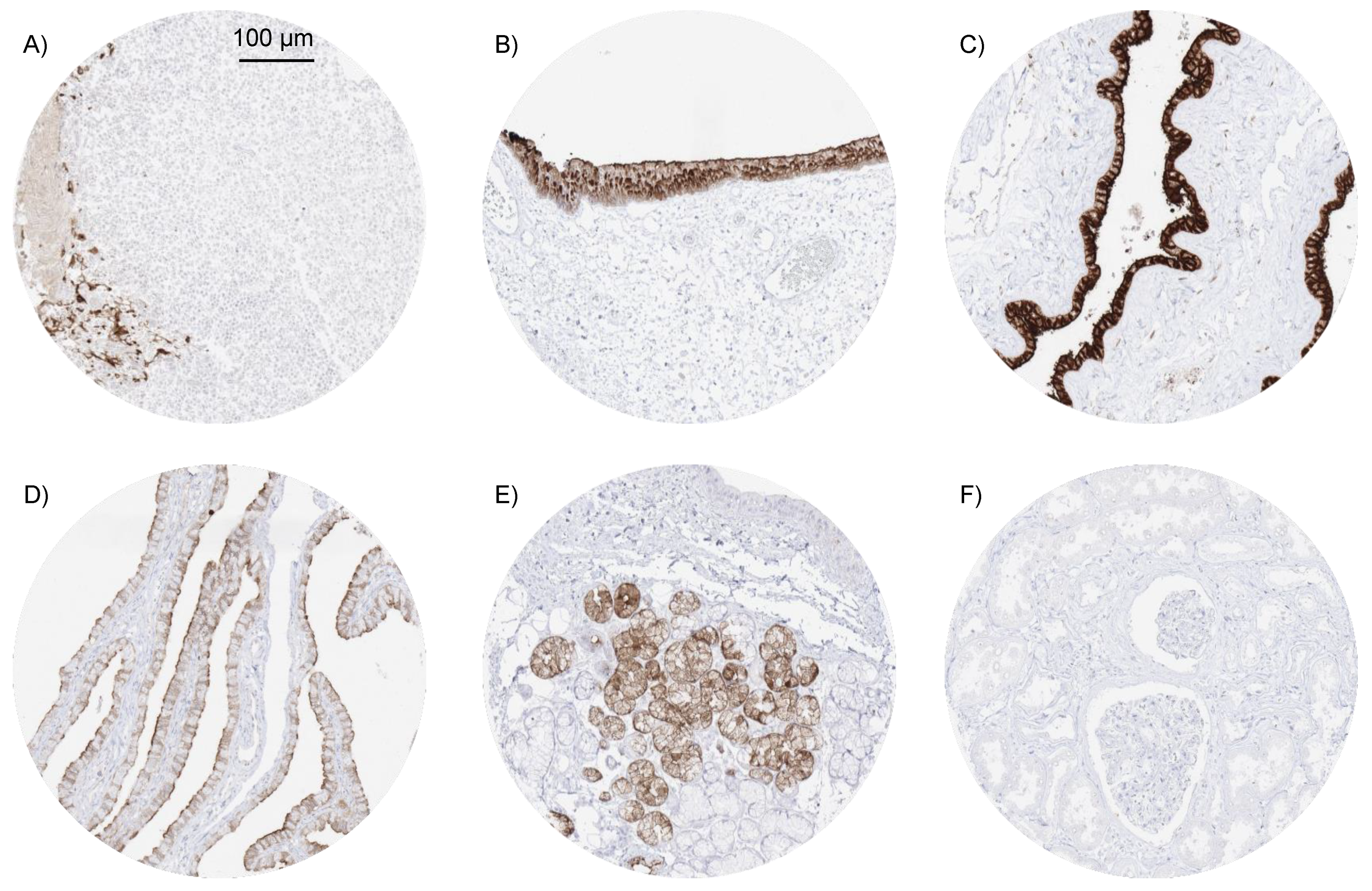
3.2. MSLN Immunostaining in Normal Tissues
3.3. MSLN Immunostaining in Neoplastic Tissues
3.4. MSLN Immunostaining, Tumor Phenotype, and Prognosis
4. Discussion
5. Conclusions
Author Contributions
Funding
Institutional Review Board Statement
Informed Consent Statement
Data Availability Statement
Acknowledgments
Conflicts of Interest
References
- Chang, K.; Pastan, I. Molecular cloning of mesothelin, a differentiation antigen present on mesothelium, mesotheliomas, and ovarian cancers. Proc. Natl. Acad. Sci. USA 1996, 93, 136–140. [Google Scholar] [CrossRef] [Green Version]
- Yamaguchi, N.; Hattori, K.; Oh-Eda, M.; Kojima, T.; Imai, N.; Ochi, N. A novel cytokine exhibiting megakaryocyte potentiating activity from a human pancreatic tumor cell line HPC-Y. J. Biol. Chem. 1994, 269, 805–808. [Google Scholar] [CrossRef]
- Urwin, D.; Lake, R.A. Structure of the Mesothelin/MPF Gene and Characterization of Its Promoter. Mol. Cell Biol. Res. Commun. 2000, 3, 26–32. [Google Scholar] [CrossRef] [PubMed]
- Chang, K.; Pastan, I.; Willingham, M.C. Isolation and characterization of a monoclonal antibody, K1, reactive with ovarian cancers and normal mesothelium. Int. J. Cancer 1992, 50, 373–381. [Google Scholar] [CrossRef]
- Inaguma, S.; Wang, Z.; Lasota, J.; Onda, M.; Czapiewski, P.; Langfort, R.; Rys, J.; Szpor, J.; Waloszczyk, P.; Okoń, K.; et al. Comprehensive immunohistochemical study of mesothelin (MSLN) using different monoclonal antibodies 5B2 and MN-1 in 1562 tumors with evaluation of its prognostic value in malignant pleural mesothelioma. Oncotarget 2017, 8, 26744–26754. [Google Scholar] [CrossRef] [PubMed] [Green Version]
- Alvarez, H.; Rojas, P.L.; Yong, K.-T.; Ding, H.; Xu, G.; Prasad, P.N.; Wang, J.; Canto, M.; Eshleman, J.R.; Montgomery, E.A.; et al. Mesothelin is a specific biomarker of invasive cancer in the Barrett-associated adenocarcinoma progression model: Translational implications for diagnosis and therapy. Nanomed. Nanotechnol. Biol. Med. 2008, 4, 295–301. [Google Scholar] [CrossRef] [PubMed] [Green Version]
- Ordóñez, N.G. Value of Mesothelin Immunostaining in the Diagnosis of Mesothelioma. Mod. Pathol. 2003, 16, 192–197. [Google Scholar] [CrossRef] [Green Version]
- Frierson, H.F.; Moskaluk, C.A.; Powell, S.M.; Zhang, H.; Cerilli, L.A.; Stoler, M.H.; Cathro, H.; Hampton, G.M. Large-scale molecular and tissue microarray analysis of mesothelin expression in common human carcinomas. Hum. Pathol. 2003, 34, 605–609. [Google Scholar] [CrossRef]
- Dennis, J.L.; Hvidsten, T.R.; Wit, E.C.; Komorowski, J.; Bell, A.K.; Downie, I.; Mooney, J.; Verbeke, C.S.; Bellamy, C.; Keith, W.N.; et al. Markers of Adenocarcinoma Characteristic of the Site of Origin: Development of a Diagnostic Algorithm. Clin. Cancer Res. 2005, 11, 3766–3772. [Google Scholar] [CrossRef] [Green Version]
- Bera, T.K.; Pastan, I. Mesothelin Is Not Required for Normal Mouse Development or Reproduction. Mol. Cell. Biol. 2000, 20, 2902–2906. [Google Scholar] [CrossRef] [Green Version]
- Gubbels, J.A.; Belisle, J.; Onda, M.; Rancourt, C.; Migneault, M.; Ho, M.; Bera, T.K.; Connor, J.; Sathyanarayana, B.K.; Lee, B.; et al. Mesothelin-MUC16 binding is a high affinity, N-glycan dependent interaction that facilitates peritoneal metastasis of ovarian tumors. Mol. Cancer 2006, 5, 50. [Google Scholar] [CrossRef] [Green Version]
- Kaneko, O.; Gong, L.; Zhang, J.; Hansen, J.K.; Hassan, R.; Lee, B.; Ho, M. A Binding Domain on Mesothelin for CA125/MUC. J. Biol. Chem. 2009, 284, 3739–3749. [Google Scholar] [CrossRef] [Green Version]
- Avula, L.R.; Rudloff, M.; El-Behaedi, S.; Arons, D.; Albalawy, R.; Chen, X.; Zhang, X.; Alewine, C. Mesothelin Enhances Tumor Vascularity in Newly Forming Pancreatic Peritoneal Metastases. Mol. Cancer Res. 2019, 18, 229–239. [Google Scholar] [CrossRef] [Green Version]
- Bharadwaj, U.; Marin-Muller, C.; Li, M.; Chen, C.; Yao, Q. Mesothelin confers pancreatic cancer cell resistance to TNF-alpha-induced apoptosis through Akt/PI3K/NF-kappaB activation and IL-6/Mcl-1 overexpression. Mol. Cancer 2011, 10, 106. [Google Scholar] [CrossRef] [PubMed] [Green Version]
- Chang, M.C.; Chen, C.-A.; Chen, P.-J.; Chiang, Y.-C.; Chen, Y.-L.; Mao, T.-L.; Lin, H.-W.; Chiang, W.-H.L.; Cheng, W.-F. Mesothelin enhances invasion of ovarian cancer by inducing MMP-7 through MAPK/ERK and JNK pathways. Biochem. J. 2012, 442, 293–302. [Google Scholar] [CrossRef] [PubMed] [Green Version]
- Chang, M.; Chen, C.; Hsieh, C.; Lee, C.; Su, Y.; Hu, Y.; Cheng, W. Mesothelin inhibits paclitaxel-induced apoptosis through the PI3K pathway. Biochem. J. 2009, 424, 449–458. [Google Scholar] [CrossRef] [PubMed] [Green Version]
- He, X.; Wang, L.; Riedel, H.; Wang, K.; Yang, Y.; Dinu, C.Z.; Rojanasakul, Y. Mesothelin promotes epithelial-to-mesenchymal transition and tumorigenicity of human lung cancer and mesothelioma cells. Mol. Cancer 2017, 16, 1–13. [Google Scholar] [CrossRef] [Green Version]
- Servais, E.L.; Colovos, C.; Rodriguez, L.; Bograd, A.J.; Nitadori, J.-I.; Sima, C.; Rusch, V.W.; Sadelain, M.; Adusumilli, P.S. Mesothelin Overexpression Promotes Mesothelioma Cell Invasion and MMP-9 Secretion in an Orthotopic Mouse Model and in Epithelioid Pleural Mesothelioma Patients. Clin. Cancer Res. 2012, 18, 2478–2489. [Google Scholar] [CrossRef] [Green Version]
- Uehara, N.; Matsuoka, Y.; Tsubura, A. Mesothelin Promotes Anchorage-Independent Growth and Prevents Anoikis via Extracellular Signal-Regulated Kinase Signaling Pathway in Human Breast Cancer Cells. Mol. Cancer Res. 2008, 6, 186–193. [Google Scholar] [CrossRef] [Green Version]
- Wang, Y.; Wang, L.; Li, D.; Wang, H.B.; Chen, Q.F. Mesothelin Promotes Invasion and Metastasis in Breast Cancer Cells. J. Int. Med Res. 2012, 40, 2109–2116. [Google Scholar] [CrossRef]
- Zheng, C.; Jia, W.; Tang, Y.; Zhao, H.; Jiang, Y.; Sun, S. Mesothelin regulates growth and apoptosis in pancreatic cancer cells through p53-dependent and -independent signal pathway. J. Exp. Clin. Cancer Res. 2012, 31, 84. [Google Scholar] [CrossRef] [PubMed] [Green Version]
- Ordóñez, N.G. Application of Mesothelin Immunostaining in Tumor Diagnosis. Am. J. Surg. Pathol. 2003, 27, 1418–1428. [Google Scholar] [CrossRef] [PubMed]
- Bauss, F.; Lechmann, M.; Krippendorff, B.-F.; Staack, R.; Herting, F.; Festag, M.; Imhof-Jung, S.; Hesse, F.; Pompiati, M.; Kollmorgen, G.; et al. Characterization of a re-engineered, mesothelin-targeted Pseudomonas exotoxin fusion protein for lung cancer therapy. Mol. Oncol. 2016, 10, 1317–1329. [Google Scholar] [CrossRef] [PubMed] [Green Version]
- Haas, A.R.; Tanyi, J.L.; O’Hara, M.H.; Gladney, W.L.; Lacey, S.F.; Torigian, D.A.; Soulen, M.C.; Tian, L.; McGarvey, M.; Nelson, A.M.; et al. Phase I Study of Lentiviral-Transduced Chimeric Antigen Receptor-Modified T Cells Recognizing Mesothelin in Advanced Solid Cancers. Mol. Ther. 2019, 27, 1919–1929. [Google Scholar] [CrossRef]
- Hassan, R.; Alley, E.; Kindler, H.; Antonia, S.J.; Jahan, T.M.; Honarmand, S.; Nair, N.; Whiting, C.C.; Enstrom, A.; Lemmens, E.; et al. Clinical Response of Live-Attenuated, Listeria monocytogenes Expressing Mesothelin (CRS-207) with Chemotherapy in Patients with Malignant Pleural Mesothelioma. Clin. Cancer Res. 2019, 25, 5787–5798. [Google Scholar] [CrossRef]
- Hassan, R.; Blumenschein, G.R., Jr.; Moore, K.N.; Santin, A.D.; Kindler, H.L.; Nemunaitis, J.J.; Seward, S.M.; Thomas, A.; Kim, S.K.; Rajagopalan, P. First-in-Human, Multicenter, Phase I Dose-Escalation and Expansion Study of Anti-Mesothelin Antibody-Drug Conjugate Anetumab Ravtansine in Advanced or Metastatic Solid Tumors. J. Clin. Oncol. 2020, 38, 1824–1835. [Google Scholar] [CrossRef]
- Hassan, R.; Kindler, H.L.; Jahan, T.; Bazhenova, L.; Reck, M.; Thomas, A.; Pastan, I.; Parno, J.; O’Shannessy, D.J.; Fatato, P.; et al. Phase II Clinical Trial of Amatuximab, a Chimeric Antimesothelin Antibody with Pemetrexed and Cisplatin in Advanced Unresectable Pleural Mesothelioma. Clin. Cancer Res. 2014, 20, 5927–5936. [Google Scholar] [CrossRef] [Green Version]
- Jiang, Q.; Ghafoor, A.; Mian, I.; Rathkey, D.; Thomas, A.; Alewine, C.; Sengupta, M.; Ahlman, M.A.; Zhang, J.; Morrow, B.; et al. Enhanced efficacy of mesothelin-targeted immunotoxin LMB-100 and anti–PD-1 antibody in patients with mesothelioma and mouse tumor models. Sci. Transl. Med. 2020, 12, eaaz7252. [Google Scholar] [CrossRef]
- Kim, H.; Gao, W.; Ho, M. Novel Immunocytokine IL12-SS1 (Fv) Inhibits Mesothelioma Tumor Growth in Nude Mice. PLoS ONE 2013, 8, e81919. [Google Scholar] [CrossRef] [Green Version]
- Lanitis, E.; Poussin, M.; Hagemann, I.S.; Coukos, G.; Sandaltzopoulos, R.; Scholler, N.; Powell, D.J. Redirected Antitumor Activity of Primary Human Lymphocytes Transduced With a Fully Human Anti-mesothelin Chimeric Receptor. Mol. Ther. 2012, 20, 633–643. [Google Scholar] [CrossRef] [Green Version]
- Lazzerini, L.; Jöhrens, K.; Sehouli, J.; Cichon, G. Favorable therapeutic response after anti-Mesothelin antibody–drug conjugate treatment requires high expression of Mesothelin in tumor cells. Arch. Gynecol. Obstet. 2020, 302, 1255–1262. [Google Scholar] [CrossRef]
- Mizukami, T.; Kamachi, H.; Fujii, Y.; Matsuzawa, F.; Einama, T.; Kawamata, F.; Kobayashi, N.; Hatanaka, Y.; Taketomi, A. The anti-mesothelin monoclonal antibody amatuximab enhances the anti-tumor effect of gemcitabine against mesothelin-high expressing pancreatic cancer cells in a peritoneal metastasis mouse model. Oncotarget 2018, 9, 33844–33852. [Google Scholar] [CrossRef] [Green Version]
- Scales, S.J.; Gupta, N.; Pacheco, G.; Firestein, R.; French, D.M.; Koeppen, H.; Rangell, L.; Barry-Hamilton, V.; Luis, E.; Chuh, J.; et al. An Antimesothelin-Monomethyl Auristatin E Conjugate with Potent Antitumor Activity in Ovarian, Pancreatic, and Mesothelioma Models. Mol. Cancer Ther. 2014, 13, 2630–2640. [Google Scholar] [CrossRef] [PubMed] [Green Version]
- Terwisscha van Scheltinga, A.G.; Ogasawara, A.; Pacheco, G.; Vanderbilt, A.N.; Tinianow, J.N.; Gupta, N.; Li, D.; Firestein, R.; Marik, J.; Scales, S.J.; et al. Preclinical Efficacy of an Antibody-Drug Conjugate Targeting Mesothelin Correlates with Quantitative 89Zr-ImmunoPET. Mol. Cancer Ther. 2017, 16, 134–142. [Google Scholar] [CrossRef] [PubMed] [Green Version]
- Weekes, C.D.; Lamberts, L.E.; Borad, M.J.; Voortman, J.; McWilliams, R.R.; Diamond, J.R.; De Vries, E.G.E.; Verheul, H.M.; Lieu, C.H.; Kim, G.P.; et al. Phase I Study of DMOT4039A, an Antibody–Drug Conjugate Targeting Mesothelin, in Patients with Unresectable Pancreatic or Platinum-Resistant Ovarian Cancer. Mol. Cancer Ther. 2016, 15, 439–447. [Google Scholar] [CrossRef] [Green Version]
- Yu, L.; Feng, M.; Kim, H.; Phung, Y.; Kleiner, D.E.; Gores, G.J.; Qian, M.; Wang, X.W.; Ho, M. Mesothelin as a Potential Therapeutic Target in Human Cholangiocarcinoma. J. Cancer 2010, 1, 141–149. [Google Scholar] [CrossRef] [PubMed] [Green Version]
- Kushitani, K.; Takeshima, Y.; Amatya, V.J.; Furonaka, O.; Sakatani, A.; Inai, K. Immunohistochemical marker panels for distinguishing between epithelioid mesothelioma and lung adenocarcinoma. Pathol. Int. 2007, 57, 190–199. [Google Scholar] [CrossRef]
- Miettinen, M.; Sarlomo-Rikala, M. Expression of calretinin, thrombomodulin, keratin 5, and mesothelin in lung carcinomas of different types: An immunohistochemical analysis of 596 tumors in comparison with epithelioid mesotheliomas of the pleura. Am. J. Surg. Pathol. 2003, 27, 150–158. [Google Scholar] [CrossRef]
- Ordonez, N.G. The immunohistochemical diagnosis of mesothelioma: A comparative study of epithelioid mesothelioma and lung adenocarcinoma. Am. J. Surg. Pathol. 2003, 27, 1031–1051. [Google Scholar] [CrossRef]
- Nahm, C.B.; Turchini, J.; Jamieson, N.; Moon, E.; Sioson, L.; Itchins, M.; Arena, J.; Colvin, E.; Howell, V.M.; Pavlakis, N.; et al. Biomarker panel predicts survival after resection in pancreatic ductal adenocarcinoma: A multi-institutional cohort study. Eur. J. Surg. Oncol. 2019, 45, 218–224. [Google Scholar] [CrossRef] [Green Version]
- Hassan, R.; Laszik, Z.G.; Lerner, M.; Raffeld, M.; Postier, R.; Brackett, D. Mesothelin is overexpressed in pancreaticobiliary adenocarcinomas but not in normal pancreas and chronic pancreatitis. Am. J. Clin. Pathol. 2005, 124, 838–845. [Google Scholar] [CrossRef] [PubMed]
- Shiraishi, T.; Shinto, E.; Mochizuki, S.; Tsuda, H.; Kajiwara, Y.; Okamoto, K.; Einama, T.; Hase, K.; Ueno, H. Mesothelin expression has prognostic value in stage IotaIota/IotaIotaIota colorectal cancer. Virchows Arch. 2019, 474, 297–307. [Google Scholar] [CrossRef]
- Illei, P.B.; Alewine, C.; Zahurak, M.; Cowan, M.L.; Montgomery, E.; Hassan, R.; Xiang, L.; Pastan, I.; Kelly, R.J. Mesothelin Expression in Advanced Gastroesophageal Cancer Represents a Novel Target for Immunotherapy. Appl. Immunohistochem. Mol. Morphol. 2016, 24, 246–252. [Google Scholar] [CrossRef] [PubMed] [Green Version]
- Magalhaes, I.; Fernebro, J.; Own, S.A.; Glaessgen, D.; Corvigno, S.; Remberger, M.; Mattsson, J.; Dahlstrand, H. Mesothelin Expression in Patients with High-Grade Serous Ovarian Cancer Does Not Predict Clinical Outcome But Correlates with CD11c+ Expression in Tumor. Adv. Ther. 2020, 37, 5023–5031. [Google Scholar] [CrossRef] [PubMed]
- Shin, S.; Park, S.; Kim, M.H.; Nam, C.M.; Kim, H.; Choi, Y.Y.; Jung, M.K.; Choi, H.J.; Rha, S.Y.; Chung, H.C. Mesothelin Expression Is a Predictive Factor for Peritoneal Recurrence in Curatively Resected Stage III Gastric Cancer. Oncologist 2019, 24, 1108–1114. [Google Scholar] [CrossRef] [PubMed] [Green Version]
- Li, Y.R.; Xian, R.R.; Ziober, A.; Conejo-Garcia, J.; Perales-Puchalt, A.; June, C.H.; Zhang, P.J.; Tchou, J. Mesothelin expression is associated with poor outcomes in breast cancer. Breast Cancer Res. Treat. 2014, 147, 675–684. [Google Scholar] [CrossRef] [PubMed] [Green Version]
- Suzuki, T.; Yamagishi, Y.; Einama, T.; Koiwai, T.; Yamasaki, T.; Fukumura‑Koga, M.; Ishibashi, Y.; Takihata, Y.; Shiraishi, T.; Miyata, Y.; et al. Membrane mesothelin expression positivity is associated with poor clinical outcome of luminal‑type breast cancer. Oncol. Lett. 2020, 20, 1. [Google Scholar] [CrossRef] [PubMed]
- Dancau, A.-M.; Simon, R.; Mirlacher, M.; Sauter, G. Tissue Microarrays. Adv. Struct. Saf. Stud. 2016, 1381, 53–65. [Google Scholar] [CrossRef]
- Kononen, J.; Bubendorf, L.; Kallionimeni, A.; Bärlund, M.; Schraml, P.; Leighton, S.B.; Torhorst, J.; Mihatsch, M.J.; Sauter, G.; Kallionimeni, O.-P. Tissue microarrays for high-throughput molecular profiling of tumor specimens. Nat. Med. 1998, 4, 844–847. [Google Scholar] [CrossRef] [PubMed]
- Lizio, M.; The FANTOM Consortium; Harshbarger, J.; Shimoji, H.; Severin, J.; Kasukawa, T.; Sahin, S.; Abugessaisa, I.; Fukuda, S.; Hori, F.; et al. Gateways to the FANTOM5 promoter level mammalian expression atlas. Genome Biol. 2015, 16, 1–14. [Google Scholar] [CrossRef] [PubMed] [Green Version]
- Lizio, M.; Abugessaisa, I.; Noguchi, S.; Kondo, A.; Hasegawa, A.; Hon, C.C.; De Hoon, M.; Severin, J.; Oki, S.; Hayashizaki, Y.; et al. Update of the FANTOM web resource: Expansion to provide additional transcriptome atlases. Nucleic Acids Res. 2019, 47, D752–D758. [Google Scholar] [CrossRef] [PubMed] [Green Version]
- Consortium, G.T. The Genotype-Tissue Expression (GTEx) project. Nat. Genet. 2013, 45, 580–585. [Google Scholar]
- Yen, M.J.; Hsu, C.-Y.; Mao, T.-L.; Wu, T.-C.; Roden, R.; Wang, T.-L.; Shih, I.-M. Diffuse Mesothelin Expression Correlates with Prolonged Patient Survival in Ovarian Serous Carcinoma. Clin. Cancer Res. 2006, 12, 827–831. [Google Scholar] [CrossRef] [Green Version]
- Liu, H.; Shi, J.; Anandan, V.; Wang, H.L.; Diehl, D.; Blansfield, J.; Gerhard, G.; Lin, F. Reevaluation and Identification of the Best Immunohistochemical Panel (pVHL, Maspin, S100P, IMP-3) for Ductal Adenocarcinoma of the Pancreas. Arch. Pathol. Lab. Med. 2012, 136, 601–609. [Google Scholar] [CrossRef] [PubMed]
- Ordóñez, N.G.; Sahin, A.A. Diagnostic utility of immunohistochemistry in distinguishing between epithelioid pleural mesotheliomas and breast carcinomas: A comparative study. Hum. Pathol. 2014, 45, 1529–1540. [Google Scholar] [CrossRef]
- Eguchi, T.; Kadota, K.; Mayor, M.; Zauderer, M.G.; Rimner, A.; Rusch, V.W.; Travis, W.D.; Sadelain, M.; Adusumilli, P.S. Cancer antigen profiling for malignant pleural mesothelioma immunotherapy: Expression and coexpression of mesothelin, cancer antigen 125, and Wilms tumor 1. Oncotarget 2017, 8, 77872–77882. [Google Scholar] [CrossRef] [Green Version]
- Einama, T.; Homma, S.; Kamachi, H.; Kawamata, F.; Takahashi, N.; Taniguchi, M.; Kamiyama, T.; Furukawa, H.; Matsuno, Y.; Tanaka, S.; et al. Luminal membrane expression of mesothelin is a prominent poor prognostic factor for gastric cancer. Br. J. Cancer 2012, 107, 137–142. [Google Scholar] [CrossRef] [Green Version]
- Baba, K.; Ishigami, S.; Arigami, T.; Uenosono, Y.; Okumura, H.; Matsumoto, M.; Kurahara, H.; Uchikado, Y.; Kita, Y.; Kijima, Y.; et al. Mesothelin expression correlates with prolonged patient survival in gastric cancer. J. Surg. Oncol. 2011, 105, 195–199. [Google Scholar] [CrossRef] [PubMed]
- Rizk, N.P.; Servais, E.L.; Tang, L.H.; Sima, C.S.; Gerdes, H.; Fleisher, M.; Rusch, V.W.; Adusumilli, P.S. Tissue and Serum Mesothelin Are Potential Markers of Neoplastic Progression in Barrett’s Associated Esophageal Adenocarcinoma. Cancer Epidemiol. Biomark. Prev. 2012, 21, 482–486. [Google Scholar] [CrossRef] [Green Version]
- Hassan, R.; Cohen, S.J.; Phillips, M.; Pastan, I.; Sharon, E.; Kelly, R.J.; Schweizer, C.; Weil, S.; Laheru, D. Phase I Clinical Trial of the Chimeric Anti-Mesothelin Monoclonal Antibody MORAb-009 in Patients with Mesothelin-Expressing Cancers. Clin. Cancer Res. 2010, 16, 6132–6138. [Google Scholar] [CrossRef] [Green Version]
- Kreitman, R.J.; Hassan, R.; Fitzgerald, D.J.; Pastan, I. Phase I Trial of Continuous Infusion Anti-Mesothelin Recombinant Immunotoxin SS1P. Clin. Cancer Res. 2009, 15, 5274–5279. [Google Scholar] [CrossRef] [PubMed] [Green Version]
- Tsujikawa, T.; Crocenzi, T.; Durham, J.N.; Sugar, E.A.; Wu, A.A.; Onners, B.; Nauroth, J.M.; Anders, R.A.; Fertig, E.J.; Laheru, D.A.; et al. Evaluation of Cyclophosphamide/GVAX Pancreas Followed by Listeria-Mesothelin (CRS-207) with or without Nivolumab in Patients with Pancreatic Cancer. Clin. Cancer Res. 2020, 26, 3578–3588. [Google Scholar] [CrossRef] [PubMed] [Green Version]
- Hassan, R.; Alewine, C.; Mian, I.; Spreafico, A.; Siu, L.L.; Gomez-Roca, C.; Delord, J.; Italiano, A.; Lassen, U.; Soria, J.; et al. Phase 1 study of the immunotoxin LMB-100 in patients with mesothelioma and other solid tumors expressing mesothelin. Cancer 2020, 126, 4936–4947. [Google Scholar] [CrossRef]
- Ito, T.; Kajino, K.; Abe, M.; Sato, K.; Maekawa, H.; Sakurada, M.; Orita, H.; Wada, R.; Kajiyama, Y.; Hino, O. ERC/mesothelin is expressed in human gastric cancer tissues and cell lines. Oncol. Rep. 2014, 31, 27–33. [Google Scholar] [CrossRef] [PubMed] [Green Version]
- Parinyanitikul, N.; Blumenschein, G.R.; Wu, Y.; Lei, X.; Chavez-MacGregor, M.; Smart, M.; Gonzalez-Angulo, A.M. Mesothelin Expression and Survival Outcomes in Triple Receptor Negative Breast Cancer. Clin. Breast Cancer 2013, 13, 378–384. [Google Scholar] [CrossRef] [Green Version]
- Wang, L.; Niu, Z.; Zhang, L.; Liu, X.; Wang, X.; Li, F.; Wang, Y. Clinicopathological Significance of Mesothelin Expression in Invasive Breast Cancer. J. Int. Med. Res. 2012, 40, 909–916. [Google Scholar] [CrossRef] [PubMed] [Green Version]
- Swierczynski, S.L.; Maitra, A.; Abraham, S.C.; Iacobuzio-Donahue, C.A.; Ashfaq, R.; Cameron, J.L.; Schulick, R.D.; Yeo, C.J.; Rahman, A.; Hinkle, D.A.; et al. Analysis of novel tumor markers in pancreatic and biliary carcinomas using tissue microarrays. Hum. Pathol. 2004, 35, 357–366. [Google Scholar] [CrossRef]
- McCarthy, D.M.; Maitra, A.; Argani, P.; Rader, A.E.; Faigel, D.O.; Van Heek, N.T.; Hruban, R.H.; Wilentz, R.E. Novel Markers of Pancreatic Adenocarcinoma in Fine-Needle Aspiration: Mesothelin and Prostate Stem Cell Antigen Labeling Increases Accuracy in Cytologically Borderline Cases. Appl. Immunohistochem. Mol. Morphol. 2003, 11, 238–243. [Google Scholar] [CrossRef]
- Argani, P.; Iacobuzio-Donahue, C.; Ryu, B.; Rosty, C.; Goggins, M.; Wilentz, R.E.; Murugesan, S.R.; Leach, S.D.; Jaffee, E.; Yeo, C.J.; et al. Mesothelin is overexpressed in the vast majority of ductal adenocarcinomas of the pancreas: Identification of a new pancreatic cancer marker by serial analysis of gene expression (SAGE). Clin. Cancer Res. 2001, 7, 3862–3868. [Google Scholar]
- Frank, R.; Li, S.; Ahmad, N.A.; Sepulveda, A.R.; Jhala, N.C. Mesothelin Expression in Pancreatic Mucinous Cysts. Am. J. Clin. Pathol. 2014, 142, 313–319. [Google Scholar] [CrossRef] [Green Version]
- Jhala, N.; Jhala, D.; Vickers, S.M.; Eltoum, I.; Batra, S.K.; Manne, U.; Eloubeidi, M.; Jones, J.J.; Grizzle, W.E. Biomarkers in Diagnosis of pancreatic carcinoma in fine-needle aspirates. Am. J. Clin. Pathol. 2006, 126, 572–579. [Google Scholar] [CrossRef] [PubMed]
- Dim, D.C.; Jiang, F.; Qiu, Q.; Li, T.; Darwin, P.; Rodgers, W.H.; Peng, H.Q. The usefulness of S100P, mesothelin, fascin, prostate stem cell antigen, and 14-3-3 sigma in diagnosing pancreatic adenocarcinoma in cytological specimens obtained by endoscopic ultrasound guided fine-needle aspiration. Diagn. Cytopathol. 2011, 42, 193–199. [Google Scholar] [CrossRef] [PubMed]
- Hassan, R.; Kreitman, R.J.; Pastan, I.; Willingham, M.C. Localization of Mesothelin in Epithelial Ovarian Cancer. Appl. Immunohistochem. Mol. Morphol. 2005, 13, 243–247. [Google Scholar] [CrossRef] [PubMed]
- Han, S.-H.; Joo, M.; Kim, H.; Chang, S. Mesothelin Expression in Gastric Adenocarcinoma and Its Relation to Clinical Outcomes. J. Pathol. Transl. Med. 2017, 51, 122–128. [Google Scholar] [CrossRef] [Green Version]
- Le, K.; Wang, J.; Zhang, T.; Guo, Y.; Chang, H.; Wang, S.; Zhu, B. Overexpression of Mesothelin in Pancreatic Ductal Adenocarcinoma (PDAC). Int. J. Med. Sci. 2020, 17, 422–427. [Google Scholar] [CrossRef] [Green Version]
- Ibrahim, D.A.; Abouhashem, N.S. Diagnostic value of IMP3 and mesothelin in differentiating pancreatic ductal adenocarcinoma from chronic pancreatitis. Pathol. Res. Pract. 2016, 212, 288–293. [Google Scholar] [CrossRef]
- Gnemmi, V.; Leroy, X.; Triboulet, J.-P.; Pruvot, F.-R.; Villers, A.; Leteurtre, E.; Buob, D. Pancreatic metastases of renal clear cell carcinoma: A clinicopathological study of 11 cases with special emphasis on the usefulness of PAX2 and mesothelin for the distinction from primary ductal adenocarcinoma of the pancreas. Anal. Quant. Cytopathol. Histopathol. 2013, 35, 157–162. [Google Scholar]
- Glass, J.P.; Parasher, G.; Arias-Pulido, H.; Donohue, R.; Cerilli, L.A.; Prossnitz, E.R. Mesothelin and GPR30 Staining Among a Spectrum of Pancreatic Epithelial Neoplasms. Int. J. Surg. Pathol. 2011, 19, 588–596. [Google Scholar] [CrossRef]
- Tabrizi, A.D.; Kalloger, S.E.; Köbel, M.; Cipollone, J.; Roskelley, C.D.; Mehl, E.; Gilks, C.B. Primary Ovarian Mucinous Carcinoma of Intestinal Type: Significance of Pattern of Invasion and Immunohistochemical Expression Profile in a Series of 31 Cases. Int. J. Gynecol. Pathol. 2010, 29, 99–107. [Google Scholar] [CrossRef]
- Kanner, W.A.; Galgano, M.T.; Stoler, M.H.; Mills, S.E.; Atkins, K.A. Distinguishing Breast Carcinoma From Müllerian Serous Carcinoma With Mammaglobin and Mesothelin. Int. J. Gynecol. Pathol. 2008, 27, 491–495. [Google Scholar] [CrossRef]
- Pu, R.T.; Pang, Y.; Michael, C.W. Utility of WT-1, p63, MOC31, mesothelin, and cytokeratin (K903 and CK5/6) immunostains in differentiating adenocarcinoma, squamous cell carcinoma, and malignant mesothelioma in effusions. Diagn. Cytopathol. 2008, 36, 20–25. [Google Scholar] [CrossRef] [Green Version]
- Leroy, X.; Farine, M.-O.; Buob, D.; Wacrenier, A.; Copin, M.-C. Diagnostic value of cytokeratin 7, CD10 and mesothelin in distinguishing ovarian clear cell carcinoma from metastasis of renal clear cell carcinoma. Histopathology 2007, 51, 874–876. [Google Scholar] [CrossRef]
- Galloway, M.L.; Murray, D.; Moffat, D.F. The use of the monoclonal antibody mesothelin in the diagnosis of malignant mesothelioma in pleural biopsies. Histopathology 2006, 48, 767–769. [Google Scholar] [CrossRef]
- Cao, D.; Ji, H.; Ronnett, B.M. Expression of mesothelin, fascin, and prostate stem cell antigen in primary ovarian mucinous tumors and their utility in differentiating primary ovarian mucinous tumors from metastatic pancreatic mucinous carcinomas in the ovary. Int. J. Gynecol. Pathol. 2005, 24, 67–72. [Google Scholar] [PubMed]
- Ordóñez, N.G. The diagnostic utility of immunohistochemistry in distinguishing between mesothelioma and renal cell carcinoma: A comparative study. Hum. Pathol. 2004, 35, 697–710. [Google Scholar] [CrossRef]
- Pan, C.-C.; Chen, P.C.-H.; Chou, T.-Y.; Chiang, H. Expression of calretinin and other mesothelioma-related markers in thymic carcinoma and thymoma. Hum. Pathol. 2003, 34, 1155–1162. [Google Scholar] [CrossRef] [PubMed]
- Nomura, R.; Fujii, H.; Abe, M.; Sugo, H.; Ishizaki, Y.; Kawasaki, S.; Hino, O. Mesothelin Expression Is a Prognostic Factor in Cholangiocellular Carcinoma. Int. Surg. 2013, 98, 164–169. [Google Scholar] [CrossRef] [PubMed] [Green Version]
- Showalter, S.L.; Huang, Y.-H.; Witkiewicz, A.; Costantino, C.L.; Yeo, C.J.; Green, J.J.; Langer, R.; Anderson, D.G.; Sawicki, J.A.; Brody, J.R. Nanoparticulate delivery of diphtheria toxin DNA effectively kills Mesothelin expressing pancreatic cancer cells. Cancer Biol. Ther. 2008, 7, 1584–1590. [Google Scholar] [CrossRef] [Green Version]
- Van Heek, N.T.; Maitra, A.; Koopmann, J.; Fedarko, N.; Jain, A.; Rahman, A.; Iacobuzio-Donahue, C.A.; Adsay, V.; Ashfaq, R.; Yeo, C.J.; et al. Gene expression profiling identifies markers of ampullary adenocarcinoma. Cancer Biol. Ther. 2004, 3, 651–656. [Google Scholar] [CrossRef] [PubMed] [Green Version]
- Liebig, B.; Brabletz, T.; Staege, M.S.; Wulfanger, J.; Riemann, D.; Burdach, S.; Ballhausen, W.G. Forced expression of deltaN-TCF-1B in colon cancer derived cell lines is accompanied by the induction of CEACAM5/6 and mesothelin. Cancer Lett. 2005, 223, 159–167. [Google Scholar] [CrossRef]
- Ordonez, N.G.; Ord, N.G. The diagnostic utility of immunohistochemistry in distinguishing between epithelioid mesotheliomas and squamous carcinomas of the lung: A comparative study. Mod. Pathol. 2006, 19, 417–428. [Google Scholar] [CrossRef] [Green Version]
- Regzedmaa, O.; Li, Y.; Li, Y.; Zhang, H.; Wang, J.; Gong, H.; Yuan, Y.; Li, W.; Liu, H.; Chen, J. Prevalence of DLL3, CTLA-4 and MSTN Expression in Patients with Small Cell Lung Cancer. OncoTargets Ther. 2019, 12, 10043–10055. [Google Scholar] [CrossRef] [PubMed] [Green Version]
- Forest, F.; Patoir, A.; Col, P.D.; Sulaiman, A.; Camy, F.; Laville, D.; Bayle-Bleuez, S.; Fournel, P.; Habougit, C. Nuclear grading, BAP1, mesothelin and PD-L1 expression in malignant pleural mesothelioma: Prognostic implications. Pathology 2018, 50, 635–641. [Google Scholar] [CrossRef] [PubMed]
- Tan, K.; Kajino, K.; Momose, S.; Masaoka, A.; Sasahara, K.; Shiomi, K.; Izumi, H.; Abe, M.; Ohtsuji, N.; Wang, T.; et al. Mesothelin (MSLN) promoter is hypomethylated in malignant mesothelioma, but its expression is not associated with methylation status of the promoter. Hum. Pathol. 2010, 41, 1330–1338. [Google Scholar] [CrossRef]
- Dainty, L.A.; Risinger, J.I.; Morrison, C.; Chandramouli, G.; Bidus, M.A.; Zahn, C.; Rose, G.S.; Fowler, J.; Berchuck, A.; Maxwell, G.L. Overexpression of folate binding protein and mesothelin are associated with uterine serous carcinoma. Gynecol. Oncol. 2007, 105, 563–570. [Google Scholar] [CrossRef] [PubMed] [Green Version]
- Yildiz, Y.; Kabadayi, G.; Yigit, S.; Kucukzeybek, Y.; Alacacioglu, A.; Varol, U.; Taskaynatan, H.; Salman, T.; Oflazoglu, U.; Akyol, M.; et al. High expression of mesothelin in advanced serous ovarian cancer is associated with poor prognosis. J. BUON 2019, 24, 1549–1554. [Google Scholar] [PubMed]
- Drapkin, R.; Crum, C.P.; Hecht, J.L. Expression of candidate tumor markers in ovarian carcinoma and benign ovary: Evidence for a link between epithelial phenotype and neoplasia. Hum. Pathol. 2004, 35, 1014–1021. [Google Scholar] [CrossRef]
- Zhao, H.; Davydova, L.; Mandich, D.; Cartun, R.W.; Ligato, S. S100A4 protein and mesothelin expression in dysplasia and carcinoma of the extrahepatic bile duct. Am. J. Clin. Pathol. 2007, 127, 374–379. [Google Scholar] [CrossRef]
- Inoue, S.; Tsunoda, T.; Riku, M.; Ito, H.; Inoko, A.; Murakami, H.; Ebi, M.; Ogasawara, N.; Pastan, I.; Kasugai, K.; et al. Diffuse mesothelin expression leads to worse prognosis through enhanced cellular proliferation in colorectal cancer. Oncol. Lett. 2020, 19, 1741–1750. [Google Scholar] [CrossRef]
- Shiraishi, T.; Shinto, E.; Nearchou, I.P.; Tsuda, H.; Kajiwara, Y.; Einama, T.; Caie, P.D.; Kishi, Y.; Ueno, H. Prognostic significance of mesothelin expression in colorectal cancer disclosed by area-specific four-point tissue microarrays. Virchows Arch. Pathol. Anat. Physiol. Klin. Med. 2020, 477, 409–420. [Google Scholar] [CrossRef]
- Foda, A.A.M.; El-Hawary, A.K.; Hamed, H. Aberrant Expression of Calretinin, D2–40 and Mesothelin in Mucinous and Non-Mucinous Colorectal Carcinomas and Relation to Clinicopathological Features and Prognosis. Pathol. Oncol. Res. 2016, 22, 725–732. [Google Scholar] [CrossRef]
- Einama, T.; Kamachi, H.; Nishihara, H.; Homma, S.; Kanno, H.; Takahashi, K.; Sasaki, A.; Tahara, M.; Okada, K.; Muraoka, S.; et al. Co-Expression of Mesothelin and CA125 Correlates With Unfavorable Patient Outcome in Pancreatic Ductal Adenocarcinoma. Pancreas 2011, 40, 1276–1282. [Google Scholar] [CrossRef] [PubMed]
- Kawamata, F.; Kamachi, H.; Einama, T.; Homma, S.; Tahara, M.; Miyazaki, M.; Tanaka, S.; Kamiyama, T.; Nishihara, H.; Taketomi, A.; et al. Intracellular localization of mesothelin predicts patient prognosis of extrahepatic bile duct cancer. Int. J. Oncol. 2012, 41, 2109–2118. [Google Scholar] [CrossRef] [Green Version]
- Kawamata, F.; Homma, S.; Kamachi, H.; Einama, T.; Kato, Y.; Tsuda, M.; Tanaka, S.; Maeda, M.; Kajino, K.; Hino, O.; et al. C-ERC/mesothelin provokes lymphatic invasion of colorectal adenocarcinoma. J. Gastroenterol. 2013, 49, 81–92. [Google Scholar] [CrossRef]
- Hanaoka, T.; Hasegawa, K.; Kato, T.; Sato, S.; Kurosaki, A.; Miyara, A.; Nagao, S.; Seki, H.; Yasuda, M.; Fujiwara, K. Correlation Between Tumor Mesothelin Expression and Serum Mesothelin in Patients with Epithelial Ovarian Carcinoma: A Potential Noninvasive Biomarker for Mesothelin-targeted Therapy. Mol. Diagn. Ther. 2017, 21, 187–198. [Google Scholar] [CrossRef] [PubMed]
- Thomas, A.; Chen, Y.; Steinberg, S.M.; Luo, J.; Pack, S.; Raffeld, M.; Abdullaev, Z.; Alewine, C.; Rajan, A.; Giaccone, G.; et al. High mesothelin expression in advanced lung adenocarcinoma is associated with KRAS mutations and a poor prognosis. Oncotarget 2015, 6, 11694–11703. [Google Scholar] [CrossRef] [PubMed] [Green Version]
- Bayoglu, I.V.; Kucukzeybek, B.B.; Kucukzeybek, Y.; Varol, U.; Yildiz, I.; Alacacioglu, A.; Akyol, M.; Demir, L.; Dirican, A.; Yildiz, Y.; et al. Prognostic value of mesothelin expression in patients with triple negative and HER2-positive breast cancers. Biomed. Pharmacother. 2015, 70, 190–195. [Google Scholar] [CrossRef]
- Muniyan, S.; Haridas, D.; Chugh, S.; Rachagani, S.; Lakshmanan, I.; Gupta, S.; Seshacharyulu, P.; Smith, L.M.; Ponnusamy, M.P.; Batra, S.K. MUC16 contributes to the metastasis of pancreatic ductal adenocarcinoma through focal adhesion mediated signaling mechanism. Genes Cancer 2016, 7, 110–124. [Google Scholar] [CrossRef] [Green Version]
- El-Behaedi, S.; Landsman, R.; Rudloff, M.; Kolyvas, E.; Albalawy, R.; Zhang, X.; Bera, T.; Collins, K.; Kozlov, S.; Alewine, C. Protein Synthesis Inhibition Activity of Mesothelin Targeting Immunotoxin LMB-100 Decreases Concentrations of Oncogenic Signaling Molecules and Secreted Growth Factors. Toxins 2018, 10, 447. [Google Scholar] [CrossRef] [Green Version]
- Chen, S.-H.; Hung, W.-C.; Wang, P.; Paul, C.; Konstantopoulos, K. Mesothelin Binding to CA125/MUC16 Promotes Pancreatic Cancer Cell Motility and Invasion via MMP-7 Activation. Sci. Rep. 2013, 3, srep01870. [Google Scholar] [CrossRef] [Green Version]
- Saper, C.B. A Guide to the Perplexed on the Specificity of Antibodies. J. Histochem. Cytochem. 2008, 57, 1–5. [Google Scholar] [CrossRef] [PubMed] [Green Version]


| MSLN Immunostaining | ||||||||
|---|---|---|---|---|---|---|---|---|
| Tumor Entity | on TMA (n) | Analyzable (n) | Negative (%) | Weak (%) | Moderate (%) | Strong (%) | Positive (%) | |
| Tumors of the skin | Pilomatrixoma | 35 | 31 | 100.0 | 0.0 | 0.0 | 0.0 | 0.0 |
| Basal cell carcinoma | 88 | 83 | 98.8 | 1.2 | 0.0 | 0.0 | 1.2 | |
| Benign nevus | 29 | 26 | 100.0 | 0.0 | 0.0 | 0.0 | 0.0 | |
| Squamous cell carcinoma of the skin | 90 | 84 | 95.2 | 4.8 | 0.0 | 0.0 | 4.8 | |
| Malignant melanoma | 48 | 44 | 100.0 | 0.0 | 0.0 | 0.0 | 0.0 | |
| Merkel cell carcinoma | 46 | 44 | 100.0 | 0.0 | 0.0 | 0.0 | 0.0 | |
| Tumors of the head and neck | Squamous cell carcinoma of the larynx | 110 | 105 | 80.0 | 11.4 | 3.8 | 4.8 | 20.0 |
| Squamous cell carcinoma of the pharynx | 60 | 57 | 80.7 | 5.3 | 8.8 | 5.3 | 19.3 | |
| Oral squamous cell carcinoma (floor of the mouth) | 130 | 121 | 83.5 | 9.1 | 5.0 | 2.5 | 16.5 | |
| Pleomorphic adenoma of the parotid gland | 50 | 35 | 57.1 | 20.0 | 5.7 | 17.1 | 42.9 | |
| Warthin tumor of the parotid gland | 49 | 45 | 46.7 | 31.1 | 11.1 | 11.1 | 53.3 | |
| Basal cell adenoma of the salivary gland | 15 | 15 | 66.7 | 33.3 | 0.0 | 0.0 | 33.3 | |
| Tumors of the lung, pleura, and thymus | Squamous cell carcinoma of the lung | 127 | 71 | 74.6 | 18.3 | 5.6 | 1.4 | 25.4 |
| Adenocarcinoma of the lung | 250 | 174 | 44.8 | 19.0 | 13.8 | 22.4 | 55.2 | |
| Small cell carcinoma of the lung | 20 | 16 | 93.8 | 0.0 | 6.3 | 0.0 | 6.3 | |
| Mesothelioma, epitheloid | 39 | 25 | 36.0 | 12.0 | 4.0 | 48.0 | 64.0 | |
| Mesothelioma, other types | 76 | 65 | 29.2 | 10.8 | 4.6 | 55.4 | 70.8 | |
| Thymoma | 29 | 28 | 96.4 | 0.0 | 0.0 | 3.6 | 3.6 | |
| Tumors of the female genital tract | Squamous cell carcinoma of the vagina | 78 | 75 | 88.0 | 4.0 | 2.7 | 5.3 | 12.0 |
| Squamous cell carcinoma of the vulva | 130 | 123 | 89.4 | 7.3 | 0.8 | 2.4 | 10.6 | |
| Squamous cell carcinoma of the cervix | 130 | 125 | 57.6 | 21.6 | 8.0 | 12.8 | 42.4 | |
| Endometrioid endometrial carcinoma | 236 | 220 | 54.5 | 22.3 | 11.8 | 11.4 | 45.5 | |
| Endometrial serous carcinoma | 82 | 69 | 43.5 | 17.4 | 10.1 | 29.0 | 56.5 | |
| Carcinosarcoma of the uterus | 48 | 46 | 50.0 | 21.7 | 10.9 | 17.4 | 50.0 | |
| Endometrial carcinoma, high grade, G3 | 13 | 13 | 61.5 | 23.1 | 0.0 | 15.4 | 38.5 | |
| Endometrial clear cell carcinoma | 8 | 7 | 28.6 | 28.6 | 14.3 | 28.6 | 71.4 | |
| Endometrioid carcinoma of the ovary | 115 | 100 | 23.0 | 28.0 | 19.0 | 30.0 | 77.0 | |
| Serous carcinoma of the ovary | 567 | 511 | 2.7 | 5.1 | 8.4 | 83.8 | 97.3 | |
| Mucinous carcinoma of the ovary | 97 | 80 | 28.8 | 37.5 | 18.8 | 15.0 | 71.3 | |
| Clear cell carcinoma of the ovary | 54 | 42 | 16.7 | 33.3 | 19.0 | 31.0 | 83.3 | |
| Carcinosarcoma of the ovary | 47 | 43 | 34.9 | 16.3 | 11.6 | 37.2 | 65.1 | |
| Brenner tumor | 9 | 9 | 55.6 | 11.1 | 0.0 | 33.3 | 44.4 | |
| Tumors of the breast | Invasive breast carcinoma of no special type | 1391 | 1138 | 93.4 | 2.8 | 0.8 | 3.0 | 6.6 |
| Lobular carcinoma of the breast | 294 | 215 | 99.1 | 0.0 | 0.9 | 0.0 | 0.9 | |
| Medullary carcinoma of the breast | 26 | 26 | 76.9 | 11.5 | 7.7 | 3.8 | 23.1 | |
| Tubular carcinoma of the breast | 27 | 19 | 94.7 | 0.0 | 0.0 | 5.3 | 5.3 | |
| Mucinous carcinoma of the breast | 58 | 46 | 89.1 | 4.3 | 0.0 | 6.5 | 10.9 | |
| Phyllodes tumor of the breast | 50 | 36 | 100.0 | 0.0 | 0.0 | 0.0 | 0.0 | |
| Tumors of the digestive system | Adenomatous polyp, low-grade dysplasia | 50 | 48 | 72.9 | 16.7 | 8.3 | 2.1 | 27.1 |
| Adenomatous polyp, high-grade dysplasia | 50 | 46 | 56.5 | 30.4 | 8.7 | 4.3 | 43.5 | |
| Adenocarcinoma of the colon | 1882 | 1186 | 58.6 | 29.0 | 6.9 | 5.5 | 41.4 | |
| Adenocarcinoma of the small intestine | 10 | 6 | 0.0 | 50.0 | 16.7 | 33.3 | 100.0 | |
| Gastric adenocarcinoma, diffuse type | 176 | 164 | 51.2 | 14.6 | 11.6 | 22.6 | 48.8 | |
| Gastric adenocarcinoma, intestinal type | 174 | 170 | 60.6 | 18.8 | 11.2 | 9.4 | 39.4 | |
| Gastric adenocarcinoma, mixed type | 62 | 59 | 50.8 | 25.4 | 10.2 | 13.6 | 49.2 | |
| Adenocarcinoma of the esophagus | 133 | 75 | 58.7 | 14.7 | 18.7 | 8.0 | 41.3 | |
| Squamous cell carcinoma of the esophagus | 124 | 71 | 78.9 | 14.1 | 4.2 | 2.8 | 21.1 | |
| Squamous cell carcinoma of the anal canal | 91 | 86 | 75.6 | 14.0 | 2.3 | 8.1 | 24.4 | |
| Cholangiocarcinoma | 114 | 105 | 78.1 | 4.8 | 4.8 | 12.4 | 21.9 | |
| Hepatocellular carcinoma | 50 | 50 | 100.0 | 0.0 | 0.0 | 0.0 | 0.0 | |
| Ductal adenocarcinoma of the pancreas | 130 | 64 | 25.0 | 12.5 | 21.9 | 40.6 | 75.0 | |
| Pancreatic/Ampullary adenocarcinoma | 58 | 26 | 19.2 | 19.2 | 23.1 | 38.5 | 80.8 | |
| Acinar cell carcinoma of the pancreas | 7 | 6 | 100.0 | 0.0 | 0.0 | 0.0 | 0.0 | |
| Gastrointestinal stromal tumor (GIST) | 50 | 43 | 100.0 | 0.0 | 0.0 | 0.0 | 0.0 | |
| Tumors of the urinary system | Non-invasive papillary urothelial carcinoma, pTa G2 low grade | 177 | 154 | 96.1 | 3.2 | 0.0 | 0.6 | 3.9 |
| Non-invasive papillary urothelial carcinoma, pTa G2 high grade | 141 | 125 | 96.8 | 3.2 | 0.0 | 0.0 | 3.2 | |
| Non-invasive papillary urothelial carcinoma, pTa G3 | 187 | 130 | 96.9 | 2.3 | 0.0 | 0.8 | 3.1 | |
| Urothelial carcinoma, pT2-4 G3 | 940 | 838 | 87.5 | 8.0 | 2.5 | 2.0 | 12.5 | |
| Small cell neuroendocrine carcinoma of the bladder | 18 | 18 | 100.0 | 0.0 | 0.0 | 0.0 | 0.0 | |
| Sarcomatoid urothelial carcinoma | 25 | 22 | 90.9 | 9.1 | 0.0 | 0.0 | 9.1 | |
| Clear cell renal cell carcinoma | 858 | 807 | 93.4 | 3.7 | 1.6 | 1.2 | 6.6 | |
| Papillary renal cell carcinoma | 255 | 242 | 91.3 | 5.4 | 1.7 | 1.7 | 8.7 | |
| Clear cell (tubulo) papillary renal cell carcinoma | 21 | 21 | 81.0 | 19.0 | 0.0 | 0.0 | 19.0 | |
| Chromophobe renal cell carcinoma | 131 | 124 | 98.4 | 0.8 | 0.8 | 0.0 | 1.6 | |
| Oncocytoma | 177 | 170 | 100.0 | 0.0 | 0.0 | 0.0 | 0.0 | |
| Tumors of the male genital organs | Adenocarcinoma of the prostate, Gleason 3 + 3 | 83 | 80 | 100.0 | 0.0 | 0.0 | 0.0 | 0.0 |
| Adenocarcinoma of the prostate, Gleason 4 + 4 | 80 | 76 | 100.0 | 0.0 | 0.0 | 0.0 | 0.0 | |
| Adenocarcinoma of the prostate, Gleason 5 + 5 | 85 | 84 | 100.0 | 0.0 | 0.0 | 0.0 | 0.0 | |
| Adenocarcinoma of the prostate (recurrence) | 330 | 241 | 100.0 | 0.0 | 0.0 | 0.0 | 0.0 | |
| Small cell neuroendocrine carcinoma of the prostate | 17 | 17 | 100.0 | 0.0 | 0.0 | 0.0 | 0.0 | |
| Seminoma | 624 | 613 | 99.7 | 0.0 | 0.0 | 0.3 | 0.3 | |
| Embryonal carcinoma of the testis | 50 | 44 | 100.0 | 0.0 | 0.0 | 0.0 | 0.0 | |
| Yolk sack tumor | 50 | 37 | 100.0 | 0.0 | 0.0 | 0.0 | 0.0 | |
| Teratoma | 50 | 43 | 83.7 | 11.6 | 4.7 | 0.0 | 16.3 | |
| Squamous cell carcinoma of the penis | 80 | 76 | 93.4 | 5.3 | 0.0 | 1.3 | 6.6 | |
| Tumors of endocrine organs | Adenoma of the thyroid gland | 114 | 104 | 100.0 | 0.0 | 0.0 | 0.0 | 0.0 |
| Papillary thyroid carcinoma | 392 | 358 | 99.2 | 0.8 | 0.0 | 0.0 | 0.8 | |
| Follicular thyroid carcinoma | 158 | 146 | 98.6 | 1.4 | 0.0 | 0.0 | 1.4 | |
| Medullary thyroid carcinoma | 107 | 91 | 100.0 | 0.0 | 0.0 | 0.0 | 0.0 | |
| Anaplastic thyroid carcinoma | 45 | 43 | 100.0 | 0.0 | 0.0 | 0.0 | 0.0 | |
| Adrenal cortical adenoma | 50 | 49 | 100.0 | 0.0 | 0.0 | 0.0 | 0.0 | |
| Adrenal cortical carcinoma | 26 | 25 | 100.0 | 0.0 | 0.0 | 0.0 | 0.0 | |
| Phaeochromocytoma | 50 | 49 | 100.0 | 0.0 | 0.0 | 0.0 | 0.0 | |
| Appendix, neuroendocrine tumor (NET) | 22 | 14 | 92.9 | 0.0 | 7.1 | 0.0 | 7.1 | |
| Colorectal, neuroendocrine tumor (NET) | 10 | 10 | 70.0 | 30.0 | 0.0 | 0.0 | 30.0 | |
| Ileum, neuroendocrine tumor (NET) | 49 | 47 | 87.2 | 8.5 | 0.0 | 4.3 | 12.8 | |
| Lung, neuroendocrine tumor (NET) | 19 | 17 | 94.1 | 5.9 | 0.0 | 0.0 | 5.9 | |
| Pancreas, neuroendocrine tumor (NET) | 102 | 95 | 98.9 | 0.0 | 0.0 | 1.1 | 1.1 | |
| Colorectal, neuroendocrine carcinoma (NEC) | 11 | 9 | 100.0 | 0.0 | 0.0 | 0.0 | 0.0 | |
| Gallbladder, neuroendocrine carcinoma (NEC) | 4 | 4 | 100.0 | 0.0 | 0.0 | 0.0 | 0.0 | |
| Pancreas, neuroendocrine carcinoma (NEC) | 13 | 12 | 100.0 | 0.0 | 0.0 | 0.0 | 0.0 | |
| Tumors of hematopoietic and lymphoid tissues | Hodgkin Lymphoma | 103 | 101 | 100.0 | 0.0 | 0.0 | 0.0 | 0.0 |
| Non-Hodgkin Lymphoma | 62 | 14 | 100.0 | 0.0 | 0.0 | 0.0 | 0.0 | |
| Small lymphocytic lymphoma, B-cell type (B-SLL/B-CLL) | 50 | 50 | 100.0 | 0.0 | 0.0 | 0.0 | 0.0 | |
| Diffuse large B cell lymphoma (DLBCL) | 114 | 114 | 100.0 | 0.0 | 0.0 | 0.0 | 0.0 | |
| Follicular lymphoma | 88 | 87 | 100.0 | 0.0 | 0.0 | 0.0 | 0.0 | |
| T-cell Non Hodgkin lymphoma | 24 | 24 | 100.0 | 0.0 | 0.0 | 0.0 | 0.0 | |
| Mantle cell lymphoma | 18 | 18 | 100.0 | 0.0 | 0.0 | 0.0 | 0.0 | |
| Marginal zone lymphoma | 16 | 16 | 100.0 | 0.0 | 0.0 | 0.0 | 0.0 | |
| Diffuse large B-cell lymphoma (DLBCL) in the testis | 16 | 16 | 100.0 | 0.0 | 0.0 | 0.0 | 0.0 | |
| Burkitt lymphoma | 5 | 2 | 100.0 | 0.0 | 0.0 | 0.0 | 0.0 | |
| Tumors of soft tissue and bone | Tenosynovial giant cell tumor | 45 | 43 | 100.0 | 0.0 | 0.0 | 0.0 | 0.0 |
| Granular cell tumor | 53 | 47 | 100.0 | 0.0 | 0.0 | 0.0 | 0.0 | |
| Leiomyoma | 50 | 43 | 100.0 | 0.0 | 0.0 | 0.0 | 0.0 | |
| Angiomyolipoma | 91 | 84 | 100.0 | 0.0 | 0.0 | 0.0 | 0.0 | |
| Angiosarcoma | 73 | 66 | 100.0 | 0.0 | 0.0 | 0.0 | 0.0 | |
| Dermatofibrosarcoma protuberans | 21 | 16 | 100.0 | 0.0 | 0.0 | 0.0 | 0.0 | |
| Ganglioneuroma | 14 | 14 | 100.0 | 0.0 | 0.0 | 0.0 | 0.0 | |
| Kaposi sarcoma | 8 | 6 | 100.0 | 0.0 | 0.0 | 0.0 | 0.0 | |
| Leiomyosarcoma | 87 | 84 | 100.0 | 0.0 | 0.0 | 0.0 | 0.0 | |
| Liposarcoma | 132 | 116 | 100.0 | 0.0 | 0.0 | 0.0 | 0.0 | |
| Malignant peripheral nerve sheath tumor (MPNST) | 13 | 12 | 100.0 | 0.0 | 0.0 | 0.0 | 0.0 | |
| Myofibrosarcoma | 26 | 26 | 100.0 | 0.0 | 0.0 | 0.0 | 0.0 | |
| Neurofibroma | 117 | 113 | 100.0 | 0.0 | 0.0 | 0.0 | 0.0 | |
| Sarcoma, not otherwise specified (NOS) | 75 | 73 | 100.0 | 0.0 | 0.0 | 0.0 | 0.0 | |
| Paraganglioma | 41 | 40 | 100.0 | 0.0 | 0.0 | 0.0 | 0.0 | |
| Primitive neuroectodermal tumor (PNET) | 23 | 19 | 100.0 | 0.0 | 0.0 | 0.0 | 0.0 | |
| Rhabdomyosarcoma | 7 | 7 | 100.0 | 0.0 | 0.0 | 0.0 | 0.0 | |
| Schwannoma | 121 | 111 | 100.0 | 0.0 | 0.0 | 0.0 | 0.0 | |
| Synovial sarcoma | 12 | 11 | 72.7 | 27.3 | 0.0 | 0.0 | 27.3 | |
| Osteosarcoma | 43 | 36 | 100.0 | 0.0 | 0.0 | 0.0 | 0.0 | |
| Chondrosarcoma | 39 | 18 | 100.0 | 0.0 | 0.0 | 0.0 | 0.0 | |
| MSLN Immunostaining | p Value | ||||||
|---|---|---|---|---|---|---|---|
| Analyzable (n) | Negative (%) | Weak (%) | Moderate (%) | Strong (%) | |||
| endometrioid endometrial carcinoma | all cancers | 171 | 55.0 | 20.5 | 12.9 | 11.7 | |
| pT1 | 108 | 55.6 | 17.6 | 14.8 | 12.0 | 0.5077 | |
| pT2 | 24 | 58.3 | 25.0 | 4.2 | 12.5 | ||
| pT3-4 | 35 | 51.4 | 28.6 | 14.3 | 5.7 | ||
| pN0 | 50 | 42.0 | 24.0 | 22.0 | 12.0 | 0.6098 | |
| pN+ | 30 | 56.7 | 20.0 | 13.3 | 10.0 | ||
| serous high grade ovarian carcinoma | all cancers | 386 | 3.1 | 5.2 | 8.8 | 82.9 | |
| pT1 | 32 | 0.0 | 12.5 | 9.4 | 78.1 | 0.3821 | |
| pT2 | 42 | 2.4 | 7.1 | 4.8 | 85.7 | ||
| pT3 | 259 | 2.7 | 3.9 | 9.3 | 84.2 | ||
| pN0 | 81 | 1.2 | 3.7 | 9.9 | 85.2 | 0.0621 | |
| pN1 | 166 | 1.8 | 6.6 | 10.8 | 80.7 | ||
| Invasive breast carcinoma of no special type | all cancers | 1072 | 93.7 | 2.7 | 0.8 | 2.8 | |
| pT1 | 545 | 95.0 | 1.8 | 1.1 | 2.0 | 0.0262 | |
| pT2 | 406 | 93.8 | 2.5 | 0.5 | 3.2 | ||
| pT3-4 | 79 | 86.1 | 8.9 | 0.0 | 5.1 | ||
| G1 | 150 | 98.0 | 1.3 | 0.0 | 0.7 | <0.0001 | |
| G2 | 540 | 97.6 | 0.7 | 0.4 | 1.3 | ||
| G3 | 381 | 86.4 | 6.0 | 1.8 | 5.8 | ||
| pN0 | 544 | 92.5 | 2.5 | 1.8 | 3.3 | 0.0390 | |
| pN+ | 398 | 95.4 | 1.7 | 0.2 | 2.8 | ||
| non triple negative | 737 | 98.2 | 0.9 | 0.1 | 0.7 | <0.0001 | |
| Triple negative | 133 | 67.7 | 14.3 | 6.0 | 12.0 | ||
| Urinary bladder carcinoma | all cancers | 925 | 90.7 | 5.8 | 1.8 | 1.6 | |
| pTa G2 low | 154 | 96.1 | 3.2 | 0.0 | 0.6 | <0.0001 | |
| pTa G2 high | 125 | 96.8 | 3.2 | 0.0 | 0.0 | ||
| pTaG3 | 130 | 96.9 | 2.3 | 0.0 | 0.8 | ||
| pT ≥2 G3 | 792 | 87.8 | 8.1 | 2.3 | 1.9 | ||
| pN0 | 312 | 87.2 | 7.4 | 2.9 | 2.6 | 0.7125 | |
| pN+ | 170 | 83.7 | 9.0 | 4.5 | 2.8 | ||
| Clear cell renal cell carcinoma | all cancers | 757 | 93.4 | 4.0 | 1.6 | 1.1 | |
| pT1 | 450 | 93.3 | 4.0 | 1.3 | 1.3 | 0.8197 | |
| pT2 | 82 | 95.1 | 2.4 | 1.2 | 1.2 | ||
| pT3-4 | 219 | 92.7 | 4.6 | 2.3 | 0.5 | ||
| ISUP 1 | 241 | 92.9 | 3.7 | 2.5 | 0.8 | 0.6913 | |
| ISUP 2 | 250 | 92.0 | 4.8 | 1.2 | 2.0 | ||
| ISUP 3 | 211 | 95.3 | 3.3 | 0.9 | 0.5 | ||
| ISUP 4 | 45 | 93.3 | 4.4 | 2.2 | 0.0 | ||
| pN0 | 127 | 93.7 | 3.1 | 2.4 | 0.8 | 0.5127 | |
| pN+ | 19 | 100.0 | 0.0 | 0.0 | 0.0 | ||
| Gastric carcinoma | all cancers | 373 | 55.0 | 19.0 | 10.2 | 15.8 | |
| pT1-2 | 63 | 57.1 | 17.5 | 12.7 | 12.7 | 0.2490 | |
| pT3 | 122 | 60.7 | 18.9 | 9.0 | 11.5 | ||
| pT4 | 122 | 45.9 | 22.1 | 10.7 | 21.3 | ||
| pN0 | 83 | 65.1 | 13.3 | 8.4 | 13.3 | 0.0697 | |
| pN+ | 222 | 48.7 | 23.0 | 11.3 | 17.1 | ||
| Colorectal adenocarcinoma | all cancers | 1619 | 58.8 | 28.9 | 6.5 | 5.8 | |
| pT1 | 68 | 66.2 | 30.9 | 1.5 | 1.5 | <0.0001 | |
| pT2 | 323 | 68.4 | 22.9 | 5.9 | 2.8 | ||
| pT3 | 894 | 55.8 | 31.4 | 7.4 | 5.4 | ||
| pT4 | 322 | 56.5 | 27.3 | 5.3 | 10.9 | ||
| pN0 | 839 | 64.2 | 25.4 | 5.7 | 4.6 | <0.0001 | |
| pN+ | 752 | 52.5 | 33.0 | 7.3 | 7.2 | ||
| MMR proficient | 1114 | 58.6 | 29.0 | 6.9 | 5.5 | 0.7017 | |
| MMR deficient | 82 | 62.2 | 25.6 | 4.9 | 7.3 | ||
| RAS wildtype | 441 | 62.4 | 28.1 | 5.4 | 4.1 | 0.0010 | |
| RAS mutation | 345 | 49.0 | 34.5 | 9.0 | 7.5 | ||
| BRAF wildtype | 122 | 60.7 | 27.9 | 8.2 | 3.3 | 0.1063 | |
| BRAF V600E mutation | 21 | 47.6 | 19.0 | 19.0 | 14.3 | ||
| adenocarcinoma of the lung | all cancers | 174 | 44.8 | 19.0 | 13.8 | 22.4 | |
| pT1 | 83 | 45.8 | 19.3 | 14.5 | 20.5 | 0.8172 | |
| pT2 | 52 | 46.2 | 21.2 | 7.7 | 25.0 | ||
| pT3 | 28 | 42.9 | 17.9 | 17.9 | 21.4 | ||
| pT4 | 9 | 44.4 | 11.1 | 33.3 | 11.1 | ||
| pN0 | 95 | 51.6 | 14.7 | 14.7 | 18.9 | 0.1194 | |
| pN1 | 57 | 36.8 | 28.1 | 10.5 | 24.6 | ||
| MSLN Immunostaining | n | HPV Status | |||
|---|---|---|---|---|---|
| Negative (%) | Positive (%) | ||||
| All cancers | negative | 427 | 56.7 | 43.3 | 0.0098 |
| positive | 93 | 41.9 | 58.1 | ||
| Oral squamous cell carcinoma | negative | 60 | 85.0 | 15.0 | 0.3061 |
| positive | 15 | 73.3 | 26.7 | ||
| Squamous cell carcinoma of the pharynx | negative | 43 | 44.2 | 55.8 | 0.0996 |
| positive | 3 | 18.2 | 81.8 | ||
| Squamous cell carcinoma of the larynx | negative | 47 | 83.0 | 17.0 | 0.4281 |
| positive | 12 | 91.7 | 8.3 | ||
| Squamous cell carcinoma of the cervix | negative | 48 | 10.4 | 89.6 | 0.6182 |
| positive | 28 | 14.3 | 85.7 | ||
| Squamous cell carcinoma of the vagina | negative | 26 | 50.0 | 50.0 | 1.0000 |
| positive | 4 | 50.0 | 50.0 | ||
| Squamous cell carcinoma of the vulva | negative | 71 | 70.4 | 29.6 | 0.0698 |
| positive | 8 | 20.0 | 80.0 | ||
| Squamous cell carcinoma of the penis | negative | 68 | 38.2 | 61.8 | 0.3435 |
| positive | 5 | 60.0 | 40.0 | ||
| Squamous cell carcinoma of the skin | negative | 37 | 97.3 | 2.7 | 0.8161 |
| positive | 1 | 100.0 | 0.0 | ||
| Squamous cell carcinoma of the anal canal | negative | 27 | 11.1 | 88.9 | 0.4237 |
| positive | 9 | 22.2 | 77.8 | ||
Publisher’s Note: MDPI stays neutral with regard to jurisdictional claims in published maps and institutional affiliations. |
© 2021 by the authors. Licensee MDPI, Basel, Switzerland. This article is an open access article distributed under the terms and conditions of the Creative Commons Attribution (CC BY) license (https://creativecommons.org/licenses/by/4.0/).
Share and Cite
Weidemann, S.; Gagelmann, P.; Gorbokon, N.; Lennartz, M.; Menz, A.; Luebke, A.M.; Kluth, M.; Hube-Magg, C.; Blessin, N.C.; Fraune, C.; et al. Mesothelin Expression in Human Tumors: A Tissue Microarray Study on 12,679 Tumors. Biomedicines 2021, 9, 397. https://doi.org/10.3390/biomedicines9040397
Weidemann S, Gagelmann P, Gorbokon N, Lennartz M, Menz A, Luebke AM, Kluth M, Hube-Magg C, Blessin NC, Fraune C, et al. Mesothelin Expression in Human Tumors: A Tissue Microarray Study on 12,679 Tumors. Biomedicines. 2021; 9(4):397. https://doi.org/10.3390/biomedicines9040397
Chicago/Turabian StyleWeidemann, Sören, Pauline Gagelmann, Natalia Gorbokon, Maximilian Lennartz, Anne Menz, Andreas M. Luebke, Martina Kluth, Claudia Hube-Magg, Niclas C. Blessin, Christoph Fraune, and et al. 2021. "Mesothelin Expression in Human Tumors: A Tissue Microarray Study on 12,679 Tumors" Biomedicines 9, no. 4: 397. https://doi.org/10.3390/biomedicines9040397
APA StyleWeidemann, S., Gagelmann, P., Gorbokon, N., Lennartz, M., Menz, A., Luebke, A. M., Kluth, M., Hube-Magg, C., Blessin, N. C., Fraune, C., Möller, K., Bernreuther, C., Lebok, P., Clauditz, T. S., Jacobsen, F., Izbicki, J. R., Jansen, K., Sauter, G., Uhlig, R., ... Simon, R. (2021). Mesothelin Expression in Human Tumors: A Tissue Microarray Study on 12,679 Tumors. Biomedicines, 9(4), 397. https://doi.org/10.3390/biomedicines9040397