Review

A Role of Stress Sensor Nrf2 in Stimulating Thermogenesis and Energy Expenditure

1
Department of Food Science and Biotechnology, Sungkyunkwan University, Suwon 16419, Korea
2
Disease Target Structure Research Center, KRIBB, Daejeon 34141, Korea
3
Dementia DTC R&D Convergence Program, KIST, Seoul 02792, Korea
*
Authors to whom correspondence should be addressed.
Biomedicines 2021, 9(9), 1196; https://doi.org/10.3390/biomedicines9091196
Submission received: 6 August 2021 / Revised: 1 September 2021 / Accepted: 8 September 2021 / Published: 10 September 2021
(This article belongs to the Special Issue Oxidative Stress and Antioxidants in Cardiovascular Diseases)

Abstract

:
During chronic cold stress, thermogenic adipocytes generate heat through uncoupling of mitochondrial respiration from ATP synthesis. Recent discovery of various dietary phytochemicals, endogenous metabolites, synthetic compounds, and their molecular targets for stimulating thermogenesis has provided promising strategies to treat or prevent obesity and its associated metabolic diseases. Nuclear factor E2 p45-related factor 2 (Nrf2) is a stress response protein that plays an important role in obesity and metabolisms. However, both Nrf2 activation and Nrf2 inhibition can suppress obesity and metabolic diseases. Here, we summarized and discussed conflicting findings of Nrf2 activities accounting for part of the variance in thermogenesis and energy metabolism. We also discussed the utility of Nrf2-activating mechanisms for their potential applications in stimulating energy expenditure to prevent obesity and improve metabolic deficits.

1. Introduction

In response to external stimuli, physiological signals are required to activate thermogenic effectors, leading to increased thermogenic respiration. Maintaining thermal homeostasis is critical for mammals to survive in cold environments during evolution [1]. During cold exposure, body temperature is maintained by generating heat through contracting muscle involuntarily (shivering thermogenesis) and increasing non-shivering thermogenesis in adipose tissues [2,3]. Brown adipose tissue (BAT) is a specialized organ for adaptive thermogenesis with high expression of uncoupling protein 1 (Ucp1) in mitochondria [4,5]. Due to the catabolizing effect of lipid and glucose stored in BAT by uncoupling, BAT has been considered as a therapeutic strategy for improving metabolic diseases and obesity [6,7].
BAT as a major organ for thermogenesis contains high mitochondrial contents which display elevated levels of reactive oxygen species (ROS) during thermogenesis [8]. Moreover, thiol redox state of BAT is shifting toward more pro-oxidant conditions during cold exposure by generating mitochondrial ROS [9,10,11]. Under these oxidative stress conditions, Nrf2, a master regulator of anti-oxidant response, is activated to remove excess ROS [12,13]. Several studies have shown Nrf2-induced effects on energy expenditure in mouse models either by inhibiting or activating Nrf2 [14,15,16,17,18]. However, Nrf2 activation or suppression exhibits conflicting results in published literature concerning its effects against obesity and metabolic dysregulations. To resolve this discrepancy, we reviewed recent findings regarding the Nrf2 activity on thermogenesis and energy metabolism and further discussed its activation for potential applications in preventing obesity and metabolic diseases.

2. Stress Inducible Nrf2 Responds to Cellular Stress

The transcription factor nuclear factor E2 p45-related factor 2 (Nrf2) is cloned for its ability to bind the NF-E2/AP repeat in the promoter of the β-globin gene [19]. It is ubiquitously expressed and dispensable for mouse development [20]. Nrf2 is a major regulator responsible for protection against oxidative stress by controlling the expression of antioxidant genes [21]. Under basal conditions, Nrf2 is maintained at low levels through cytoplasmic retention by Kelch-like ECH-associated protein 1 (Keap1) and constitutive degradation by the ubiquitin-proteasome system in the cytoplasm [22].
ROS can be produced by various cellular stresses including cold, exercise, high energy diets, and chemical activators that activate Nrf2 to regulate antioxidation, detoxification, mitochondrial biogenesis, and energy expenditure (Figure 1). In response to oxidative stress, Nrf2 dissociates from Keap1 and translocates to the nucleus [23]. In the nucleus, Nrf2 binds to the antioxidant response element (ARE), a cis-acting sequence found in the 5′-flanking region of the genes involved in antioxidant responses. Target genes include catalytic and modulator subunits of glutamate-cysteine ligase and glutathione reductase which are enzymes involved in the synthesis and reduction of glutathione (GSH) from oxidized GSSG [24,25]. Nrf2 also mediates the transcriptional activation of detoxifying enzymes including cytochrome P450-dependent oxygenases, NAD(P)H quinone oxidoreductase1 (Nqo1), glutathione S-transferase (GST), and heme oxygenase1 (Ho1) [26,27,28]. Nqo1 detoxifies quinones to prevent the generation of reactive semiquinones and hydrogen peroxide. GST conjugates hydrophobic electrophiles and lipid hydroperoxides with gluthathione for their excretion and Ho1 catalyzes heme catabolism. In addition, drug transporters involved in drug efflux and anti-apoptotic proteins for cell survival are also regulated by Nrf2 [23,24,29]. As ubiquitous Nrf2 plays a critical role in cell protection from oxidative stress, it has been implicated in various human diseases including cancer, respiratory, neurodegenerative, and metabolic diseases [30].

2.1. Oxidative Stress

Since a small amount of ROS can cause harmful effects on cellular functions [31], cells have developed defense mechanisms against oxidative stress using various enzymatic and nonenzymatic mechanisms [32]. However, when the production of ROS overwhelms the cellular anti-oxidant ability, it causes damage to cellular proteins, lipids, and DNA [33]. In addition to disturbing redox balance by increasing oxidized macromolecules, ROS can elicit oxidative stress by decreasing redox buffering capacity of GSH, a non-protein antioxidant species. Since Nrf2 is activated during oxidative stress to provide cellular protective mechanisms, it seems practical to activate Nrf2-mediated antioxidant pathway to remove injurious oxidative stress and improve pathological conditions.

2.2. Xenobiotic, Phytochemical and Electrophilic Stress

Xenobiotic electrophiles such as naphthoquinone, crotonaldehyde, methylmercury, acrylamide, and benzoquinone cause extensive and non-selective modification of cellular proteins associated with cytotoxicity and cytoprotection [34,35]. Endogenously produced nitrated oleic acid, 4-8-nitroguanosine 3′,5′-cyclic monophosphate (8-nitrocGMP), estrogen quinone, acrolein, hydroxynonenal, and 15-deoxy-delta(12,14)-prostaglandin J2 are expected to activate Nrf2 through chemical modifications on thiols of Keap1 [36,37,38].
Various phytochemicals derived from edible or medicinal plants, artificial synthetic chemicals, vitamins, and endogenous metabolites can function as Nrf2 activators. Phytochemicals, sulforaphane from broccoli sprouts, silymarin and silibinin from Silybum marianum (milk thistle), sesamol from sesame seeds, epigallocatechin gallate (EGCG) from green tea, quercetin from vegetables and fruits, and curcumin from rhizome of Curcuma longa (turmeric) are naturally occurring antioxidants [39]. Other phytochemicals including luteolin, apigenin, myricetin, rutin, hesperetin, naringenin, daidzein, genistein, butein, and resveratrol have also been shown to possess antioxidative activities [39]. Synthetic BHA (tert-butyl-4-hydroxyanisole), BHT (3,5-di-tert-butyl-4-hydroxytoluene), and tBHQ (tert-butylhydroquinone), vitamin E, tocopherols, and tocotrienols also exhibit antioxidation effects [39,40,41]. In addition to chemical protection by phenolic antioxidants, these antioxidants can also provide cellular protection by inducing Nrf2-mediated defensive enzymes and antiapoptotic proteins [39,42]. Synthetic antioxidants, tocopherols, and phytochemicals in cells can disrupt the interaction between Keap1 and Nrf2 to drive ARE-mediated cytoprotective gene transcription.

2.3. Metabolic Stress

Excess ROS has been associated with various chronic diseases including obesity, cancer, respiratory, and neurodegenerative diseases. The ROS levels can be managed by endogenous and supplemented antioxidants. Dietary antioxidants including vitamins C and E, carotenoids, and polyphenols can increase the antioxidant system and slow down disease progression. Epidemiologic studies showed negative association between dietary intake of antioxidants such as carotenoids, vitamin C, and vitamin E and incidence of metabolic diseases, asthma, obstructive pulmonary disease, and Crohn’s disease [43,44,45].
ROS production increases as a result of “excess nutrient” [46,47]. Upon excessive calorie intake, ROS production can be above the physiological threshold because a large number of oxidizable substrates converge in the mitochondria, leading to redox imbalance in human and animal models [46,47,48,49,50,51,52].
ROS produced during exercise also activates Nrf2 signaling [53]. Several animal studies have demonstrated that exercise up-regulates either Nrf2 protein expression and phase II enzyme amounts, or enzyme activity in skeletal muscles, kidney and brain, or in combination [54]. Similarly, Wang et al. [55] showed a positive correlation between exercise-induced H2O2 levels and Nrf2 expression. Furthermore, it has been shown that physical exercise-induced mitochondrial biogenesis in skeletal muscle requires Nrf2 expression [56,57]. It is thus believed that Nrf2 can mediate metabolic effects during physical exercise in addition to its protective roles against oxidative stress.
Mild hypothermia can augment the stress response pathway by activating Nrf2 and inducing expression of its target genes, resulting in resistance to oxidative stress [58]. Cold stress also activates beta-3 adrenergic receptor (β3-AR) in brown adipose tissues to maintain whole-body temperature, probably by stimulating Ucp1 [59,60]. Interestingly, it seems that β3-AR-induced Ucp1 expression and oxygen consumption in diet-induced obese mice are partly dependent on Nrf2 [61]. These findings suggest the existence of β3-AR-Nrf2-Ucp1 axis in controlling thermogenesis and oxidative stress during cold exposure.
Collectively, metabolic stress induced by hypothermia, excess of nutrients, and exercise can increase ROS levels and result in activation of Nrf2 to regulate energy metabolism and prevent ROS-induced cellular damages (Figure 1).

3. Mitochondrial ROS Signaling Induces Thermogenesis

Mitochondria are important cellular organelles contributing to the generation of ROS through respiratory chain and other metabolic machineries [62,63]. Increased intracellular ROS causes damage to cellular components and acts in cellular signaling pathways [33,64]. Mitochondrial ROS have also emerged as signaling molecules that mediate thermogenesis in adipocytes [8,65,66,67]. The important role of mitochondrial ROS in thermogenesis has been demonstrated by Boudina and co-workers in an experiment using mice with SOD deleted specifically in adipocytes (AdSOD2KO) [68]. Mitochondrial superoxide dismutase (SOD2) is the primary enzyme functioning for dismutating mitochondrial superoxide to hydrogen peroxide. SOD deletion in adipocytes elevated superoxide levels, enhanced browning in WAT and mitochondrial uncoupling in BAT [68].
It has been shown that acute activation of BAT thermogenesis by applying either cold exposure (4 °C) or β3-adrenergic stimulus is accompanied by elevated mitochondrial ROS levels [65,69]. Consistent with this phenomenon, depletion of mitochondrial ROS using mitochondria-targeting antioxidant MitoQ prior to cold exposure causes hypothermia and reduction of energy expenditure [8,65]. In this study, Chouchani et al. showed that increased BAT mitochondrial ROS supported thermogenesis in a Ucp1-dependent manner as the inhibitory effects of Mito Q in energy expenditure was not observed in Ucp1 KO mice. Similarly, elevated oxygen consumption upon β3-adrenergic stimulation was significantly decreased when limiting thiol oxidation by NAC treatment [65]. Mechanistically, BAT mitochondrial ROS during thermogenesis oxidatively modified Cys253 residue on Ucp1 to increase its sensitivity to adrenergic activation.
The level of glutathione (GSH), an antioxidant against ROS, is negatively correlated with activation of the thermogenic program. Mitochondria isolated from BAT were found to have highly oxidized status (GSH/GSSG ratio) and this GSH pool was even more oxidized in mitochondria from cold-exposed mice [9]. Pharmacological GSH depletion also induced uncoupling respiration in BAT and stimulated white-to-brown conversion in WAT [70,71]. In line with this, forced reduction of GSH level by Gclm deletion in mice protected from HFD-induced excessive weight gain and adipose deposition [72].
Independent of β-adrenergic signaling, succinate a mitochondrial TCA cycle intermediate released from skeletal muscle is selectively accumulated in 4 °C-activated BAT, leading to increased thermogenic respiration [66]. Succinate treatment in brown adipocytes rapidly increased mitochondrial ROS levels and hyperoxidized cysteine residue on peroxiredoxin3 to sulfonic acid. They also found that succinate stimulated brown adipocyte thermogenesis through succinate dehydrogenase (SDH) mediated oxidation activity. Therefore, redox modification of mitochondrial metabolic proteins including Ucp1 and peroxiredoxin3 can acutely increase their activities and thermogenic respiration.

4. The Role of Nrf2 in Energy Expenditure

In addition to the defensive roles against oxidative stress, Nrf2 is a significant player in energy metabolism [73,74]. To show Nrf2 effects in energy metabolism and obesity, Nrf2 can be pharmacologically regulated by its activators or inhibitors. Recently, dietary phytochemicals and small molecules targeting Nrf2 have been extensively reviewed elsewhere [75,76,77,78]. Genetic loss of Nrf2 or Keap1 can lead to constitutive inhibition or activation of Nrf2, respectively. Increased body weight and oxidative stress by high fat diet feeding can be prevented by activating Nrf2 or ablating Nrf2 [17,79,80]. However, the published data on the roles of Nrf2 are not consistent as shown below. In this section, we summarize the current research on the role of Nrf2 inhibition and activation in obesity and energy expenditure.

4.1. Nrf2 Inhibition Increases Thermogenesis and Energy Expenditure

Recently, it was described that Nrf2 plays a role in controlling adipogenesis [73,81,82,83,84,85,86]. Several studies have shown that mice lacking Nrf2 can be protected from high fat diet induced obesity despite similar food intake [81,82,87,88]. It appears that anti-obese effects shown in Nrf2 KO mice are due to increased energy expenditure by increasing their metabolic rates. Experiments done by Meakin et al. [89] showed that white adipose tissue mass accumulation was less apparent in Nrf2 KO mice after 8–10 weeks of high fat diet feeding and that Nrf2 KO mice exhibited higher oxygen consumption. Similarly, Schneider et al. [17] provided evidence that Nrf2 deficient mice displayed mitigation of HFD-induced weight gain and that this resistance to diet induced obesity was associated with 20–30% increase in energy expenditure. Despite increased energy metabolism, both wild type and Nrf2 KO mice showed similar food intake and RER, suggesting that these mice did not shift their energy source. Increased oxygen consumption and Ucp1 expression were observed in adipose tissues of Nrf2 deficient mice. In addition, they found that treatment with an antioxidant downregulated Ucp1 expression in Nrf2 deficient mouse embryonic fibroblasts [17]. These findings suggest that increased oxidative stress due to Nrf2 deletion might be responsible for increased cellular respiration and Ucp1 expression, resulting in change of energetics. Collectively, Nrf2 knockout (KO) mice often display a leaner phenotype than wild type mice (Table 1).
In one study using adipocyte specific Nrf2 knockout (ANKO) mice, levels of Ucp1 were found to be almost 75% higher in iWAT but four times lower in eWAT of ANKO mice without showing significant changes in weight gain or energy expenditure [92]. These data imply that the increase of Ucp1 expression in iWAT may not be enough to drive an increase in systemic energy expenditure observed in whole body Nrf2 KO mice. In addition, other studies showed that the absence of Nrf2 did not prevent HFD-induced obesity [90,91]. The controversial outcomes on the role of Nrf2 inhibition in HFD-induced weight gains could be the results of differences in genetic background, sources of the diet, age of the mice on the HFD diets, or diet composition. Nevertheless, these observations point out that the Nrf2 pathway might be an effective means to increase energy expenditure and treat obesity.

4.2. Nrf2 Activation Stimulates Energy Metabolism and Prevents Obesity

Effects of Nrf2 activation on metabolic disease including obesity have been investigated using Nrf2 chemical activators [16,79,80,93]. Pharmacological Nrf2 activators such as oltipraz, sulforaphane, curcumin, and 1-(2-cyano-3, 12-dioxooleana-1,9(11)-dien-28-oyl) imidazole(CDDO-Im) are known to induce the expression of Nrf2 both in vitro and in vivo. Effects of Nrf2 activators on obesity were first reported by Shin et al. [14]. The authors showed that CDDO-Im prevented body weight gain upon high fat diet feeding. However, this effect on weight gain was completely lost in Nrf2 KO mice. Moreover, repeated treatment of CDDO-Im increased oxygen consumption and energy expenditure, suggesting that Nrf2 activation by CDDO-Im could induce energy metabolism. Subsequently, several natural and synthetic Nrf2 activators have been found to be effective against obesity. Sulforaphane, an isothiocyanate derived from cruciferous vegetable, is one of the most potent natural Nrf2 inducers. Nagata et al. [15] investigated effects of glucoraphanin, a stable precursor of sulforaphane, in obese mice and found that oral administration of glucoraphanin can significantly decrease weight gains and increased energy expenditure and Ucp1 expression in HFD-fed mice. They further showed that effects of glucoraphanin on weight-gain, whole body energy expenditure, and protein expression of Ucp1 in WAT were abolished in Nrf2 KO mice. Thus, other chemicals (i.e., oltipraz, sesamol, and curcumin) that can activate Nrf2 are presumably good candidates for preventing obesity and improving metabolic diseases possibly through Nrf2 activation [16,79,80,93] (Table 2).
Nrf2 activation can be managed by genetic disruption of the Keap1 gene, a Nrf2 negative regulator [74,83,84,90]. Uruno et al. [18] showed that enhancing Nrf2 signaling by employing hypomorphic allele of Keap1 (Keap1 KD) in mice allows graded expression of Keap1. They also showed that genetic activation of Nrf2 signaling prevented weight gain on normal and high fat diet feeding. Furthermore, the reduction of body weight gain in Keap1 KD was dependent on Nrf2 under both standard diet and high calorie diet-fed conditions. They also demonstrated that energy consumption related gene expression and oxygen consumption in BAT and skeletal muscle of Keap1 KD mice were increased, although tissue weight and locomotor activity were similar. These results imply that changes of energy consumption-related gene expression can lead to increased oxygen consumption and protect from diet-induced obesity and metabolic dysregulations (Table 2). Similarly, Xu et al. [83] reported that HFD-induced body weight gains and lipid accumulation in white adipose tissue were decreased in Keap1-KD mice. However, they also showed that increased Nrf2 activity induced insulin resistance in ob/ob (Lepob/ob) genetic background. Zhang et al. [90] showed no significant effect of enhanced Nrf2 activity on body weight gains and insulin sensitivity. Furthermore, Keap1-KD mice, on treatment with HFD for long term (24 weeks), exhibited higher body weight and white adipose tissue mass compared to C57BL/6 mice [84].
The cause of conflicting results may also be due to the differences in the experimental conditions such as diet composition, feeding duration, or genetic background. Considering multiple functions of Keap1, it is also possible that Keap1 plays a role in Nrf2 independent fashion. Future studies are needed to clearly define the role of Keap1-Nrf2 in obesity. Together, even though studies have yielded some conflicting evidence, most of studies have shown that Nrf2 activation can prevent obesity and metabolic diseases.

5. How to Resolve Similar Effects of Nrf2 Activation and Inhibition in Preventing Obesity?

Published studies have demonstrated that Nrf2 plays a role in energy metabolism and thermogenesis. However, outcomes on obesity by either Nrf2 activators or inhibitors seem to be confusing. Experiments using Nrf2 KO and Keap1 KD mice as well as pharmacological Nrf2 activator showed contradictory roles of Nrf2 signaling in regulating energy metabolism and thermogenesis [17,18,89,92]. Here, we discuss possible causes for this discrepancy.
How to reconcile this dispute? High ROS levels in Nrf2 KO mice are known to promote Ucp1 activity to increase energy expenditure. Mitochondrial ROS induces heat production in BAT upon meal consumption [65]. Inhibition of oxidation by NAC treatments decreased adrenergic induction of Ucp1 and energy expenditure [70]. Reduction of GSH level and induced oxidized condition increased thermogenic program and drove respiration in brown adipocytes [91]. Based on these findings, it is speculated that high oxidative stress in Nrf2 deficient mice would be responsible for the stimulation of Ucp1 mediated thermogenesis and energy expenditure. Molecular basis for these observations can be explained in that ROS can induce Ucp1 modifications, increase Ucp1 activity, and stimulate energy expenditure during acute adrenergic stimulation.
Alternatively, redox imbalance (oxidative stress) can occur when ROS production exceeds the antioxidant ability. This imbalance is involved in several diseases including metabolic diseases and cancer [94]. Conflicting results of Nrf2 KO mice in obesity are possibly due to high levels of ROS within cells since Nrf2 is a key factor for controlling ROS levels. Production of uncontrollable ROS levels especially in the condition of HFD fed Nrf2 KO obese mice may generate metabolic tissue damage and lead to disruption of whole-body metabolism [17,89,92]. Similarly, in Nrf2 KO mice, sustained high level of ROS under chronic adrenergic stimulation might trigger mitochondrial dysfunction in particularly highly oxidized tissues such as BAT and muscles. Impairments of thermogenic activity in highly stressed Nrf2 KO mice even in ambient temperature would induce BAT/muscle-independent thermogenic mechanisms to maintain body temperature. The BAT/muscle-independent thermogenic mechanisms can be less efficient and consume energy, consequently preventing weight gains upon high calorie diet feeding [3].
Of note, increased Ucp1 expression may also contribute to increased energy expenditure in Nrf2 activated mice. Pharmacological Nrf2 activation via Ucp1 proximal promoter can induce Ucp1 transcription. This plays a part in adrenergic activation induced energy expenditure [39,61]. Similarly, glucoraphanin administrated mice exhibited elevated Ucp1 expression and enhanced energy expenditure [15]. These results are consistent with phenotypes shown in Keap1 haplo-insufficient mice [18]. Therefore, these data suggest that increased Ucp1 activity by sustained ROS in Nrf2 KO mice and induced Ucp1 expression in Nrf2 gain-of-function mice both converge into increase of energy expenditure and prevention of weight gain.

6. Nrf2 Activation as Strategies to Enhance Energy Expenditure in Obese Conditions

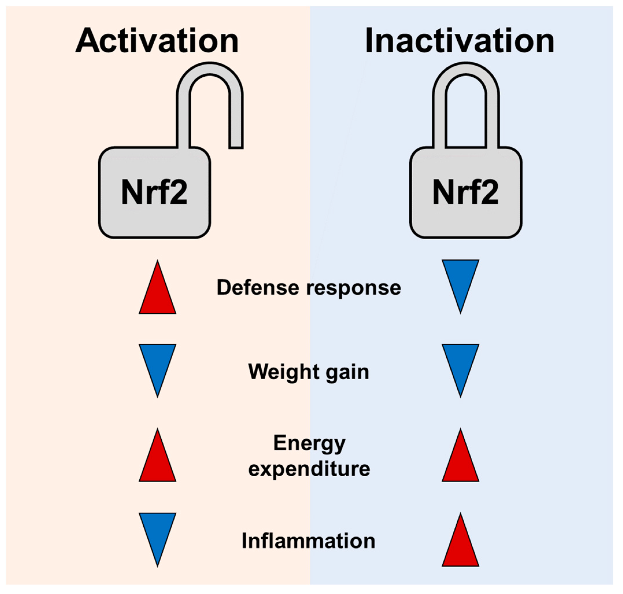
The primary function of Nrf2 is to provide antioxidative and cytoprotective roles [95]. Beyond cytoprotection, Nrf2 has potential to combat obesity and type 2 diabetes [96]. Nrf2 and ROS can regulate each other. Both display physiological responses, causing Nrf2 to have roles in energy metabolism in a context-dependent manner. Nrf2 suppression or activation exhibits similar anti-obese effects upon HFD feeding. However, as discussed above, unchecked high ROS levels and inflammatory insults in the Nrf2 KO mice are likely to exhibit deleterious physiological responses [97]. Thus, lean phenotype of HFD fed Nrf2 KO mice might be metabolically unhealthy. In addition, chemical Nrf2 activators can stimulate mitochondrial biogenesis in muscle, brain, kidney, and heart [98,99]. By contrast, Nrf2 inhibition by siRNA or Nrf2 knockouts impairs mitochondrial biogenesis and suppresses Pgc-1a, indicating that Nrf2 activation compared to Nrf2 inhibition is more effective in improving energy metabolism [99]. As Nrf2 is shown to negatively control inflammation, Nrf2 inhibition further raises concerns of inflamed status [100]. Therefore, Nrf2 activation might be a better strategy to control ROS levels, metabolism, and energy expenditure than Nrf2 inhibition (Figure 2).
It is tempting to envision that Nrf2 activation might be a safer way than Nrf2 inhibition to defend cells against oxidative stress and increase energy expenditure in obese conditions. However, this activation strategy to induce energy expenditure may also require caution. ROS are signaling molecules in various physiological processes. Consequences of ROS production are contradictory depending on ROS levels [94]. In line with this speculation, treatments with high doses of activators or genetic activation may exhibit unexpected physiological outcomes due to altered ROS levels or Nrf2 activities. Mild Nrf2 activation is likely to extend life span in multiple organisms [101], whereas hyperactivation confers a short-life span. In flies, one copy deletion of Keap1 gene increased life span and protected against stress while complete deletion was lethal during development [102,103,104]. Similarly, moderate activation of Nrf2 by a chemical RU486 increased life span while persistent Nrf2 overexpression in flies or high-copy transgenic SKN-1 in nematodes reduced longevity [103,105]. In rodents, sustained Nrf2 activation by Keap1 deficiency in hematopoietic stem cells caused stem cell exhaustion [106]. Furthermore, Nrf2 activators such as xenobiotics and phytochemicals at high concentrations displayed cellular cytotoxicity by forming DNA, protein, and lipid adducts [34,36].
In addition, Nrf2 activity in certain diseases should be considered. Nrf2 has a role in activating cellular antioxidant response. In addition, it acts as a major regulator of cell survival. Thus, activation of Nrf2-mediated defense can protect against various diseases including cancers [107]. A variety of natural and synthetic compounds that can increase the activity of Nrf2 have been tested for treating diseases. However, studies have demonstrated that activation of Nrf2 can not only promote cell survival by eliminating ROS in normal cells, but can also support cancer cells to survive better by protecting them from oxidative stress, chemotherapeutic reagents, and radiotherapy [108,109]. Indeed, some Nrf2 activating phytochemicals can facilitate cancer cell growth through reduction of ROS levels [39]. Conversely, Nrf2 suppression in cancer cells can restore sensitivity of cancer cells to chemotherapy [110]. Treatment with Nrf2 inhibitor (i.e., apigenin or luteolin) and doxorubicin has shown synergistic anti-tumor effect [111,112,113]. This phenomenon makes it necessary to better understand Nrf2 signaling for treating certain diseases. Taken together, temporal and spatial Nrf2 activation along with nutritional status and physical exercise should be considered for thermogenic responses and energy expenditure. Beneficial effects of temporal, spatial, and degrees of Nrf2 activation associated with energy metabolism and obesity should be further investigated.

7. Conclusions

Recent studies have revealed that various chemicals including phytochemicals, synthetic small molecules, or metabolic intermediates can increase energy expenditure. Significant numbers of these small molecules have been indicated or suggested as Nrf2 activators. Given that obesity is associated with high levels of ROS and oxidative stress, these Nrf2 activators can be applied particularly to stressful conditions to remove oxidative stress, induce thermogenesis, and enhance energy expenditure. Therefore, the utility of currently available Nrf2 activators such as drugs and diet supplementation should be further investigated to stimulate energy expenditure in humans, especially under genetic and environmental metabolic dysregulations.

Author Contributions

Conceptualization, U.J.Y. and K.W.P.; literature collections, S.-H.C. and J.-S.L.; writing—original draft preparation, S.-H.C. and U.J.Y.; writing—review and editing, K.W.P. and J.-S.L.; figures and tables, S.-H.C. and U.J.Y.; supervision, K.W.P.; funding acquisition, U.J.Y. and K.W.P. All authors have read and agreed to the published version of the manuscript.

Funding

This study was funded by grants (NRF-2020R1A2B5B02001592 to K.W.P., NRF- 2020R1I1A1A01074938 to U.J.Y., and NRF-2019R1A2C1010661 to J.-S.L.) from the Basic Science Research Program through the National Research Foundation of Korea (NRF) funded by the Ministry of Education, Science, and Technology, Republic of Korea. This study was also partly supported by The Health Fellowship Foundation to S.-H.C and KRIBB Research Initiative Program (KGM5352113, KGM2112133) to J.-S.L.

Institutional Review Board Statement

Not applicable.

Informed Consent Statement

Not applicable.

Conflicts of Interest

The funders had no role in the design of the study; in the collection, analyses, or interpretation of data; in the writing of the manuscript, or in the decision to publish the results.

References

  1. Crompton, A.W.; Taylor, C.R.; Jagger, J.A. Evolution of homeothermy in mammals. Nature 1978, 272, 333–336. [Google Scholar] [CrossRef]
  2. Betz, M.J.; Enerbäck, S. Targeting thermogenesis in brown fat and muscle to treat obesity and metabolic disease. Nat. Rev. Endocrinol. 2018, 14, 77–87. [Google Scholar] [CrossRef]
  3. Chang, S.-H.; Song, N.-J.; Choi, J.H.; Yun, U.J.; Park, K.W. Mechanisms underlying UCP1 dependent and independent adipocyte thermogenesis. Obes. Rev. 2019, 20, 241–251. [Google Scholar] [CrossRef]
  4. Foster, D.O.; Frydman, M.L. Brown Adipose Tissue: The Dominant Site of Nonshivering Thermogenesis in the Rat. Galanin 1978, 32, 147–151. [Google Scholar] [CrossRef]
  5. Divakaruni, A.S.; Brand, M. The Regulation and Physiology of Mitochondrial Proton Leak. Physiology 2011, 26, 192–205. [Google Scholar] [CrossRef]
  6. Saito, M. Human brown adipose tissue: Regulation and anti-obesity potential. Endocr. J. 2014, 61, 409–416. [Google Scholar] [CrossRef] [PubMed] [Green Version]
  7. Whittle, A.; Relat, J.; Vidal-Puig, A. Pharmacological strategies for targeting BAT thermogenesis. Trends Pharmacol. Sci. 2013, 34, 347–355. [Google Scholar] [CrossRef] [PubMed]
  8. Chouchani, E.T.; Kazak, L.; Spiegelman, B.M. Mitochondrial reactive oxygen species and adipose tissue thermogenesis: Bridging physiology and mechanisms. J. Biol. Chem. 2017, 292, 16810–16816. [Google Scholar] [CrossRef] [PubMed] [Green Version]
  9. Mailloux, R.J.; Adjeitey, C.N.; Xuan, J.Y.; Harper, M. Crucial yet divergent roles of mitochondrial redox state in skeletal muscle vs. brown adipose tissue energetics. FASEB J. 2012, 26, 363–375. [Google Scholar] [CrossRef] [PubMed]
  10. Stier, A.; Bize, P.; Habold, C.; Bouillaud, F.; Massemin, S.; Criscuolo, F. Mitochondrial uncoupling prevents cold-induced oxidative stress: A case study using UCP1 knock-out mice. J. Exp. Biol. 2013, 217 Pt 4, 624–630. [Google Scholar] [CrossRef] [Green Version]
  11. De Quiroga, G.B.; Lopez-Torres, M.; Pérez-Campo, R.; Abelenda, M.; Nava, M.P.; Puerta, M.L. Effect of cold acclimation on GSH, antioxidant enzymes and lipid peroxidation in brown adipose tissue. Biochem. J. 1991, 277, 289–292. [Google Scholar] [CrossRef] [PubMed] [Green Version]
  12. Ma, Q. Role of Nrf2 in Oxidative Stress and Toxicity. Annu. Rev. Pharmacol. Toxicol. 2013, 53, 401–426. [Google Scholar] [CrossRef] [PubMed] [Green Version]
  13. Baldelli, S.; Aquilano, K.; Ciriolo, M.R. Punctum on two different transcription factors regulated by PGC-1α: Nuclear factor erythroid-derived 2-like 2 and nuclear respiratory factor Biochim. Biophys. Acta 2013, 1830, 4137–4146. [Google Scholar] [CrossRef] [PubMed]
  14. Shin, S.; Wakabayashi, J.; Yates, M.S.; Wakabayashi, N.; Dolan, P.M.; Aja, S.; Liby, K.; Sporn, M.B.; Yamamoto, M.; Kensler, T.W. Role of Nrf2 in prevention of high-fat diet-induced obesity by synthetic triterpenoid CDDO-Imidazolide. Eur. J. Pharmacol. 2009, 620, 138–144. [Google Scholar] [CrossRef] [Green Version]
  15. Nagata, N.; Xu, L.; Kohno, S.; Ushida, Y.; Aoki, Y.; Umeda, R.; Fuke, N.; Zhuge, F.; Ni, Y.; Nagashimada, M.; et al. Glucoraphanin Ameliorates Obesity and Insulin Resistance Through Adipose Tissue Browning and Reduction of Metabolic Endotoxemia in Mice. Diabetes 2017, 66, 1222–1236. [Google Scholar] [CrossRef] [PubMed] [Green Version]
  16. Lee, D.H.; Chang, S.-H.; Yang, D.K.; Song, N.-J.; Yun, U.J.; Park, K.W.; Bell, A.; Chiappe, L.M. Sesamol Increases Ucp1 Expression in White Adipose Tissues and Stimulates Energy Expenditure in High-Fat Diet-Fed Obese Mice. Nutrients 2020, 12, 1459. [Google Scholar] [CrossRef]
  17. Schneider, K.; Valdez, J.; Nguyen, J.; Vawter, M.; Galke, B.; Kurtz, T.W.; Chan, J.Y. Increased Energy Expenditure, Ucp1 Expression, and Resistance to Diet-induced Obesity in Mice Lacking Nuclear Factor-Erythroid-2-related Transcription Factor-2 (Nrf2). J. Biol. Chem. 2016, 291, 7754–7766. [Google Scholar] [CrossRef] [Green Version]
  18. Uruno, A.; Furusawa, Y.; Yagishita, Y.; Fukutomi, T.; Muramatsu, H.; Negishi, T.; Sugawara, A.; Kensler, T.W.; Yamamoto, M. The Keap1-Nrf2 System Prevents Onset of Diabetes Mellitus. Mol. Cell. Biol. 2013, 33, 2996–3010. [Google Scholar] [CrossRef] [Green Version]
  19. Moi, P.; Chan, K.; Asunis, I.; Cao, A.; Kan, Y.W. Isolation of NF-E2-related factor 2 (Nrf2), a NF-E2-like basic leucine zipper transcriptional activator that binds to the tandem NF-E2/AP1 repeat of the beta-globin locus control region. Proc. Natl. Acad. Sci. USA 1994, 91, 9926–9930. [Google Scholar] [CrossRef] [PubMed] [Green Version]
  20. Chan, K.; Lu, R.; Chang, J.C.; Kan, Y.W. NRF2, a member of the NFE2 family of transcription factors, is not essential for murine erythropoiesis, growth, and development. Proc. Natl. Acad. Sci. USA 1996, 93, 13943–13948. [Google Scholar] [CrossRef] [Green Version]
  21. Mitsuishi, Y.; Motohashi, H.; Yamamoto, M. The Keap1-Nrf2 system in cancers: Stress response and anabolic metabolism. Front. Oncol. 2012, 2, 200. [Google Scholar] [CrossRef] [Green Version]
  22. McMahon, M.; Itoh, K.; Yamamoto, M.; Hayes, J. Keap1-dependent Proteasomal Degradation of Transcription Factor Nrf2 Contributes to the Negative Regulation of Antioxidant Response Element-driven Gene Expression. J. Biol. Chem. 2003, 278, 21592–21600. [Google Scholar] [CrossRef] [Green Version]
  23. Motohashi, H.; Yamamoto, M. Nrf2–Keap1 defines a physiologically important stress response mechanism. Trends Mol. Med. 2004, 10, 549–557. [Google Scholar] [CrossRef] [PubMed]
  24. Hayes, J.D.; Dinkova-Kostova, A.T. The Nrf2 regulatory network provides an interface between redox and intermediary metabo-lism. Trends Biochem. Sci. 2014, 39, 199–218. [Google Scholar] [CrossRef]
  25. Cuadrado, A.; Rojo, A.I.; Wells, G.; Hayes, J.D.; Cousin, S.P.; Rumsey, W.L.; Attucks, O.C.; Franklin, S.; Levonen, A.-L.; Kensler, T.W.; et al. Therapeutic targeting of the NRF2 and KEAP1 partnership in chronic diseases. Nat. Rev. Drug Discov. 2019, 18, 295–317. [Google Scholar] [CrossRef] [PubMed] [Green Version]
  26. Itoh, K.; Chiba, T.; Takahashi, S.; Ishii, T.; Igarashi, K.; Katoh, Y.; Oyake, T.; Hayashi, N.; Satoh, K.; Hatayama, I.; et al. An Nrf2/small Maf heterodimer mediates the induction of phase II detoxifying enzyme genes through antioxidant response elements. Biochem. Biophys. Res. Commun. 1997, 236, 313–322. [Google Scholar] [CrossRef] [PubMed]
  27. Sedlak, T.W.; Saleh, M.; Higginson, D.; Paul, B.D.; Juluri, K.R.; Snyder, S.H. Bilirubin and glutathione have complementary antioxidant and cytoprotective roles. Proc. Natl. Acad. Sci. USA 2009, 106, 5171–5176. [Google Scholar] [CrossRef] [Green Version]
  28. Wu, K.C.; Cui, J.Y.; Klaassen, C.D. Beneficial Role of Nrf2 in Regulating NADPH Generation and Consumption. Toxicol. Sci. 2011, 123, 590–600. [Google Scholar] [CrossRef] [Green Version]
  29. Niture, S.K.; Khatri, R.; Jaiswal, A.K. Regulation of Nrf2-an update. Free Radic. Biol. Med. 2014, 66, 36–44. [Google Scholar] [CrossRef] [Green Version]
  30. Egea, J.; González-Rodríguez, Á.; Gómez-Guerrero, C.; Moreno, J.A. Editorial: Role of Nrf2 in Disease: Novel Molecular Mechanisms and Therapeutic Approaches. Front. Pharmacol. 2019, 10, 1149. [Google Scholar] [CrossRef]
  31. Sies, H.; Berndt, C.; Jones, D.P. Oxidative Stress. Annu. Rev. Biochem. 2017, 86, 715–748. [Google Scholar] [CrossRef]
  32. He, L.; He, T.; Farrar, S.; Ji, L.; Liu, T.; Ma, X. Antioxidants Maintain Cellular Redox Homeostasis by Elimination of Reactive Oxygen Species. Cell. Physiol. Biochem. 2017, 44, 532–553. [Google Scholar] [CrossRef]
  33. Alfadda, A.A.; Sallam, R.M. Reactive Oxygen Species in Health and Disease. J. Biomed. Biotechnol. 2012, 2012, 936486. [Google Scholar] [CrossRef]
  34. Kumagai, Y.; Akiyama, M.; Unoki, T. Adaptive Responses to Electrophilic Stress and Reactive Sulfur Species as their Regulator Molecules. Toxicol. Res. 2019, 35, 303–310. [Google Scholar] [CrossRef] [Green Version]
  35. Birben, E.; Sahiner, U.M.; Sackesen, C.; Erzurum, S.; Kalayci, O. Oxidative stress and antioxidant defense. World Allergy Organ. J. 2012, 5, 9–19. [Google Scholar] [CrossRef] [Green Version]
  36. Suzuki, T.; Yamamoto, M. Stress-sensing mechanisms and the physiological roles of the Keap1–Nrf2 system during cellular stress. J. Biol. Chem. 2017, 292, 16817–16824. [Google Scholar] [CrossRef] [Green Version]
  37. Ahmed, K.A.; Sawa, T.; Akaike, T. Protein cysteine S-guanylation and electrophilic signal transduction by endogenous ni-tro-nucleotides. Amino Acids 2011, 41, 123–130. [Google Scholar] [CrossRef] [PubMed]
  38. Kobayashi, E.; Suzuki, T.; Yamamoto, M. Roles Nrf2 Plays in Myeloid Cells and Related Disorders. Oxidative Med. Cell. Longev. 2013, 2013, 529219. [Google Scholar] [CrossRef] [PubMed] [Green Version]
  39. Suraweera, T.L.; Rupasinghe, H.P.; Dellaire, G.; Xu, Z. Regulation of Nrf2/ARE Pathway by Dietary Flavonoids: A Friend or Foe for Cancer Management? Antioxidants 2020, 9, 973. [Google Scholar] [CrossRef] [PubMed]
  40. Wattenberg, L.W. Protective effects of 2(3)-tert-butyl-4-hydroxyanisole on chemical carcinogenesis. Food Chem. Toxicol. 1986, 24, 1099–1102. [Google Scholar] [CrossRef]
  41. Iverson, F. Phenolic antioxidants: Health protection branch studies on butylated hydroxyanisole. Cancer Lett. 1995, 93, 49–54. [Google Scholar] [CrossRef]
  42. Otsuki, A.; Yamamoto, M. Cis-element architecture of Nrf2–sMaf heterodimer binding sites and its relation to diseases. Arch. Pharmacal Res. 2020, 43, 275–285. [Google Scholar] [CrossRef] [PubMed]
  43. Andersen, L.F.; Jacobs, D.R.; Gross, M.D.; Schreiner, P.J.; Williams, O.D.; Lee, D.-H. Longitudinal associations between body mass index and serum carotenoids: The CARDIA study. Br. J. Nutr. 2006, 95, 358–365. [Google Scholar] [CrossRef] [Green Version]
  44. Wallström, P.; Wirfält, E.; Lahmann, P.H.; Gullberg, B.; Janzon, L.; Berglund, G. Serum concentrations of beta-carotene and alpha-tocopherol are associated with diet, smoking, and general and central adiposity. Am. J. Clin. Nutr. 2001, 73, 777–785. [Google Scholar] [CrossRef] [Green Version]
  45. Allen, S.; Britton, J.R.; Leonardi-Bee, J. Association between antioxidant vitamins and asthma outcome measures: Systematic review and meta-analysis. Thorax 2009, 64, 610–619. [Google Scholar] [CrossRef] [PubMed] [Green Version]
  46. Masschelin, P.M.; Cox, A.R.; Chernis, N.; Hartig, S.M. The Impact of Oxidative Stress on Adipose Tissue Energy Balance. Front. Physiol. 2020, 10, 1638. [Google Scholar] [CrossRef] [PubMed]
  47. Wellen, K.; Thompson, C.B. Cellular Metabolic Stress: Considering How Cells Respond to Nutrient Excess. Mol. Cell 2010, 40, 323–332. [Google Scholar] [CrossRef] [Green Version]
  48. Lettieri-Barbato, D. Redox control of non-shivering thermogenesis. Mol. Metab. 2019, 25, 11–19. [Google Scholar] [CrossRef]
  49. Samuel, V.T.; Liu, Z.-X.; Qu, X.; Elder, B.D.; Bilz, S.; Befroy, D.; Romanelli, A.J.; Shulman, G. Mechanism of Hepatic Insulin Resistance in Non-alcoholic Fatty Liver Disease. J. Biol. Chem. 2004, 279, 32345–32353. [Google Scholar] [CrossRef] [Green Version]
  50. Daly, M.E.; Vale, C.; Walker, M.; Alberti, K.G.; Mathers, J.C. Dietary carbohydrates and insulin sensitivity: A review of the evidence and clinical implications. Am. J. Clin. Nutr. 1997, 66, 1072–1085. [Google Scholar] [CrossRef] [PubMed]
  51. Basciano, H.; Federico, L.; Adeli, K. Fructose, insulin resistance, and metabolic dyslipidemia. Nutr. Metab. 2005, 2, 5. [Google Scholar] [CrossRef] [PubMed] [Green Version]
  52. Furukawa, S.; Fujita, T.; Shimabukuro, M.; Iwaki, M.; Yamada, Y.; Nakajima, Y.; Nakayama, O.; Makishima, M.; Matsuda, M.; Shimomura, I. Increased oxidative stress in obesity and its impact on metabolic syndrome. J. Clin. Investig. 2004, 114, 1752–1761. [Google Scholar] [CrossRef] [PubMed]
  53. Kasai, S.; Shimizu, S.; Tatara, Y.; Mimura, J.; Itoh, K. Regulation of Nrf2 by Mitochondrial Reactive Oxygen Species in Physiology and Pathology. Biomolecules 2020, 10, 320. [Google Scholar] [CrossRef] [Green Version]
  54. Done, A.J.; Traustadóttir, T. Nrf2 mediates redox adaptations to exercise. Redox Biol. 2016, 10, 191–199. [Google Scholar] [CrossRef] [Green Version]
  55. Wang, P.; Li, C.G.; Qi, Z.; Cui, D.; Ding, S. Acute exercise induced mitochondrial H2O2 production in mouse skeletal muscle: Association with p(66Shc) and FOXO3a signaling and antioxidant enzymes. Oxid. Med. Cell. Longev. 2015, 2015, 536456. [Google Scholar] [CrossRef]
  56. Zoladz, J.A.; Koziel, A.; Broniarek, I.; Woyda-Płoszczyca, A.M.; Ogrodna, K.; Majerczak, J.; Celichowski, J.; Szkutnik, Z.; Jarmuszkiewicz, W. Effect of temperature on fatty acid metabolism in skeletal muscle mitochondria of untrained and endurance-trained rats. PLoS ONE 2017, 12, e0189456. [Google Scholar] [CrossRef] [Green Version]
  57. Coleman, V.; Sa-Nguanmoo, P.; Koenig, J.; Schulz, T.J.; Grune, T.; Klaus, S.; Kipp, A.P.; Ost, M. Partial involvement of Nrf2 in skeletal muscle mitohormesis as an adaptive response to mitochondrial uncoupling. Sci. Rep. 2018, 8, 2446. [Google Scholar] [CrossRef] [PubMed]
  58. Eskla, K.-L.; Porosk, R.; Reimets, R.; Visnapuu, T.; Vasar, E.; Hundahl, C.A.; Luuk, H. Hypothermia augments stress response in mammalian cells. Free. Radic. Biol. Med. 2018, 121, 157–168. [Google Scholar] [CrossRef]
  59. Cypess, A.M.; Weiner, L.S.; Roberts-Toler, C.; Elía, E.F.; Kessler, S.H.; Kahn, P.A.; English, J.; Chatman, K.; Trauger, S.A.; Doria, A.; et al. Activation of human brown adipose tissue by a beta3-adrenergic receptor agonist. Cell Metab. 2015, 21, 33–38. [Google Scholar] [CrossRef] [Green Version]
  60. Collins, S. β-Adrenoceptor Signaling Networks in Adipocytes for Recruiting Stored Fat and Energy Expenditure. Front. Endocrinol. 2011, 2, 102. [Google Scholar] [CrossRef] [Green Version]
  61. Chang, S.H.; Jang, J.Y.; Oh, S.; Yoon, J.H.; Jo, D.G.; Yun, U.J.; Park, K.W. Nrf2 induces Ucp1 expression in adipocytes in response to beta3-AR stimulation and enhances oxygen consumption in high-fat diet-fed obese mice. BMB Rep. 2021, 54, 419–424. [Google Scholar] [CrossRef]
  62. Murphy, M.P. How mitochondria produce reactive oxygen species. Biochem. J. 2009, 417, 1–13. [Google Scholar] [CrossRef] [Green Version]
  63. Collins, Y.; Chouchani, E.T.; James, A.M.; Menger, K.E.; Cochemé, H.M.; Murphy, M.P. Mitochondrial redox signalling at a glance. J. Cell Sci. 2012, 125 Pt 4, 801–806. [Google Scholar] [CrossRef] [Green Version]
  64. Murphy, M.P.; Holmgren, A.; Larsson, N.-G.; Halliwell, B.; Chang, C.J.; Kalyanaraman, B.; Rhee, S.G.; Thornalley, P.; Partridge, L.; Gems, D.; et al. Unraveling the Biological Roles of Reactive Oxygen Species. Cell Metab. 2011, 13, 361–366. [Google Scholar] [CrossRef] [Green Version]
  65. Chouchani, E.T.; Kazak, L.; Jedrychowski, M.P.; Lu, G.Z.; Erickson, B.; Szpyt, J.; Pierce, K.A.; Laznik-Bogoslavski, D.; Vetrivelan, R.; Clish, C.; et al. Mitochondrial ROS regulate thermogenic energy expenditure and sulfenylation of UCP. Nature 2016, 532, 112–116. [Google Scholar] [CrossRef] [Green Version]
  66. Mills, E.L.; Pierce, K.A.; Jedrychowski, M.P.; Garrity, R.; Winther, S.; Vidoni, S.; Yoneshiro, T.; Spinelli, J.B.; Lu, G.Z.; Kazak, L.; et al. Accumulation of succinate controls activation of adipose tissue thermogenesis. Nature 2018, 560, 102–106. [Google Scholar] [CrossRef]
  67. Echtay, K.S.; Roussel, D.; St-Pierre, J.; Jekabsons, M.B.; Cadenas, S.; Stuart, J.A.; Harper, J.A.; Roebuck, S.J.; Morrison, A.; Pickering, S.; et al. Superoxide activates mitochondrial uncoupling proteins. Nat. Cell Biol. 2002, 415, 96–99. [Google Scholar] [CrossRef] [PubMed]
  68. Han, Y.H.; Buffolo, M.; Pires, K.M.; Pei, S.; Scherer, P.E.; Boudina, S. Adipocyte-Specific Deletion of Manganese Superoxide Dismutase Protects from Diet-Induced Obesity Through Increased Mitochondrial Uncoupling and Biogenesis. Diabetes 2016, 65, 2639–2651. [Google Scholar] [CrossRef] [Green Version]
  69. Chouchani, E.T.; Kazak, L.; Spiegelman, B.M. New Advances in Adaptive Thermogenesis: UCP1 and Beyond. Cell Metab. 2019, 29, 27–37. [Google Scholar] [CrossRef] [PubMed]
  70. Barbato, D.L.; Tatulli, G.; Cannata, S.M.; Bernardini, S.; Aquilano, K.; Ciriolo, M.R. Glutathione Decrement Drives Thermogenic Program in Adipose Cells. Sci. Rep. 2015, 5, 13091. [Google Scholar] [CrossRef] [PubMed] [Green Version]
  71. Ortega, S.P.; Chouchani, E.T.; Boudina, S. Stress turns on the heat: Regulation of mitochondrial biogenesis and UCP1 by ROS in adipocytes. Adipocyte 2017, 6, 56–61. [Google Scholar] [CrossRef] [Green Version]
  72. Kendig, E.L.; Chen, Y.; Krishan, M.; Johansson, E.; Schneider, S.N.; Genter, M.B.; Nebert, D.W.; Shertzer, H.G. Lipid metabolism and body composition in Gclm (−/−) mice. Toxicol. Appl. Pharmacol. 2011, 257, 338–348. [Google Scholar] [CrossRef] [Green Version]
  73. Seo, H.-A.; Lee, I.-K. The Role of Nrf2: Adipocyte Differentiation, Obesity, and Insulin Resistance. Oxid. Med. Cell. Longev. 2013, 2013, 184598. [Google Scholar] [CrossRef] [Green Version]
  74. Zhang, Z.; Zhou, S.; Jiang, X.; Wang, Y.-H.; Li, F.; Wang, Y.-G.; Zheng, Y.; Cai, L. The role of the Nrf2/Keap1 pathway in obesity and metabolic syndrome. Rev. Endocr. Metab. Disord. 2014, 16, 35–45. [Google Scholar] [CrossRef]
  75. Robledinos-Antón, N.; Fernández-Ginés, R.; Manda, G.; Cuadrado, A. Activators and Inhibitors of NRF2: A Review of Their Potential for Clinical Development. Oxid. Med. Cell. Longev. 2019, 2019, 9372182. [Google Scholar] [CrossRef] [PubMed]
  76. Yang, J.; Fu, X.; Liao, X.; Li, Y. Nrf2 Activators as Dietary Phytochemicals Against Oxidative Stress, Inflammation, and Mitochondrial Dysfunction in Autism Spectrum Disorders: A Systematic Review. Front. Psychiatry 2020, 11, 561998. [Google Scholar] [CrossRef] [PubMed]
  77. Zhu, Y.; Yang, Q.; Liu, H.; Song, Z.; Chen, W. Phytochemical compounds targeting on Nrf2 for chemoprevention in colorectal cancer. Eur. J. Pharmacol. 2020, 887, 173588. [Google Scholar] [CrossRef]
  78. Lu, M.-C.; Ji, J.-A.; Jiang, Z.-Y.; You, Q.-D. The Keap1-Nrf2-ARE Pathway as a Potential Preventive and Therapeutic Target: An Update. Med. Res. Rev. 2016, 36, 924–963. [Google Scholar] [CrossRef]
  79. Yu, Z.; Shao, W.; Chiang, Y.; Foltz, W.; Zhang, Z.; Ling, W.; Fantus, I.G.; Jin, T. Oltipraz upregulates the nuclear factor (erythroid-derived 2)-like 2 (corrected)(NRF2) antioxidant system and prevents insulin resistance and obesity induced by a high-fat diet in C57BL/6J mice. Diabetologia 2011, 54, 922–934. [Google Scholar] [CrossRef] [Green Version]
  80. He, H.-J.; Wang, G.-Y.; Gao, Y.; Ling, W.-H.; Guo-Yu, W.; Jin, T.-R. Curcumin attenuates Nrf2 signaling defect, oxidative stress in muscle and glucose intolerance in high fat diet-fed mice. World J. Diabetes 2012, 3, 94–104. [Google Scholar] [CrossRef] [PubMed]
  81. Pi, J.; Leung, L.; Xue, P.; Wang, W.; Hou, Y.; Liu, D.; Yehuda-Shnaidman, E.; Lee, C.; Lau, J.; Kurtz, T.W.; et al. Deficiency in the Nuclear Factor E2-related Factor-2 Transcription Factor Results in Impaired Adipogenesis and Protects against Diet-induced Obesity. J. Biol. Chem. 2010, 285, 9292–9300. [Google Scholar] [CrossRef] [Green Version]
  82. Xue, P.; Hou, Y.; Chen, Y.; Yang, B.; Fu, J.; Zheng, H.; Yarborough, K.; Woods, C.G.; Liu, D.; Yamamoto, M.; et al. Adipose Deficiency of Nrf2 in ob/ob Mice Results in Severe Metabolic Syndrome. Diabetes 2012, 62, 845–854. [Google Scholar] [CrossRef] [PubMed] [Green Version]
  83. Xu, J.; Kulkarni, S.; Donepudi, A.C.; More, V.R.; Slitt, A.L. Enhanced Nrf2 Activity Worsens Insulin Resistance, Impairs Lipid Accumulation in Adipose Tissue, and Increases Hepatic Steatosis in Leptin-Deficient Mice. Diabetes 2013, 61, 3208–3218. [Google Scholar] [CrossRef] [PubMed] [Green Version]
  84. More, V.R.; Xu, J.; Shimpi, P.C.; Belgrave, C.; Luyendyk, J.P.; Yamamoto, M.; Slitt, A.L. Keap1 knockdown increases markers of metabolic syndrome after long-term high fat diet feeding. Free Radic. Biol. Med. 2013, 61, 85–94. [Google Scholar] [CrossRef] [PubMed] [Green Version]
  85. Chorley, B.N.; Campbell, M.R.; Wang, X.; Karaca, M.; Sambandan, D.; Bangura, F.; Xue, P.; Pi, J.; Kleeberger, S.R.; Bell, D.A. Identification of novel NRF2-regulated genes by ChIP-Seq: Influence on retinoid X receptor alpha. Nucleic Acids Res. 2012, 40, 7416–7429. [Google Scholar] [CrossRef] [PubMed] [Green Version]
  86. Li, S.; Eguchi, N.; Lau, H.; Ichii, H. The Role of the Nrf2 Signaling in Obesity and Insulin Resistance. Int. J. Mol. Sci. 2020, 21, 6973. [Google Scholar] [CrossRef]
  87. Chartoumpekis, D.V.; Ziros, P.G.; Psyrogiannis, A.I.; Papavassiliou, A.G.; Kyriazopoulou, V.E.; Sykiotis, G.; Habeos, I.G. Nrf2 Represses FGF21 During Long-Term High-Fat Diet-Induced Obesity in Mice. Diabetes 2011, 60, 2465–2473. [Google Scholar] [CrossRef] [Green Version]
  88. Zhang, L.; Dasuri, K.; Fernandez-Kim, S.-O.; Bruce-Keller, A.J.; Keller, J. Adipose-specific ablation of Nrf2 transiently delayed high-fat diet-induced obesity by altering glucose, lipid and energy metabolism of male mice. Am. J. Transl. Res. 2016, 8, 5309–5319. [Google Scholar]
  89. Meakin, P.; Chowdhry, S.; Sharma, R.S.; Ashford, F.B.; Walsh, S.V.; McCrimmon, R.; Dinkova-Kostova, A.; Dillon, J.; Hayes, J.D.; Ashford, M. Susceptibility of Nrf2-Null Mice to Steatohepatitis and Cirrhosis upon Consumption of a High-Fat Diet Is Associated with Oxidative Stress, Perturbation of the Unfolded Protein Response, and Disturbance in the Expression of Metabolic Enzymes but Not with Insulin Resistance. Mol. Cell. Biol. 2014, 34, 3305–3320. [Google Scholar] [CrossRef] [Green Version]
  90. Zhang, Y.-K.J.; Wu, K.C.; Liu, J.; Klaassen, C.D. Nrf2 deficiency improves glucose tolerance in mice fed a high-fat diet. Toxicol. Appl. Pharmacol. 2012, 264, 305–314. [Google Scholar] [CrossRef] [Green Version]
  91. Tanaka, Y.; Aleksunes, L.M.; Yeager, R.L.; Gyamfi, M.A.; Esterly, N.; Guo, G.L.; Klaassen, C.D. NF-E2-Related Factor 2 Inhibits Lipid Accumulation and Oxidative Stress in Mice Fed a High-Fat Diet. J. Pharmacol. Exp. Ther. 2008, 325, 655–664. [Google Scholar] [CrossRef] [PubMed]
  92. Chartoumpekis, D.V.; Palliyaguru, D.L.; Wakabayashi, N.; Fazzari, M.; Khoo, N.K.H.; Schopfer, F.J.; Sipula, I.; Yagishita, Y.; Michalopoulos, G.K.; O’Doherty, R.M.; et al. Nrf2 deletion from adipocytes, but not hepatocytes, potentiates systemic metabolic dysfunction after long-term high-fat diet-induced obesity in mice. Am. J. Physiol. Endocrinol. Metab. 2018, 315, E180–E195. [Google Scholar] [CrossRef] [PubMed] [Green Version]
  93. Weisberg, S.; Leibel, R.; Tortoriello, D.V. Dietary Curcumin Significantly Improves Obesity-Associated Inflammation and Diabetes in Mouse Models of Diabesity. Endocrinology 2008, 149, 3549–3558. [Google Scholar] [CrossRef] [PubMed] [Green Version]
  94. Yang, S.; Lian, G. ROS and diseases: Role in metabolism and energy supply. Mol. Cell. Biochem. 2020, 467, 1–12. [Google Scholar] [CrossRef] [PubMed]
  95. Shaw, P.; Chattopadhyay, A. Nrf2–ARE signaling in cellular protection: Mechanism of action and the regulatory mechanisms. J. Cell. Physiol. 2020, 235, 3119–3130. [Google Scholar] [CrossRef] [PubMed]
  96. Schneider, K.S.; Chan, J.Y. Emerging Role of Nrf2 in Adipocytes and Adipose Biology. Adv. Nutr. 2013, 4, 62–66. [Google Scholar] [CrossRef] [Green Version]
  97. Dinkova-Kostova, A.T.; Abramov, A.Y. The emerging role of Nrf2 in mitochondrial function. Free Radic. Biol. Med. 2015, 88 Pt B, 179–188. [Google Scholar] [CrossRef] [Green Version]
  98. Hayashi, G.; Jasoliya, M.; Sahdeo, S.; Saccà, F.; Pane, C.; Filla, A.; Marsili, A.; Puorro, G.; Lanzillo, R.; Morra, V.B.; et al. Dimethyl fumarate mediates Nrf2-dependent mitochondrial biogenesis in mice and humans. Hum. Mol. Genet. 2017, 26, 2864–2873. [Google Scholar] [CrossRef]
  99. Gureev, A.P.; Shaforostova, E.A.; Popov, V. Regulation of Mitochondrial Biogenesis as a Way for Active Longevity: Interaction Between the Nrf2 and PGC-1α Signaling Pathways. Front. Genet. 2019, 10, 435. [Google Scholar] [CrossRef] [Green Version]
  100. Ahmed, S.M.U.; Luo, L.; Namani, A.; Wang, X.J.; Tang, X. Nrf2 signaling pathway: Pivotal roles in inflammation. Biochim. Biophys. (BBA) Acta Mol. Basis Dis. 2017, 1863, 585–597. [Google Scholar] [CrossRef]
  101. Ristow, M.; Schmeisser, S. Extending life span by increasing oxidative stress. Free Radic. Biol. Med. 2011, 51, 327–336. [Google Scholar] [CrossRef] [Green Version]
  102. Castillo-Quan, J.I.; Li, L.; Kinghorn, K.; Ivanov, D.; Tain, L.; Slack, C.; Kerr, F.; Nespital, T.; Thornton, J.; Hardy, J.; et al. Lithium Promotes Longevity through GSK3/NRF2-Dependent Hormesis. Cell Rep. 2016, 15, 638–650. [Google Scholar] [CrossRef] [Green Version]
  103. Sykiotis, G.P.; Bohmann, D. Keap1/Nrf2 Signaling Regulates Oxidative Stress Tolerance and Lifespan in Drosophila. Dev. Cell 2008, 14, 76–85. [Google Scholar] [CrossRef] [PubMed] [Green Version]
  104. Tsakiri, E.; Gumeni, S.; Iliaki, K.K.; Benaki, D.; Vougas, K.; Sykiotis, G.; Gorgoulis, V.; Mikros, E.; Scorrano, L.; Trougakos, I.P. Hyperactivation of Nrf2 increases stress tolerance at the cost of aging acceleration due to metabolic deregulation. Aging Cell 2019, 18, e12845. [Google Scholar] [CrossRef] [PubMed]
  105. Tullet, J.; Hertweck, M.; An, J.H.; Baker, J.; Hwang, J.Y.; Liu, S.; Oliveira, R.D.P.; Baumeister, R.; Blackwell, T.K. Direct Inhibition of the Longevity-Promoting Factor SKN-1 by Insulin-like Signaling in C. elegans. Cell 2008, 132, 1025–1038. [Google Scholar] [CrossRef] [Green Version]
  106. Murakami, S.; Suzuki, T.; Harigae, H.; Romeo, P.-H.; Yamamoto, M.; Motohashi, H. NRF2 Activation Impairs Quiescence and Bone Marrow Reconstitution Capacity of Hematopoietic Stem Cells. Mol. Cell. Biol. 2017, 37, e00086-17. [Google Scholar] [CrossRef] [PubMed] [Green Version]
  107. Görlach, A.; Dimova, E.Y.; Petry, A.; Martínez-Ruiz, A.; Hernansanz-Agustín, P.; Rolo, A.P.; Palmeira, C.M.; Kietzmann, T. Reactive oxygen species, nutrition, hypoxia and diseases: Problems solved? Redox Biol. 2015, 6, 372–385. [Google Scholar] [CrossRef] [PubMed]
  108. Kim, J.; Keum, Y.-S. NRF2, a Key Regulator of Antioxidants with Two Faces towards Cancer. Oxid. Med. Cell. Longev. 2016, 2016, 2746457. [Google Scholar] [CrossRef] [Green Version]
  109. Milkovic, L.; Zarkovic, N.; Saso, L. Controversy about pharmacological modulation of Nrf2 for cancer therapy. Redox Biol. 2017, 12, 727–732. [Google Scholar] [CrossRef] [Green Version]
  110. Probst, B.L.; McCauley, L.; Trevino, I.; Wigley, W.C.; Ferguson, D.A. Cancer Cell Growth Is Differentially Affected by Constitutive Activation of NRF2 by KEAP1 Deletion and Phar-macological Activation of NRF2 by the Synthetic Triterpenoid, RTA. PLoS ONE 2015, 10, e0135257. [Google Scholar] [CrossRef] [Green Version]
  111. Gao, A.M.; Ke, Z.P.; Wang, J.N.; Yang, J.Y.; Chen, S.Y.; Chen, H. Apigenin sensitizes doxorubicin-resistant hepatocellular carcinoma BEL-7402/ADM cells to doxorubicin via inhibiting PI3K/Akt/Nrf2 pathway. Carcinogenesis 2013, 34, 1806–1814. [Google Scholar] [CrossRef] [PubMed]
  112. Korga, A.; Ostrowska-Lesko, M.; Jozefczyk, A.; Iwan, M.; Wojcik, R.; Zgorka, G.; Herbet, M.; Vilarrubla, G.G.; Dudka, J. Apigenin and hesperidin augment the toxic effect of doxorubicin against HepG2 cells. BMC Pharmacol. Toxicol. 2019, 20, 22. [Google Scholar] [CrossRef] [PubMed] [Green Version]
  113. Sabzichi, M.; Hamishehkar, H.; Ramezani, F.; Sharifi, S.; Tabasinezhad, M.; Pirouzpanah, M.; Ghanbari, P.; Samadi, N. Luteolin-loaded phytosomes sensitize human breast carcinoma MDA-MB 231 cells to doxorubicin by suppressing Nrf2 mediated signalling. Asian Pac. J. Cancer Prev. 2014, 15, 5311–5316. [Google Scholar] [CrossRef] [PubMed] [Green Version]
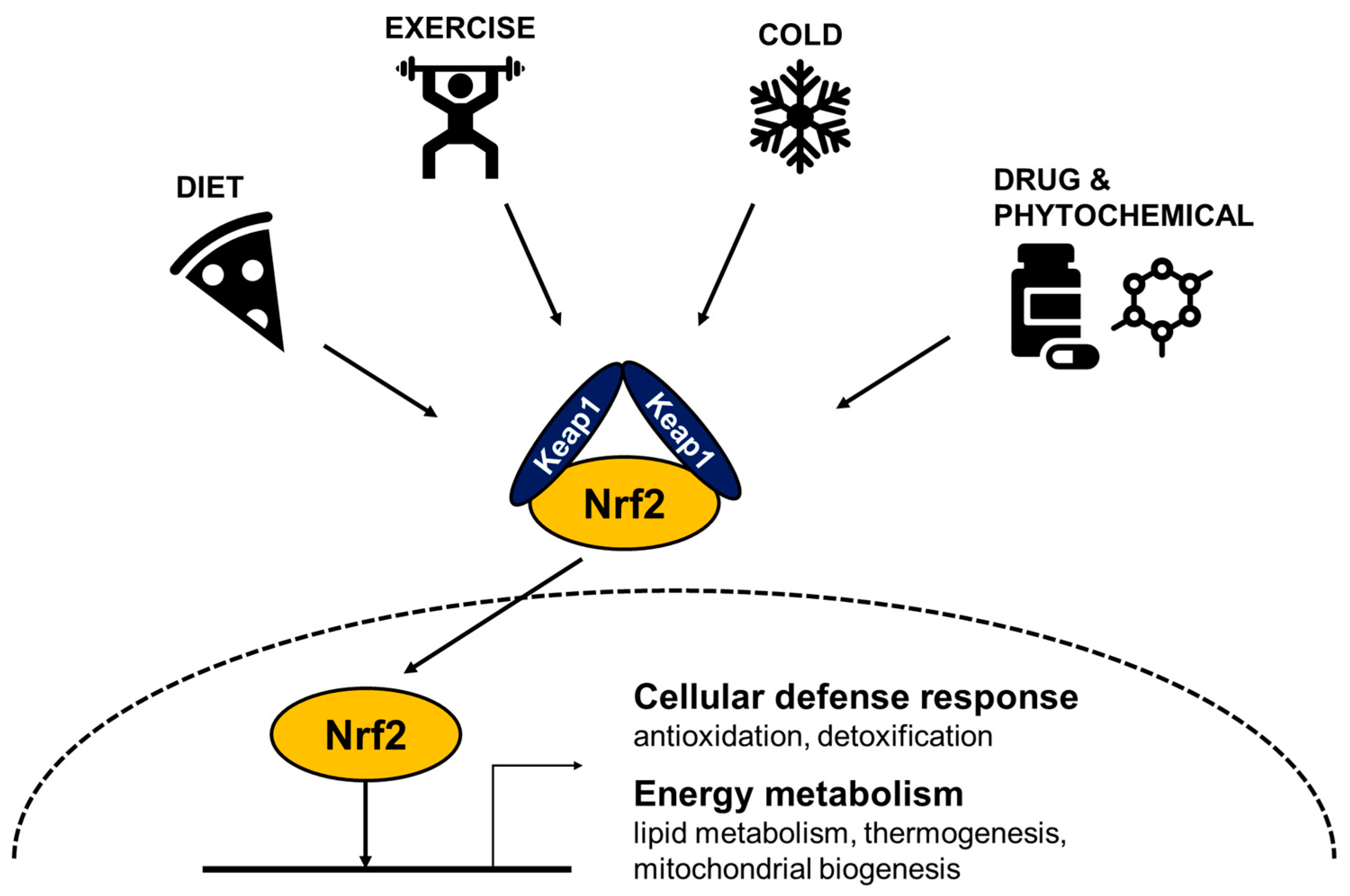
Figure 1. Cellular stresses activate Nrf2 to induce defense systems, reduce body weight, and increase energy expenditure. Schematic diagram of Nrf2-induced responses against various cellular stresses. Cold exposure, physical exercise, food intake, and xenobiotic chemicals activate Nrf2 to transcribe its target genes involved in antioxidation, detoxification, mitochondrial biogenesis, and energy expenditure. ROS can be produced by cold, exercise, high energy diets, and chemical activators that can disrupt Kelch-like ECH-associated protein 1 (Keap1) and Nrf2 interaction to increase Nrf2 abundance and subsequent nuclear translocation of Nrf2. In the nucleus, Nrf2 forms a heterodimer with a small Maf protein and binds to antioxidant response element (ARE) in the promoter region to drive transcription of antioxidative genes. Its target genes include glutamate-cysteine ligase catalytic subunit (GCLC), glutathione reductase, ABCC-family efflux transporter genes, NAD(P)H quinone oxidoreductase1 (NQO1), heme oxygenase1 (HO1), and possibly mitochondrial genes including nuclear respiratory factor 1 (Nrf1), Peroxisome proliferator-activated receptor-gamma coactivator-1a (Pgc1α), and uncoupling protein 1 (Ucp1).
Biomedicines 09 01196 g001
Figure 2. Comparative effects of Nrf2 activation and Nrf2 inhibition on defense responses, weight reduction, and increases of energy expenditure. Schematic diagram shows effects of Nrf2—gain and loss—of function on energy expenditure and weight reduction. Various cellular stresses can activate Nrf2 to provide defense mechanisms in antioxidation and detoxification. Nrf2 inhibition increases energy expenditure and decreases weight gain in a high calorie diet feeding. However, Nrf2 inhibition potentially fails to protect cells from oxidative/electrophilic stress and inflammatory insults. By contrast, chemical Nrf2 activators can stimulate mitochondrial biogenesis in tissues while Nrf2 inhibition impairs mitochondrial biogenesis and energy expenditure.
Biomedicines 09 01196 g002
Table 1. The effects of Nrf2 inhibition in energy metabolism.
MouseBackgroundDiet
(Start/Duration)
Body MassIROxidative StressInflammationEE (Ucp1)Ref.
Nrf2 KOC57BL6;129SVHFD
(3–4 w/12 w)
[81]
Nrf2 KOC57BL6HFD
(9–10 w/180 d)
[87]
Nrf2 KOC57BL6HFWD (12 w/12 w)↓ (GSH in liver) [90]
Nrf2 KOob/obSD (4 w/11 w) ↓ (WAT) [82]
Nrf2 KOC57BL6HFD (8 w/4 w) ↑ (MDA in liver) [91]
Nrf2 KOC57BL6HFD
(8–10 w/16–20 w)
↑ (GSSG, MDA in liver)↑ (liver)[89]
Nrf2 KOC57BL6HFD (12 w/6 w)↓ (GSH/GSSG in WAT) ↑ (↔RER) (↑in WAT)[17]
Adipo-Nrf2 KOC57BL6HFD (6 w/14 w) ↓ RER in SD[88]
Adipo-Nrf2 KOAlbino C57BL6HFD (8 w/170 d) ↔ (↑iWAT ↓eWAT)[92]
KO, knockout; ↑, increased; ↓, decreased; ↔; no change; w, week; d, day; IR, insulin resistance; SD, standard diet; HFWD, high fat Western diet; HFD, high fat diet; ob/ob, leptin deficient obese mice; GSH, reduced glutathione (reduced); GSSG, glutathione disulfide (oxidized); MDA, malondialdehyde; RER, respiratory exchange ratio; EE, energy expenditure; eWAT, epididymal white adipose tissue; iWAT, inguinal white adipose tissue; WAT, white adipose tissue; BAT, brown adipose tissue.
Table 2. The effects of Nrf2 activators in energy metabolism.
Treatment
/Mouse
BackgroundDiet
(Start/Duration)
Body MassIROxidative StressInflammationEE (Ucp1)Ref.
Pharmacological activation
CDDO-ImC57BL6HFD
(6–7 w/95 d)
[14]
GlucoraphaninC57BL6HFD
(8 w/14 w)
↓ (liver,
eWAT)
↑ (↑WAT, ↔BAT)[15]
OltiprazC57BL6HFD
(5 w/28 w)
↑ (GSH/GSSG)↓ (eWAT) [79]
CurcuminC57BL6HFD
(9 w/18 w)
↓ (muscle)
↔ (adipose and liver)
[80]
CurcuminC57BL6HFD
(3–5 w/15 w)
↓ (liver, WAT) [93]
Curcuminob/obSD
(8–10 w/14–18 w)
↓ (liver, WAT) [93]
SesamolC57BL6HFD
(8 w/12 w)
↓ (iWAT)↑ (↑iWAT)[16]
Genetic activation
Keap1 KDC57BL6 HFD
(9 w/36 d)
[83]
Keap1 KDob/obSD (4 w/8 w) [83]
Keap1 KDC57BL6HFWD (12 w/12 w) [90]
Keap1 KDC57BL6HFD
(3 w/24 w)
↑ (liver, WAT)[84]
Keap1 flox/−ICRSD/HCD
(4 w/12 w)
↑ (↔BAT)[18]
↑, increased; ↓, decreased; ↔; no change; w, week; d, day; IR, insulin resistance; HFD, high fat diet; HCD, high carbohydrate diet; HFWD, high fat western diet; SD, standard diet; CDDO-Im, 1-(2-cyano-3, 12-dioxooleana-1,9(11)-dien-28-oyl) imidazole; ob/ob, leptin deficient obese mice; Keap1, Kelch-like ECH-associated protein 1; KD, knockdown; Keap1 flox/−, Keap1 gene hypomorphic knockdown; GSH, reduced glutathione (reduced); GSSG, glutathione disulfide (oxidized); eWAT, epididymal white adipose tissue; iWAT, inguinal white adipose tissue; BAT, brown adipose tissue.
Publisher’s Note: MDPI stays neutral with regard to jurisdictional claims in published maps and institutional affiliations.