
Exploring a Paradigm Shift in Primary Teeth Root Canal Preparation: An Ex Vivo Micro-CT Study


 , ,
, ,
Abstract
:1. Introduction
2. Materials and Methods
2.1. Experimental Teeth
2.2. Tooth Instrumentation
- (1)
- MI (manual instrumentation) group (n = 9): For the root canal instrumentation procedure, the operator used the balanced force technique [23] with K-files (Dentsply Maillefer, Tulsa, OK, USA). Initially, a size 10 K-file was inserted into the canal, and the working length determined and confirmed by a radiograph. The file was rotated in a clockwise and counterclockwise motion with light pressure to create a glide path for larger files. The canal was then recapitulated with smaller hand files to remove debris and verify the working length. The operator then selected a size-15 K-file and repeated the process of preparation and recapitulation. This process was repeated with progressively larger K-files up to size 30, until the desired apical size and taper were achieved. Recapitulation with smaller hand files and irrigation with a sodium hypochlorite solution were performed throughout the procedure. The operator used balanced force and controlled movements to minimize the risk of iatrogenic errors, such as canal transportation or perforation.
- (2)
- KS (Kedo S) group (n = 9): The teeth were instrumented with a Kedo-S system (16 mm) (Reeganz Dental Care Private Limited, Chennai, India). The system included the usage of three files: (a) white (17/08), (b) yellow (20/04), and (c) red (25/04). For the root canal instrumentation procedure, the operator used a Kedo-S rotary file system (Reeganz Dental Care Private Limited, Chennai, India). After establishing access to the root canal system and determining the working length, the operator inserted the appropriate size Kedo-S file to the working length and used a gentle filing motion in clockwise and counterclockwise directions to create a glide path. Recapitulation with smaller hand files was performed to remove debris and verify the working length. This process was repeated with progressively larger Kedo-S files until the desired apical size and taper were achieved. Throughout the procedure, irrigation with sodium hypochlorite solution was used to clean the canal and remove debris. The operator used controlled and gentle movements to minimize the risk of iatrogenic errors, such as canal transportation or perforation.
- (3)
- PTG (ProTaper Gold) group (n = 10): For the root canal instrumentation procedure, the operator used a ProTaper Gold rotary file system (Dentsply Maillefer, Ballaigues, Switzerland). After establishing access to the root canal system and determining the working length, the operator used a size 10 hand file to establish a glide path. The S1 file was then used to shape the coronal portion of the canal. The SX file was used to finish shaping the canal or to bypass ledges or obstructions. The operator then used the S1, S2, and F1 files in a sequential manner to shape the middle and apical portions of the canal. Recapitulation with smaller hand files was performed to remove debris and verify the working length. The operator then used the F2 file to achieve the desired apical size and taper. Throughout the procedure, irrigation with a sodium hypochlorite solution was used to clean the canal and remove debris. The operator used controlled and gentle movements to minimize the risk of iatrogenic errors, such as canal transportation or perforation.
- (4)
- Control (n = 8) group: no instrumentation and irrigation were performed in the root canals. The control group aimed to evaluate the reliability of dentin volume evaluation using micro-CT.
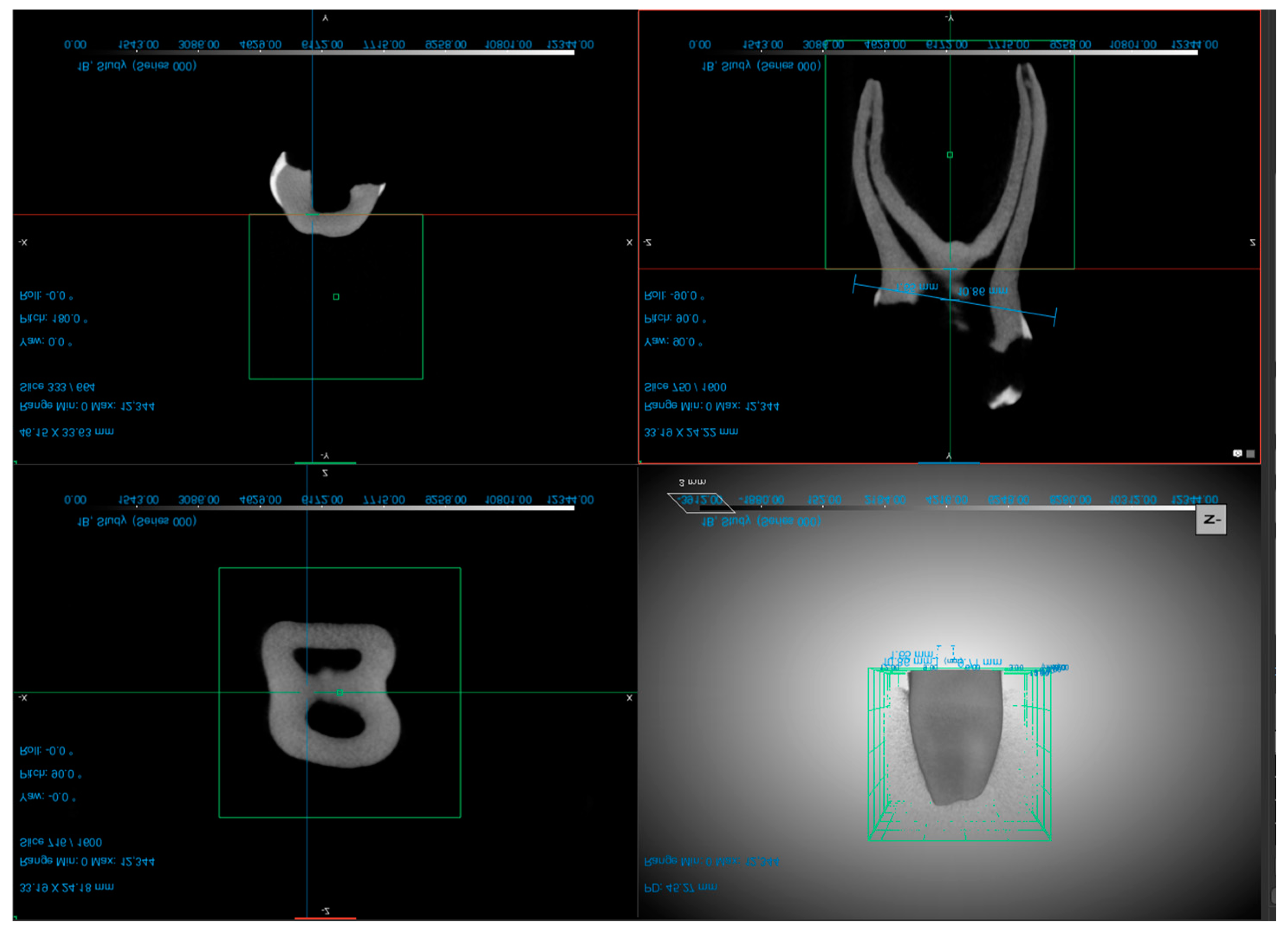
2.3. Micro-CT Scanning
2.4. Preparation Time and Errors
2.5. Statistical Analysis
2.6. Reliability
3. Results
3.1. Reliability Analysis
3.2. Root Canal Preparation Time
3.3. Root Canal Volume
4. Discussion
5. Conclusions
Author Contributions
Funding
Institutional Review Board Statement
Informed Consent Statement
Data Availability Statement
Conflicts of Interest
References
- Aly Ahmed, H. Pulpectomy procedures in primary molar teeth. Eur. J. Gen. Dent. 2014, 3, 3–10. [Google Scholar] [CrossRef]
- Fuks, A.B.; Peretz, B. (Eds.) Pediatric endodontics: Past and present perspectives and future directions. In Pediatric Endodontics; Springer International Publishing: Cham, Switzerland, 2016; pp. 1–5. ISBN 978-3-319-27551-2. [Google Scholar]
- Natchiyar, N.; Asokan, S.; Priya, P.R.G.; Kumar, T.D.Y. Comparison of Clinical and Radiographic Success of Rotary with Manual Instrumentation Techniques in Primary Teeth: A Systematic Review. Int. J. Clin. Pediatr. Dent. 2021, 14, 8–13. [Google Scholar] [CrossRef] [PubMed]
- Nisar, P.; Katge, F.; Bhanushali, P.; Deshpande, S.; Poojari, M.; Shetty, S. Comparative in vitro evaluation of remaining dentine thickness following instrumentation with hand and rotary endodontic files during pulpectomy in primary molars: A systematic review. Eur. Arch. Paediatr. Dent. 2023, 24, 15–32. [Google Scholar] [CrossRef] [PubMed]
- Katge, F.; Chimata, V.K.; Poojari, M.; Shetty, S.; Rusawat, B. Comparison of cleaning Efficacy and Instrumentation Time between Rotary and Manual Instrumentation Techniques in Primary Teeth: An in vitro Study. Int. J. Clin. Pediatr. Dent. 2016, 9, 124–127. [Google Scholar] [CrossRef]
- Bergmans, L.; Van Cleynenbreugel, J.; Wevers, M.; Lambrechts, P. Mechanical root canal preparation with NiTi rotary instruments: Rationale, performance and safety. Status report for the American Journal of Dentistry. Am. J. Dent. 2001, 14, 324–333. [Google Scholar]
- Peters, O.A.; de Azevedo Bahia, M.G.; Pereira, E.S.J. Contemporary root canal preparation: Innovations in biomechanics. Dent. Clin. N. Am. 2017, 61, 37–58. [Google Scholar] [CrossRef]
- Kurthukoti, A.J.; Sharma, P.; Swamy, D.F.; Shashidara, R.; Swamy, E.B. Computed tomographic morphometry of the internal anatomy of mandibular second primary molars. Int. J. Clin. Pediatr. Dent. 2015, 8, 202–207. [Google Scholar] [CrossRef]
- Esentürk, G.; Akkas, E.; Cubukcu, E.; Nagas, E.; Uyanik, O.; Cehreli, Z.C. A micro-computed tomographic assessment of root canal preparation with conventional and different rotary files in primary teeth and young permanent teeth. Int. J. Paediatr. Dent. 2020, 30, 202–208. [Google Scholar] [CrossRef]
- Musale, P.K.; Mujawar, S.A.V. Evaluation of the efficacy of rotary vs. hand files in root canal preparation of primary teeth in vitro using CBCT. Eur. Arch. Paediatr. Dent. 2014, 15, 113–120. [Google Scholar] [CrossRef]
- Chugh, V.K.; Patnana, A.K.; Chugh, A.; Kumar, P.; Wadhwa, P.; Singh, S. Clinical differences of hand and rotary instrumentations during biomechanical preparation in primary teeth-A systematic review and meta-analysis. Int. J. Paediatr. Dent. 2021, 31, 131–142. [Google Scholar] [CrossRef]
- Chernyshikhin, S.V.; Pelevin, I.A.; Karimi, F.; Shishkovsky, I.V. The study on resolution factors of LPBF technology for manufacturing superelastic niti endodontic files. Materials 2022, 15, 6556. [Google Scholar] [CrossRef] [PubMed]
- Barr, E.S.; Kleier, D.J.; Barr, N.V. Use of nickel-titanium rotary files for root canal preparation in primary teeth. Pediatr. Dent. 2000, 22, 77–78. [Google Scholar]
- Bertrand, M.F.; Lupi-Pégurier, L.; Médioni, E.; Muller, M.; Bolla, M. Curved molar root canal preparations using Hero 642 rotary nickel-titanium instruments. Int. Endod. J. 2001, 34, 631–636. [Google Scholar] [CrossRef] [PubMed]
- Hülsmann, M.; Schade, M.; Schäfers, F. A comparative study of root canal preparation with HERO 642 and Quantec SC rotary Ni-Ti instruments. Int. Endod. J. 2001, 34, 538–546. [Google Scholar] [CrossRef] [PubMed]
- Kim, H.C.; Kim, H.J.; Lee, C.J.; Kim, B.M.; Park, J.K.; Versluis, A. Mechanical response of nickel-titanium instruments with different cross-sectional designs during shaping of simulated curved canals. Int. Endod. J. 2009, 42, 593–602. [Google Scholar] [CrossRef] [PubMed]
- Swaminathan, K.; Rakkesh, K.M.; Haridoss, S. Computed Tomographic Assessment of Remaining Dentin and Risk of Perforation after Kedo-S and Mtwo Rotary Instrumentation in Root Canals of Primary Teeth: An In Vitro Study. Int. J. Clin. Pediatr. Dent. 2022, 15, S87–S91. [Google Scholar] [CrossRef]
- Haridoss, S.; Rakkesh, K.M.; Swaminathan, K. Transportation and Centering Ability of Kedo-S Pediatric and Mtwo Instruments in Primary Teeth: A Cone-beam Computed Tomography Study. Int. J. Clin. Pediatr. Dent. 2022, 15, S30–S34. [Google Scholar] [CrossRef] [PubMed]
- Jeevanandan, G. Kedo-S Paediatric Rotary Files for Root Canal Preparation in Primary Teeth—Case Report. J. Clin. Diagn. Res. 2017, 11, ZR03–ZR05. [Google Scholar] [CrossRef] [PubMed]
- Swain, M.V.; Xue, J. State of the art of Micro-CT applications in dental research. Int. J. Oral Sci. 2009, 1, 177–188. [Google Scholar] [CrossRef]
- Siqueira, J.F.; Pérez, A.R.; Marceliano-Alves, M.F.; Provenzano, J.C.; Silva, S.G.; Pires, F.R.; Vieira, G.C.S.; Rôças, I.N.; Alves, F.R.F. What happens to unprepared root canal walls: A correlative analysis using micro-computed tomography and histology/scanning electron microscopy. Int. Endod. J. 2018, 51, 501–508. [Google Scholar] [CrossRef]
- Schneider, S.W. A comparison of canal preparations in straight and curved root canals. Oral Surg. Oral Med. Oral Pathol. 1971, 32, 271–275. [Google Scholar] [CrossRef] [PubMed]
- Roane, J.B.; Sabala, C.L.; Duncanson, M.G. The “balanced force” concept for instrumentation of curved canals. J. Endod. 1985, 11, 203–211. [Google Scholar] [CrossRef] [PubMed]
- Crespo, S.; Cortes, O.; Garcia, C.; Perez, L. Comparison between rotary and manual instrumentation in primary teeth. J. Clin. Pediatr. Dent. 2008, 32, 295–298. [Google Scholar] [CrossRef] [PubMed]
- Barasuol, J.C.; Alcalde, M.P.; Bortoluzzi, E.A.; Duarte, M.A.H.; Cardoso, M.; Bolan, M. Shaping ability of hand, rotary and reciprocating files in primary teeth: A micro-CT study in vitro. Eur. Arch. Paediatr. Dent. 2021, 22, 195–201. [Google Scholar] [CrossRef]
- Gagliardi, J.; Versiani, M.A.; de Sousa-Neto, M.D.; Plazas-Garzon, A.; Basrani, B. Evaluation of the shaping characteristics of protaper gold, protaper NEXT, and protaper universal in curved canals. J. Endod. 2015, 41, 1718–1724. [Google Scholar] [CrossRef]
- Fuks, A.B. Current concepts in vital primary pulp therapy. Eur. J. Paediatr. Dent. 2002, 3, 115–120. [Google Scholar]
- Lim, S.S.; Stock, C.J. The risk of perforation in the curved canal: Anticurvature filing compared with the stepback technique. Int. Endod. J. 1987, 20, 33–39. [Google Scholar] [CrossRef]
- Panchal, V.; Jeevanandan, G.; Erulappan, S.M. Comparison between the Effectiveness of Rotary and Manual Instrumentation in Primary Teeth: A Systematic Review. Int. J. Clin. Pediatr. Dent. 2019, 12, 340–346. [Google Scholar] [CrossRef]
- Jeevanandan, G.; Govindaraju, L. Clinical comparison of Kedo-S paediatric rotary files vs manual instrumentation for root canal preparation in primary molars: A double blinded randomised clinical trial. Eur. Arch. Paediatr. Dent. 2018, 19, 273–278. [Google Scholar] [CrossRef]
- Ghahramani, Y.; Mohammadi, N.; Zangooei-Booshehri, M.; Shirdel, S. Comparing the amount of removed dentin thickness in root canal treated primary molar teeth using different instrumentation techniques: In-vitro study using CBCT. Eur. Arch. Paediatr. Dent. 2022, 23, 255–260. [Google Scholar] [CrossRef]
- Seema, T.; Ahammed, H.; Parul, S.; Cheranjeevi, J. Comparative Evaluation of Dentin Removal and Taper of Root Canal Preparation of Hand K File, ProTaper Rotary File, and Kedo S Rotary File in Primary Molars Using Cone-beam Computed Tomography. Int. J. Clin. Pediatr. Dent. 2020, 13, 332–336. [Google Scholar] [CrossRef] [PubMed]


| Preparation Method | Instrument Type | n (Teeth) | Mean (s) | ±SD | Min. | Max. | p-Value * |
|---|---|---|---|---|---|---|---|
| Manual | MI | 9 | 788.11 | 127.486 | 660 | 971 | <0.0001 |
| Rotor | KS | 9 | 277.11 | 67.953 | 180 | 418 | |
| PTG | 10 | 387.10 | 85.239 | 243 | 543 |
| Preparation Method | Instrument Type | n (Canals) | Evaluation | Mean (mm3) | ±SD | Min. | Max. |
|---|---|---|---|---|---|---|---|
| Period | |||||||
| Manual | MI | 18 | Initial | 8.13 | 3.607 | 3.59 | 17.85 |
| Final | 9.28 | 4.118 | 3.9 | 19.33 | |||
| Change | 1.15 | 1.119 | 0.47 | 3.66 | |||
| Rotor | KS | 18 | Initial | 6.92 | 4.958 | 1.18 | 18.36 |
| Final | 9.13 | 6.123 | 1.6 | 27.37 | |||
| Change | 2.22 | 2.134 | 0.14 | 9.01 | |||
| PTG | 20 | Initial | 5.03 | 2.715 | 1.28 | 11.89 | |
| Final | 7.11 | 3.214 | 3.15 | 13.14 | |||
| Change | 2.08 | 1.110 | 0.65 | 4.64 |
Disclaimer/Publisher’s Note: The statements, opinions and data contained in all publications are solely those of the individual author(s) and contributor(s) and not of MDPI and/or the editor(s). MDPI and/or the editor(s) disclaim responsibility for any injury to people or property resulting from any ideas, methods, instructions or products referred to in the content. |
© 2023 by the authors. Licensee MDPI, Basel, Switzerland. This article is an open access article distributed under the terms and conditions of the Creative Commons Attribution (CC BY) license (https://creativecommons.org/licenses/by/4.0/).
Share and Cite
Schachter, D.; Blumer, S.; Sarsur, S.; Peretz, B.; Sella Tunis, T.; Fadela, S.; Kharouba, J.; Elbahary, S. Exploring a Paradigm Shift in Primary Teeth Root Canal Preparation: An Ex Vivo Micro-CT Study. Children 2023, 10, 792. https://doi.org/10.3390/children10050792
Schachter D, Blumer S, Sarsur S, Peretz B, Sella Tunis T, Fadela S, Kharouba J, Elbahary S. Exploring a Paradigm Shift in Primary Teeth Root Canal Preparation: An Ex Vivo Micro-CT Study. Children. 2023; 10(5):792. https://doi.org/10.3390/children10050792
Chicago/Turabian StyleSchachter, Dora, Sigalit Blumer, Sara Sarsur, Benjamin Peretz, Tatiana Sella Tunis, Shada Fadela, Johnny Kharouba, and Shlomo Elbahary. 2023. "Exploring a Paradigm Shift in Primary Teeth Root Canal Preparation: An Ex Vivo Micro-CT Study" Children 10, no. 5: 792. https://doi.org/10.3390/children10050792
APA StyleSchachter, D., Blumer, S., Sarsur, S., Peretz, B., Sella Tunis, T., Fadela, S., Kharouba, J., & Elbahary, S. (2023). Exploring a Paradigm Shift in Primary Teeth Root Canal Preparation: An Ex Vivo Micro-CT Study. Children, 10(5), 792. https://doi.org/10.3390/children10050792