
Intra- and Juxta-Articular Osteoid Osteoma Mimicking Arthritis: Case Series and Literature Review
 , , ,
, , ,
Abstract
:1. Introduction
2. Materials and Methods
3. Results
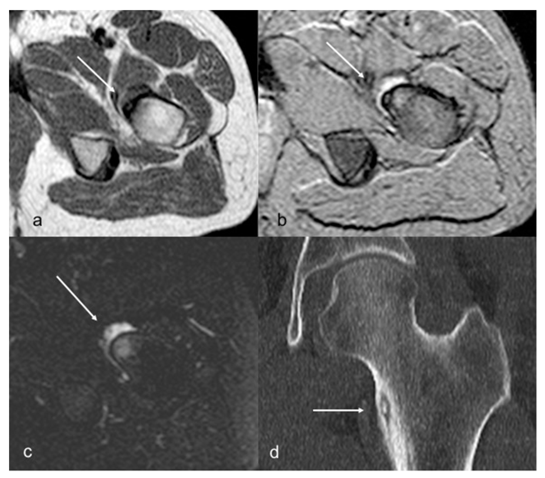
3.1. Case Series
3.2. Literature review
4. Discussion
5. Conclusions
Author Contributions
Funding
Institutional Review Board Statement
Informed Consent Statement
Data Availability Statement
Conflicts of Interest
References
- Jaffe, H.L. Osteoid-Osteoma. Proc. R. Soc. Med. 1953, 46, 1007–1012. [Google Scholar] [CrossRef]
- Kattapuram, S.V.; Kushner, D.C.; Phillips, W.C.; Rosenthal, D.I. Osteoid Osteoma: An Unusual Cause of Articular Pain. Radiology 1983, 147, 383–387. [Google Scholar] [CrossRef] [PubMed]
- Pikoulas, C.; Mantzikopoulos, G.; Thanos, L.; Passomenos, D.; Dalamarinis, C.; Glampedaki-Dagianta, K. Unusually Located Osteoid Osteomas. Eur. J. Radiol. 1995, 20, 120–125. [Google Scholar] [CrossRef] [PubMed]
- Allen, S.D.; Saifuddin, A. Imaging of Intra-Articular Osteoid Osteoma. Clin. Radiol. 2003, 58, 845–852. [Google Scholar] [CrossRef]
- O’Connell, J.X.; Nanthakumar, S.S.; Nielsen, G.P.; Rosenberg, A.E. Osteoid Osteoma: The Uniquely Innervated Bone Tumor. Mod. Pathol. 1998, 11, 175–180. [Google Scholar]
- Bottner, F.; Roedl, R.; Wortler, K.; Grethen, C.; Winkelmann, W.; Lindner, N. Cyclooxygenase-2 Inhibitor for Pain Management in Osteoid Osteoma. Clin. Orthop. Relat. Res. 2001, 393, 258–263. [Google Scholar] [CrossRef] [PubMed]
- Ciabattoni, G.; Tamburrelli, F.; Greco, F. Increased Prostacyclin Biosynthesis in Patients with Osteoid Osteoma. Eicosanoids 1991, 4, 165–167. [Google Scholar]
- Szendroi, M.; Köllo, K.; Antal, I.; Lakatos, J.; Szoke, G. Intraarticular Osteoid Osteoma: Clinical Features, Imaging Results, and Comparison with Extraarticular Localization. J. Rheumatol. 2004, 31, 957–964. [Google Scholar]
- Corrêa, D.G.; Costa, F.M. Osteoid Osteoma Mimicking Arthritis. J. Clin. Rheumatol. 2023, 29, e1–e2. [Google Scholar] [CrossRef]
- Breda, L.; Sassano, G.; La Bella, S.; Gentile, C.; Sansone, L.; Chiarelli, F. Intra-Articular Osteoid Osteoma Mimicking Juvenile Psoriatic Dactylitis. Clin. Exp. Rheumatol. 2021, 40, 1058. [Google Scholar] [CrossRef]
- Abd El Baky, H.; Thomas, R.D.; Kuechle, J.; Abdul-Aziz, R. Acetabula Osteoid Osteoma Mimicking Juvenile Idiopathic Arthritis and Chronic Recurrent Multifocal Osteomyelitis. Case Rep. Rheumatol. 2020, 8810735. [Google Scholar] [CrossRef] [PubMed]
- Vlaic, J.; Lamot, L.; Simunic, S.; Harjacek, M.; Bojic, D. Unusual Localization and Presentation of Osteoid Osteoma Mimicking Juvenile Spondyloarthritis: A Case Report. BMC Musculoskelet. Disord. 2019, 20, 17. [Google Scholar] [CrossRef] [PubMed]
- Cakar, E.; Durmus, O.; Kiralp, M.Z.; Dincer, U. An Unusual Case of Osteoid Osteoma Misdiagnosed as Inflammatory Joint Disease and Complex Regional Pain Syndrome I. Acta Reum. Port. 2009, 34, 670–671. [Google Scholar]
- Zeng, H.; He, H.; Tong, X.; Wang, Z.; Luo, R.; Liu, Q. Osteoid Osteoma of the Proximal Femur: Pitfalls in Diagnosis and Performance of Open Surgical Resection. Front. Surg. 2022, 9, 922317. [Google Scholar] [CrossRef]
- Knežević, I.; Bojanić, I. Comparison of Arthroscopy versus Percutaneous Radiofrequency Thermal Ablation for the Management of Intra- and Juxta-Articular Elbow Osteoid Osteoma: Case Series and a Literature Review. BMC Musculoskelet. Disord. 2022, 23, 287. [Google Scholar] [CrossRef] [PubMed]
- Traore, S.Y.; Dumitriu, D.I.; Docquier, P.-L. Intra-Articular Osteoid Osteoma Mimicking Juvenile Arthritis. Case Rep. Orthop. 2014, 2014, 912609. [Google Scholar] [CrossRef]
- Albisinni, U.; Bazzocchi, A.; Bettelli, G.; Facchini, G.; Castiello, E.; Cavaciocchi, M.; Battista, G.; Rotini, R. Treatment of Osteoid Osteoma of the Elbow by Radiofrequency Thermal Ablation. J. Shoulder Elb. Surg. 2014, 23, e1–e7. [Google Scholar] [CrossRef]
- Filippiadis, D.K.; Velonakis, G.; Kostantos, C.; Kouloulias, V.; Brountzos, E.; Kelekis, N.; Kelekis, A. Computed Tomography-Guided Radiofrequency Ablation of Intra-Articular Osteoid Osteoma: A Single Centre’s Experience. Int. J. Hyperth. 2017, 33, 670–674. [Google Scholar] [CrossRef]
- Georgoulis, A.D.; Soucacos, P.N.; Beris, A.E.; Xenakis, T.A. Osteoid Osteoma in the Differential Diagnosis of Persistent Joint Pain. Knee Surg. Sport. Traumatol. Arthrosc. 1995, 3, 125–128. [Google Scholar] [CrossRef]
- Payo-Ollero, J.; Moreno-Figaredo, V.; Llombart-Blanco, R.; Alfonso, M.; San Julián, M.; Villas, C. Osteoid Osteoma in the Ankle and Foot. An Overview of 50 Years of Experience. Foot Ankle Surg. 2021, 27, 143–149. [Google Scholar] [CrossRef]
- Cassar-Pullicino, V.N.; Mccall, I.W.; Wan, S. Intra-Articular Osteoid Osteoma. Clin. Radiol. 1992, 45, 153–160. [Google Scholar] [CrossRef] [PubMed]
- Goldberg, V.M.; Jacobs, B. Osteoid Osteoma of the Hip in Children. Clin. Orthop. Relat. Res. 1975, 106, 41–47. [Google Scholar] [CrossRef] [PubMed]
- May, C.J.; Bixby, S.D.; Anderson, M.E.; Kim, Y.J.; Yen, Y.-M.; Millis, M.B.; Heyworth, B.E. Osteoid Osteoma About the Hip in Children and Adolescents. J. Bone Jt. Surg. 2019, 101, 486–493. [Google Scholar] [CrossRef] [PubMed]
- Peyser, A.; Applbaum, Y.; Simanovsky, N.; Safran, O.; Lamdan, R. CT-Guided Radiofrequency Ablation of Pediatric Osteoid Osteoma Utilizing a Water-Cooled Tip. Ann. Surg. Oncol. 2009, 16, 2856–2861. [Google Scholar] [CrossRef]
- Song, M.H.; Yoo, W.J.; Cho, T.-J.; Chung, C.Y.; Park, M.S.; Cheon, J.-E.; Choi, I.H. Clinical and Radiological Features and Skeletal Sequelae in Childhood Intra-/Juxta-Articular versus Extra-Articular Osteoid Osteoma. BMC Musculoskelet. Disord. 2015, 16, 3. [Google Scholar] [CrossRef]
- Cohen, M.D.; Harrington, T.M.; Ginsburg, W.W. Osteoid Osteoma: 95 Cases and a Review of the Literature. Semin. Arthritis Rheum. 1983, 12, 265–281. [Google Scholar] [CrossRef]
- Germann, T.; Weber, M.-A.; Lehner, B.; Kintzele, L.; Burkholder, I.; Kauczor, H.-U.; Rehnitz, C. Intraarticular Osteoid Osteoma: MRI Characteristics and Clinical Presentation before and after Radiofrequency Ablation Compared to Extraarticular Osteoid Osteoma. Rofo 2020, 192, 1190–1199. [Google Scholar] [CrossRef]
- Carneiro, B.C.; Da Cruz, I.A.N.; Ormond Filho, A.G.; Silva, I.P.; Guimarães, J.B.; Silva, F.D.; Nico, M.A.C.; Stump, X.M.G.R.G. Osteoid osteoma: The great mimicker. Insights Imaging 2021, 12, 32. [Google Scholar] [CrossRef]
- Pan, X.; Qiao, J.; Liu, Z.; Sun, X.; Zhu, Z. Spontaneous correction of scoliosis after curettage of spinal osteoid osteoma: How and when? J. Orthop. Surg. Res. 2022, 17, 534. [Google Scholar] [CrossRef]
- Civino, A.; Bovis, F.; Ponzano, M.; Alighieri, G.; Prete, E.; Sorrentino, S.; Magni-Manzoni, S.; Vinti, L.; Romano, M.; Santoro, N.; et al. Development and Initial Validation of the ONCOREUM Score to Differentiate Childhood Cancer with Arthropathy from Juvenile Idiopathic Arthritis. J. Pediatr. 2023, 253, 219–224.e3. [Google Scholar] [CrossRef]
- Duman, İ.; Aydemir, K.; Tan, A.K.; Dinçer, K.; Kalyon, T.A. An Unusual Case of Osteoid Osteoma Clinically Mimicking Sacroiliitis. Clin. Rheumatol. 2007, 26, 1158–1160. [Google Scholar] [CrossRef]
- Hosalkar, H.S.; Garg, S.; Moroz, L.; Pollack, A.; Dormans, J.P. The Diagnostic Accuracy of MRI versus CT Imaging for Osteoid Osteoma in Children. Clin. Orthop. Relat. Res. 2005, 433, 171–177. [Google Scholar] [CrossRef] [PubMed]
- Woods, E.R.; Martel, W.; Mandell, S.H.; Crabbe, J.P. Reactive Soft-Tissue Mass Associated with Osteoid Osteoma: Correlation of MR Imaging Features with Pathologic Findings. Radiology 1993, 186, 221–225. [Google Scholar] [CrossRef] [PubMed]
- Campanacci†, M.; Ruggieri, P.; Gasbarrini, A.; Ferraro, A.; Campanacci, L. Osteoid Osteoma: Direct visual identification and intralesional excision of the nidus with minimal removal of bone. J. Bone Jt. Surgery. Br. Vol. 1999, 81-B, 814–820. [Google Scholar] [CrossRef]
- Ghanem, I. The Management of Osteoid Osteoma: Updates and Controversies. Curr. Opin. Pediatr. 2006, 18, 36–41. [Google Scholar] [CrossRef]
- Rosenthal, D.I.; Hornicek, F.J.; Torriani, M.; Gebhardt, M.C.; Mankin, H.J. Osteoid Osteoma: Percutaneous Treatment with Radiofrequency Energy. Radiology 2003, 229, 171–175. [Google Scholar] [CrossRef]
- Nourissat, G.; Kakuda, C.; Dumontier, C. Arthroscopic Excision of Osteoid Osteoma of the Elbow. Arthrosc. J. Arthrosc. Relat. Surg. 2007, 23, 799.e1–799.e4. [Google Scholar] [CrossRef] [PubMed]


| Patient No. | Gender | Age at Diagnosis (Years) | Location | Symptoms at Onset | Night Pain | Pain with Activities | Relief with NSAIDs | Joint Swelling | Limitation of Motion | Time to Diagnosis (Months) | Initial Misdiagnosis | Therapy |
|---|---|---|---|---|---|---|---|---|---|---|---|---|
| 1 | F | 13 | right femoral neck, intra-articular, subperiosteal | pain, anterior thigh | no | no | N/A | - | yes | 2 | no | CT-guided RFA |
| 2 | F | 10 | right femoral neck, intra-articular, cortical | recurrent pain, right hip and knee | yes | no | yes | - | yes | 24 | recurrent hip synovitis | N/A |
| 3 | M | 18 | left femoral neck, juxta-articular subcortical | pain, left hip | yes | no | yes | - | yes | 5 | ileo-psoas enthesopathy | N/A |
| 4 | F | 15 | left acetabulum, intra-articular cortical | pain, left hip | no | yes | no | - | yes | 3 | no | N/A |
| 5 | F | 18 | right tibia, upper third, juxta-articular subcortical | pain, right knee | no | yes | yes | no | yes | 1 | arthritis | observation |
| 6 | M | 17 | left tibia, upper third, juxta-articular subcortical | pain, left knee | yes | no | yes | no | no | 8 | no | CT-guided RFA |
| 7 | F | 15 | right distal fibula, juxta-articular cortical | pain, right knee and leg | yes | yes | yes | no | no | 4 | amplification pain syndrome | N/A |
| 8 | F | 14 | right humerus, olecranon fossa, intra-articular cortical | pain, right elbow | yes | yes | yes | yes | yes | 9 | post-traumatic arthritis | CT-guided RFA |
| 9 | M | 15 | right second toe, distal phalanx, juxta-articular cortical | swelling and pain, second toe | yes | no | yes | yes | no | 12 | psoriatic arthritis | surgery resection |
| 10 | F | 5 | pedicle L1, intra-articular cortical | back pain, scoliosis | yes | yes | no | - | yes | 2 | traumatic vertebral fracture | surgery resection |
| Study, Year | Sample (Patients ≤ 18 Years Old) | Average Age at Diagnosis (Years) | Location | Night Pain | Relief with NSAIDs | Joint Swelling | Limitation of Motion | Average Time to Diagnosis (Years) | Arthritis as Misdiagnosis |
|---|---|---|---|---|---|---|---|---|---|
| Pikoulas et al., 1995 [3] | 6 | 15 | femoral neck (2/6), tibia (1/6), acetabulum (2/6), acromion (1/6) | 2/6 | 5/6 | 0/6 | 1/6 | 1 | N/A |
| Szendroi et al., 2004 [8] | 8 | 15 | (1/11), calcaneus (1/11) | 1/8 | 6/8 | N/A | 8/8 | 1,2 | N/A |
| Knezevic et al., 2022 [15] | 4 | 13 | olecranon fossa (4) | 2/4 | 3/4 | 2/4 | 3/4 | 1,3 | 3/4 |
| Traore et al., 2014 [16] | 4 | 11 | talus (4/8), acetabulum (2/8), femur (2/8) | 2/4 | 1/4 | 1/4 | 4/4 | 0,4 | 4/4 |
| Albisinni et al., 2014 [17] | 4 | 14 | talus (1/4), femoral neck (3/4) | N/A | N/A | N/A | N/A | 1,1 | N/A |
| Filippiadis et al., 2017 [18] | 6 | 12 | hip (5/6), foot (1/6) | N/A | N/A | N/A | 6/6 | N/A | N/A |
| Georgoulis et al., 1995 [19] | 9 | 15 | femur (6/9), lumbar spine (1/9), carpus (1/9), patella (1/9) | N/A | 9/9 | N/A | N/A | 1,6 | 2/9 |
| Payo-Ollero et al., 2019 [20] | 5 | 14 | calcaneus (4/5), talus (1/5) | 5/5 | 5/5 | 0/5 | 0/5 | 1,8 | 1/5 |
| Cassar-Pullicino et al., 1992 [21] | 4 | 13 | femur (1/4), talus (1/4), ulna (1/4), MTF (1/4) | N/A | 1/4 | 3/4 | 4/4 | 1,3 | 2/4 |
| Goldberg et al., 1975 [22] | 31 | N/A | femur (30), acetabulum (1) | 19/31 | 21/31 | N/A | 25/31 | 1,7 | 7/31 |
| May et al., 2019 [23] | 50 | 12 | femur (43), acetabulum (7) | 45/50 | 43/50 | N/A | 17/50 | 0,6 | 2/50 |
| Peyser et al., 2009 [24] | 5 | N/A | femur (3/5), calcaneus (1/5), talus (1/5) | N/A | N/A | N/A | N/A | N/A | N/A |
| Song et al., 2015 [25] | 11 | 10 | femur (8/11), prox humerus (1/11), dist humerus | 4/11 | 4/11 | N/A | 8/11 | 1 | 2/11 |
Disclaimer/Publisher’s Note: The statements, opinions and data contained in all publications are solely those of the individual author(s) and contributor(s) and not of MDPI and/or the editor(s). MDPI and/or the editor(s) disclaim responsibility for any injury to people or property resulting from any ideas, methods, instructions or products referred to in the content. |
© 2023 by the authors. Licensee MDPI, Basel, Switzerland. This article is an open access article distributed under the terms and conditions of the Creative Commons Attribution (CC BY) license (https://creativecommons.org/licenses/by/4.0/).
Share and Cite
Civino, A.; Diomeda, F.; Giordano, L.; Damasio, M.B.; Perrone, S.; Gallizzi, R.; Ravelli, A.; Piscitelli, P.; Maggio, M.C. Intra- and Juxta-Articular Osteoid Osteoma Mimicking Arthritis: Case Series and Literature Review. Children 2023, 10, 829. https://doi.org/10.3390/children10050829
Civino A, Diomeda F, Giordano L, Damasio MB, Perrone S, Gallizzi R, Ravelli A, Piscitelli P, Maggio MC. Intra- and Juxta-Articular Osteoid Osteoma Mimicking Arthritis: Case Series and Literature Review. Children. 2023; 10(5):829. https://doi.org/10.3390/children10050829
Chicago/Turabian StyleCivino, Adele, Federico Diomeda, Luca Giordano, Maria Beatrice Damasio, Sandra Perrone, Romina Gallizzi, Angelo Ravelli, Prisco Piscitelli, and Maria Cristina Maggio. 2023. "Intra- and Juxta-Articular Osteoid Osteoma Mimicking Arthritis: Case Series and Literature Review" Children 10, no. 5: 829. https://doi.org/10.3390/children10050829