Article

Impact of Biogenic Ag-Containing Nanoparticles on Germination Rate, Growth, Physiological, Biochemical Parameters, and Antioxidants System of Tomato (Solanum tuberosum L.) In Vitro

by
Abdalrhaman M. Salih
*,
Ahmed A. Qahtan
,
Fahad Al-Qurainy
and
Bander M. Al-Munqedhi
Botany and Microbiology Department, College of Science King Saud University, P.O. Box 2455, Riyadh 11451, Saudi Arabia
*
Author to whom correspondence should be addressed.
Processes 2022, 10(5), 825; https://doi.org/10.3390/pr10050825
Submission received: 3 March 2022 / Revised: 14 April 2022 / Accepted: 19 April 2022 / Published: 22 April 2022
(This article belongs to the Special Issue Green Synthesis of Metallic Nanomaterials and Their Applications)

Abstract

:
Tomatoes (Solanum tuberosum L.) are considered one of the most effective and nutritious foods in the human diet. Moreover, the fruit of a tomato is rich in phytochemical compounds such as carotenoids, vitamins, and phenolics which are beneficial to human health. The objective of this present research was to investigate the impact of biogenic Ag-containing nanoparticles on seed germination rate and germination speed index, the development of the stem and root system, and physio-biochemical parameters. Ag-containing nanoparticles were synthesized biologically using a silver nitrate solution and aqueous seed extract of Juniperus procera, which acted as a capping and reducing agent. The impact of different concentrations (0.0, 2.5, 5, 10, and 25 mg/L) of Ag-containing NPs on seed germination rate, biomass accumulation, phenolic compounds, total protein, enzymes activity, and total soluble sugar of tomatoes (Solanum tuberosum L.) in vitro has been tested. The obtained result demonstrated that Ag-containing nanoparticles have a significant impact on tomato seed germination rate, germination speed index, and the development of stem and root systems. As well as photosynthetic pigments, total protein, enzymes activity, phenolic compounds, and total soluble sugar. We concluded that Ag-containing NPs can be a promising nano-fertilizer for improving crop growth and production.

1. Introduction

Tomatoes (Solanum lycopersicum L.) are widely distributed as a vegetable crop consumed in a different form. It is nutrient-enriched containing proteins, fibres, carbohydrates, calories, water, a large number of vitamins, and 37 minerals [1]. Moreover, tomatoes are the second most important vegetable or fruit crop next to potatoes (Solanum tuberosum L.). About 182.3 million tons of tomato fruits are produced on 4.85 million hectares each year [2]. Additionally, tomatoes are considered one of the most effective and nutritious foods in the human diet. The fruit of a tomato is rich in phytochemical compounds such as carotenoids, vitamins, and phenolic compounds. These compounds have high antioxidant activity which is beneficial to human health [3]. Nanotechnology is described as a technology related to material, systems, and processes that operates on a scale of 100 nm. Moreover, nanotechnology can be used as an alternative technology in the different fields of science [4]. Furthermore, nanotechnology has played a vital role in modern agriculture and has become a promising technology with incredible potential to resolve the deficit in plant nutrition. It has been reported that nano-fertilizer can be used as macro or micronutrients with different application methods. This provides a slow and sustainable nutrient release boosting the maximum nutrient utilization with a comparatively higher production. Thus, it has increasingly been used to enhance crop productivity [5,6,7]. Silver nanoparticles (AgNPs) are used as a nutrient for plants due to their many unique properties which can be used to increase the nutrient uptake efficiency of plants. The AgNPs provide an improved uptake of nutrients from the soil [7]. In context, it has been stated that AgNPs could be used to boost seed germination in many plants [8]. Additionally, it has been reported that exposure of a plant to silver nanoparticles can affect plant physiological, biochemical, and metabolic reactions. Therefore, AgNPs alter plant growth, water and nutrient absorption, respiration, photosynthesis, and transpiration rate, production of reactive oxygen species (ROS), and antioxidant responses [9,10,11]. Nanoparticles provide nutrients to plants in an available form, thus increasing the uptake of nutrients by plants, and boosting crop productivity [12]. According to [13], 25 ppm of AgNPs gives a significant improvement in wheat vegetative growth as well as grain productivity. Additionally, a recent study [14] has reported that 20 ppm of silver nanoparticles improved the morphological traits and yield of onions (Allium cepa L.) However, only a few studies investigated the role of AgNPs on the growth and development of crop plants [15]. Hence, the main objective of this research was to investigate the impact of biogenic Ag-containing NPs on germination rate, germination speed index, growth development, bioactive compounds, and antioxidant system of tomato crop as nano-fertilizer. Ag-containing NPs were biologically synthesized using an aqueous seed extract of Juniperus procera as a reducing and capping agent and silver nitrate solution. The impact of biosynthesized Ag-containing nanoparticles on tomato development was tested by estimating germination rate, germination speed index, photosynthetic pigments, and enzymatic and non-enzymatic antioxidants. We concluded that biogenic Ag-containing nanoparticles have a positive impact on tomato crop development. Additionally, this study could provide a potential alternative fertilizer that would improve crop productivity with a higher nutrition value. Further, to better understand the impact of Ag-containing nanoparticles on crop development, gene expression should be performed.

2. Materials and Methods

2.1. Biosynthesis of Ag-Containing NPs

Ag-containing NPs were synthesized biologically using seeds of Juniperus procera and a silver nitrate solution, following the method described by [16] with light modifications. First, 50 mL of 1 mm solution of silver nitrate was added to 100 mL of seed extract (1:2) (v/v). Next, it was heated to minimize photo-activation of the silver nitrate.

2.2. Characterization of Ag-Containing NPs

Different techniques have been used for the characterization of biosynthesized silver nanostructure. The optical absorption spectrum of Biosynthesized Ag-containing NPs (AgNPs) was measured by UV-visible spectrophotometer in the range of 200 to 800 nm. For FTIR patterns of the biosynthesized Ag-containing NPs (AgNPs), a Fourier transmission infrared spectrometer (Perkin Elmer, Norwalk, CT, USA) was used at 4000–400 cm−1, whereas a scanning electron microscope (SEM) was used to measure the morphology features and particle size of biosynthesized Ag-containing NPs.

2.3. Media Preparation and AgNP Treatment

Murashige and Skoog Media (MS) were used with a supplement of sucrose (30 g/L), which was used as a source of carbon and agar (7 g/L), and the pH was adjusted to be (5.7). Different concentrations (0.0, 2.5, 5, 10, and 25 mg/L) of AgNPs were added to the plant media before autoclaving at 121 °C for 20 min. Next, certified tomato seeds were cultured into MS Media under controlled conditions. The experiment was done into triplicate with 10 explants per jar. Next, it was incubated in a growth chamber for 45 days at 25 °C ± 1, with 14/10 h illumination periods.

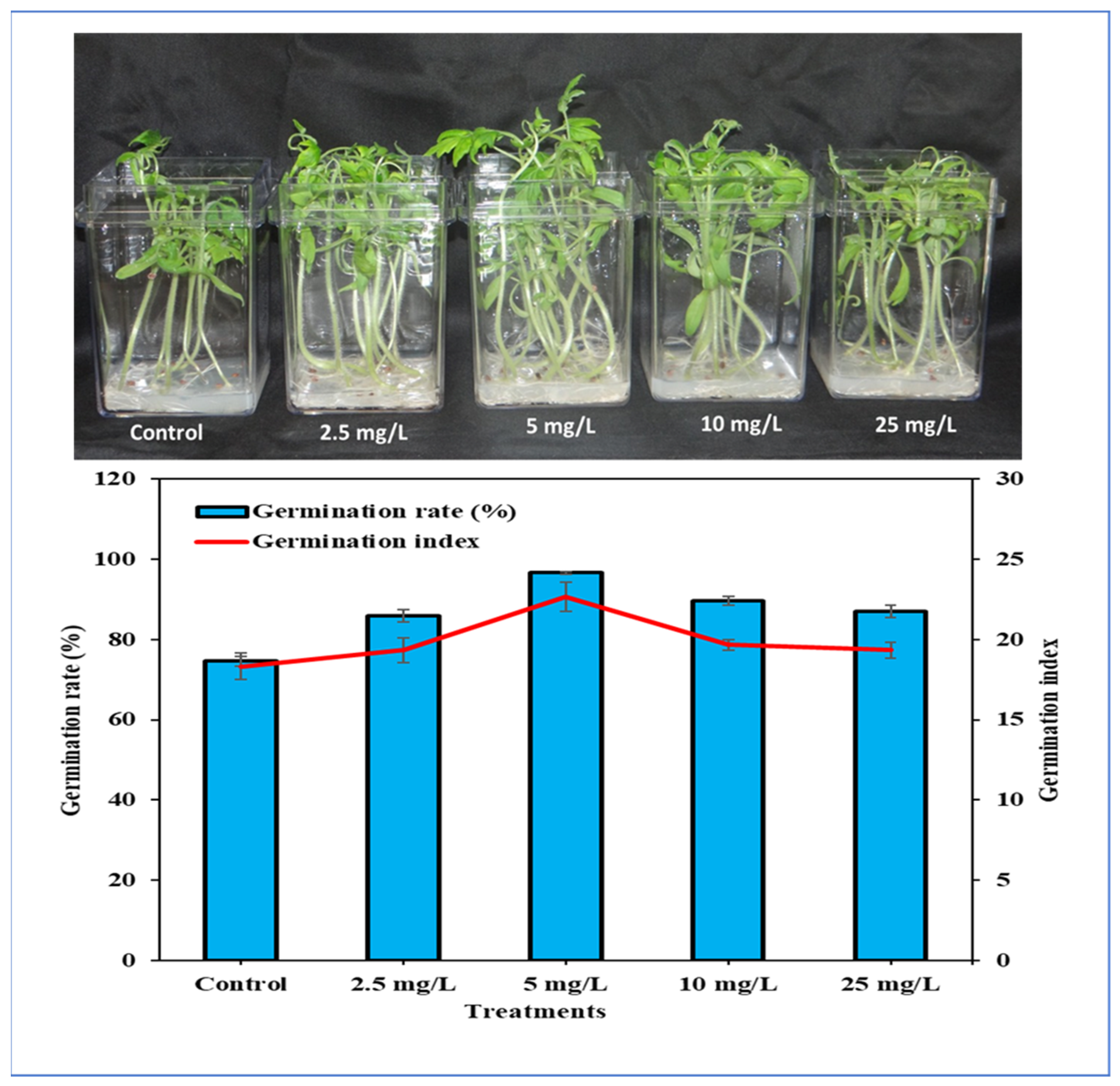
2.4. Seed Germination Assay

2.4.1. Seed Germination Rate (%)

The seed germination percentage (GP) was calculated using the following equation:
GP (%) = (n/N) × 100
where n is the total number of germinated seeds at the end of the experiment and N = the total number of seeds cultivated for the germination test [17].

2.4.2. Germination Index (GI)

The germination index (GI) was determined using the method described by [18].
(7 × n1) + (6 × n2) + ⋯ + (1 × n10)
where n1, n2, n3, etc., are the number of germinated seeds on days 1, 2, 3, etc.

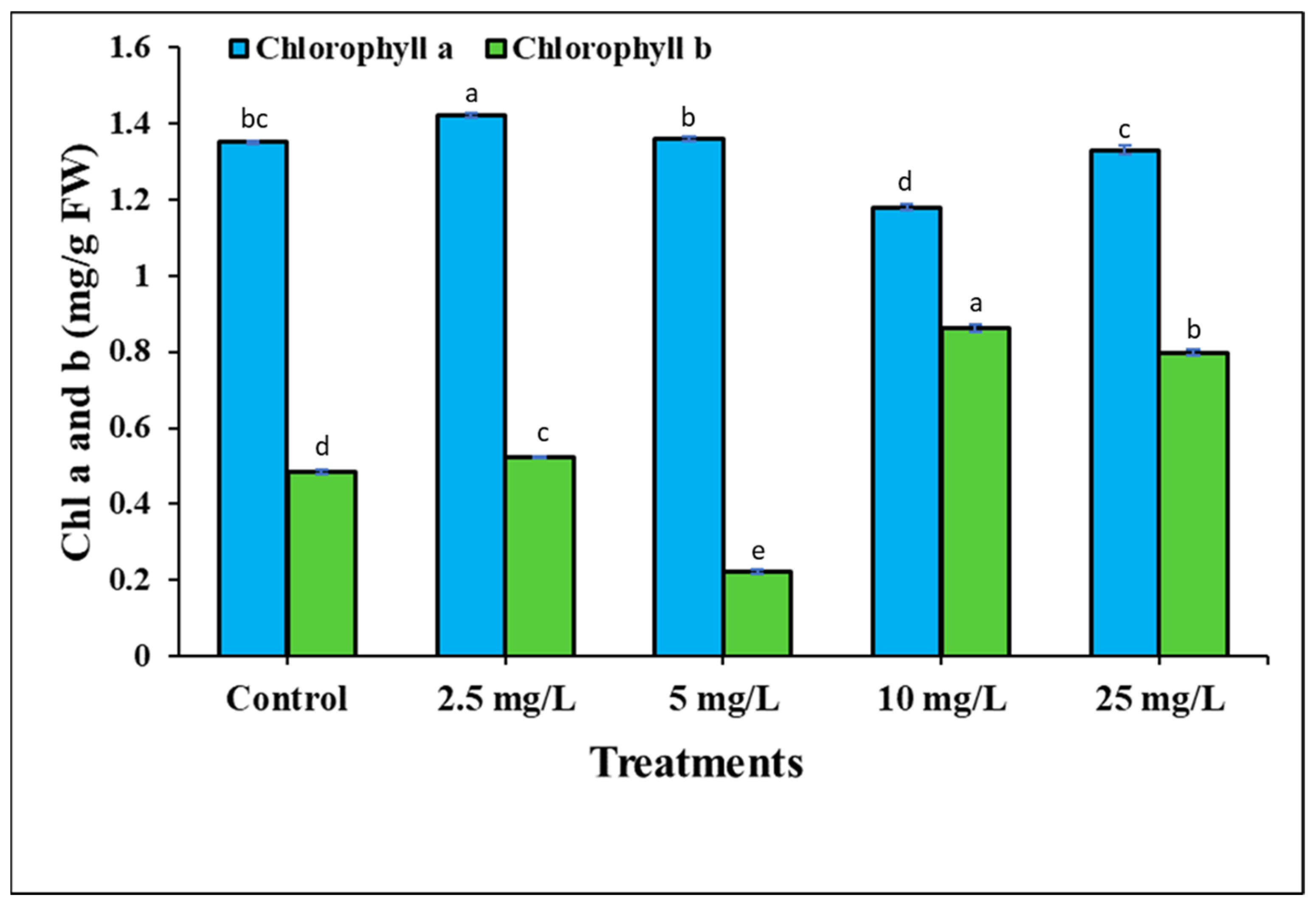
2.5. Photosynthetic Pigments Estimation

The content of photosynthetic pigments a and b in the tomato leaves was estimated using the Arnon method [19]. Acetone (80%) (v/v) was used for pigment extraction; 100 mg of fresh leaves was homogenized in 2 mL acetone solution 80% and stored at 4 °C for 24 h. Next, the mixture was centrifuged for 15 min at 20,000× g, Further, supernatant was collected and stored at 4 °C. The wavelength of the pigments was measured at 663 and 645 nm using a UV-Vis spectrophotometer.

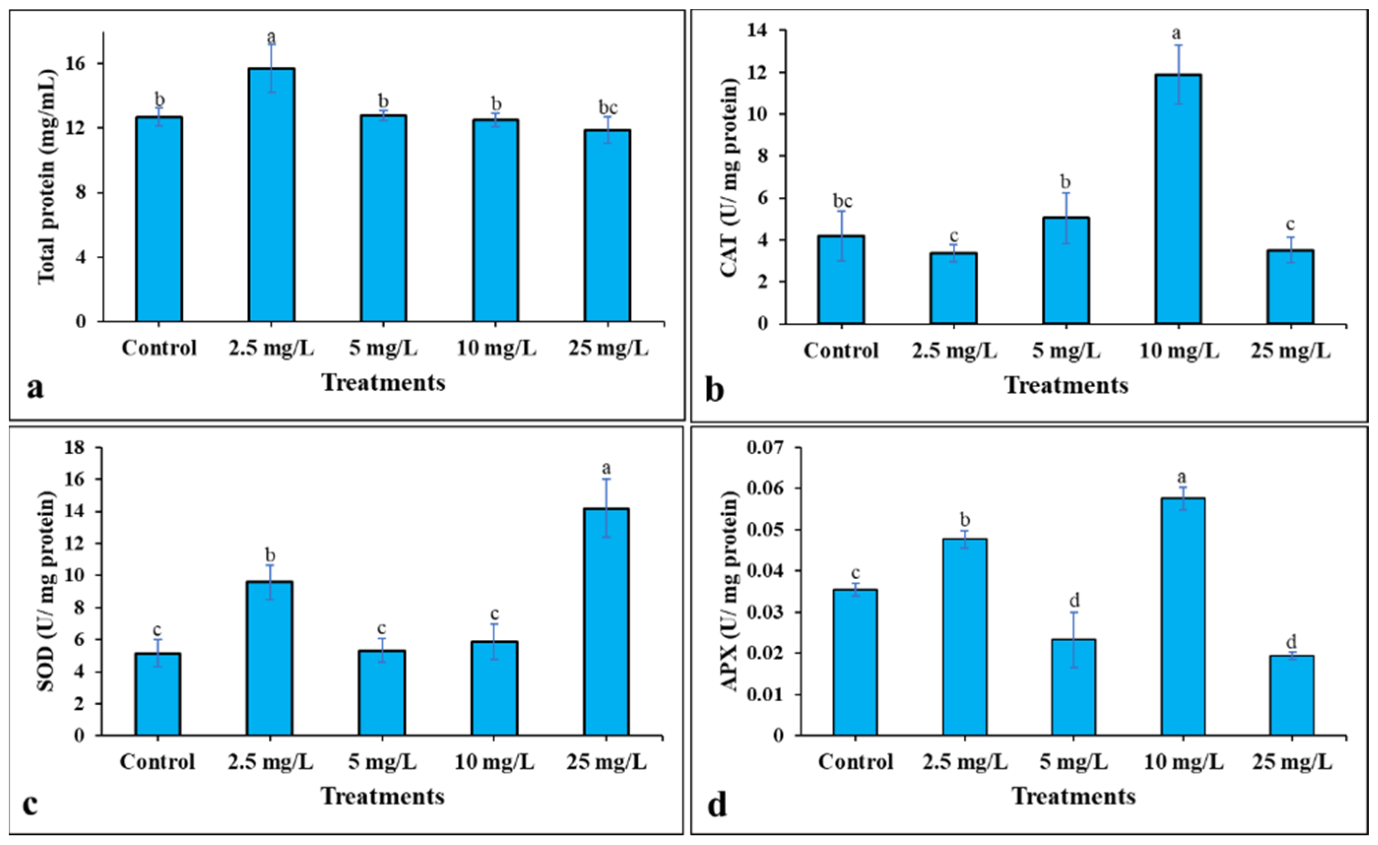
2.6. Antioxidant Enzymes Activity Estimation

2.6.1. Total Protein Content Estimation

Plant material was grounded and homogenized using liquid nitrogen and dissolved in a mixture consisting of 100 mm of sodium phosphate buffer (pH 7.0) containing 1% PVP and 0.5% Triton X-100 (v/v). Next, the mixture was centrifuged for 15 min at 20,000× g at 4 °C. The supernatant was collected, while the total protein was estimated using a Nanodrop following the method in [20].

2.6.2. Superoxide Dismutase Estimation

The superoxide dismutase (SOD, EC 1.15.1.1) activity was estimated following the Marklund and Marklund [21] method. The mixture of reaction contained 1.0 mL of 0.25 mm pyrogallol, 1.9 mL of 0.1 M sodium phosphate buffer (pH 7.0), and 100 μL of extracted protein. Next, the wavelength was recorded at 420 nm. The SOD activity (U g − 1 protein) was calculated as the amount of enzyme needed for 50% inhibition of pyrogallol oxidation.

2.6.3. Catalase Estimation

The catalase (CAT, EC 1.11.1.6) activity was estimated following the method in [22]. The reaction mixture contained 1 mL of 0.059 M H2O2 in 0.1 M phosphate buffer (pH 7.0), 1.9 mL of Milli-Q water, and 100 μL of extracted protein, while the wavelength was quantified at 240 nm. The catalase activity was expressed as unit g−1 of protein.

2.6.4. Ascorbate Peroxidase Estimation

The activity of ascorbate peroxidase (EC 1.11.1.11) was determined according to [23]. The reaction mixture contained 1 mL of 0.1 M sodium phosphate buffer (pH 7.4), 1 mL Milli-Q water, 100 µL ETDA (0.1 mm), 100 µL of H2O2, and 100 µL extracted protein. The wavelength was recorded at 290 nm.

2.7. Determination of Phenolic Compounds

2.7.1. Determination of Total Phenolic Content (TPC)

The total phenolic content was estimated using the Ainsworth [24] method. First, 0.1 mL of the plant extracted material was mixed with 100 µL of the Folin–Ciocalteu reagent and 300 µL of sodium carbonate solution (20%). Next, the mixture was incubated in the dark at room temperature for 30 min. The wavelength was measured at 765 nm using UV–visible spectrophotometer. TPC was estimated from the linear equation (y = 0.0014x + 0.0114 with R2 = 0.999) based on the standard curve prepared with gallic acid (25–400 µg/mL). The TPC was expressed as mg/g gallic acid equivalent (GAE) of dry weight.

2.7.2. Determination of Total Flavonoid Content (TFC)

The estimation of the total flavonoid content was carried out using the method in [25]. A volume of 0.5 mL of methanol extract and a volume of 0.5 mL of 2% AlCl3 water solution was mixed. After 2 h in the dark at room temperature, the wavelength was measured at 420 nm. The TFC was estimated from the calibration curve using the quercetin standard (50–400 µg/mL) and the following equation (y  =  0.0012x  +  0.0595) based on the calibration curve. TFC was expressed as quercetin (mg/g DW).

2.7.3. Determination of Total Tannin Content (TTC)

The total tannin content was measured by using the Folin–Ciocalteu method described by [26] with light modifications. A total of 100 µL of the extracted plant material was added to a tube (2 mL) containing 1.5 mL of deionized water and 100 µL of Folin–Ciocalteu phenol reagent for 8 min. Then, 300 µL of 35% sodium carbonate solution was added to the mixture. Next, the mixture was shaken well and kept in the dark, at room temperature for 20 min. The wavelength was recorded at 700 nm. TTC was estimated using the following equation (Y = 0.0054 − 0.0252 with R2 = 9937) based on the calibration curve prepared with tannic acid. TTC was expressed in terms of mg/g DW.

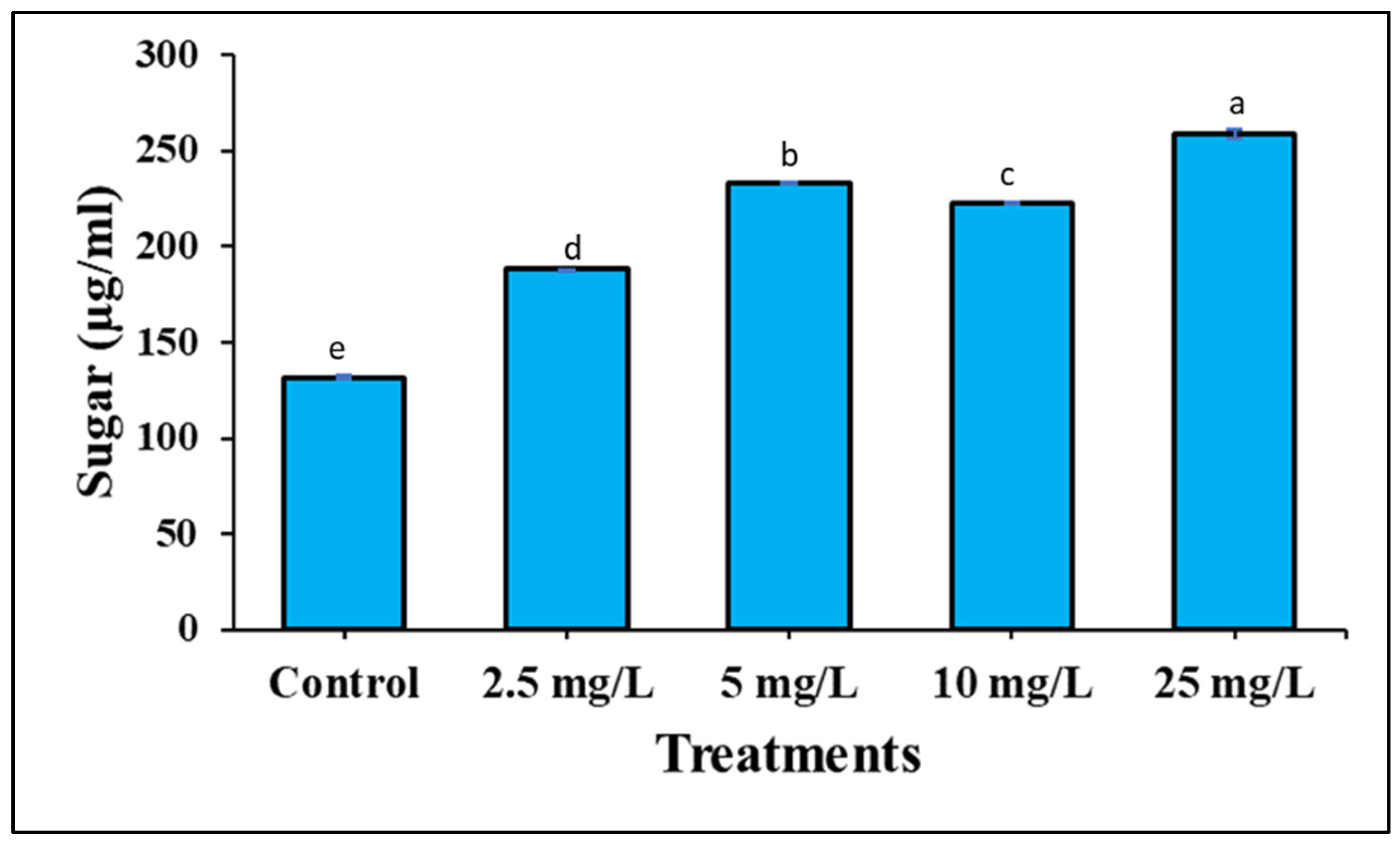
2.8. Estimation of Total Sugar

The total sugar content in the sample was estimated using the method described by [27]. First, 1 mL of phenol (5%) and 5 mL of sulphuric acid (96%) were added sequentially to the sample. The test tubes containing the reaction mixture were incubated for 20 min in a water bath set at 30 °C. The wavelength was recorded spectrophotometrically at 490 nm. Then, the total sugar was estimated following the equation (y = 10.334x − 0.0906 with R2 = 0.993) based on the calibration curve prepared with the glucose standard (0.025–0.3 µg/mL). The total soluble sugar content was defined as glucose (mg/g DW).

2.9. Statistical Analysis

The experiment was performed in triplicate, while the results which are reported in the tables and figures are the means value ± (SD) using SPSS, one-way ANOVA for estimated statistical significance differences at (p < 0.05).

3. Results and Discussion

3.1. Green Synthesis and Characterization of Ag-Containing Nanoparticles

Ag-containing NPs were synthesized biologically using an aqueous seed extract of Juniperus procera as a capping and reducing agent and silver nitrate solution. For the biosynthesis of Ag-containing NPs, 1 mm solution of silver nitrate was added to the aqueous seed extract (1:2) (v/v). Next, the mixture was incubated at 40 °C until the colour of the mixture changed from light yellowish to brown (Figure 1a). This changing in colour of the mixture indicated the reduction of Ag+ to Ag0 in the AgNO3 solution [28,29], which confirmed Ag ion reduction and the formation of silver nanoparticles [28,29]. This is the first sign and notable indication of the formation of silver nanoparticles [30]. Next, the biosynthesis of Ag-containing NPs was confirmed by various characterization techniques (UV, FTIR, and SEM) to ascertain the shape, size, morphology, and functionalization of nanoparticles. For UV-visible spectroscopy analysis, biosynthesized Ag-containing NPs were dissolved in deionized water and detected using a UV-visible spectrophotometer. The UV-visible spectrum showed that strong broad peaks were observed at 400 and 262 nm (Figure 1b) and no more major peak shifts were observed during the reactions and measurement. The peak at 400 nm indicated the appearance of Ag in the sample, while the band at 262 nm indicated the presence of bioactive compounds of seed extract in the mixed solution. Therefore, our findings are in agreement with [31], who reported that the UV spectra of AgNPs was found to be 400 nm. Additionally, it has been stated that the UV spectrum of Ag is in the range of 400–500 nm due to surface plasmon resonance (SPR) [32]. Moreover, UV spectroscopy is an appropriate approach to confirm the formation of silver nanoparticles [33]. FTIR analysis of biosynthesized Ag-containing NPs showed bands at 861.50, 1604.85, 1766.81, and 3313.57 cm (Figure 1c). The band shown at 861.50 cm is assigned to C=CH2, the peak at 1604.85 cm is attributed to N-H bend primary amines in protein, the peak at 1766.81 cm corresponds to carbonyl stretching vibrations, and the band at 3313.57 cm is obtained from O-H stretching H-bonded phenols and alcohols. Therefore, the FTIR result indicated that biogenic Ag-containing NPs were surrounded by bioactive compounds and protein. The morphology and size of biosynthesized Ag-containing NPs were investigated using a scanning electron microscope (SEM). The result has shown that biosynthesized Ag-containing NPs were spherical in shape and the average size of the particles was 100 nm (Figure 1d). However, energy-dispersive X-ray spectroscopy (EDX) was not performed in this study for elemental mapping or elements distribution of the nanoparticles (NPs). Nevertheless, the method which has been used in this research is reliable and proven for AgNPs synthesis [16,34,35]. On other hand, the biosynthesis of AgNPs was not fully investigated to prove that nanoparticles are composed entirely of silver. Therefore, the term Ag-containing nanoparticles has been used instead of silver nanoparticles.

3.2. The Effect of Ag-Containing NPs on Tomato Germination Rate, Growth and Development

Silver nanoparticles have unique chemical and physical properties and are now being intensively introduced to the agriculture field as inducers and nano-fertilizers. However, only a few studies have been conducted to show a beneficial impact of AgNPs on the growth and development of crop plants [15]. The exposure of plants to AgNPs can affect plant metabolic reactions by inducing plant growth, water and nutrient uptake, respiration, photosynthesis, and transpiration rate, production of reactive oxygen species (ROS), and antioxidant responses [9,10,11]. The optimum dose of nanoparticles is expected to improve crop productivity. Additionally, the usage of an optimum dose of nanoparticles might minimize the risk of the presence of nanoparticles in the plant environment [36]. Here, in this study, the impact of different concentrations of Ag-containing NPs (0.0, 2.5, 5, 10, 25 mg) on the seed germination index and germination rate was tested. In addition, their effects on stem and root development and physiological parameters were investigated. As we can see in the detailed results, the biosynthesized Ag-containing NP application had a significant impact on all traits including germination rate, germination speed index, and shoot and root system development. The obtained results show that Ag-containing NPs have a significant effect on tomato development (Figure 2). Among the different concentrations tested in this experiment, the 5 mg dose of Ag-containing NPs recorded the highest germination rate (96.7%), followed by 10 mg (89.67%), 25 mg (87%), 2.5 mg (86%), and control (74.67%) (Figure 2). It has been stated that germination rate was increased significantly in response to AgNPs [36], which is in agreement with our finding. On the other hand, recent studies have reported that a plant’s response to AgNPs, enhancement, or inhibition of growth, depends on the AgNP dose. The exposure of Arabidopsis thaliana plants to 1 or 2.5 mg/L of AgNPs increased seedling biomass [37]. Additionally, the germination speed index was calculated using the method described by [18], which revealed that 5 mg of biogenic Ag-containing NPs generated the highest value of germination index (22.67), followed by 10 mg (19.67), 25 mg (19.33), 2.5 mg (19.33), and control (18.33) (Figure 2). In agreement with our findings, [38] stated that the germination index (GI) and germination rate (GR) of the investigated seeds greatly increased in response to AgNPs compared to those untreated seeds. Silver nanoparticles can enhance germination index and seed germination rate with different scenarios. For example, AgNPs create small pores by interacting with the cell walls of the seed coat, thus enhancing water uptake by the seeds. The fast water uptake by seeds can induce higher metabolic activity and hydrolysis of starch during the initial phase of seed imbibition. Silver nanoparticles might interact with α-amylase as NPs-amylase complex to fasten the hydrolysis of starch, and thus, seeds can generate higher available sugars to support embryo development. In addition, AgNPs might mediate the generation of reactive oxygen species (ROS) which act as signalling molecules and participate in cell wall loosening and endosperm weakening [38]. Moreover, the effect of different concentrations of Ag-containing NPs on parameters such as stem and root length and fresh and dry weight were estimated. The recorded result showed that Ag-containing NPs have a significant effect compared to non-treated plants, as shown in Table 1. These morphological findings were supported by physiological analysis such as chlorophyll a and b, as well as enzymes activity. The result demonstrated that Ag-containing NPs have an impact on photosynthetic pigments (Figure 3). Moreover, total protein content and the activity of SOD, CAT and APX were up-regulated in response to Ag-containing NPs (Figure 4). The addition of AgNPs improved plant growth indices, biochemical parameters, and the activity of antioxidant enzymes in Brassica juncea, Zea mays, and Phaseolus vulgaris [17,39]. In addition to the morphological parameters, AgNPs have played an important role which increased plant height, fresh and dry weight, and the length of root and shoot of Vigna radiata, as reported by [40]. The improvement of plant growth response to AgNPs might be due to the action of ethylene being blocked [41]. Additionally, Syu et al. suggested that the effect of AgNPs on the morphological and physiological parameters of plants is related to the morphology of the nanoparticles that are used [42]. However, there are different views about the mechanism of action of silver nanoparticles to trigger the positive effect on plant growth. For example, some researchers suggested that nanoparticles enter the seed coat and have a useful impact on the seed germination processes. Additionally, nanoparticles might induce and increase water absorption by seeds [43].

3.3. The Effect of Ag-Containing NPs on Phenolic Compounds and Total Soluble Sugar

The application of nanomaterials is currently focused on the enhancement of bioactive compound production from plants using NPs as elicitors [44]. In this study, we explored the impact of different concentrations of biogenic Ag-containing NPs on phenolic compounds; the total phenolic content (TPC), total flavonoid content (TFC), and total tannin content (TTC) of tomato crops were investigated. The result demonstrated that Ag-containing NPs have a significant impact on phenolic compound production compared to non-treated plants, as shown in (Table 2). For TPC production, a dose of 5 mg of Ag-containing nanoparticles generated the highest value (281.7 mg GAE/g DW), while for TFC production, 2.5 mg registered a higher yield (127.8 (mg QE/g DW) of TFC, as well as for TTC production (76.94 mg TAE/g DW) (Table 2). In accordance, significant changes have been observed for five phenolic compounds under silver nanoparticle treatment [45]. Additionally, it was reported that silver nanoparticles could be an efficient elicitor to increase the phytochemical production [46]. Furthermore, the concentration of osmolytes, such as total soluble sugar, was also determined under different concentrations of Ag-containing nanoparticles. For soluble sugar production, 25 mg of Ag-containing NPs recorded the highest yield of soluble sugar (Figure 5). The total soluble sugar content of seedlings increased in response to AgNPs [38].

4. Conclusions

Silver nanoparticles have unique chemical and physical properties and are now being intensively introduced to the agriculture field as inducers and fertilizers. Ag-containing nanoparticles had a positive impact on the germination rate, germination speed index, stem and root system, and physio-biochemical traits indices compared to non-treated plants. This resulted in a higher germination rate, germination speed index, average of stem length, root length, photosynthetic pigments, total protein content, phenolic compounds, and enzyme activity. We concluded that the addition of biosynthesized silver nanoparticles to the plant media in vitro can play a vital role in enhancing plant growth and biomass production. Further, to better understand the impact of Ag-containing nanoparticles on crop development, gene expression should be done and the toxicity of Ag must be considered.

Author Contributions

A.M.S. and B.M.A.-M. proposed and planned the work; A.A.Q. performed the experiments; A.M.S. made the biogenic Ag-containing nanoparticles; A.A.Q., A.M.S. and B.M.A.-M. contributed to the methodology; A.A.Q. analysed the data and figures; A.M.S. wrote the full manuscript; F.A.-Q. was the supervisor of LAB. All authors have read and agreed to the published version of the manuscript.

Funding

This research was funded by (RSP-2021/73) at King Saud University, Riyadh, Saudi Arabia.

Institutional Review Board Statement

Not applicable.

Informed Consent Statement

Not applicable.

Data Availability Statement

Data are available from the corresponding author.

Acknowledgments

The authors extend their appreciation to researchers supporting project number (RSP-2021/73) at King Saud University, Riyadh, Saudi Arabia.

Conflicts of Interest

The authors declare no conflict of interest.

References

  1. Tanveer, K.; Gilani, S.; Hussain, Z.; Ishaq, R.; Adeel, M.; Ilyas, N. Effect of salt stress on tomato plant and the role of calcium. J. Plant Nutr. 2020, 43, 28–35. [Google Scholar] [CrossRef]
  2. FAOSTAT. Food and Agriculture Organization of the United Nations—Statistic Division. Available online: https://www.fao.org/faostat/en/#data (accessed on 15 April 2019).
  3. Muzolf-Panek, M.; Kleiber, T.; Kaczmarek, A. Effect of increasing manganese concentration in nutrient solution on the antioxidant activity, Vitamin C, lycopene and polyphenol contents of tomato fruit. Food Addit. Contam. Part A 2017, 34, 379–389. [Google Scholar] [CrossRef] [PubMed]
  4. Ditta, A. How Helpful is nanotechnology in Agricultural? Adv. Nat. Sci. Nanosci. Nanotechnol. 2012, 3, 033002. [Google Scholar] [CrossRef]
  5. Tarafder, C.; Daizy, M.; Alam, M.M.; Ali, M.R.; Islam, M.J.; Islam, R.; Ahommed, M.S.; Aly Saad Aly, M.; Khan, M.Z.H. Formulation of a hybrid nanofertilizer for slow and sustainable release of micronutrients. ACS Omega 2020, 5, 23960–23966. [Google Scholar] [CrossRef] [PubMed]
  6. Rop, K.; Karuku, G.N.; Mbui, D.; Njomo, N.; Michira, I. Evaluating the effects of formulated nano-NPK slow release fertilizer composite on the performance and yield of maize, kale and capsicum. Ann. Agric. Sci. 2019, 64, 9–19. [Google Scholar] [CrossRef]
  7. Anand, R.; Bhagat, M. Silver nanoparticles (AgNPs): As nanopesticides and nanofertilizers. MOJ Biol. Med. 2019, 4, 19–20. [Google Scholar] [CrossRef]
  8. Duhan, J.S.; Kumar, R.; Kumar, N.; Kaur, P.; Nehra, K.; Duhan, S. Nanotechnology: The new perspective in precision agriculture. Biotechnol. Rep. 2017, 15, 11–23. [Google Scholar] [CrossRef]
  9. Hossain, Z.; Mustafa, G.; Komatsu, S. Plant Responses to Nanoparticle Stress. Int. J. Mol. Sci. 2015, 16, 26644–26653. [Google Scholar] [CrossRef] [Green Version]
  10. Tripathi, D.K.; Tripathi, A.; Shweta; Singh, S.; Singh, Y.; Vishwakarma, K.; Yadav, G.; Sharma, S.; Singh, V.K.; Mishra, R.K.; et al. Uptake, Accumulation and Toxicity of Silver Nanoparticle in Autotrophic Plants, and Heterotrophic Microbes: A Concentric Review. Front. Microbiol. 2017, 8, 7. [Google Scholar] [CrossRef]
  11. Qian, H.; Peng, X.; Han, X.; Ren, J.; Sun, L.; Fu, Z. Comparison of the toxicity of silver nanoparticles and silver ions on the growth of terrestrial plant model Arabidopsis thaliana. J. Environ. Sci. 2013, 25, 1947–1956. [Google Scholar] [CrossRef]
  12. Guru, T.; Veronica, N.; Thatikunta, R.; Reddy, S. Crop nutrition management with nano fertilizers. Int. J. Environ. Sci. Technol. 2015, 1, 4–6. [Google Scholar]
  13. Jhanzab, H.M.; Razzaq, A.; Jilani, G.; Rehman, A.; Hafeez, A.; Yasmeen, F. Silver nano-particles enhance the growth, yield and nutrient use efficiency of wheat. Int. J. Agron. Agric. Res. 2015, 7, 15–22. [Google Scholar]
  14. Fouda, M.M.; Abdelsalam, N.R.; El-Naggar, M.E.; Zaitoun, A.F.; Salim, B.M.; Bin-Jumah, M.; Allam, A.; Abo-Marzoka, S.A.; Kandil, E.E. Impact of high throughput green synthesized silver nanoparticles on agronomic traits of onion. Int. J. Biol. Macromol. 2020, 149, 1304–1317. [Google Scholar] [CrossRef]
  15. Mehmood, A. Brief overview of the application of silver nanoparticles to improve growth of crop plants. IET Nanobiotechnol. 2018, 12, 701–705. [Google Scholar] [CrossRef] [PubMed]
  16. Ahmed, S.; Saifullah Ahmad, M.; Swami, B.L.; Ikram, S. Green synthesis of silver nanoparticles using Azadirachta indica aqueous leaf extract. J. Radiat. Res. Appl. Sci. 2016, 9, 1–7. [Google Scholar] [CrossRef] [Green Version]
  17. Hojjat, S.S.; Kamyab, M. The effect of silver nanoparticle on Fenugreek seed germination under salinity levels. Russ. Agric. Sci. 2017, 43, 61–65. [Google Scholar] [CrossRef]
  18. Benech Arnold, R.; Fenner, M.; Edwards, P. Changes in germinability, ABA content and ABA embryonic sensitivity in developing seeds of Sorghum bicolor (L.) Moench. induced by water stress during grain filling. New Phytol. 1991, 118, 339–347. [Google Scholar] [CrossRef]
  19. Arnon, D.I. Copper enzymes in isolated chloroplasts. Polyphenoloxidase in Beta vulgaris. Plant Physiol. 1949, 24, 1. [Google Scholar] [CrossRef] [Green Version]
  20. Jogeswar, G.; Pallela, R.; Jakka, N.M.; Reddy, P.S.; Rao, J.V.; Sreenivasulu, N.; Kishor, P.B.K. Antioxidative response in different sorghum species under short-term salinity stress. Acta Physiol. Plant. 2006, 28, 465–475. [Google Scholar] [CrossRef]
  21. Marklund, S.; Marklund, G. Involvement of the Superoxide Anion Radical in the Autoxidation of Pyrogallol and a Convenient Assay for Superoxide Dismutase. Eur. J. Biochem. 1974, 47, 469–474. [Google Scholar] [CrossRef]
  22. Claiborne, A. Catalase activity. In Methods for Oxygen Research; Greenwald, R.A., Ed.; CRC Press: Boca Raton, FL, USA, 1985. [Google Scholar]
  23. Nakano, Y.; Asada, K. Hydrogen Peroxide is Scavenged by Ascorbate-specific Peroxidase in Spinach Chloroplasts. Plant Cell Physiol. 1981, 22, 867–880. [Google Scholar] [CrossRef]
  24. Ainsworth, E.A.; Gillespie, K.M. Estimation of total phenolic content and other oxidation substrates in plant tissues using Folin–Ciocalteu reagent. Nat. Protoc. 2007, 2, 875–877. [Google Scholar] [CrossRef]
  25. Ordonez, A.A.L.; Gomez, J.D.; Vattuone, M.A.; Lsla, M.I. Antioxidant activities of Sechium edule (Jacq.) Swartz extracts. Food Chem. 2006, 97, 452–458. [Google Scholar] [CrossRef]
  26. Rodrigues, C.I.; Marta, L.; Maia, R.; Miranda, M.; Ribeirinho, M.; Máguas, C. Application of solid-phase extraction to brewed coffee caffeine and organic acid determination by UV/HPLC. J. Food Compos. Anal. 2007, 20, 440–448. [Google Scholar] [CrossRef]
  27. Dubois, M.; Gilles, K.A.; Hamilton, J.K.; Rebers, P.T.; Smith, F. Colorimetric method for determination of sugars and related substances. Anal. Chem. 1956, 28, 350–356. [Google Scholar] [CrossRef]
  28. Khalil, M.M.H.; Ismail, E.H.; El-Baghdady, K.Z.; Mohamed, D. Green synthesis of silver nanoparticles using olive leaf extract and its antibacterial activity. Arab. J. Chem. 2014, 7, 1131–1139. [Google Scholar] [CrossRef] [Green Version]
  29. Chandran, S.P.; Chaudhary, M.; Pasricha, R.; Ahmad, A.; Sastry, M. Synthesis of gold nanotriangles and silver nanoparticles using Aloevera plant extract. Biotechnol. Prog. 2006, 22, 577–583. [Google Scholar] [CrossRef]
  30. Banerjee, P.; Satapathy, M.; Mukhopahayay, A.; Das, P. Leaf extract mediated green synthesis of silver nanoparticles from widely available Indian plants: Synthesis, characterization, antimicrobial property and toxicity analysis. Bioresour. Bioprocess. 2014, 1, 3. [Google Scholar] [CrossRef] [Green Version]
  31. Bu, Y.; Lee, S.W. The characteristic AgcoreAushell nanoparticles as SERS substrates in detecting dopamine molecules at various pH ranges. Int. J. Nanomed. 2015, 10, 47. [Google Scholar] [CrossRef] [Green Version]
  32. Sastry, M.; Mayya, K.; Bandyopadhyay, K. pH Dependent changes in the optical properties of carboxylic acid derivatized silver colloidal particles. Colloids Surfaces A Physicochem. Eng. Asp. 1997, 127, 221–228. [Google Scholar] [CrossRef]
  33. Zou, J.; Xu, Y.; Hou, B.; Wu, D.; Sun, Y. Controlled growth of silver nanoparticles in a hydrothermal process. China Particuology 2007, 5, 206–212. [Google Scholar] [CrossRef]
  34. Masum, M.; Islam, M.; Siddiqa, M.M.; Ali, K.A.; Zhang, Y.; Abdallah, Y.; Ibrahim, E.; Qiu, W.; Yan, C.; Li, B. Biogenic Synthesis of Silver Nanoparticles Using Phyllanthus emblica Fruit Extract and Its Inhibitory Action Against the Pathogen Acidovorax oryzae Strain RS-2 of Rice Bacterial Brown Stripe. Front. Microbiol. 2019, 10, 820. [Google Scholar] [CrossRef]
  35. Hemlata, P.R.M.; Singh, A.P.; Tejavath, K.K. Biosynthesis of Silver Nanoparticles Using Cucumis prophetarum Aqueous Leaf Extract and Their Antibacterial and Antiproliferative Activity Against Cancer Cell Lines. ACS Omega 2020, 5, 5520. [Google Scholar] [CrossRef] [Green Version]
  36. Almutairi, Z.M.; Alharbi, A. Effect of Silver Nanoparticles on Seed Germination of Crop Plants. Int. J. Nucl. Quantum Eng. 2015, 9, 594–598. [Google Scholar] [CrossRef]
  37. Kaveh, R.; Li, Y.-S.; Ranjbar, S.; Tehrani, R.; Brueck, C.L.; Van Aken, B. Changes in Arabidopsis thaliana Gene Expression in Response to Silver Nanoparticles and Silver Ions. Environ. Sci. Technol. 2013, 47, 10637–10644. [Google Scholar] [CrossRef]
  38. Mahakham, W.; Sarmah, A.K.; Maensiri, S.; Theerakulpisut, P. Nanopriming technology for enhancing germination and starch metabolism of aged rice seeds using phytosynthesized silver nanoparticles. Sci. Rep. 2017, 7, 8263. [Google Scholar] [CrossRef]
  39. Sharma, P.; Bhatt, D.; Zaidi, M.G.H.; Saradhi, P.P.; Khanna, P.K.; Arora, S. Silver Nanoparticle-Mediated Enhancement in Growth and Antioxidant Status of Brassica juncea. Appl. Biochem. Biotechnol. 2012, 167, 2225–2233. [Google Scholar] [CrossRef]
  40. Saeideh, N.; Rashid, J. Effect of silver nanoparticles and Pb(NO3)2 on the yield and chemical composition of mung bean (Vigna radiata). J. Stress Physiol. Biochem. 2014, 10, 316–325. [Google Scholar]
  41. Gruyer, N.; Dorais, M.; Bastien, C.; Dassylva, N.; Triffault-Bouchet, G. Interaction between silver nanoparticles and plant growth. In International Symposium on New Technologies for Environment Control, Energy-Saving and Crop Production in Greenhouse and Plant 1037; Son, J.E., Lee, I.b., Oh, M.M., Eds.; International Society for Horticultural Science: Leuven, Belgium, 2014; pp. 795–800. [Google Scholar] [CrossRef]
  42. Syu, Y.-Y.; Hung, J.-H.; Chen, J.-C.; Chuang, H.-W. Impacts of size and shape of silver nanoparticles on Arabidopsis plant growth and gene expression. Plant Physiol. Biochem. 2014, 83, 57–64. [Google Scholar] [CrossRef] [PubMed]
  43. Zheng, L.; Hong, F.; Lu, S.; Liu, C. Effect of nano-TiO2 on strength of naturally aged seeds and growth of spinach. Biol. Trace Elem. Res. 2005, 104, 83–91. [Google Scholar] [CrossRef]
  44. Hezaveh, T.A.; Rahmani, F.; Alipour, H.; Pourakbar, L. Effects of foliar application of ZnO nanoparticles on secondary metabolite and micro-elements of camelina (Camelina sativa L.) under salinity stress. J. Stress Physiol. Biochem. 2020, 16, 54–69. [Google Scholar]
  45. Kruszka, D.; Sawikowska, A.; Selvakesavan, R.K.; Krajewski, P.; Kachlicki, P.; Franklin, G. Silver nanoparticles affect phenolic and phytoalexin composition of Arabidopsis thaliana. Sci. Total Environ. 2020, 716, 135361. [Google Scholar] [CrossRef]
  46. Chung, I.-M.; Rajakumar, G.; Thiruvengadam, M. Effect of silver nanoparticles on phenolic compounds production and biological activities in hairy root cultures of Cucumis anguria. Acta Biol. Hung. 2018, 69, 97–109. [Google Scholar] [CrossRef] [Green Version]
Figure 1. Results of different approaches used for silver nanostructure characterization: (a) colour change showed the reduction of Ag+ to Ag0 in AgNO3 solution; (b) UV-Visible spectrum of biogenic Ag-containing NPs; (c) FTIR Pattern of Ag-containing NPs; (d) SEM image of Ag-containing NPs.
Processes 10 00825 g001
Figure 2. The impact of Ag-containing nanoparticle concentrations on seed germination rate (%) and germination index of tomatoes (Solanum lycopersicum L.) in vitro. The presented data are mean values based on three replicates. Means in the same column with different letters are significantly different at (p  <  0.05).
Processes 10 00825 g002
Figure 3. The impact of Ag-containing NP concentrations on photosynthetic pigments (Chlorophyll a and Chlorophyll b) of tomatoes (Solanum lycopersicum L.) in vitro. The presented data are mean values based on three replicates. Means in the same column with different letters are significantly different at (p  <  0.05).
Processes 10 00825 g003
Figure 4. The impact of different concentrations of Ag-containing NPs on total protein and enzyme activity: (a) total protein, (b) catalase activity, (c) SOD activity, and (d) APX activity of tomatoes (Solanum lycopersicum L.) in vitro. The presented data are mean values based on three replicates. Means in the same column with different letters are significantly different at (p  <  0.05).
Processes 10 00825 g004
Figure 5. The impact of different concentrations of Ag-containing NPs on total soluble sugar of tomatoes (Solanum lycopersicum L.) in vitro. The presented data are mean values based on three replicates. Average in the same column with different letters are significantly different at (p <  0.05).
Processes 10 00825 g005
Table 1. The impact of different concentrations of Ag-containing NPs on morphological development (stem length, stem fresh weight, root length, and root fresh weight) of tomatoes (Solanum lycopersicum L.) in vitro.
Stem Length (cm)Stem FW (mg)Root Length (cm)Root FW (mg)
Control8.77 ± 0.62 b337 ± 2.95 b8.33 ± 1.27 ac98.7 ± 04.7 c
2.5 mg/L11.5 ± 1.11 ab504 ± 1.68 ab9.20 ± 0.61 a72.1 ± 1.34 d
5 mg/L12.4 ± 0.56 a637 ± 1.12 a8.33 ± 0.59 ac127 ± 2.82 b
10 mg/L11.0 ± 0.57 ab394 ± 2.45 ab9.03 ± 1.43 a184 ± 1.67 a
25 mg/L9.60 ± 1.4 ab336 ± 2.17 b5.30 ± 0.71 c68.1 ± 1.36 d
The data present the average of stem length, stem fresh weight, root length, and root fresh weight ± standard deviation (SD). a,b,c,d Means in the same column with different letters are significantly different at (p < 0.05).
Table 2. The impact of different concentrations of Ag-containing NPs on phenolic compounds of tomatoes (Solanum lycopersicum L.) in vitro.
TreatmentsPhenol (mg GAE/g DW)Flavonoid (mg QE/g DW)Tannin (mg TAE/g DW)
Control194.6 ± 0.41 e118.0 ± 0.28 d56.51 ± 0.061 e
2.5 mg/L266.0 ± 0.41 b127.8 ± 0.24 a76.94 ± 0.11 a
5 mg/L281.7 ± 0.41 a121.5 ± 0.26 c76.64 ± 0.06 b
10 mg/L231.5 ± 0.23 d125.1 ± 0.19 b64.97 ± 0.06 d
25 mg/L263.9 ± 0.41 c121.0 ± 0.15 c72.62 ± 0.06 c
The data present the average of TFC, TFC, and TTC ± standard deviation (SD). a,b,c,d,e Means in the same column with different letters are significantly different (p < 0.05).
Publisher’s Note: MDPI stays neutral with regard to jurisdictional claims in published maps and institutional affiliations.

