Article

Skin-Whitening and Antiwrinkle Proprieties of Maackia amurensis Methanolic Extract Lead Compounds

Skin & Natural Products Lab., Kolmar Korea, 61 Heolleung-ro 8-gil, Seoul 06800, Korea
*
Author to whom correspondence should be addressed.
These authors contributed equally to this work.
Processes 2022, 10(5), 855; https://doi.org/10.3390/pr10050855
Submission received: 15 March 2022 / Revised: 18 April 2022 / Accepted: 25 April 2022 / Published: 26 April 2022

Abstract

:
(1) Background: This study aimed to investigate the feasibility of using Maackia amurensis branch extract as a cosmetic ingredient with skin-whitening and antiwrinkle effects. (2) Methods: The skin-whitening effect of M. amurensis branch extract was confirmed by investigating α-melanocyte-stimulating hormone (α-MSH)-induced melanin synthesis and melanogenic protein expression in B16F1 cells. The antiwrinkle effect of M. amurensis branch extract was verified by assessing matrix metalloproteinase (MMP)-1 expression and soluble collagen content in CCD-986sk cells. The major compounds in M. amurensis branch extract were identified through isolation and characterization and confirmed by high-performance liquid chromatography analysis. (3) Results: M. amurensis branch extract significantly inhibited α-MSH-induced melanin synthesis by 49%, 42%, and 18% at 50, 37.5, and 25 μg/mL concentrations, respectively, compared with the negative control (NC). M. amurensis branch extract also significantly reduced the expression of the microphthalmia-associated transcription factor, tyrosinase-related protein (TRP)-1, TRP-2, and tyrosinase in B16F1 cells. Furthermore, M. amurensis branch extracts decreased ultraviolet A-induced MMP-1 expression and increased soluble collagen synthesis in CCD-986sk cells. In addition, the major compounds present in M. amurensis branch extract were found to be formononetin, genistein, trans-resveratrol, piceatannol, and tectoridin. (4) Conclusions: M. amurensis branch extract has skin-whitening and antiwrinkle properties. Therefore, it can be used as an ingredient in functional cosmetics with skin-whitening and antiwrinkle effects.

1. Introduction

With the increasing interest in health, environment, and beauty, extensive studies have been conducted to explore natural materials with the potential to be used as the ingredients in functional cosmetics [1,2]. Functional cosmetics have various features such as skin-whitening effects, antiwrinkle effects, protection from ultraviolet (UV) rays, and relief from acne and atopic dermatitis [3]. Skin pigmentation and wrinkles, in particular, are prominent indicators of skin aging. Because these indicators are not aesthetically pleasing, several studies are being conducted on skin-whitening and antiwrinkle agents [4].
The skin-whitening effect of cosmetic products is associated with the regulation of melanin synthesis, the main factor determining skin color [5]. Melanin plays a role in protecting the skin from UV rays; however, overproduction of melanin induces various pigmentation phenomena, such as melasma, freckles, nevus, age spots, and uneven skin tone [6,7]. Melanin synthesis occurs via the response of melanocytes to various intrinsic and extrinsic factors such as UV, melanocyte-stimulating hormone (MSH), various growth factors, and cytokines [8]. Melanin synthesis is catalyzed by three melanocyte-specific enzymes: tyrosinase (TYR), tyrosinase-related protein (TRP)-1, and TRP-2. The tyrosinase family genes—TYR, TRP-1, and TRP-2—are regulated by microphthalmia-associated transcription factor (MITF) [9,10]. Therefore, the expression of various proteins involved in melanin biosynthesis is measured to identify chemicals or natural products with a skin-whitening effect.
Wrinkle formation is associated with the regulation of collagen and matrix metalloproteinase (MMP) expression [11]. UV rays, the main cause of wrinkles, damage collagen and elastin, the fibrous proteins of the skin. Particularly, ultraviolet A rays (UVA, 320–400 nm) are known to penetrate the dermis of the skin and induce MMP expression and elastin and collagen degradation, resulting in deep wrinkle formation [12]. Thus, antiwrinkle effects can be confirmed through the increase of soluble collagen and the inhibition of UVA-induced MMP-1 expression.
In this study, we selected Maackia amurensis branch extract, which has skin-whitening and antiwrinkle effects, by screening various botanical materials from the Plant Extract Bank of the Korea Research Institute of Bioscience and Biotechnology. M. amurensis is a deciduous tree broadly distributed in Korea, northeast China, Japan, and the Russian Far East. The stem bark of this plant has been used as a folk remedy for hyperthyroidism, arthritis, cancer, and cholecystitis [13]. The main components of the Maackia spp. are reported to be flavonoids (daidzein, ononin, retusin, calycosin, 8-O-methylretusin, afromosin, afromosin-7-O-glucoside, tectorigenin, isolupalbigenin, and liquiritigenin) and alkaloids (cytisine, N-formylcytisine, N-(3-oxobutyl)cytisine, (−)-epibaptifoline, and N-methylcytisine) [14,15,16], and total polyphenol and flavonoid contents have also been reported [17]. In particular, the lectins of M. amurensis reportedly affect the binding of Helicobacter pylori to gastric carbohydrates [18] and tectoridin has been reported to play the role of phytoestrogen [13]. However, the skin-whitening and antiwrinkle effects of M. amurensis branch extract remain unclear. Therefore, this study examined the effects of M. amurensis branch extract on α-MSH-induced melanin synthesis in B16F1 cells and UVA-induced MMP-1 expression in CCD-986sk cells.

2. Materials and Methods

2.1. Chemicals and Reagents

Iscove’s Modified Dulbecco’s Medium (IMDM), fetal bovine serum (FBS), antibiotic–antimycotic (100X), and Hank’s balanced salt solution were purchased from Gibco (Carlsbad, CA, USA). Dulbecco’s modified Eagle medium (DMEM) was obtained from Thermo Scientific HyClone (Logan, UT, USA). Dimethyl sulfoxide (DMSO) was purchased from Wako Pure Chemical Industries Ltd. (Osaka, Japan), and 3-(4,5-dimethylthiazol-2-yl)-2,5-diphenyltetrazolium bromide (MTT), phosphate-buffered saline (PBS), CaCl2, acetic acid, neutral red, formaldehyde, sodium hydroxide, and α-MSH were purchased from Sigma Chemical Co. (St. Louis, MO, USA). PRO-PREP protein extraction solution was purchased from iNtRON Biotechnology (Seongnam, Korea). Primary antibodies for MITF, TRP-1, TRP-2, tyrosinase, and β-actin and anti-mouse, anti-goat, and anti-rabbit secondary antibodies were obtained from Santa Cruz Biotechnology Inc., (Santa Cruz, CA, USA). MMP-1 was purchased from Invitrogen (Carlsbad, CA, USA).

2.2. Experimental Procedures

Column chromatography was conducted using 70–230 mesh silica gel (Merck, Darmstadt, Germany) and thin-layer chromatography (TLC) analysis on silica gel 60 F254 plates (Merck, Darmstadt, Germany). Preparative high-performance liquid chromatography (prep-HPLC) was carried out on the YMC LC-forte/R series (YMC, Kyoto, Japan). Nuclear magnetic resonance (NMR) spectra were obtained using Bruker Ascend 400 (Bruker, Rheinstetten, Germany). High-performance liquid chromatography (HPLC) analysis was performed using a reverse-phase HPLC instrument (Shimadzu, Kyoto, Japan) equipped with an LC-40D solvent pump coupled to an SPD-M40 UV/VIS detector. HPLC grade solvents were used for HPLC analysis. Formononetin, genistein, trans-resveratrol, and tectoridin were purchased from Sigma-Aldrich (St. Louis, MI, USA) and piceatannol was purchased from ChromaDex (Los Angeles, CA, USA).

2.3. Plant Materials and Sample Preparation

The branch of M. amurensis was purchased from Kangwonyakcho (Wonju, Korea). A voucher specimen (No. KKM-BMT-12-164) has been deposited in the Skin & Natural Products Lab of HK Kolmar, South Korea. The methanolic extract of dried branches of M. amurensis was dissolved in DMSO and filtered through a 0.45-µm pore membrane. The physiological activity of the sample was then evaluated

2.4. Cell Culture

To verify the skin-whitening effect, B16F1 cells (ATCC® CRL-6323™) were purchased from the American Type Culture Collection (ATCC) and cultured in DMEM containing 10% FBS and 1% antibiotic-antimycotic (100X) under 5% CO2 at 37 °C [19].
To verify its anti-aging activities, including improvement in skin elasticity and wrinkles, the dermal fibroblasts CCD-986Sk cells (ATCC® CRL-1947™) were purchased from ATCC and cultured in IMDM supplemented with 10% FBS and 1% antibiotic-antimycotic (100X) under 5% CO2 at 37 °C [20].

2.5. Cell Viability

A neutral red uptake assay was performed to investigate the effect of M. amurensis branch extract on B16F1 cell viability [21]. The cells were seeded on 96-well plates at a concentration of 5 × 10⁴ cells/mL and incubated for 24 h. The cells were then treated with various concentrations of M. amurensis branch extract and cultured for 72 h, after which the medium was removed. The neutral red solution was dispensed into each well and the plate was incubated for 2 h. The solution was subsequently removed and 100 µL of 1% CaCl2 and 1% formaldehyde solution were added and mixed for 1 min. This solution was then removed and 100 µL of 1% acetic acid and 50% ethanol solution was added to each well and mixed for 15 min. Cell viability was measured using a microplate reader (Bio-Rad Laboratories, Hercules, CA, USA) at an absorbance of 570 nm.
MTT assay was performed to measure the effect of M. amurensis branch extract on CCD-986sk cell viability [22]. The cells were seeded on 96-well plates at a concentration of 5 × 10⁴ cells/mL and incubated for 24 h at 37 °C under 5% CO2. The cells were then treated with various concentrations of M. amurensis branch extract and cultured for 24 h. Subsequently, 5 mg/mL MTT solution was added and the cells were cultured for 24 h. The culture medium was removed and DMSO was added and allowed to react at room temperature (RT) for 15 min. Cell viability was then measured using a microplate reader (Bio-Rad Laboratories) at an absorbance of 570 nm.

2.6. Melanin Content Assay

Melanin content assay was determined using a previously described method with modifications [23]. B16F1 cells were seeded on 6-well plates at a concentration of 5 × 10⁴ cells/mL and incubated for 24 h at 37 °C under 5% CO2. Subsequently, the cells were treated with various concentrations of M. amurensis branch extract for 72 h in the presence or absence of 0.2 µM α-MSH. Cells were washed twice with PBS and then harvested. Collected cells were centrifuged at 10,000 rpm for 10 min. The supernatant was removed, and the samples were treated with 200 μL of 1 N NaOH containing 10% DMSO at 70 °C for 1 h. The reaction solution was transferred to a 96-well plate and absorbance was measured using a microplate reader (Bio-Rad Laboratories) at an absorbance of 490 nm.

2.7. Western Blot Analysis

B16F1 cells were seeded on 6-well plates at a concentration of 5 × 10⁴ cells/mL and incubated at 37 °C under 5% CO2 for 24 h. The cells were treated with various concentrations of M. amurensis branch extract for 72 h in the presence or absence of 0.2 µM α-MSH.
CCD-986sk cells were seeded in 100-mm dishes at a concentration of 5 × 10⁴ cells/mL and incubated at 37 °C for 24 h under 5% CO2. After UVA (15 J/cm2) irradiation, the cells were treated with various concentrations of M. amurensis branch extract for 24 h.
Protein extraction from cells was performed using PRO-PREP protein extraction solution, followed by centrifugation at 13,000 rpm for 10 min to obtain the supernatant. Protein concentration was quantified using the Bradford assay (Bio-Rad Laboratories) and western blot analysis was performed. The same amount of protein sample was separated using 10% sodium dodecyl sulfate-polyacrylamide gel electrophoresis, followed by transfer onto a polyvinylidene difluoride membrane using the Trans-Blot Turbo Transfer System (Bio-Rad Laboratories). The membrane was blocked using 5% skim milk solution for 1 h at RT and incubated overnight at 4 °C with a primary antibody (1:1000). The membrane was washed five times for 5 min with 1X Tris-buffered saline with Tween (TBST; 0.3% Tween 20, w/v) and allowed to react with a secondary antibody (1:5000) for 2 h at RT. It was then washed five times for 5 min with 1X TBST. The protein bands were visualized using the Chemiluminescence Documentation system (ATTO, Tokyo, Japan) using Immobilon Western Chemiluminescent HRP Substrate China Manufacturer (Merck Millipore, Darmstadt, Germany).

2.8. Soluble Collagen Assay

The soluble collagen was determined using a previously described method with modifications [24]. CCD-986sk cells were seeded on 6-well plates at a concentration of 5 × 10⁴ cells/mL and incubated at 37 °C for 24 h under 5% CO2. The cells were treated with 100 μg/mL ascorbic acid or various concentrations of M. amurensis branch extract for 24 h. Following this, the supernatant was collected and soluble collagen was measured using the Sircol collagen assay kit (Biocolor, Carrickfergus, County Antrim, UK) according to the manufacturer’s instructions.

2.9. Extraction and Isolation of Compounds

Dried branches of M. amurensis weighing 2.7 kg were cut into pieces. Immersion extraction was performed with methanol (MeOH) (30 L × 3, 72 h) at RT, with occasional stirring. The MeOH solution was combined and filtered through a 0.45-µm pore membrane. The filtrate was concentrated using a rotary vacuum evaporator at 40 °C and was freeze-dried to obtain a powdery MeOH extract (180.0 g). After the extract was dissolved in distilled water, successive solvent partition was performed with n-hexane, dichloromethane (DCM), ethyl acetate (EtOAc), and n-butanol (each 2.5 L × 3) to gain portions of 1.36, 4.56, 92.28, and 37.96 g, respectively. The EtOAc portion (20.0 g) was chromatographed over a silica gel open column eluted with a DCM-MeOH gradient solvent to obtain 17 fractions (Fr. E1–E17). Isolation and purification were performed for Fr. E3 (586.5 mg), Fr. E5 (702.6 mg), Fr. E9 (4.32 g), and Fr. E10 (2.88 g), which are the main fractions of the EtOAc portion based on the HPLC chromatogram, TLC analysis, and the amount of obtained fractions. Fr. E5 was purified using prep-HPLC on an Agilent Prep-HT eclipse XDB-C18 column (250 × 21.2 mm, 5 µm; flow rate, 11 mL/min; 25%–70% ACN in H2O for 60 min; λmax 254 nm) to yield Compound 2 (65.7 mg, tR = 25.6 min). Fr. E10 was subjected to prep-HPLC on an Agilent Prep-HT eclipse XDB-C18 column (250 × 21.2 mm, 5 µm; flow rate, 10 mL/min; 20%–60% ACN in H2O for 40 min; λmax 254 nm) to give Compound 4 (53.9 mg, tR = 14.7 min). Compounds 1 (25.5 mg) and 3 (67.2 mg) were isolated from Fr. E3 and E9, respectively, by recrystallization. Based on the HPLC chromatogram, the major compound in the MeOH extract was separated from the n-butanol portion (15.0 g). Six fractions (Fr. B1–B6) were obtained by silica gel column chromatography of the n-butanol portion eluted with a DCM-MeOH gradient. Prep-HPLC on an Agilent Prep-HT eclipse XDB-C18 column (250 × 21.2 mm, 5 µm; flow rate, 11 mL/min; isocratic 35% MeOH in H2O for 40 min; λmax 254 nm) of Fr. B6 (1.58 g) yielded Compound 5 (216.2 mg, tR = 28.0 min).

2.10. HPLC Analysis

Formononetin, genistein, trans-resveratrol, piceatannol, and tectoridin were used as standard compounds to detect their presence in the methanol extract of M. amurensis branches. Analysis was performed using an HPLC System on a YMC C18 reverse-phase column (4.6 × 250 mm, 5 µm) using an H2O/ACN gradient from 20% to 60% B in A over 30 min. The flow rate was 0.8 mL/min and the sample injection volume was 10 μL. The UV detector was monitored at 254 nm and the temperature of the column oven was 35 °C.

2.11. Statistical Analysis

All experiments were repeated three times. Data were expressed as mean ± standard deviation (SD). Statistical analysis was conducted using the statistics program SPSS 24 software (IBM Corporation, Armonk, NY, USA). Statistical differences between the two groups were determined using a one-way analysis of variance. A p-value of <0.05 was considered statistically significant.

3. Results and Discussion

3.1. Effect of M. amurensis Branch Extract on Cell Viability in B16F1 Cells

First, we confirmed the effect of M. amurensis branch extract on cell viability in B16F1 melanoma cells. The principle of the neutral red assay is based on the detection of viable cells by the uptake of the neutral red dye. Live cells can take up neutral red via active transport and incorporate the dye into their lysosomes, but non-living cells cannot take it up [25]. Consequently, cell viability can be determined through the amount of the dye released in the cell under acidified extracted conditions. The result showed no cytotoxicity to the cells at a concentration of ≤75 μg/mL (Figure 1). Therefore, experiments were performed at a concentration range of 25–50 μg/mL to verify the skin-whitening effect.

3.2. Effect of M. amurensis Branch Extract on Melanin Contents in B16F1 Cells

To verify the skin-whitening effect of M. amurensis branch extract, its effect on melanin synthesis in B16F1 cells was measured. Arbutin was used as a positive control because it is well known to reduce melanin synthesis by inhibiting tyrosinase [26]. Melanin synthesis was measured at 50, 37.5, and 25 μg/mL concentrations of M. amurensis branch extract in the presence or absence of α-MSH. The results showed that M. amurensis branch extract suppressed α-MSH-induced melanin synthesis in a dose-dependent manner (Figure 2). M. amurensis branch extract significantly inhibited melanin synthesis by 49%, 42%, and 18% at 50, 37.5, and 25 μg/mL concentrations, respectively, compared with the negative control (NC). On the other hand, arbutin, the positive control, reduced melanin synthesis by 61% at 500 μg/mL. Several studies have reported the inhibition of melanin synthesis by various plant extracts. For example, Capsella bursa-pastoris and Perilla frutescens var. crispa extracts showed approximately 50% suppression of melanin synthesis at 50 μg/mL, which is similar to that by M. amurensis branch extract [27]. Capsella bursa-pastoris and Perilla frutescens var. crispa contain many compounds with antioxidant activity. Flavonoids with antioxidant activity isolated from Capsella bursa-pastoris are quercetin, chrysoeriol, kaempferol, and iso-rhamnetin [28]. Antioxidants isolated from Perilla frutescens Britton var. crispa (Thunb.) are vinyl caffeate, 3, 4-dihydroxybenzaldehyde, 3′, 4′, 5, 7-tetrahydroxy-flavone, caffeic acid, 6, 7-dihydroxycoumarin, and rosmarinic acid [29].

3.3. Effect of M. amurensis Branch Extract on Melanogenic Protein Expression in B16F1 Cells

When the melanin synthesis pathway is activated, the transcription factor MITF is expressed, which results in the expression of TRP-1, TRP-2, and tyrosinase, which are involved in melanin synthesis [30]. The expression of MITF, TRP-1, TRP-2, and tyrosinase was measured to confirm whether M. amurensis branch extract affects the melanin synthesis pathway. After treating B16F1 cells with M. amurensis extract at 50, 37.5, 25 µg/mL concentrations in the presence or absence of α-MSH, the expression of MITF, TRP-1, and TRP-2 was measured using western blot analysis (Figure 3). Compared with NC, 50, 37.5, and 25 µg/mL M. amurensis branch extracts inhibited MITF expression by 46%, 46%, and 25%, respectively, whereas arbutin inhibited MITF expression by 33% (Figure 3A). Although no statistical significance was observed in the expression of TRP-1, compared with NC, 50, 37.5, 25 µg/mL M. amurensis extracts disrupted TRP-1 expression by 30%, 29%, and 17%, respectively, whereas 500 μg/mL arbutin reduced TRP-1 expression by 36% (Figure 3B). M. amurensis branch extract decreased TRP-2 expression by 54%, 38%, and 21% at 50, 37.5, and 25 µg/mL concentrations, respectively, whereas arbutin decreased TRP-2 expression by 46% at 500 μg/mL concentration (Figure 3C). Moreover, compared with NC, M. amurensis branch extracts inhibited tyrosinase expression by 50%, 41%, and 14% at 50, 37.5, and 25 µg/mL concentrations, respectively, whereas arbutin suppressed tyrosinase expression by 35% at 500 μg/mL (Figure 3D). Collectively, the 50 and 37.5 µg/mL concentrations of M. amurensis branch extract significantly inhibited the α-MSH-induced expression of MITF and TRP-2. Tyrosinase was significantly reduced at 37.5 µg/mL concentration. Tyrosine, an amino acid used in melanin synthesis, is converted to 3,4-dihydroxyphenylalanine (DOPA) quinone through DOPA by tyrosinase, TRP-1, and TRP-2. Subsequently, it is converted to DOPA chrome through autoxidation and enzymatic reactions, and melanin is eventually biosynthesized [31]. Overall, M. amurensis branch extract is considered to inhibit the expression of MITF in this manner, which suppresses the expression of tyrosinase, TRP-1, and TRP-2, leading to the inhibition of melanin production. The findings of our study indicate that M. amurensis branch extract is a promising cosmeceutical with skin-whitening properties.

3.4. Effect of M. amurensis Branch Extract on Cell Viability in CCD-986Sk Cells

We confirmed the effect of M. amurensis extract on the cell viability of CCD-986sk cells. MTT assay is a colorimetric assay with the conversion principle of the water-soluble yellow dye MTT [3-(4,5-dimethylthiazol-2-yl)-2,5-diphenyltetrazolium bromide] to insoluble purple formazan by mitochondrial reductases [32]. From the results, M. amurensis branch extract was found to show no cytotoxicity at 37.5, 25, and 12.5 µg/mL concentrations (Figure 4). Therefore, further experiments were performed with 37.5, 25, and 12.5 μg/mL concentrations of M. amurensis branch extract.

3.5. Effect of M. amurensis Branch Extract on Soluble Collagen in CCD-986sk Cells

Wrinkles appear on the skin owing to the loss of elasticity resulting from the degradation of collagen in the dermis [33,34]. Many studies have been reported that with age, wrinkles increase and soluble collagen decreases in mouse and human skin [35]. We examined whether M. amurensis branch extract affects collagen synthesis, which is responsible for the antiwrinkle effect. Ascorbic acid, which significantly stimulate collagen biosynthesis, was used as a positive control [36]. Collagen synthesis was measured in CCD-986sk cells in the presence of M. amurensis branch extract at 37.5, 25, and 12.5 μg/mL concentrations. M. amurensis branch extract significantly increased the level of total soluble collagen by 133%, 149%, and 124% at 37.5, 25, and 12.5 µg/mL concentrations, respectively (Figure 5). Ascorbic acid increased the level of total soluble collagen by 267% at 100 μg/mL. Thus, the results showed that 37.5, 25, and 12.5 μg/mL concentrations of M. amurensis branch extract have a collagen synthesis effect.

3.6. Effect of M. amurensis Branch Extract on MMP-1 Expression in CCD-986sk Cells

In fibroblasts stimulated by UV rays, MMPs such as collagenase, elastase, and gelatinase decompose and damage the length and distribution of collagen fibers, resulting in wrinkles and reduced elasticity [37]. MMPs are mainly classified into three types: collagenases (MMP-1, MMP-8, and MMP-13), gelatinases (MMP-2 and MMP-9), and stromelysins (MMP-3 and MMP-10) [38]. In particular, induced by UVA, MMP-1 collapses the tertiary structure of collagen and plays a key role in the formation of wrinkles [39]. Therefore, to verify the antiwrinkle effect of M. amurensis branch extract, its effect on the expression MMP-1 was investigated. EGCG is well known to defend human fibroblasts against UVA damage by downregulating the transcription activity of Jun and the expression of MMP-1, so we used it as a positive control [40]. After UVA irradiation at 15 J/cm2, CCD-986sk cells were treated with M. amurensis branch extract at concentrations of 37.5, 25, and 12.5 µg/mL, and the effect on MMP-1 expression was assessed using western blot analysis (Figure 6). The results showed that M. amurensis branch extract significantly reduced UVA-induced MMP-1 expression at concentrations of 37.5 and 25 µg/mL. M. amurensis branch extract suppressed MMP-1 expression by 62%, 51%, and 26% at concentrations of 37.5, 25, and 12.5 µg/mL, respectively, whereas epigallocatechin gallate, which was used as the positive control, suppressed MMP-1 expression by 37% compared with NC. Therefore, M. amurensis branch extract is expected to have antiwrinkle effects through the synthesis of collagen and inhibition of MMP-1 expression resulting from external environmental stresses such as UVA.

3.7. Structural Elucidation of Isolated Compounds

We used chromatographic methods to identify the major compounds present in M. amurensis branch extract. The result showed that five known compounds (1–5)—three isoflavonoid-type compounds and two stilbene-type compounds—were isolated from M. amurensis branch extract. Their chemical structures were determined by comparing NMR spectroscopic data (1H and 13C) with data published previously. Compounds 1–5 were identified as formononetin (1) [41], genistein (2) [42], trans-resveratrol (3) [43], piceatannol (4) [44], and tectoridin (5) [45] (Figure 7).
Formononetin (1) has been reported to be present in plants of the genus Maackia such as M. fauriei (H. Lév.) Takeda [46] and M. tenuifolia (Hemsl.) Hand.-Mazz [47]. It has also been found in various plants such as Trifolium pratense L. [48], Astragalus membranaceus Fisch. ex Bunge [49], and Dalbergia frutescens (Vell.) Britton [50]. It was previously reported to exhibit weak mushroom tyrosinase inhibition activity (IC50 770 µM) [43]. Genistein (2) has been reported to be present in Maackia plants such as M. fauriei (H. Lév.) Takeda [46] and M. tenuifolia (Hemsl.) Hand.-Mazz [44]. It can also be obtained from Acalypha fruticosa Forssk. [51], Pueraria lobata (Willd.) Ohwi [52], and Butea superba Roxb [53]. A previous report confirmed that it showed low mushroom tyrosinase inhibition (IC50 > 500 µM) and cellular melanin formation suppression (IC50 = 57.83 ± 0.5 µM) activities [54]. This compound was also reported to exhibit collagenase inhibition activity (IC50 = 98.74 ± 4.25 µM) [55]. Trans-resveratrol (3) has been obtained from Arachis hypogaea L. [48], Polygonum cuspidatum Siebold & Zucc. [56], and Smilax aspera L [57]. It has been shown to significantly decrease α-MSH-induced melanin production and TRP-1, TRP-2, and MITF expression in B16F10 cells [58]. It has also been reported that it has antiwrinkle effects through the inhibition of UV-induced MMP-1 expression and skin thickening [59]. Piceatannol (4) is found in plants of the genus Maackia, such as M. tenuifolia (Hemsl.) Hand.-Mazz [45], and other plants, such as Euphorbia lagascae Spreng. [60], Rheum undulatum L. [61], and Passiflora edulis Sims [62]. Previously, it was revealed that this compound inhibits mushroom tyrosinase activity (IC50 = 1.53 µM) and α-MSH-induced tyrosinase activity and melanin production [63]. Furthermore, it attenuated UVB-induced MMP-1 activity [64]. Tectoridin (5) has been reported to be present in several species of Pueraria thunbergiana Benth. [44], Belamcanda chinensis (L.) Redouté [65], and Iris crocea Jacq [66]. It reduces melanin content and tyrosinase and MITF expression [67]. These five compounds have been previously reported to be present in this plant [15,68,69,70]. Among these, Compounds 1, 2, and 5 were isoflavonoid-type compounds and Compounds 3 and 4 were stilbene-type compounds.

3.8. HPLC Results

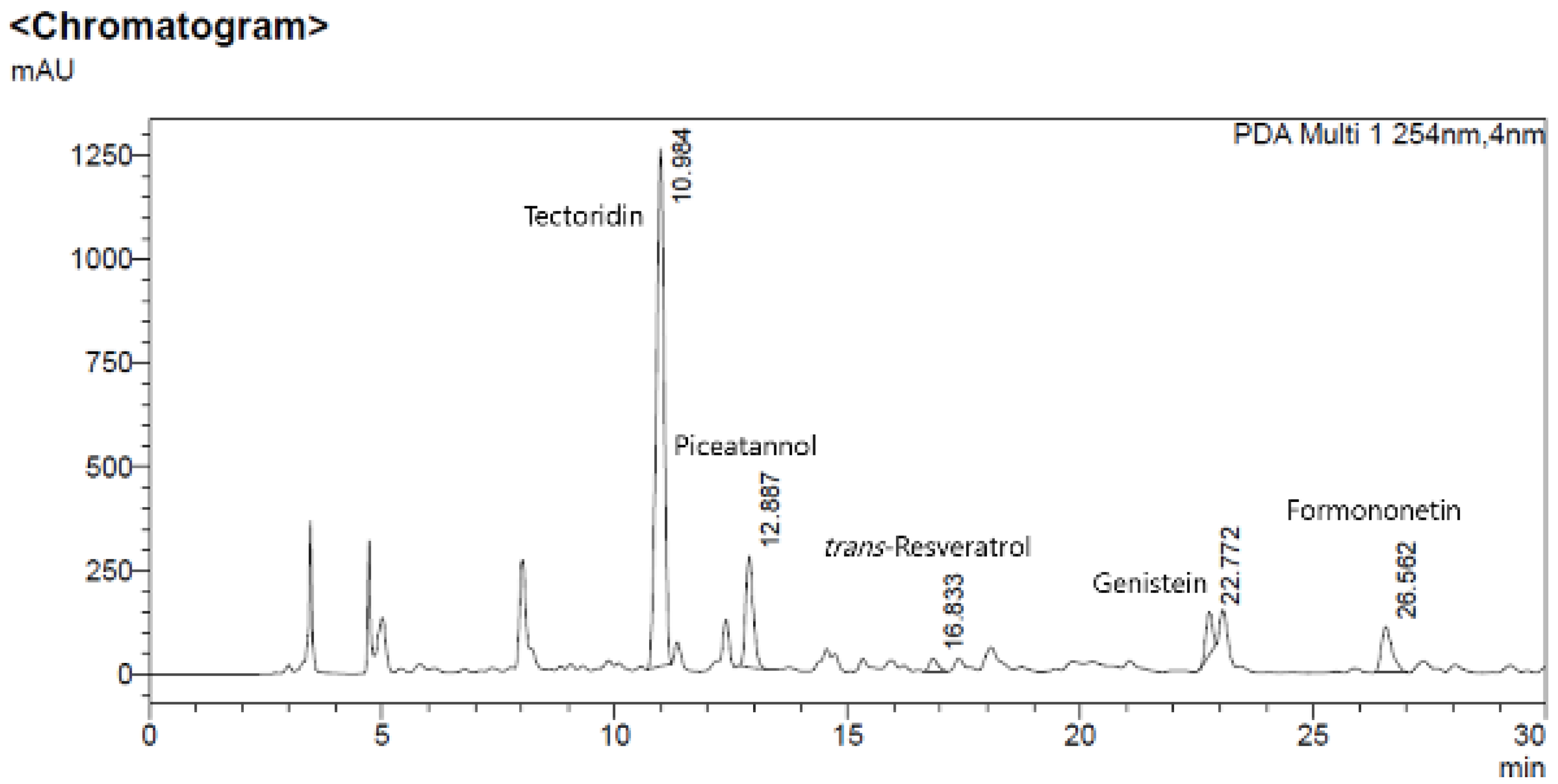
HPLC analysis was conducted to show that M. amurensis methanolic branch extract components and five standard compounds (formononetin, genistein, trans-resveratrol, piceatannol, and tectoridin) exhibited the same retention time. As shown in Figure 8, HPLC analysis of formononetin, genistein, trans-resveratrol, piceatannol, and tectoridin showed single peaks at 26.637, 22.803, 16.898, 12.937, and 10.997 min, respectively. Further, five major peaks of M. amurensis methanolic branch extract (26.562, 22.772, 16.833, 12.887, and 10.984 min) had the same retention time as formononetin, genistein, trans-resveratrol, piceatannol, and tectoridin, respectively. These results suggested that M. amurensis methanolic branch extract mainly contains formononetin, genistein, trans-resveratrol, piceatannol, and tectoridin.
The skin-whitening and antiwrinkle effects of M. amurensis branch extract may be attributed to these major compounds—formononetin, genistein, trans-resveratrol, piceatannol, and tectoridin—having these effects.

4. Conclusions

This study was conducted to evaluate the skin-whitening and antiwrinkle effects of M. amurensis branch extract and to identify the major components of this extract. The skin-whitening effect of M. amurensis branch extract was confirmed by investigating melanin content and melanogenic protein expression. M. amurensis branch extract exerted a skin-whitening effect via the suppression of melanin synthesis by inhibiting the expression of MITF, tyrosinase, TRP-1, and TRP-2 in B16F1 melanoma cells. Additionally, the antiwrinkle effect of M. amurensis branch extract was confirmed by investigating soluble collagen and MMP-1 expression. M. amurensis branch extract exerted its antiwrinkle effect by increasing the level of soluble collagen via the inhibition of MMP-1 expression in CCD-986sk cells. Five compounds—formononetin, genistein, trans-resveratrol, piceatannol, and tectoridin—were isolated from M. amurensis branch extract by chromatography, and HPLC analysis revealed that these compounds were the main compounds present in the branches of M. amurensis. Formononetin, genistein, trans-resveratrol, piceatannol, and tectoridin were reported to have a whitening effect, and the antiwrinkle effects of genistein and piceatannol were also reported. Therefore, the skin-whitening and antiwrinkle effects of M. amurensis branch extract may be attributed to these main compounds having these effects. In conclusion, our study findings indicate that M. amurensis branch extract has skin whitening and anti-wrinkle effects, and therefore great potential as a natural cosmetic ingredient. Moreover, it is believed that with further study, M. amurensis can be applied as an ingredient in the production of cosmetics, food, and pharmaceutical materials.

Author Contributions

Data curation, S.-K.K.; formal analysis, Y.-A.K. and S.-H.P.; investigation, G.-K.P., Y.-A.K. and S.-H.P.; project administration, B.P.; resources, Y.-A.K. and S.-H.P.; validation, J.-G.K., W.J. and B.-Y.K.; writing—original draft, J.-G.K., G.-K.P. and W.J.; writing—review and editing, J.-G.K. and G.-K.P. All authors have read and agreed to the published version of the manuscript.

Funding

This research was funded by Ministry of Trade, Industry and Energy grant number [10043192].

Institutional Review Board Statement

Not applicable.

Informed Consent Statement

Not applicable.

Data Availability Statement

Data available in a publicly accessible repository.

Acknowledgments

This study was supported by a grant of the WC300 R&D Project under the Ministry of Trade, Industry and Energy (Grant No. 10043192). We would like to thank the Plant Extract Bank of the Korea Research Institute of Bioscience and Biotechnology for providing the native plants.

Conflicts of Interest

The authors declare no conflict of interest.

References

  1. Lee, C.-W.; Kim, H.-A.; Yoon, H.-R.; Jeon, T.-Y. Establishment of seaweed fermentation process for cosmetic material research. J. Korea Acad.-Ind. Coop. Soc. 2019, 20, 14–19. [Google Scholar]
  2. Chermahini, S.H.; Majid, F.A.A.; Sarmidi, M.R. Cosmeceutical value of herbal extracts as natural ingredients and novel technologies in anti-aging. J. Med. Plants Res. 2011, 5, 3074–3077. [Google Scholar]
  3. Kim, H.-S. A Study on the Trends of the Natural UV Protection Materials Related to Skin Beauty. J. Korean Appl. Sci. Technol. 2021, 38, 107–117. [Google Scholar]
  4. Rodrigues, F.; de la Luz Cádiz-Gurrea, M.; Nunes, M.A.; Pinto, D.; Vinha, A.F.; Linares, I.B.; Oliveira, M.B.P.; Carretero, A.S. Cosmetics. In Polyphenols: Properties, Recovery, and Applications; Elsevier: Amsterdam, The Netherlands, 2018; pp. 393–427. [Google Scholar]
  5. Lim, Y.-J.; Lee, E.H.; Kang, T.H.; Ha, S.K.; Oh, M.S.; Kim, S.M.; Yoon, T.-J.; Kang, C.; Park, J.-H.; Kim, S.Y. Inhibitory effects of arbutin on melanin biosynthesis of α-melanocyte stimulating hormone-induced hyperpigmentation in cultured brownish guinea pig skin tissues. Arch. Pharm. Res. 2009, 32, 367–373. [Google Scholar] [CrossRef]
  6. Liu-Smith, F.; Poe, C.; Farmer, P.J.; Meyskens, F.L., Jr. Amyloids, melanins and oxidative stress in melanomagenesis. Exp. Dermatol. 2015, 24, 171–174. [Google Scholar] [CrossRef] [Green Version]
  7. Bhawan, J.; Gonzalez-Serva, A.; Nehal, K.; Labadie, R.; Lufrano, L.; Thorne, E.G.; Gilchrest, B.A. Effects of tretinoin on photodamaged skin: A histologic study. Arch. Dermatol. 1991, 127, 666–672. [Google Scholar] [CrossRef]
  8. Jin, M.L.; Park, S.Y.; Kim, Y.H.; Park, G.; Son, H.-J.; Lee, S.-J. Suppression of α-MSH and IBMX-induced melanogenesis by cordycepin via inhibition of CREB and MITF, and activation of PI3K/Akt and ERK-dependent mechanisms. Int. J. Mol. Med. 2012, 29, 119–124. [Google Scholar]
  9. Jeon, N.-J.; Kim, Y.-S.; Kim, E.-K.; Dong, X.; Lee, J.-W.; Park, J.-S.; Shin, W.-B.; Moon, S.-H.; Jeon, B.-T.; Park, P.-J. Inhibitory effect of carvacrol on melanin synthesis via suppression of tyrosinase expression. J. Funct. Foods 2018, 45, 199–205. [Google Scholar] [CrossRef]
  10. Kameyama, K.; Sakai, C.; Kuge, S.; Nishiyama, S.; Tomita, Y.; Ito, S.; Wakamatsu, K.; Hearing, V.J. The expression of tyrosinase, tyrosinase-related proteins 1 and 2 (TRP1 and TRP2), the silver protein, and a melanogenic inhibitor in human melanoma cells of differing melanogenic activities. Pigment Cell Res. 1995, 8, 97–104. [Google Scholar] [CrossRef]
  11. Jung, S.K.; Lee, K.W.; Kim, H.Y.; Oh, M.H.; Byun, S.; Lim, S.H.; Heo, Y.-S.; Kang, N.J.; Bode, A.M.; Dong, Z. Myricetin suppresses UVB-induced wrinkle formation and MMP-9 expression by inhibiting Raf. Biochem. Pharmacol. 2010, 79, 1455–1461. [Google Scholar] [CrossRef] [Green Version]
  12. Buechner, N.; Schroeder, P.; Jakob, S.; Kunze, K.; Maresch, T.; Calles, C.; Krutmann, J.; Haendeler, J. Changes of MMP-1 and collagen type Iα1 by UVA, UVB and IRA are differentially regulated by Trx-1. Exp. Gerontol. 2008, 43, 633–637. [Google Scholar] [CrossRef]
  13. Shim, M.; Bae, J.Y.; Lee, Y.J.; Ahn, M.J. Tectoridin from Maackia amurensis modulates both estrogen and thyroid receptors. Phytomedicine 2014, 21, 602–606. [Google Scholar] [CrossRef]
  14. Li, X.; Wang, D.; Xia, M.-Y.; Wang, Z.-h.; Wang, W.-N.; Cui, Z. Cytotoxic prenylated flavonoids from the stem bark of Maackia amurensis. Chem. Pharm. Bull. 2009, 57, 302–306. [Google Scholar] [CrossRef] [Green Version]
  15. Oh, J.M.; Jang, H.-J.; Kim, W.J.; Kang, M.-G.; Baek, S.C.; Lee, J.P.; Park, D.; Oh, S.-R.; Kim, H. Calycosin and 8-O-methylretusin isolated from Maackia amurensis as potent and selective reversible inhibitors of human monoamine oxidase-B. Int. J. Biol. Macromol. 2020, 151, 441–448. [Google Scholar] [CrossRef]
  16. Li, X.; Wang, D.; Cui, Z. A new cytisine-type alkaloid from the stem bark of Maackia amurensis. Nat. Prod. Res. 2010, 24, 1499–1502. [Google Scholar] [CrossRef]
  17. Kim, G.-S.; Chang, J.-P.; Doh, E.-S.; Kil, K.-J.; Yoo, J.-H. Stem bark of Maackia amurensis extract according to extraction solvent. Korea J. Herbol. 2016, 31, 43–48. [Google Scholar] [CrossRef]
  18. Radziejewska, I.; Borzym-Kluczyk, M.; Leszczynska, K. Lotus tetragonolobus, Ulex europaeus, Maackia amurensis, and Arachis hypogaea (peanut) lectins influence the binding of Helicobacter pylori to gastric carbohydrates. Adv. Clin. Exp. Med. 2018, 27, 807–811. [Google Scholar] [CrossRef]
  19. Huang, H.-C.; Chou, Y.-C.; Wu, C.-Y.; Chang, T.-M. [8]-Gingerol inhibits melanogenesis in murine melanoma cells through down-regulation of the MAPK and PKA signal pathways. Biochem. Biophys. Res. Commun. 2013, 438, 375–381. [Google Scholar] [CrossRef] [Green Version]
  20. Kim, H.O.; Shin, K.R.; Jang, B.-C.; Kim, Y.C. Action mechanism of anti-wrinkle effect of Rhamnus yoshinoi methanol extract in human dermal fibroblast and keratinocyte cell lines. Toxicol. Res. 2020, 36, 69–77. [Google Scholar] [CrossRef]
  21. Strzępek-Gomółka, M.; Gaweł-Bęben, K.; Angelis, A.; Antosiewicz, B.; Sakipova, Z.; Kozhanova, K.; Głowniak, K.; Kukula-Koch, W. Identification of mushroom and murine tyrosinase inhibitors from Achillea biebersteinii Afan. extract. Molecules 2021, 26, 964. [Google Scholar] [CrossRef]
  22. Park, H.-J.; Cho, J.-H.; Hong, S.-H.; Kim, D.-H.; Jung, H.-Y.; Kang, I.-K.; Cho, Y.-J. Whitening and anti-wrinkle activities of ferulic acid isolated from Tetragonia tetragonioides in B16F10 melanoma and CCD-986sk fibroblast cells. J. Nat. Med. 2018, 72, 127–135. [Google Scholar] [CrossRef]
  23. Ishikawa, M.; Kawase, I.; Ishii, F. Glycine inhibits melanogenesis in vitro and causes hypopigmentation in vivo. Biol. Pharm. Bull. 2007, 30, 2031–2036. [Google Scholar] [CrossRef] [Green Version]
  24. Aramwit, P.; Kanokpanont, S.; De-Eknamkul, W.; Kamei, K.; Srichana, T. The effect of sericin with variable amino-acid content from different silk strains on the production of collagen and nitric oxide. J. Biomater. Sci. Polym. Ed. 2009, 20, 1295–1306. [Google Scholar] [CrossRef]
  25. Korting, H.C.; Schindler, S.; Hartinger, A.; Kerscher, M.; Angerpointner, T.; Maibach, H.I. MTT-assay and neutral red release (NRR)-assay: Relative role in the prediction of the irritancy potential of surfactants. Life Sci. 1994, 55, 533–540. [Google Scholar] [CrossRef]
  26. Sugimoto, K.; Nishimura, T.; Nomura, K.; Sugimoto, K.; Kuriki, T. Syntheses of arbutin-α-glycosides and a comparison of their inhibitory effects with those of α-arbutin and arbutin on human tyrosinase. Chem. Pharm. Bull. 2003, 51, 798–801. [Google Scholar] [CrossRef] [PubMed] [Green Version]
  27. Hwang, J.-H.; Lee, B.M. Inhibitory effects of plant extracts on tyrosinase, L-DOPA oxidation, and melanin synthesis. J. Toxicol. Environ. Health Part A 2007, 70, 393–407. [Google Scholar] [CrossRef]
  28. Kubínová, R.; Spačková, V.; Svajdlenka, E.; Lučivjanská, K. Antioxidant activity of extracts and HPLC analysis of flavonoids from Capsella bursa-pastoris (L.) Medik. Ceska A Slov. Farm. Cas. Ceske Farm. Spol. A Slov. Farm. Spol. 2013, 62, 174–176. [Google Scholar]
  29. Masahiro, T.; Risa, M.; Harutaka, Y.; Kazuhiro, C. Novel antioxidants isolated from Perilla frutescens Britton var. crispa (Thunb.). Biosci. Biotechnol. Biochem. 1996, 60, 1093–1095. [Google Scholar] [CrossRef] [Green Version]
  30. Chen, Y.-S.; Lee, S.-M.; Lin, C.-C.; Liu, C.-Y. Hispolon decreases melanin production and induces apoptosis in melanoma cells through the downregulation of tyrosinase and microphthalmia-associated transcription factor (MITF) expressions and the activation of caspase-3,-8 and-9. Int. J. Mol. Sci. 2014, 15, 1201–1215. [Google Scholar] [CrossRef]
  31. Kim, B.Y.; Park, S.H.; Park, B.J.; Kim, J.J. Whitening effect of Androsace umbellata extract. J. Soc. Cosmet. Sci. Korea 2015, 41, 21–26. [Google Scholar]
  32. Hanelt, M.; Gareis, M.; Kollarczik, B. Cytotoxicity of mycotoxins evaluated by the MTT-cell culture assay. Mycopathologia 1994, 128, 167–174. [Google Scholar] [CrossRef] [PubMed]
  33. Baumann, L. Skin ageing and its treatment. J. Pathol. A J. Pathol. Soc. Great Br. Irel. 2007, 211, 241–251. [Google Scholar] [CrossRef] [PubMed]
  34. Ham, S.A.; Kang, E.S.; Yoo, T.; Lim, H.H.; Lee, W.J.; Hwang, J.S.; Paek, K.S.; Seo, H.G. Dalbergia odorifera Extract Ameliorates UVB-Induced Wrinkle Formation by Modulating Expression of Extracellular Matrix Proteins. Drug Dev. Res. 2015, 76, 48–56. [Google Scholar] [CrossRef] [PubMed]
  35. Takema, Y.; Hattori, M.; Aizawa, K. The relationship between quantitative changes in collagen and formation of wrinkles on hairless mouse skin after chronic UV irradiation. J. Dermatol. Sci. 1996, 12, 56–63. [Google Scholar] [CrossRef]
  36. Gęgotek, A.; Bielawska, K.; Biernacki, M.; Zaręba, I.; Surażyński, A.; Skrzydlewska, E. Comparison of protective effect of ascorbic acid on redox and endocannabinoid systems interactions in in vitro cultured human skin fibroblasts exposed to UV radiation and hydrogen peroxide. Arch. Dermatol. Res. 2017, 309, 285–303. [Google Scholar] [CrossRef] [Green Version]
  37. Kim, Y.A.; Kim, D.H.; Yu, J.M.; Park, C.B.; Park, B.J. Anti-wrinkle effects of extracts and solvent fractions from Nymphoides peltata on CCD-986sk. J. Appl. Biol. Chem. 2017, 60, 357–362. [Google Scholar] [CrossRef] [Green Version]
  38. Shin, Y.H.; Song, C.-K. Antioxidant and metalloproteinase inhibitory activities of ethanol extracts from Lespedeza cuneata G. don. Korean J. Environ. Agric. 2017, 36, 263–268. [Google Scholar] [CrossRef] [Green Version]
  39. Fisher, G.J.; Quan, T.; Purohit, T.; Shao, Y.; Cho, M.K.; He, T.; Varani, J.; Kang, S.; Voorhees, J.J. Collagen fragmentation promotes oxidative stress and elevates matrix metalloproteinase-1 in fibroblasts in aged human skin. Am. J. Pathol. 2009, 174, 101–114. [Google Scholar] [CrossRef] [Green Version]
  40. Song, X.-z.; Xia, J.-p.; Bi, Z.-g. Effects of (-)-epigallocatechin-3-gallate on expression of matrix metalloproteinase-1 and tissue inhibitor of metalloproteinase-1 in fibroblasts irradiated with ultraviolet A. Chin. Med. J. 2004, 117, 1838–1841. [Google Scholar]
  41. Kinjo, J.-E.; Furusawa, J.-I.; Baba, J.; Takeshita, T.; Yamasaki, M.; Nohara, T. Studies on the constituents of Pueraria lobata. III. Isoflavonoids and related compounds in the roots and the voluble stems. Chem. Pharm. Bull. 1987, 35, 4846–4850. [Google Scholar] [CrossRef] [Green Version]
  42. Yoon, J.S.; Sung, S.H.; Park, J.H.; Kim, Y.C. Flavonoids fromSpatholobus suberectus. Arch. Pharmacal Res. 2004, 27, 589–592. [Google Scholar] [CrossRef]
  43. Lee, Y.-Y.; Kwon, S.-H.; Kim, H.-J.; Park, H.-J.; Yang, E.-J.; Kim, S.-K.; Yoon, Y.-H.; Kim, C.-G.; Park, J.-W.; Song, K.-S. Isolation of oleanane triterpenes and trans-resveratrol from the root of peanut (Arachis hypogaea). J. Korean Soc. Appl. Biol. Chem. 2009, 52, 40–44. [Google Scholar] [CrossRef]
  44. Young Han, S.; Suck Lee, H.; Hye Choi, D.; Woon Hwang, J.; Mo Yang, D.; Jun, J.-G. Efficient Total Synthesis of Piceatannol via (E)-Selective Wittig–Horner Reaction. Synth. Commun. 2009, 39, 1425–1432. [Google Scholar] [CrossRef]
  45. Han, T.; Cheng, G.; Liu, Y.; Yang, H.; Hu, Y.-T.; Huang, W. In vitro evaluation of tectoridin, tectorigenin and tectorigenin sodium sulfonate on antioxidant properties. Food Chem. Toxicol. 2012, 50, 409–414. [Google Scholar] [CrossRef]
  46. Kim, J.M.; Ko, R.K.; Jung, D.S.; Kim, S.S.; Lee, N.H. Tyrosinase inhibitory constituents from the stems of Maackia fauriei. Phytother. Res. Int. J. Devoted Pharmacol. Toxicol. Eval. Nat. Prod. Deriv. 2010, 24, 70–75. [Google Scholar] [CrossRef]
  47. Jiafeng, Z.; Dayuan, Z. Chemical constituents of the roots of Maackia tenuifolia (Leguminosae). Acta Bot. Sin. 1999, 41, 997–1001. [Google Scholar]
  48. Mu, H.; Bai, Y.-H.; Wang, S.-T.; Zhu, Z.-M.; Zhang, Y.-W. Research on antioxidant effects and estrogenic effect of formononetin from Trifolium pratense (red clover). Phytomedicine 2009, 16, 314–319. [Google Scholar] [CrossRef]
  49. Zhang, J.; Liu, L.; Wang, J.; Ren, B.; Zhang, L.; Li, W. Formononetin, an isoflavone from Astragalus membranaceus inhibits proliferation and metastasis of ovarian cancer cells. J. Ethnopharmacol. 2018, 221, 91–99. [Google Scholar] [CrossRef]
  50. Khan, I.; Avery, M.; Burandt, C.; Goins, D.; Mikell, J.; Nash, T.; Azadegan, A.; Walker, L. Antigiardial activity of isoflavones from Dalbergia frutescens bark. J. Nat. Prod. 2000, 63, 1414–1416. [Google Scholar] [CrossRef]
  51. Surendhiran, D.; Karthiga, J.; Nirmala, S.; Sirajunnisa, A.R. Isolation of genistein from Acalypha fruticosa and studying its antibacterial activity by inhibition of bacterial DNA and protein. J. Omedicine. Toxicol. 2011, 5, 87–96. [Google Scholar]
  52. Jang, D.S.; Kim, J.M.; Lee, Y.M.; Kim, Y.S.; Kim, J.-H.; Kim, J.S. Puerariafuran, a new inhibitor of advanced glycation end products (AGEs) isolated from the roots of Pueraria lobata. Chem. Pharm. Bull. 2006, 54, 1315–1317. [Google Scholar] [CrossRef] [Green Version]
  53. Ma, K.; Ishikawa, T.; Seki, H.; Furihata, K.; Ueki, H.; Narimatsu, S.; Higuchi, Y.; Chaichantipyuth, C. Isolation of new isoflavonolignans, butesuperins A and B, from a Thai miracle herb, Butea superba. Heterocycles-Sendai Inst. Heterocycl. Chem. 2005, 65, 893–900. [Google Scholar] [CrossRef]
  54. Park, J.-S.; Kim, D.H.; Lee, J.K.; Lee, J.Y.; Kim, D.H.; Kim, H.K.; Lee, H.-J.; Kim, H.C. Natural ortho-dihydroxyisoflavone derivatives from aged Korean fermented soybean paste as potent tyrosinase and melanin formation inhibitors. Bioorganic. Med. Chem. Lett. 2010, 20, 1162–1164. [Google Scholar] [CrossRef] [PubMed]
  55. Widodo, W.S.; Widowati, W.; Ginting, C.N.; Lister, I.; Armansyah, A.; Girsang, E. Comparison of antioxidant and anti-collagenase activity of genistein and epicatechin. Pharm. Sci. Res. 2019, 6, 6. [Google Scholar]
  56. Kukrić, Z.Z.; Topalić-Trivunović, L.N. Antibacterial activity of cis-and trans-resveratrol isolated from Polygonum cuspidatum rhizome. Acta Period. Technol. 2006, 37, 131–136. [Google Scholar] [CrossRef]
  57. Harba, A.H.; abu Zargab, M.; Abdallaa, S. Effects of Trans-Resveratrol, Isolated from Smilax Aspera, on Smooth Muscle, Blood Pressure, and Inflammation in Rats and Nociceptionin Mice. Jordan J. Biol. Sci. 2009, 2, 69–76. [Google Scholar]
  58. Lee, T.H.; Seo, J.O.; Baek, S.-H.; Kim, S.Y. Inhibitory effects of resveratrol on melanin synthesis in ultraviolet B-induced pigmentation in Guinea pig skin. Biomol. Ther. 2014, 22, 35. [Google Scholar] [CrossRef] [Green Version]
  59. Kim, J.; Oh, J.; Averilla, J.N.; Kim, H.J.; Kim, J.S.; Kim, J.S. Grape peel extract and resveratrol inhibit wrinkle formation in mice model through activation of Nrf2/HO-1 signaling pathway. J. Food Sci. 2019, 84, 1600–1608. [Google Scholar] [CrossRef]
  60. Duarte, N.; Kayser, O.; Abreu, P.; Ferreira, M.J.U. Antileishmanial activity of piceatannol isolated from Euphorbia lagascae seeds. Phytother. Res. Int. J. Devoted Pharmacol. Toxicol. Eval. Nat. Prod. Deriv. 2008, 22, 455–457. [Google Scholar]
  61. Oh, S.-J.; Baek, N.-I.; Kim, H.-Y. Piceatannol, Antioxidant Compound Isolated from the Root of Rheum undulatum L. Appl. Biol. Chem. 2001, 44, 208–210. [Google Scholar]
  62. Kitada, M.; Ogura, Y.; Maruki-Uchida, H.; Sai, M.; Suzuki, T.; Kanasaki, K.; Hara, Y.; Seto, H.; Kuroshima, Y.; Monno, I. The effect of piceatannol from passion fruit (Passiflora edulis) seeds on metabolic health in humans. Nutrients 2017, 9, 1142. [Google Scholar] [CrossRef] [Green Version]
  63. Yokozawa, T.; Kim, Y.J. Piceatannol inhibits melanogenesis by its antioxidative actions. Biol. Pharm. Bull. 2007, 30, 2007–2011. [Google Scholar] [CrossRef] [Green Version]
  64. Maruki-Uchida, H.; Kurita, I.; Sugiyama, K.; Sai, M.; Maeda, K.; Ito, T. The protective effects of piceatannol from passion fruit (Passiflora edulis) seeds in UVB-irradiated keratinocytes. Biol. Pharm. Bull. 2013, 36, 845–849. [Google Scholar] [CrossRef] [Green Version]
  65. Jeong, G.-S.; An, R.-B.; Oh, S.-H.; Kang, D.-G.; Lee, H.-S.; Kim, Y.-C. Cytoprotective activity of Belamcanda chinensis rhizome against glutamate-induced oxidative injury in HT22 cells. Nat. Prod. Sci. 2007, 13, 101–104. [Google Scholar]
  66. Bhat, G.A.; Mir, F.; Shawl, A.S.; Ganai, B.A.; Kamili, A.N.; Masood, A.; Tantry, M.A. Crocetenone, a new rotenoid with an unusual trans-fused ring system from Iris crocea. Nat. Prod. Commun. 2015, 10, 503–504. [Google Scholar] [CrossRef] [Green Version]
  67. Ahn, Y.J.; Chang, Y.H.; Lee, S.Y.; Jin, M.H. A study on the whitening effects of Pueraria thomsonii extract and its three tectorigenin derivatives. J. Soc. Cosmet. Sci. Korea 2019, 45, 49–56. [Google Scholar]
  68. Kim, S.B.; Hwang, S.H.; Wang, Z.; Yu, J.M.; Lim, S.S. Rapid identification and isolation of inhibitors of rat lens aldose reductase and antioxidant in Maackia amurensis. BioMed Res. Int. 2017, 2017, 1–10. [Google Scholar]
  69. Utkina, N.; Kulesh, N. Antioxidant activity of polyphenols and polyphenol complex from the far-eastern tree Maackia amurensis. Pharm. Chem. J. 2012, 46, 488–491. [Google Scholar] [CrossRef]
  70. Park, W.S.; Bae, J.-Y.; Kim, H.J.; Kim, M.G.; Lee, W.-K.; Kang, H.-L.; Baik, S.-C.; Lim, K.M.; Lee, M.K.; Ahn, M.-J. Anti-Helicobacter pylori compounds from Maackia amurensis. Nat. Prod. Sci. 2015, 21, 49–53. [Google Scholar]
Figure 1. Effect of Maackia amurensis branch extract on B16F1 melanoma cell viability. B16F1 melanoma cells were treated with various concentrations of M. amurensis branch extract. M. amurensis branch extract did not show cytotoxicity at concentrations of ≤75 μg/mL. Data are presented as mean ± standard deviation (SD) of three independent experiments.
Processes 10 00855 g001
Figure 2. Effect of Maackia amurensis branch extract on α-melanocyte-stimulating hormone (α-MSH)-induced melanin synthesis in B16F1 melanoma cells. B16F1 cells were treated with arbutin or various concentrations (50, 37.5, and 25 μg/mL) of M. amurensis branch extract for 72 h in the presence or absence of 0.2 µM α-MSH. M. amurensis branch extract suppressed α-MSH-induced melanin synthesis in a dose-dependent manner. Data are presented as mean ± SD of three independent experiments. # p < 0.05 between blank and NC groups. * p-value between NC and M. amurensis branch extract groups (* p < 0.05 and *** p < 0.001). B, blank; N, negative control, P, positive control (arbutin).
Processes 10 00855 g002
Figure 3. Effect of Maackia amurensis branch extract on α-MSH-induced melanogenic protein expression in B16F1 melanoma cells. B16F1 cells were treated with arbutin or various concentrations (50, 37.5, and 25 μg/mL) of M. amurensis branch extract for 72 h in the presence or absence of 0.2 µM α-MSH. After treatment, western blotting analysis of microphthalmia-associated transcription factor (MITF), tyrosinase, tyrosinase-related protein (TRP)-1, and TRP-2 was performed. M. amurensis branch extract significantly inhibited the α-MSH-induced expression of (A) MITF, (C) TRP-2, and (D) tyrosinase. (B) M. amurensis branch extract inhibited TRP-1 expression, albeit not significantly. Data are presented as mean ± SD of three independent experiments. # p < 0.05 between blank and NC groups. * p-value between NC and M. amurensis branch extract groups (* p < 0.05, ** p < 0.01, and *** p < 0.001). B, blank; N, negative control; P, positive control (arbutin).
Processes 10 00855 g003
Figure 4. Effect of Maackia amurensis branch extract on CCD-986Sk cell viability. CCD-986sk cells were treated with various concentrations of M. amurensis branch extract. A. M. amurensis branch extract concentrations of >50 µg/mL showed cytotoxicity. B. M. amurensis branch extract showed no cytotoxicity at 37.5, 25, and 12.5 μg/mL concentrations. Data are presented as mean ± SD of three independent experiments.
Processes 10 00855 g004
Figure 5. Effect of Maackia amurensis branch extract on soluble collagen in CCD-986sk cells. CCD-986sk cells were treated with ascorbic acid (100 μg/mL) or various concentrations (37.5, 25, and 12.5 μg/mL) of M. amurensis branch extract for 24 h. M. amurensis branch extract significantly increased the level of soluble collagen. Data are presented as mean ± SD of three independent experiments. p-value between control and M. amurensis branch extract groups (** p < 0.01 and *** p < 0.001).
Processes 10 00855 g005
Figure 6. Effect of Maackia amurensis branch extract on UVA-induced matrix metalloproteinase (MMP)-1 expression in CCD-986Sk cells. CCD-986Sk cells were treated with epigallocatechin gallate or various concentrations (37.5, 25, and 12.5 μg/mL) of M. amurensis branch extract for 24 h in the presence or absence of UVA irradiation (15 J/cm2). After treatment, a western blotting analysis of MMP-1 was performed. M. amurensis branch extract significantly inhibited the UVA-induced expression of MMP-1. Data are presented as mean ± SD of three independent experiments. # p < 0.05 between blank and NC groups. * p-value between NC and M. amurensis branch extract groups (* p < 0.05 and ** p < 0.01). B, blank; N, negative control; P, positive control (epigallocatechin gallate; EGCG).
Processes 10 00855 g006
Figure 7. Chemical structures of Compounds 1–5.
Processes 10 00855 g007
Figure 8. HPLC chromatogram of M. amurensis methanolic branch extract and five standard compounds (formononetin, genistein, trans-resveratrol, piceatannol, and tectoridin).
Processes 10 00855 g008
Publisher’s Note: MDPI stays neutral with regard to jurisdictional claims in published maps and institutional affiliations.