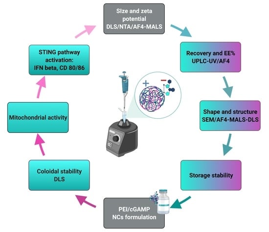
1. Introduction
Over the years, nanomaterials as drug delivery systems have attracted the attention of researchers and scientists, due to their ability to improve efficacy and reduce the toxicity of treatments, the possibility of targeted delivery of the drug within tissues and the cell cytosol (gene therapy), and the enhancement of drug stability and solubility [
1]. Although the US Food and Drug Administration (FDA) has issued guidance for nanomaterials in food, cosmetics and animal food, only a draft guidance was issued for nanomedicine. Additionally, the FDA recommends a case-by-case evaluation of safety for each specific nanosystem [
2]. Although nanocomplexes (NCs) are considered to be in the nano-size range of 1 to 100 nm [
3], the size of the NCs can go up to several hundred nanometers if it is related to the NC’s therapeutic activity [
1].
Cyclic GMP-AMP (cGAMP), a dinucleotide, is a ligand of the innate immunity stimulation of interferon gene (STING) pathway [
4]. This pathway is present in all cells and is activated by cytosolic dsDNA (microbial or tumor) [
5]. Recently, cGAMP has been widely investigated for the purpose of cancer immunotherapy, where the targeted antigen presenting cells (APC) will, upon presence of cGAMP, activate the intracellular STING pathway, produce interferon beta and eventually cause an increase in tumor-specific CD8+ T cells [
5,
6,
7]. Different natural cGAMPs have been isolated, such as 2′3′-cGAMP or 3′3′-cGAMP and administered either i.v. or i.t. in mice [
8]. Due to the difficulty for negatively charged molecules to enter cells, higher doses of cGAMP were used causing side effects such as T cell apoptosis [
9,
10]. Moreover, as a dinucleotide, cGAMP is prone to degradation by blood hydrolases [
6]. There is therefore a need for a carrier which would both protect the dinucleotide and enable successful transfection of cGAMP.
Polyethylenimine (PEI) has become the gold standard as a carrier for oligonucleotide delivery due to its remarkable pH-buffering capacity and ability to electrostatically condense genetic material enabling its protection and cellular uptake [
11]. We thus decided to complex cGAMP with PEI, spontaneously forming NCs. Once internalized by the cell, PEI causes osmotic swelling and final disruption of the endosome, leading to the endosomal escape of the drug cargo. Depending on the type and number of amino groups within the PEI chain, this disruption effect is more or less pronounced. There are two types of PEI structures, the branched one, having more primary amino groups, and the linear one, having more secondary amino groups. Molecular weight also plays an important role in the buffering capacity of PEI: the higher the molecular weight, the higher the transfection efficacy. This should be balanced with the toxicity increasing with the number of available positively charged (primary) amino groups [
11,
12]. We therefore investigated both branched (1.2 and 10 kDa) and linear PEI (4 and 25 kDa) in order to determine the best balance between toxicity and efficacy, and to ensure adequate physicochemical stability.
The targeted NCs’ properties also depend on the route of administration [
13]. We aimed here at an intratumoral (i.t.) administration, avoiding potential T-cell apoptosis upon i.v. administration of the required high dose of cGAMP [
14].
A critical step for i.t. administration is to trigger endocytosis by the tumor microenvironment. Noteworthy, the optimal size for uptake would depend on the targeted cells of the tumor microenvironment: (i) phagocytic cells for which up to now contradictory data on optimal target size for uptake have been reported, generally favoring NCs larger than 200 nm and [
15,
16] (ii) non-phagocytic cells for which uptake increases for diameters below 50 nm [
15,
16]. Additionally, it was shown that the toxicity is inversely proportional to the size of the NCs, with higher toxicity observed for smaller NCs [
17].
With regard to the shape of the NCs, phagocytic cells preferably take up elongated shape particles (resembling bacteria), as compared to non-phagocytic cells for which the uptake rate increases for smaller and spherical NCs.
Finally, the N/P charge ratio is also an important parameter to investigate. Based on the available literature, higher absolute charge of NCs is favorable for better transfection efficacy by APC [
15,
18]. Still, it is important to take into account that excess of positively charged polymers such as PEI, amino groups can cause toxicity, as they interact with different cell compartments, such as cell or mitochondrial membranes [
19]. In preliminary experiments, we assessed a broad range of ratios (1/5 to 8/1) showing that high N/P ratios were causing high toxicity and low N/P ratios were not stable. Therefore, we focused on 1/1 and 2/1 N/P ratios in the context of this study.
Due to the lack of regulations in nanomedicine, two world organizations—Nanotechnology Characterization Laboratory of the US National Cancer Initiative (NCL-NCI) and European Nanomedicine Characterization Laboratory—have been establishing standard protocols for nanomaterial characterization using adequate analytical methods [
3,
20]. This implies a detailed understanding of the critical quality attributes (CQAs), as well as additional toxicological tests towards regulatory acceptable methods. Based on EU-NCL guidelines [
20], the main physicochemical CQAs for preclinical characterization are size, size distribution, charge/zeta potential (ZP), stability in relevant media and over time, encapsulation efficiency and aggregation behavior. These parameters will strongly impact toxicity, efficacy and immunogenicity [
1,
13,
21,
22,
23]. It is therefore critical to perform a thorough preclinical physicochemical characterization of CQA for a system such as PEI/cGAMP NCs [
24].
In order to obtain solid data on those crucial parameters, several techniques have been employed to establish possible quality control protocols [
20,
25,
26,
27,
28]. For examining the size of PEI/cGAMP NCs in the 1–1000 nm range, International Standard Organization (ISO) guidelines and health agencies recommend conventional analytical techniques such as dynamic light scattering (DLS ISO 22412:2017), nanotracking particle analysis (NTA ISO 19430:2016) and electron microscopy (EM ISO/TC 202/SC 4). Additionally, Asymmetrical Flow Field Flow Fractionation (AF4 ISO/TS 21362:2018) was recently accepted by ISO as a high-resolution fractionation technique. For mostly monomodal distribution of PEI/cGAMP NCs, DLS is able to provide reliable data. Nevertheless, in case of very high polydispersity index (PDI) values, e.g., in the presence of aggregates, DLS fails to describe the distribution showing the need for orthogonal techniques [
29]. NTA and scanning electron microscopy (SEM) are suitable approaches based on particle-by-particle measurements [
20,
30]. Nevertheless, as both of these techniques give a number-averaged size distribution, whereas DLS gives an intensity-averaged one, it is important to mathematically transform DLS results from intensity to number (volume) average for comparison with SEM and NTA [
31,
32]. For that purpose, the sample’s refractive index (RI) and absorbance must be known, and PEI/cGAMP NCs are assumed to be spherical [
31]. Additionally, more advanced techniques such as online AF4-DLS provide information on the size of each population separately [
28].
Drug loading is another challenging property to assess. Based on concentration detectors such as UV (AF4-UV-DLS), drug loading (recovery (R)) can be obtained for each of the populations [
28]. To obtain more solid data, AF4-UV results are compared with standard methods such as UPLC-UV to confirm encapsulation efficiency (EE) [
33,
34]. Finally, AF4 coupled with online DLS may provide information on the shape factor. Shape factor or molecular conformation represents the average radii of gyration (Rg-mass distribution of NCs) obtained by AF4-MALS (multiangle static light scattering) divided by the hydrodynamic radii Rh obtained by AF4-DLS [
35,
36]. Based on the shape factor, NCs can be classified as micro (nano)gels for values <0.7, homogeneous spheres for 0.775, branched molecules (1–1.5), random coils (1.5–2.1) and rod-like, elongated structures for values >2. In addition, electron microscopy can serve as a qualitative technique to compare shapes [
35]. Another important property impacting colloidal stability is the zeta potential (ZP), which can be measured by both DLS and NTA [
37,
38].
The aim of this work was to formulate positively charged PEI/cGAMP NCs preferably larger than 200 nm for optimal uptake following i.t. administration, with the lowest toxicity/efficacy ratio, and a shape favoring phagocytosis. Moreover, due to the lack of specific regulations appointing for the characterization of CQA, several orthogonal techniques were employed, and protocols proposed to obtain reliable data on size, shape, zeta potential and drug content of the NCs.
3. Results and Discussion
The characterization of the physicochemical properties of PEI/cGAMP nanocomplexes, as other nanoformulations, requires an orthogonal approach. In the following we discuss results obtained for these NCs with respect to their stability, size, charge, shape, encapsulation efficiency, in vitro efficacy and toxicity.
3.1. Physicochemical Stability of PEI/cGAMP NCs
The goal of these experiments was to identify a stable formulation for PEI/cGAMP NCs of varying polymer nature and N/P ratio in relevant media (
Figure 1a–d). Moreover, complexation was examined by measuring PEI/cGAMP NCs zeta potential (
Figure 1f) at the pH at which they were prepared (
Figure 1e).
Influence of polymer nature and media on the NCs’ size and PDI. PEI/cGAMP NCs prepared using branched PEI shown high size and PDI variations depending on the media (water, PBS, DMEM,
Figure 1a,b vs.
Figure 1c,d). In contrast, NCs prepared with linear PEI had similar characteristics in different media. Moreover, branched PEI/cGAMP NCs prepared in PBS were significantly (
p < 0.0001, n = 9) smaller compared to linear PEI NCs (
Figure S1a). Finally, it appears that linP25/cGAMP NCs prepared in PBS were the most stable having the lowest standard deviations in size and PDI values.
Surface charge and pH of NCs. Since the positive charge plays an important role in complexation with negatively charged cGAMP, we examined the zeta potential of PEI/cGAMP NCs and PEIs alone as a control. Linear PEI/cGAMP NCs had higher zeta potential values compared to branched PEIs, most probably due to the preparation pH (
Figure 1e,f). At high pH, branched PEI is less charged (pKa 9.1) and thus the zeta potential is reduced. Based on the molar charge N/P ratio calculation, it was expected to obtain nearly neutralized PEI/cGAMP NCs in case of 1/1 ratio and positive charge in case of 2/1 ratio after complexation. Accordingly, zeta potential for the linP25/cGAMP 2/1 ratio (7.7 ± 6 mV) was twice that of 1/1 ratio (4 ± 2.3 mV). PEI/cGAMP NCs made of branched PEIs followed the same trend. Significant differences in zeta potential values were observed only in the case of linear PEI 25 in the comparison of empty to loaded PEI/cGAMP NCs (
p < 0.0001, n = 3). This difference was confirmed by NTA measurements, although higher zeta potential values (
p < 0.0001, n = 3) were measured with this approach (
Figure S1b). This is suggested to be due to the increased electrophoretic mobility of the measurements obtained by NTA compared to DLS (
p < 0.0001) (
Figure S1c). Electrophoretic mobility is inversely proportional to the electric field strength, which was in the case of DLS 40 V/cm and NTA 20 V/cm [
37]. Thus, besides the particle-by-particle measurement obtained by NTA, different amplitudes and modulation of the electric field might be the reason for the higher values. These data suggest successful complexation having our N/P ratio calculation in accordance with the data obtained, 2/1 NCs being twice as highly charged as 1/1 NCs, as expected.
Based on the media stability data showing no significant difference in size and size distribution in water, PBS and culture medium (DMEM), linear PEIs compared to the branched ones have an advantage as a carrier for cGAMP (
Figure 1a–d). The number of amino groups exposed to the solvent in branched PEI is less controlled due to conformational variability, which might explain the lack of stability and size variations in different media. Moreover, it was reported that pH plays an important role, having at lower pH more protonated groups and better binding, favoring again linear PEIs which were able to be buffered compared to the branched ones (
Figure 1e) [
46]. Stability of the NCs in DMEM is important for ensuing biological studies. Moreover, linear PEI 25 is the only polymer having statistically lower zeta potential values once complexed with cGAMP, suggesting successful complexation.
3.2. Toxicity/Efficacy Ratio of PEI/cGAMP NCs
Besides stability profile in different media, surface charge and pH, toxicity and activation of the STING pathway (IFN beta level) were important properties to be examined in order to pick the cGAMP polymers for further studies (
Figure 2).
NCs impact on mitochondrial enzymatic activity. It is well accepted that PEI toxicity is time, cell type and dose dependent [
47]. In this study, significant differences in mitochondrial activity were detected between time points 30 min, 1 h, 2 h and 24 h (
Figure S2a). There was no significant difference between BMDC and RAW cells, but BMDC tended to be more sensitive than RAW cells in terms of the lower mitochondrial activity measured upon NCs incubation (
Figure S2b). After 24 h of exposure, both free branched PEI and NCs prepared using branched PEI were significantly more toxic compared to the negative control (
p < 0.05, n = 9). There was a trend of NCs made of branched PEI to be more toxic compared to NCs made of linear PEI (
Figure 2a). Several factors may explain this difference in toxicity, with pH as a first factor. Preparations of NCs made of branched PEI had a higher pH compared to linPEI/cGAMP NCs (
Figure 1e). Moreover, surface chemistry plays an important role, with branched PEI being more toxic compared to linear PEI due the presence of a higher number of primary amino groups [
17,
48]. Another important parameter influencing cytotoxicity may be the size of PEI/cGAMP NCs. The smaller the PEI/cGAMP NCs, the higher their cytotoxicity [
17]. In our case, NCs made of branched PEI were indeed smaller than linear PEI NCs (
Figure S2c).
In order to select the best formulation taking into account its activity, we examined the secretion of IFN beta, the final product of the STING pathway activation and the expression of CD80/86 markers in BMDC.
IFN beta level induced by NCs. Incubation with NCs made of linP25 resulted in the highest IFN beta expression (
Figure 2b, 2.5-fold increase compared to cGAMP in the case of 1/1 ratio and 3-fold for 2/1). Moreover, only in the case of linear PEI 25 kDa, a significant increase in IFN beta response was observed over time (Ratio 1/1: 2 h 24 h vs. 1 h and 2 h
p < 0.001; ratio 2/1: 2 h 24 h vs. 1 h and 2 h
p < 0.0001, n = 9) (
Figure S2d). Branched PEI did not induce significant IFN-β expression, probably due to its toxicity (
Figure 2a,b).
CD80/86 expression on BMDC induced by NCs. To further investigate the carrier of choice, we examined the expression of cell markers CD 80 and CD 86, important for providing co-stimulation to T cells [
49]. Due their significant toxicity, NCs made of branched PEI were not further investigated. The highest expression of both CD86 and CD80 was observed with 2/1 NCs prepared from PEI 25 (1.9-fold higher CD 80 and 1.6-fold higher CD86 higher expression compared to the control, naked cGAMP).
It is important to notice that the cGAMP dose (2 µg) used in this work is five times lower than the average one reported in the literature [
14]. Our aim was to at least double the activation of STING pathway at this low dose compared to naked cGAMP. LinP25 and LinP4 as carriers were compared due their acceptable toxicity, cationic nature needed for transfection and ability to control the pH (
Figure 1e,f). Besides the influence of molecular weight on efficacy already reported in the literature [
50], we speculate on possible NCs’ size correlation with bioactivity. NCs 2/1 linP25 in DMEM (
Figure 1b) had a trend to be larger (ca. 600 nm) than NCs 2/1 linP4 in DMEM (ca. 400 nm). Moreover, based on Gustafson et al., phagocytosis is favored for larger particles [
16]. Therefore, the trend towards higher STING pathway activation for NCs 2/1 linP25 might be due to their size. Additionally, variations (STD) in IFN beta, CD80 and CD86 expression (
Figure 2b) might be partially explained by high STD in size (
Figure 1a,b). Finally, we selected linear PEI 25 for further studies due to (i) its acceptable toxicity on BMDC and RAW cells, (ii) promotion of IFN beta/CD80/86 expression level compared to free cGAMP, (iii) NCs’ stability in different media, (iv) positive charge and (v) the pH 6 of the preparation, which corresponds to the tumor microenvironment.
3.3. cGAMP Extinction Coefficient Determination and Method Optimization for AF4 Measurements
We determined the extinction coefficient of cGAMP (
Figure 3a) in order to compare NCs’ recovery with and without applying cross flow during the run (
Figure 3b) and to allow the quantification of cGAMP complexed in the NCs. We first checked that the UV signal at 256 nm arises mostly from cGAMP by injecting cGAMP alone (5 µg) and linP25 alone at two concentrations corresponding to 1/1 (0.625 µg) and 2/1 (1.25 µg) ratios (
Figure S3a). While applying the cross flow, the linP25/cGAMP may stick to the membrane. Therefore, we had to establish an efficient cleaning protocol besides BSA conditioning (
Figure 3c).
Extinction coefficient cGAMP. First, we determined the extinction coefficient of cGAMP in order to obtain recovery results as precise as possible. Extinction coefficient at 256 nm of cGAMP was calculated as an average of all extinction coefficients adjusted on Postnova software corresponding to different recoveries (0, 0.05, 0.1, 0.15 and 0.2 mg/mL), with a final value of 16.82 ± 0.88 mL/(mg·cm). This value was lower than reported for a parent molecule c-diGMP (34.9 mL/(mg·cm) [
12], maybe due to different mobile phases used. Linearity was established between different concentrations of cGAMP, which proved the reliability of the UV detector (R2 = 0.991) (
Figure 3a). Unfortunately, the RI signal suffered from higher SD, and RI linearity was not confirmed for cGAMP (R2 = 0.96) (
Figure S3c). Therefore, we based our cGAMP recovery results on UV detection, and continued following RI results only qualitatively in order to check if linP25/cGAMP remained complexed.
Recovery of cGAMP with and without cross flow. By injecting three times cGAMP alone without cross flow (
Figure 3b, pink curve/fractogram), recovery was around 100% (5.06 ± 0.33 μg recovered). Once the cross flow was applied (
Figure 3b, overlapping orange, blue and green graphs) cGAMP recovery was 61.7% (1.9 ± 0.2 μg recovered), probably due to losses through the membrane with a high molecular weight cut off (10 kDa), with respect to the molecular weight of cGAMP (718.28 g/mol) [
51]. UV was thus used for further studies on cGAMP recovery as a concentration detector, considering a maximum recovery Rcf of 60% under crossflow.
Cleaning protocol. In order to avoid overloading of the PEI/cGAMP sample, there was a need for every sample injection to be preceded by and followed by “cleaning” phases as follows: 1. acetic buffer pH 4 0.1 M; 2. water; 3. sample; and 4. SDS 0.1%. Comparison of injections 4 and 2 shows that after the sample run, there was still cGAMP remaining on the membrane that needed to be removed before the next measurement run (
Figure 3c).
3.4. Orthogonal Techniques for Size Comparison (DLS, NTA and AF4-DLS) of Empty Linear PEI 25 and PEI/cGAMP NCs
The aim of the experiment was to examine the size of 1/1 and 2/1 NCs compared to the parent polymer linP25 (
Figure 4). Size was obtained through different techniques (DLS z-average and number size, NTA and AF4-MALS-UV-DLS Dg and Dh).
Size and PDI of 1/1 and 2/1 NCs vs. linP25. Based on DLS measurements (both z- and number averages), both 1/1 and 2/1 NCs were significantly smaller than linP25 (
p < 0.0001) and PDI was lower, suggesting polymer condensation upon the addition of cGAMP (
Figure 4). Moreover, 2/1 NCs were significantly bigger compared to 1/1 (
p < 0.0001), as expected for a more extensive dinucleotide complexation. The trend was confirmed by NTA and AF4-DLS size measurements, where linP25 was bigger compared to NCs. Altogether, we can conclude that cGAMP complexes linP25, forming smaller PEI/cGAMP NCs at lower PDI values compared to PEI alone.
Comparison of sizing techniques. Both z-average values of NCs were significantly higher than number-average values (
p < 0.0001), which together with PDI values (0.4) suggests that we might not have a monomodal distribution. Nevertheless, there was no significant difference in between NTA and DLS number-average size values for the loaded NCs (
Figure 4). On the contrary, in the case of free polymer (linP25), DLS number-average values were statistically higher than NTA size values and polydispersity high, suggesting polymer aggregation.
There was a significant difference (p < 0.0001) between z-average by DLS and Dh calculated by AF4-DLS, which may be due to the selected population eluted and separated by AF4. As for the gyration size measured by MALS, the difference between Dg (2xRg) and Dh (2xRh) arise from their definitions. Rh is the hydrodynamic radii, i.e., the size of the solvated particle, and the gyration radii is calculated as the average distance from the center of mass to the molecule’s surface. Moreover, in the next section, we will discuss in more detail the ratio between the two radii, called shape factor. The trend towards higher STD in case of 2/1 NCs compared to 1/1 was shown by all sizing techniques and might be due to the fact that lower fraction of bound PEI decreases colloidal stability.
Effect of NCs’ concentration. Measurements were performed at 0.01 mg/mL of cGAMP in NCs corresponding to a sufficient concentration for STING pathway activation. To first confirm that DLS measurements were consistent between 0.01 and 0.1 mg/mL, we compared the derived count rate and z-average (
Figure S4a,b). It was shown that the values measured at a 0.01 mg/mL concentration matched those at the higher particle concentration of 0.1 mg/mL, concluding that a different concentration does not affect physicochemical parameters of NCs.
In terms of cGAMP complexation, both 1/1 and 2/1 LinP25/cGAMP NCs are smaller than empty NCs (linP25), as shown by several orthogonal techniques used (DLS, NTA, AF4-MALS-DLS for size) at different concentrations. (
Figure 4 and
Figure S1b). Moreover, zeta potential was measured my two orthogonal techniques NTA and DLS (
Figure S1b) and for both 1/1 and 2/1 NCs significantly lower zeta potential compared to empty linP25 was shown. These data strongly suggest a successful complexation of cGAMP by linP25. However, there is still a need to perform encapsulation efficiency to confirm at what extent there is complexation between the two.
A general approach for the characterization of gel-like nanocomplexes would start with the size measurements using intensity-based average (z-average), DLS. In the case of high PDI values (>0.08 corresponding to mid-range distribution based on Malvern Panalytical guidlines), we propose to use particle-by-particle methods such as NTA. In order to compare DLS and NTA, intensity-based results are transformed to number based, provided the particle shape is spherical and refractive index is known. Nevertheless, if results from intensity-based DLS and NTA are significantly different, it might point out the presence of large agglomerates or aggregates, as DLS intensity-based technique is highly sensitive to large particles. For accurate analysis of a multimodal size distribution, it is beneficial to separate small particles from agglomerates. AF4 coupled to DLS was employed for this purpose, measuring gyration diameter Dg and hydrodynamic radii Dh.
In our case, NCs are having a higher PDI value of 0.4, and intensity-based results being significantly higher than number-based NTA, we performed AF4-DLS analysis to separate the different populations. Based on NTA most of the NCs both for 1/1 and 2/1 were in the 200 nm range, and once separated by AF4-DLS the size decreased to 50 nm. We speculate that loose NC aggregates–floccules might form under static DLS conditions. The DLS/NTA difference is even more marked for 2/1 NCs, probably due to more NCs flocculating together, possibly resulting from higher concentration of free PEI. We hypothesize that those formations are either flocculate or agglomerate randomly.
3.5. Shape, Encapsulation Efficiency and Recovery of linP25/cGAMP NCs
The aim of this study was to determine the encapsulation efficiency of cGAMP obtained by UPLC-UV compared to its recovery R0 (no cross flow) and Rcf (with cross flow) by AF4-UV (
Figure 5c and
Figure S3). The two N/P ratios investigated were 1/1 and 2/1. In addition, the shape of the PEI/cGAMP NCs was examined. We compared the shape of the PEI/cGAMP NCs observed by electron microscopy (
Figure 5e) to the shape factor obtained by real time AF4-UV-DLS (
Figure 5a,b,d).
Recovery and EE of cGAMP in NCs. UV absorption of cGAMP was higher than linP25 (
Figure S3a) at a concentration used in the NCs, while the RI of linP25 was the same as for NCs (
Figure S3e,f). Based on
Figure 5a,b and
Figure S3e,f, we can hypothesize that we have NCs’ complexation suggested by UV and RI spectra overlapping. In order to confirm the complexation, we compared the recovery of cGAMP in NCs under cross flow (Rcf) to EE-indirect measurement by UPLC-UV. For the latter, we collected the centrifuged and filtered fraction (free cGAMP, not bound to linP25) using a membrane with the same cut off as the AF4 membrane. Moreover, we applied two different centrifugation speeds (6000×
g and 12,000×
g over 10 min,
Figure S3g) in order to check whether complexation is dependent on shear forces. For both ratios, there was a significant difference (
p < 0.0001) between Rcf (around 30%) and EE (around 60%) at both 6 and 12,000×
g (around 60%) (
Figure 5c). The centrifugation speed did not affect EE. By applying cross flow, we only obtained one elution peak (
Figure 5a,b and
Figure S3e,f). Noteworthy, only one peak was eluted from the polydisperse system. This might be due to the effect of the cross flow, forcing the loose, larger aggregates to stick to the slightly negatively charged membrane. In order to investigate this hypothesis, we performed non-cross flow measurements to examine R0. As expected, R0 was significantly higher (
p < 0.0001) compared to both NCs analyzed under mechanical stresses (Rcf—cross flow studies, centrifugation), supporting the hypothesis (
Figure S3g). We will thus base our result on EE%.
Gyration and hydrodynamic radii, shape factor. LinP25/cGAMP NCs were corresponding to elongated structures based on the measured shape factor of 3 ± 1.2 for 1/1 and 4.1 ± 1.8 for 2/1 NCs (
Figure 5d). We speculate that NCs are rod-like structures, having at 2/1 ratio more elongated shape than at a ratio of 1/1. In contrast, its structure resembles more that of a gel as determined by SEM (
Figure 5e). These findings are directly in line with previous reports on PEI-based nanogels for gene therapy [
11]. In addition, in the case of the 2/1 ratio we can observe more gelified and rigid structures by SEM, both after dehydration or lyophilization of the sample (
Figure 5e down left and right).
LinP25 and cGAMP are forming elongated scaffolds and eventually nanogels, being complexed as shown by size, zeta potential, shape and drug loading measurements. Both 1/1 and 2/1 N/P ratio shown EE around 60% and elongated structures (
Figure 4 and
Figure 5c,d). However, the 2/1 NCs were significantly larger as shown by DLS (
Figure 4), more positively charged (
Figure 1f and
Figure S1b), showing occasionally large floccules, higher IFN beta level and CD80, 86 expression (
Figure 2b,c) compared to 1/1 NCs. Thus 2/1 were kept for further investigation. However, high STD from biological assays for 2/1 might be due to loosely bound floccules and accordingly high STD in size. Possibly, one of the future approaches for stabilization of such complexes would be the increase in viscosity of the external phase.
3.6. Storage Stability
The goal of these experiments was to investigate the stability of particles of different preparation methods for the storage, for instance in view of further in vivo studies. Particles were prepared in water and stored at 4 °C (
Figure S4c) or stored in a dry form (dehydrated or lyophilized) and rehydrated (
Figure 6a). Moreover, INF β expression of lyophilized or dehydrated forms after rehydration was compared to the fresh form (
Figure 6b).
Stability of the NCs’ suspension and storage of dry forms. The NCs show limited stability in water suspension, with a non-significant trend towards size increase over 1 week (1/1 NCs) or 1 month (2/1 NCs) (
Figure S4c). Therefore, in view of translating to in vivo application, we assessed dehydration and lyophilization for long-term storage of the nanoparticles. Compared to freshly prepared NCs, both the particle size and IFN beta levels were not significantly different compared to the ones dehydrated or lyophilized. Nevertheless, there was a trend of lyophilized samples having a slightly lower expression of IFN beta and forming smaller NCs compared to both freshly prepared and dehydrated samples. Therefore, the preferred process for storage stability would be to dehydrate the sample. Our future goal is to check the use of possible cryoprotectants and examine the effects of time and temperature on our dehydrated samples.
4. Conclusions
Due to analytical challenges and lack in regulatory standards when determining the exact size and zeta potential by a single technique and the difficulty of the characterization of polydisperse samples such as PEI/cGAMP polyectrolyte nanocomplexes, characterization by orthogonal techniques is required. The aim of this work was to select NCs for i.t. administration which are the most stable in terms of constant size in different media, absence of aggregation, storage stability and the best efficacy/toxicity ratio. In order to separate different size populations, online coupled AF4 with DLS was employed and as expected, for all NCs’ preparations Dh (AF4-DLS) was significantly lower compared to z-average (DLS) after separation of possible aggregates by AF4-DLS. Although differences in size were measured by different techniques (DLS, NTA and AF4-DLS), all techniques did not have the same value, but the same trend, indicated by loaded NCs being smaller than empty carriers, suggesting a polyelectrolyte complexation. Zeta potential values confirmed the complexation, with cGAMP loaded NCs having a significantly lower charge. Encapsulation efficiency data measured by two techniques, UPLC-UV and AF4-UV, were significantly different having a value of around 60–70% obtained by UPLC-UV and around 30% by AF4-UV. As NCs were eluted only in one peak once cross flow was applied, having almost 100% recovery without cross flow, we speculate that NCs might stick to the oppositely charged membrane. The shape observed by SEM and the AF4-DLS shape factor >2 suggests an elongated rod-like structure, which would be preferred for the phagocytosis of NCs, which would have a shape similar to bacteria. Finally, samples stored as dehydrated or lyophilized dry forms did neither lose their activity nor have a significant change in their size and PDI.
Based on in-depth orthogonal analytical techniques of PEI/cGAMP NCs characterization, we would select as an immunotherapy carrier the 2/1 linear PEI 25 kDa/cGAMP NCs prepared in PBS, based on its comparably low cytotoxicity, highest IFN beta expression, CD80 and CD86 marker expression on BMDC, larger size, positive surface charge and bacteria-like elongated shape. In addition, this formulation showed a high encapsulation rate for cGAMP (60%). Nevertheless, there are few challenges remaining to be solved, such as a choice of more neutral-based AF4 membrane and additional stabilization of complexes.


