Article

Codonopsis laceolata Water Extract Ameliorates Asthma Severity by Inducing Th2 Cells’ and Pulmonary Epithelial Cells’ Apoptosis via NF-κB/COX-2 Pathway

1
College of Oriental Medicine, Dongshin University, Naju-si 58245, Korea
2
Department of Pathology, Dongguk University, Goyang-si 10326, Korea
3
Research Institute, Wellphyto Co., Ltd., K1 Office Town 415, Gwangju 69506, Korea
4
College of Pharmacy, Mokpo National University, Muan-gun 58554, Korea
5
Biomedicine, Health & Life Convergence Sciences, BK21 Four, College of Pharmacy, Mokpo National University, Muan-gun 58554, Korea
*
Authors to whom correspondence should be addressed.
These authors contributed equally to this work.
Processes 2022, 10(7), 1249; https://doi.org/10.3390/pr10071249
Submission received: 20 May 2022 / Revised: 8 June 2022 / Accepted: 18 June 2022 / Published: 23 June 2022

Abstract

:
Asthma is an incurable pulmonary disease with several symptoms, including abnormal breathing, coughing, and sleep apnea, which can lead to death, and the population of asthma patients has been increasing worldwide. There are many adverse effects in current drugs, and thus, we have tried to develop anti-asthmatic agents from natural products such as Codonopsis laceolata. To define the anti-asthmatic effect and the mechanism of Codonopsis laceolata, an animal study was conducted considering different cell counts of BALF, serum IgE levels, morphological changes in the pulmonary system, the Th2 cell transcription factor (GATA-3), and the apoptotic pathway (NF-κB/COX-2). Codonopsis laceolata significantly suppressed the representative asthmatic changes, such as airway remodeling, mucous hypersecretion, epithelial hyperplasia, and inflammatory cell infiltration, in the respiratory system. It suppressed the levels of GATA-3, IL-4, and IL-13. The down-regulation of Th2-related factors, such as GATA-3, IL-4, and IL-13, results from the stimulated apoptosis of Th2 cells and epithelial cells via a decrease in the levels of NF-κB and COX-2. We concluded that Codonopsis laceolata might be a promising anti-asthmatic drug.

1. Introduction

Asthma is an incurable chronic inflammatory disease of the pulmonary system, which is caused by a wide range of allergens [1]. It has several symptoms, such as wheezing, fast and difficult breathing, coughing, and sleep apnea, which can lead to death [2], and representative morphological changes, such as airway remodeling, inflammatory cell infiltration, mucous hypersecretion, and epithelial cell dysfunction/hyperplasia [3,4]. In 2019, it was estimated that there were 262 million asthma patients, and 461 thousand deaths were caused by asthma [5]. Specifically, asthma is most severe in children and the elderly and is the main cause of hospitalization in children [6]. In 2019, the healthy life expectancy (HALE) and the average life expectancy (LE) increased; for males, HALE and LE were 70.9 and 62.5 years and reached 75.9 and 64.9 years for females, respectively, and as time goes on, the percentage of the elderly population will significantly increase [7]. As asthma occurrence is related to age, the population of asthma patients could increase.
In our surrounding environment, there are many asthma-inducible allergens, and the various carriers can introduce allergens to us, such as Der p1 and Der p2 from house dust mite (Dermatophagoides pteronyssinus and Dermatophagoides farina), Fel d1 from cat (Felis domesticus), Can f1 from dog (Canis familiaris), Bla g1, Gla g2 and Per a1 from cockroach (Blatella germanica, Periplaneta americana), Mus m1 from mouse (Mus musculus), Rat n1 from rat (Rattus norvegicus), Bet v1 from birch tree (Betula verrucose), Phl p1 and Phl p5 from timothy (Pheum pretense), and pollen/mold [8]. These allergens induce immune imbalances, such as eosinophilia (over-differentiation of eosinophils), which is caused by IL-5 [9]; neutrophilia in some asthma patients [10]; type 1 hyperresponsiveness related to the IgE surge, which is caused by the activation of IL-4 and IL-13 [11]; breaking the cytokine balance, such as Th1 cell-related cytokines (IFN-γ and IL-12) [12], Th2 cell-related factors (IL-4, IL-5, IL-13), and Th17-related ones (IL-6 and TNF-α) [13]; and finally, the imbalance of Th1 and Th2 cells [14]. The imbalance of Th1 cells and Th2 ones means that in healthy persons the level of Th1 cells is higher than in asthma patients, but in the opposite situation, asthma occurrence Th1 cells might decrease, while the level of Th2 ones might increase.
In asthma patients, the level of Th2 cell-related cytokines such as IL-4, IL-5, and IL-13 increased compared to the healthy persons [15], but the level of Th1 cell-related cytokines such as IFN-γ and IL-12 decreased relatively [16]. T-bet is a transcription factor for Th1 cells and specifically produces IFN-γ in a positive feedback manner [17], but GATA-3 is a Th2 cell transcription factor that has a controversial effect, such as stimulating Th2 cell differentiation and inhibiting Th1 cell differentiation [18]. IFN-γ inhibits various Th2-related effects such as mucous secretion and eosinophilia [19]; IL-12 modulates Th1 cell differentiation and is released by B cells, macrophages, and dendritic cells [20]; IL-4 and IL-13 stimulate mucous hypersecretion, IgE over-production, eosinophilia, and airway remodeling [15]; IL-5 induces eosinophilia and airway hyperresponsiveness [9]; IL-6, which increases in asthma patients, is related to the inflammation process and stimulates IL-4 production/Th17 cell differentiation but inhibits Th1 cell differentiation [21]; and TNF-α has several functions related to asthma, such as a chemoattractant for neutrophils and eosinophils and Th2 cell activation/migration to damage in the lung [22].
The representative morphological changes in asthma patients’ lungs are hyperplasia/over-differentiation of the several cell types, such as epithelial cells, goblet cells, airway smooth muscle cells and Th2 cells, and these are related to the cell death (apoptosis) [23]. The status of the cell types differs significantly over time of observation [24], and in the recent animal studies, the level of apoptosis of several cell types increased the number of apoptotic cells in the respiratory system, which could be suggested as a biomarker for evaluating the asthma stage [25,26].
Asthma is an incurable disease, and anti-asthmatic drugs can be classified into two categories—controllers to prevent asthma occurrence and relievers to suppress asthma exacerbation [1]. The controllers involve anti-inflammatory drugs, such as corticosteroids and leukotriene receptor antagonists, and immunomodulators, such as Th2 cell-related down-regulators and IgE suppressors [27]. The relievers include β2-adrenergic receptor agonists [28]. The inhaled β2-adrenergic receptor agonist and corticosteroid combination used with a nebulizer has been commonly used as an anti-asthmatic drug, but if there is no inhaler tool such as a nebulizer, it is difficult to deliver drugs to the asthma patient [29]. Many problems, such as death, hospitalization, and exacerbation, related to the β2-adrenergic drug for asthma treatment have been reported for several decades [30], and the adverse effects of corticosteroids as a combined therapy for asthma have been reported such as growth retardation in children, immunosuppression, high blood pressure, hyperglycemia, delayed wound healing, metabolic disease, glaucoma, and cataracts [31].
Codonopsis laceolata (C. laceolata) has been used as a culinary material or a traditional medicine material for a long time in eastern Asia (Korea, Chinese, and Japan), and its biological effects have been studied for a long time. C. laceolata has an anti-proliferation effect against the HT-29 human colon cancer cell line via G0/G1 arrest and apoptosis [32], preventive effects against high-fat diet-induced obesity and hyperlipidemia [33], inhibitive effects against ethanol-induced hepatic lipogenesis and inflammation via TLR-mediated modulation [34], and regulatory effects against LPS-induced macrophage-mediated immune response (inflammation) [35].
In this study, we evaluated the anti-asthmatic effect of C. laceolata based on the therapeutic mechanisms and the possibility of it as a drug candidate for anti-asthmatic treatment.

2. Materials and Methods

2.1. Codonopsis laceolata (C. laceolata) Extract Preparation

In this study, the C. laceolata used was the same as that in our previously published study [36], which briefly explained that Diploid C. laceolate root was provided from Wellphyto Co. (Gwangju, Korea) and deposited in Mokpo National University (MNUCSS-CL-01). Twenty grams of C. laceolata root were extracted with boiled water for 4 h and freeze-dried. In order to analyze the components, a high-pressure liquid chromatography (HPLC) assay was conducted; lobetyolin was found, and the concentration of that was 0.17 ± 0.001 w/w.

2.2. Animal Experiment and Ethics Statement

The animal schedule was the same in our previous study, excluding the C. laceolata dosing groups [3]. In order to confirm the study results, 2 studies were conducted, and 96 laboratory animals (BALB/c mice) were purchased for 2 studies from Samtako Korea (Osan, Korea). The animals were classified into 6 groups; vehicle group (sterilized tap water, CON), ovalbumin-induced asthma group (OVA), positive control group (1 mg/kg/day dexamethasone with OVA, DEX), 50 mg/kg/day C. laceolata treated group with OVA, 150 mg/kg/day C. laceolata treated group with OVA, 300 mg/kg/day C. laceolata treated group with OVA. In order to induce OVA on days 1 and 8, mice were intraperitoneally injected with 20 μg ovalbumin (Sigma-Aldrich, St. Louis, MO, USA) and 1 mg aluminum hydroxide hydrate (Sigma-Aldrich) in 500 μL normal saline for sensitizing, and from day 21 to day 25, they were exposed to 5% ovalbumin in normal saline for 30 min using a nebulizer (3 mL/min, NE-U17, OMRON Co. Ltd., Kyoto, Japan). During the 5 days, all mice were treated depending on the classification group.
This animal study was approved by the Institutional Animal Care and Use Committee at Chonnam National University (the approval number, CNU IACUC-YB-2016-15).

2.3. Bronchioalveolar Fluid (BALF) and Serum Analysis

The BALF and serum analytic methods were the same as in our previously published study [3]. On day 26, all mice were anesthetized with 50 mg/kg Zoletin (Virbac, Fort Worth, TX, USA), then half of the mice in each group were cannulated with a flexible feeding needle through the trachea for collecting BALF, and the BALF was collected three times using 0.4 mL of phosphate-buffered saline (PBS) each time. The collected samples were centrifuged at 848× g for 5 min at 4 °C (hanil M15R, Hanil Scientific Inc., Kimpo, Korea). The downed cells were suspended again in PBS for the white blood cell count and differential cell counts. The cell counts were evaluated by the Hemavet Multispecies Hematology System (Drew Scientific Inc., Waterbury, CT, USA), and in order to qualitatively measure the change of the inflammatory cells, the Diff-Quick stain (ThermoFisher Scientific) was conducted according to the manufacturer’s guide.
In order to measure the level of serum IgE, an enzyme-linked immunosorbent assay kit (555248, BD Bioscience, San Jose, CA, USA) was used according to the manufacturer’s guide. The results were obtained by a microplate reader (Multiskan Sky, ThermoFisher Scientific).

2.4. Histopathological Anlaysis

Histopathological evaluation, such as hematoxylin and eosin (H&E) stain and periodic acid chiff (PAS) stain, was conducted similarly to our previous study [3]. Fixed lung tissues with 10% (v/v) formaldehyde solution were dehydrated in graded ethanol from 99.9% to 70% and were paraffin-embedded. Embedded cells were longitudinally sectioned as 4 μm and were stained using H&E for morphological evaluation and with PAS for measuring the level of glycoproteins. The photos were acquired with the Axioscope A1 (Carl Zeiss, Gottingen, Germany), and through the H&E stained results, the histopathological severity was analyzed. According to our previous study, in order to evaluate the level of morphological change, the quantitative score from 0 to 3 was based on the representative changes in the asthmatic pulmonary system, including mucous hypersecretion (0, none; 1, little releasing; 2, half-packed in whole duct; 3, packed mucous), epithelial cell hyperplasia (0, none; 1, corrugated wall; 2, folded epithelium; 3, severely folded epithelium) and inflammatory cell infiltration (0, none; 1, few leukocytes; 2, moderate number of leukocytes; 3, large number of leukocytes) [3].

2.5. Immunofluorescent Analysis

In order to evaluate the expression level of specific proteins, immunofluorescent staining was conducted as followed by our previous study [37]. Four-micrometer-long sectioned lung tissues of 4 groups, such as CON, OVA, DEX, and the 300 mg/kg/day C. lanceolate treated group, were immunofluorescently stained using several primary antibodies, such as GATA-3 (TA305795, OriGene, Rockville, MD, USA), CD278 for Th2 cells (ab224644, Abcam, Cambridge, UK), TUNEL for apoptotic cells (Click-iT™ Plus TUNEL Assay, Invitrogen, Waltham, MA, USA), NF-κB (51-0500, Invitrogen), COX-2 (PA1-9032, Invitrogen), and DAPI for the nucleus (62249, ThermoFisher Scientific, Waltham, MA, USA). The primary antibodies were bound on the specific proteins for 1 h at room temperature, the secondary antibodies, such as Alexa Fluor 555-conjugated IgG (A-21127, ThermoFischer Scientific), were incubated for 2 h, and the photos were acquired with a K1-Fluo confocal microscope (Nanoscope System, Daejeon, Korea).

2.6. Real Time-Poly Chain Reaction (RT-PCR) Analysis

The analysis of cDNA levels, such as IL-4 and IL-13, was conducted based on our previous study [3]. RNAs in the lung sample were extracted with the RNeasy Mini Kit (Qiagen, Hilden, Germany) according to the manufacturer’s instructions. One hundred RNA were used for a reacting template, and the sequences of primers for RT-PCR were as follows; IL-4 forward 5′-ACAGGAGAAGGGACGCCAT-3′, IL-4 reverse 5′-GAAGCCCTACAGACGAGCTCA-3′; IL-13 forward 5′-CAGCCCTCAGCCATGAAATA-3′, and IL-13 reverse 5′-CTTGAGTGTGTAACAGGCCATTCT-3′. The cycle for analysis consisted of denaturation at 95 °C for 5 s and annealing/extension at 65 °C for 30 s for 40 cycles, and the results were acquired by qTOWER 2.2 (Analytik Jena GmbH, Jena, Germany).

2.7. Enzyme-Linked Immunosorbent Assay (ELISA)

The protein levels of IL-4 (OptEIA, BD Bioscience, Franklin Lakes, NJ, USA) and IL-13 (AbFrontier Cymax, AbFrontier, Seoul, Korea) were measured according to the manufacturer’s protocols [3]. All lung samples were broken-down with lysis buffer that was made with a protease inhibitor cocktail and a RIPA buffer (ThermoFisher Scientific) and were centrifuged at 9000× g for 15 min at room temperature. After that, the supernatant in each sample was collected, and the levels were analyzed using a microplate reader (Multiskan Sky, ThermoFisher Scientific).

2.8. Statistical Analysis

Results were represented as mean ± standard deviation (SD), and the difference of each group was compared by a one-way analysis of variance followed by Dunnett’s multiple comparison test. The p-value was 0.05 or less.

3. Results

3.1. Codonopsis laceolata Extract Effectively Suppressed the Population Increments of White Blood Cells (WBC) and Lymphocytes (LY) in Bronchioalveolar Fluid (BALF) and the Upregulated Level of Serum IgE, Which Are Induced by Ovalbumin Treatment

C. laceolata dose-dependently decreased not only the numbers of WBCs but also the population of lymphocytes compared to that of the ovalbumin treatment group in BALF (Figure 1A,B). Specifically, C. laceolata treatment significantly inhibited the proliferation of inflammatory cells, which were caused by ovalbumin treatment in a dose-dependent manner (Figure 1C). IgE is one of the important biomarkers for evaluating hyperresponsiveness, and in asthma patients, the level of that significantly increases [11]. C. laceolata treatment dose-dependently decreased the level of serum IgE, which was increased by ovalbumin treatment, and specifically, the level of IgE in the 300 mg/kg/day C. laceolata treatment group was similar to that in the dexamethasone treatment group (Figure 1D).

3.2. Codonopsis laceolata Extract Significantly Prevented the Representative Morphological Changes in the Respiratory System, Which Was Induced by Ovalbumin Treatment Such as Airway Remodeling, Mucous Hypersecretion, Pulmonary Epithelial Cell Hyperplasia, and Inflammatory Cell Infiltration

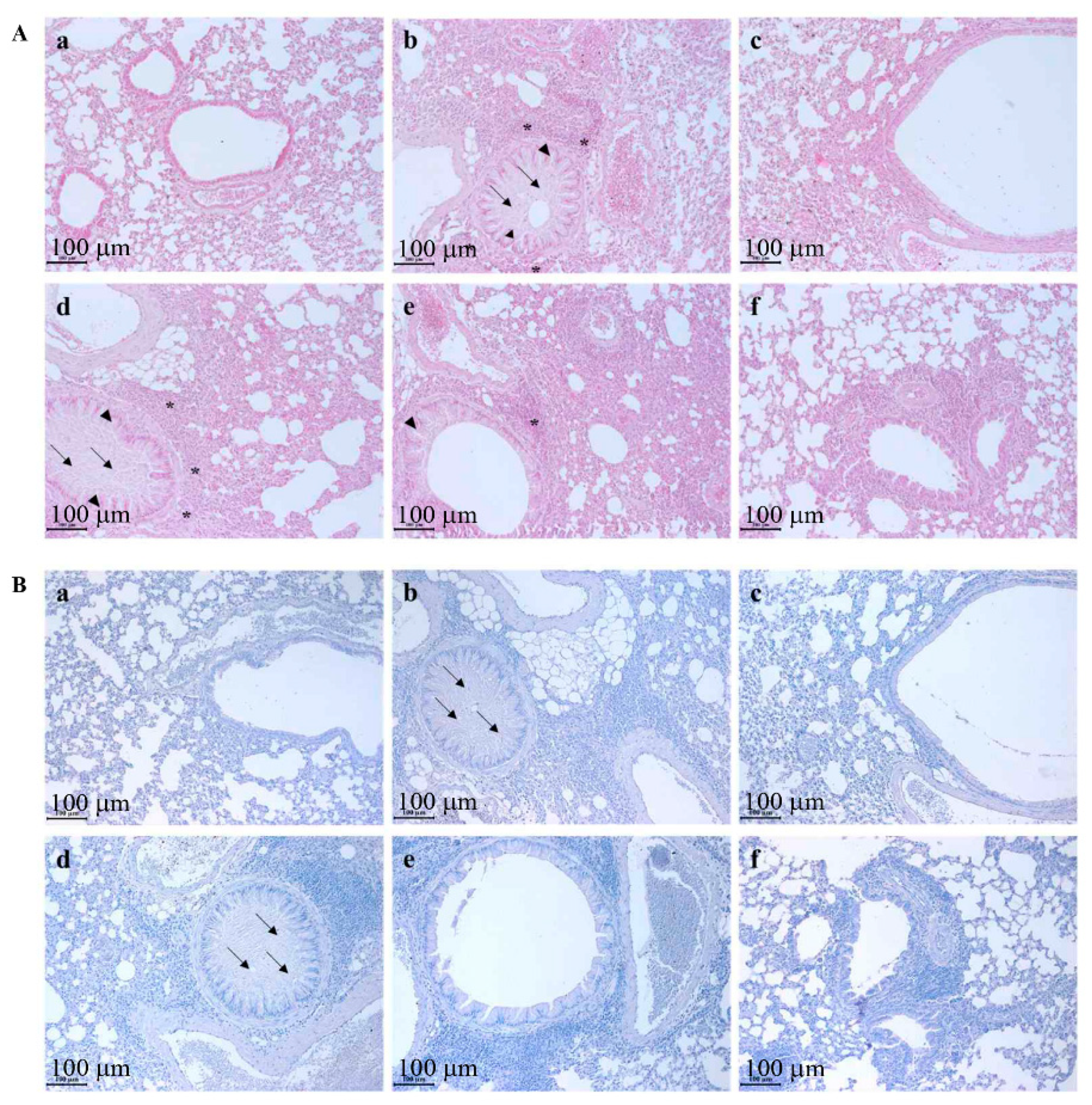
Ovalbumin effectively induced the representative morphological changes in the pulmonary system, which could be observed in asthma patients, such as airway remodeling, mucous hypersecretion, pulmonary epithelial cell hyperplasia, and inflammatory cell infiltration near the bronchioalveolar ducts and the vessels [3,4] (Figure 2(Ab,Bb)) compared to that of the control group (Figure 2(Aa,Ba)). C. laceolata treatment dose-dependently prevented the ovalbumin-induced morphological changes (Figure 2(Ad–f,Bd–f)). Although the inflammatory cell infiltration could be observed both in the 150 mg/kg/day C. laceolata treatment group and in the 300 mg/kg/day C. laceolata treatment group, the histopathological changes were almost completely prevented (Figure 2(Ae,f,Be,f)). Specifically, the mucous secretion levels (Figure 2(Be,f)) were similar to that in the dexamethasone treatment group (Figure 2(Bc)).
In order to clarify the prevention effect of C. laceolata against asthmatic changes, quantitative scoring was conducted according to our previous study [3]. In the vehicle control, the quantitative scores of histopathological changes (mean ± standard deviation) such as mucous hypersecretion, epithelial cell hyperplasia, and inflammatory cell infiltration were 0.1 ± 0.08, 0.2 ± 0.21, and 0.2 ± 0.13, respectively. However, the ovalbumin increased each item to 2.6 ± 0.34, 2.5 ± 0.21, and 2.6 ± 0.34, respectively, and dexamethasone treatment effectively decreased each score to 0.2 ± 0.11, 0.6 ± 0.25, and 0.5 ± 0.16, respectively. C. laceolata treatment dose-dependently prevented the morphological changes, and specifically, in the 300 mg/kg/day C. laceolata treatment group, the epithelial cell hyperplasia (0.9 ± 0.29) and the inflammatory cell infiltration (0.8 ± 0.31) were similar to that of the dexamethasone treatment group.
Table 1 is the quantitative score chart of the histopathological changes in the lung. The score was from 0 to 3 based on the morphological changes, including mucous hypersecretion (0, none; 1, little releasing; 2, half-packed in whole duct; 3, packed mucous), epithelial cell hyperplasia (0, none; 1, corrugated wall; 2, folded epithelium; 3, severely folded epithelium) and inflammatory cell infiltration (0, none; 1, few leukocytes; 2, moderate number of leukocytes; 3, large number of leukocytes). The score showed mean ± standard deviation. CON, vehicle group; OVA, ovalbumin-induced asthma group; DEX, positive control group (1 mg/kg/day dexamethasone with OVA); 50 mg/kg/day C. laceolata, 50 mg/kg/day C. laceolata treated group with OVA; 150 mg/kg/day C. laceolata, 150 mg/kg/day C. laceolata treated group with OVA; 300 mg/kg/day C. laceolata, 300 mg/kg/day C. laceolata treated group with OVA. * p < 0.05 vs. CON; ** p < 0.001 vs. CON; $ p < 0.05 vs. OVA; $$ p < 0.001 vs. OVA; # p < 0.05 vs. DEX; ## p < 0.001 vs. DEX; @ p < 0.05 vs. 50 mg/kg/day C. laceolata; @@ p < 0.001 vs. 50 mg/kg/day C. laceolata; % p < 0.05 vs. 150 mg/kg/day C. laceolata.

3.3. Codonopsis laceolata Extract Completely Controlled the Levels of cDNA and Protein of IL-4 and IL-13 via the Inactivation of Th2 Cell Transcription Factor, GATA-3

Asthma is a chronic inflammatory disease causing hyperresponsiveness in the pulmonary system through the imbalance of Th1 factors and Th2 factors, and the level of Th2 cell-related factors is much higher in asthma patients than in normal people [14]. GATA-3 is the typical Th2 cell transcription factor, the levels of which increased in asthma patients [18]. In order to evaluate C. laceolata‘s effect on the change of Th2 cell-related factors, the change of the Th2 cell transcription factor, GATA-3, was measured using immunofluorescent analysis (Figure 3A). In the ovalbumin treatment group, GATA-3 translocated from the cytoplasm to the nucleus compared to that in the control group, but dexamethasone treatment reversed the location of GATA-3 that was caused by ovalbumin treatment. C. laceolata treatment with 300 mg/kg/day prevented the GATA-3 translocation from the cytoplasm to the nucleus, prohibiting its function as a Th2 cell transcription factor. C. laceolata treatment effectively prevented the expression levels of IL-4 cDNA (Figure 3B) and IL-4 protein (Figure 3C), and specifically, in the 150 mg/kg/day C. laceolata treatment group and the 300 mg/kg/day C. laceolata treatment group, the declined levels of IL-4 protein were similar to that in the dexamethasone treatment group. In all of the C. laceolata treatment groups, the cDNA levels of IL-13 were similar to that in the dexamethasone treatment group (Figure 3D), and the C. laceolata treatment dose-dependently decreased the protein levels of IL-13 (Figure 3E). Specifically, the levels of IL-13 both in the 150 mg/kg/day C. laceolata treatment group and the 300 mg/kg/day C. laceolata treatment group were similar to that in the dexamethasone treatment group. That meant that the suppression effects of C. laceolata treatment on Th2 cell-related cytokines, such as IL-4 and IL-13, were very significant. (Figure 3C,E).

3.4. Codonopsis laceolata Extract Induced Apoptosis of Th2 Cells and Pulmonary Epithelial Cells via the NF-κB/COX-2 Pathway

Asthma is caused by the imbalance of Th1 cells and Th2 cells, and specifically, an increase in Th2 cells is a very important factor in causing asthma [14]. The hyperplasia/over-differentiation of some cells, such as pulmonary epithelial cells, Th2 cells, and airway smooth muscle cells, can usually be observed in asthma patients [23], but the characteristics of the cell types are very different based on the length of observation [24]. Codonopsis laceolata extract significantly induced the death of Th2 cells and respiratory epithelial cells, and produced similar levels of apoptotic cells as the dexamethasone treatment group (Figure 4A). However, it was difficult to find the apoptotic cells both in the ovalbumin treatment group and in the control vehicle treatment group. Ovalbumin treatment inhibited apoptosis in respiratory cells via the NF-κB/COX-2 pathway. In the ovalbumin treatment group, NF-κB was translocated from the cytoplasm to the nucleus compared to the result of the control vehicle treatment group, and the expression of COX-2 protein was highest among the experimental groups (Figure 4B). Both the dexamethasone treatment and Codonopsis laceolata extract suppressed the NF-κB translocation to the nucleus and the expression of COX-2 protein.

4. Discussion

Anti-asthmatic drugs can be classified into two categories—relievers, such as short-acting β2-agonists/anticholinergics, and controllers, such as long-acting β2-agonists, but a combined therapy, such as reliever/controller and corticosteroids, is usually used [28]. The current strategy of anti-asthmatic drug development involves searching for safe levels/doses of immunomodulatory or anti-inflammatory agents. The immunomodulatory agents involve the specific cytokine inhibitors, such as IL-4, IL-5, and IL-13, as anti-IL-4 and IL-13 can block eosinophilia, and IgE releasing via inactivating eosinophils and IL-5 can prevent severe eosinophilic asthma [38]. Anti-IgE agents, such as omalizumab, are immunomodulatory and are called the ‘magic bullet’ for allergic asthma treatment as they can suppress the exacerbation of asthma [39]. In our lab, we have been concentrating on finding Th2 cell-suppressing agents from natural products [13,40,41,42].
In our previous study, luteolin had been revealed as a major component of C. laceolata root, and it had an anti-hyperuricemic effect [36]. Another group reported that luteolin had an anti-allergic effect, although the results stated that it had a cytokine suppressive effect, such as IL-4, IL-5, and IL-13, but they could not confirm its anti-allergic mechanism [43]. Although the major compound in C. laceolata root was confirmed as luteolin, and the therapeutic effect for allergic asthma was evaluated, the biological mechanism could not be identified, and thus, an investigation of the anti-asthmatic mechanism of C. laceolata extract should be conducted.
Asthma might be caused by the imbalance of Th1 and Th2 factors [14], and specifically in asthma, Th2-related factors, such as IL-4 and IL-13, increase significantly [13,15]. IL-4 induces GATA-3’s transcriptional activity via a positive feedback mechanism and causes enhanced IgE secretion from B cells, eosinophilia, mucous hypersecretion, and airway remodeling [38]. IL-13 could stimulate asthma via various effects such as IgE secretion, eosinophilia stimulation, mucous hypersecretion, and airway remodeling via the NF-κB pathway [44,45]. Because of the importance of the inhibition effect of IL-4 and IL-13 against asthma occurrence, research for a dual vaccination of anti-IL-4 and anti-IL-13 against asthma has increased [46]. C. laceolata treatment effectively suppressed the expression of IL-4 and IL-13 via down-regulating GATA-3 levels (Figure 3), which means that if the anti-asthmatic potency of C. laceolata could be very high, there is the potential for C. laceolata to be a new anti-asthmatic agent. Interestingly, the cDNA level of IL-4 in the 150 mg/kg/day C. laceolata treatment group was similar to that in the dexamethasone treatment group, and the cDNA level of IL-13 in the 150 mg/kg/day C. laceolata treatment group was lower than that in the dexamethasone treatment group, but both of the protein levels, IL-4 and IL-13, were similar to that in the dexamethasone treatment group. However, we could not figure out the cause of the difference, and thus in a further study, we should define the reason.
Apoptosis (programmed cell death) is a very important physiological process, for example, in the development and aging of bio-organisms, and is one of the defense systems, contributing to for example, immune reactions or methods against toxins or disease occurrence [47]. Currently, it could be classified into three pathways such as the extrinsic pathway, the intrinsic pathway, and the perforin/granzyme pathway. The extrinsic pathway is related to the transmembrane receptor, and the intrinsic pathway is related to the mitochondria but not to the transmembrane receptor. The perforin/granzyme pathway is related to the transmembrane pore-forming molecules, perforin and the serine proteases, granzyme A and granzyme B [48]. In the extrinsic pathway, the typical transmembrane is a tumor necrosis factor receptor (TNFR), such as TNFR1, cluster of differentiation 95 (CD95)/Fas, or TNF-related apoptosis-inducing ligand receptor (TRAILR), and it is related to the NF-κB/COX-2 pathway and p53 pathway [49]. NF-κB is one of the transcription factors and could induce COX-2 for cell proliferation [50], and the IκB/NF-κB pathway is an important signal for balancing life and death [51]. Specifically, asthma is deeply related to hyperplasia/over-differentiation of special types of cells, such as pulmonary epithelial cells, Th2 cells, and airway smooth muscle cells [23], and the tendency of the cell type (apoptotic cells or normal cells) is based on the length of observation [24]. Vignola’s group reported that although there was no difference in the expression of apoptosis among asthma patients, normal persons, and corticosteroid-treated persons, there were differences in several other factors such as NF-κB, PCNA, and CD40-L [52]. In asthma patients and in asthma animal models, because the asthma-related cells could be over-differentiated, the population of apoptotic cells decreases [53,54,55], but in contrast to the other studies, Isik’s group reported that in an animal model, the apoptotic cells increased [56]. In our study, the level of apoptotic cells in the ovalbumin treatment group was very low, and the result was similar to that in the vehicle control group (Figure 4A). However, the apoptotic population both in the dexamethasone treatment group and in the 300 mg/kg/day C. laceolata treatment group significantly increased—specifically, the apoptotic cells located in the parenchymal region and in the pulmonary epithelial region. That meant that dexamethasone or C. laceolata stimulated Th2 cells and epithelial cells to undergo apoptosis. The pathway of apoptosis in dexamethasone treatment and C. laceolata treatment was revealed as the NF-κB/COX-2 pathway, as the NF-κB translocation and COX-2 expression were blocked (Figure 4B). We concluded that C. laceolata could be a promising drug against asthma via the NF-κB/COX-2 pathway-mediated apoptosis of Th2 cells and pulmonary epithelial cells.

Author Contributions

Conceptualization, S.-S.C. and D.-H.P.; methodology, K.M.H., H.-O.B. and S.-H.B.; software, S.-H.B.; investigation, S.-H.B. and K.M.H.; data curation, S.-H.B.; writing—original draft preparation, S.-H.B. and K.M.H.; writing—review and editing, S.-S.C. and D.-H.P.; supervision, D.-H.P. All authors have read and agreed to the published version of the manuscript.

Funding

This research received no external funding.

Institutional Review Board Statement

The study was conducted according to the guidelines of the Declaration of Helsinki and approved by the Institute of Animal Care and Use Committee of Chonnam National University (Approval No. CNU IACUC-YB-2016-15).

Conflicts of Interest

The authors declare no conflict of interest.

References

  1. Slejko, J.F.; Ghushchyan, V.H.; Sucher, B.; Globe, D.R.; Lin, S.L.; Globe, G.; Sullivan, P.W. Asthma control in the United States, 2008–2010: Indicators of poor asthma control. J. Allergy Clin. Immunol. 2014, 133, 1579–1587. [Google Scholar] [CrossRef]
  2. Ioachimescu, O.C.; Janocko, N.J.; Ciavatta, M.M.; Howard, M.; Warnock, M.V. Obstructive lung disease and obstructive sleep apnea (OLDOSA) cohort study: 10-year assessment. J. Clin. Sleep Med. 2020, 16, 267–277. [Google Scholar] [CrossRef]
  3. Lee, S.Y.; Kim, M.H.; Kim, S.H.; Ahn, T.; Kim, S.W.; Kwak, Y.S.; Cho, I.H.; Nah, S.Y.; Cho, S.S.; Park, K.M.; et al. Korean red ginseng affects ovalbumin-induced asthma by modulating IL-12, IL-4, and IL-6 levels and the NF-κB/COX-2 and PGE2 pathways. J. Ginseng Res. 2021, 45, 482–489. [Google Scholar] [CrossRef]
  4. Van den Bosch, W.; James, A.L.; Tiddens, H.A.W.M. Structure and function of small airways in asthma patients revisited. Eur. Respir. Rev. 2021, 30, 200186. [Google Scholar] [CrossRef]
  5. World Health Organization. Asthma. Fact Sheet in 3 May 2021. Available online: https://www.who.int/news-room/fact-sheets/detail/asthma (accessed on 22 March 2022).
  6. Ferrante, G.; Grutta, S.L. The burden of pediatric asthma. Front. Pediatr. 2018, 6, 186. [Google Scholar] [CrossRef] [Green Version]
  7. World Health Organization. World Health Statistics 2021: Monitoring Health for the SDGs, Sustainable Development Goals. Available online: https://apps.who.int/iris/handle/10665/342703 (accessed on 22 March 2022).
  8. Baxi, S.N.; Phipatanakul, W. The role of allergen exposure and avoidance in asthma. Adolesc. Med. State Art Rev. 2010, 21, 57-ix. [Google Scholar]
  9. Kouro, T.; Takatsu, K. IL-5- and eosinophil-mediated inflammation: From discovery to therapy. Int. Immunol. 2009, 21, 1303–1309. [Google Scholar] [CrossRef] [Green Version]
  10. Monteseirin, J. Neutrophils and asthma. J. Investig. Allergol. Clin. Immunol. 2009, 19, 340–354. [Google Scholar]
  11. Platts-Mills, T.A.E. The role of immunoglobulin E in allergy and asthma. Am. J. Respir. Crit. Care Med. 2001, 164, S1–S5. [Google Scholar] [CrossRef]
  12. Zedan, M.M.; El-Chennawi, F.A.; Fouda, A.E. Interleukin-12 and peripheral blood invariant natural killer T cells as an axis in childhood asthma pathogenesis. Iran. J. Allergy Asthma Immunol. 2010, 9, 43–48. [Google Scholar]
  13. Bok, S.H.; Cho, S.S.; Bae, C.S.; Kang, B.; Son, H.S.; Park, D.H. Socheongryongtang modulates asthma-related changes via modulation of TNF-α and T-bet & IFN-γ in an asthma murine model. Processes 2020, 8, 1167. [Google Scholar] [CrossRef]
  14. Boonpiyathad, T.; Sozener, Z.C.; Akdis, M.; Akdis, C.A. The role of Treg cell subsets in allergic disease. Asian Pac. J. Allergy Immunol. 2020, 38, 139–149. [Google Scholar] [CrossRef]
  15. Larche, M.; Robinson, D.S.; Kay, A.B. The role of T lymphocytes in the pathogenesis of asthma. J. Allergy Clin. Immunol. 2003, 111, 450–463. [Google Scholar] [CrossRef]
  16. Romagnani, S. T-cell subsets (Th1 versus Th2). Ann. Allergy Asthma Immunol. 2000, 85, 9–21. [Google Scholar] [CrossRef]
  17. Lighvani, A.A.; Frucht, D.M.; Jankovic, D.; Yamane, H.; Aliberti, J.; Hissong, B.D.; Nguyen, B.V.; Gadina, M.; Sher, A.; Paul, W.E.; et al. T-bet is rapidly induced by interferon-γ in lymphoid and myeloid cells. Proc. Natl. Acad. Sci. USA 2001, 98, 15137–15142. [Google Scholar] [CrossRef] [Green Version]
  18. Yagi, R.; Zhu, J.; Paul, W.E. An updated view on transcription factor GATA3-mediated regulation of Th1 and Th2 cell differentiation. Int. Immunol. 2011, 23, 415–420. [Google Scholar] [CrossRef] [Green Version]
  19. Mitchell, C.; Provost, K.; Niu, N.; Homer, R.; Cohn, L. Interferon-gamma acts on the airway epithelium to inhibit local and systemic pathology in allergic airway disease. J. Immunol. 2011, 187, 3815–3820. [Google Scholar] [CrossRef] [Green Version]
  20. Chung, F. Anti-inflammatory cytokines in asthma and allergy: Interleukin-10, interleukin-12, interferon-γ. Mediat. Inflamm. 2001, 10, 51–59. [Google Scholar] [CrossRef] [Green Version]
  21. Gubernatorova, E.O.; Namakanova, O.A.; Gorshkova, E.A.; Medvedovskaya, A.D.; Nedospasov, S.A.; Drutskaya, M.S. Novel anti-cytokine strategies for prevention and treatment of respiratory allergic diseases. Front. Immunol. 2021, 12, 601842. [Google Scholar] [CrossRef]
  22. Walter, M.J.; Morton, J.D.; Kajiwara, N.; Agapov, E.; Holtzman, M.J. Viral induction of a chronic asthma phenotype and genetic segregation from the acute response. J. Clin. Investig. 2002, 110, 165–175. [Google Scholar] [CrossRef]
  23. Johnson, P.R.A.; Roth, M.; Tamm, M.; Hughes, M.; Ge, Q.; King, G.; Burgess, J.; Black, J.L. Airway smooth muscle cell proliferation is increased in asthma. Am. J. Respir. Crit. Care Med. 2001, 164, 474–477. [Google Scholar] [CrossRef] [PubMed]
  24. O’Sullivan, M.P.; Tyner, J.W.; Holtzman, M.J. Apoptosis in the airways. Another balancing act in the epithelial program. Am. J. Respir. Cell Mol. Biol. 2003, 29, 3–7. [Google Scholar] [CrossRef] [PubMed]
  25. Datta, A.; Moitra, S.; Faruk, S.M.O.; Das, P.K.; Mondal, S.; Hazra, I.; Basu, A.K.; Tripathi, S.K.; Chaudhuri, S. Unravelling the apoptotic mechanisms in T-lymphocytes in an animal model for pollen induced airway allergy and studying the impact of specific immunotherapy. Immunobiology 2019, 224, 183–195. [Google Scholar] [CrossRef] [PubMed]
  26. Zhang, J.; Zhou, X.; Zhu, J. Beauveria attenuates asthma by inhibiting inflammatory response and inducing lymphocytic cell apoptosis. Oncotarget 2016, 7, 74557–74568. [Google Scholar] [CrossRef] [Green Version]
  27. Nagai, H. Recent research and developmental strategy of anti-asthma drugs. Pharmacol. Ther. 2012, 133, 70–78. [Google Scholar] [CrossRef]
  28. Fahy, J.V. Type 2 inflammation in asthma—Present in most, absent in many. Nat. Rev. Immunol. 2015, 15, 57–65. [Google Scholar] [CrossRef]
  29. Crompton, G. A brief history of inhaled asthma therapy over the last fifty years. Prim. Care Respir. J. 2006, 15, 326–331. [Google Scholar] [CrossRef] [Green Version]
  30. Blake, K.; Lima, J. Pharmacogenomics of long-acting β2-agonsits. Expert Opin. Drug Metab. Toxicol. 2015, 11, 1733–1751. [Google Scholar] [CrossRef]
  31. Ciriaco, M.; Ventrice, P.; Russo, G.; Scicchitano, M.; Mazzitello, G.; Scicchitano, F.; Russo, E. Corticosteroid-related central nervous system side effects. J. Pharmacol. Pharmacother. 2013, 4 (Suppl. 1), S94–S98. [Google Scholar] [CrossRef] [Green Version]
  32. Wang, L.; Xu, M.L.; Hu, J.H.; Rasmussen, S.K.; Wang, M.H. Codonopsis lanceolate extract induces G0/G1 arrest and apoptosis in human colon tumor HT-29 cells—Involvement of ROS generation and polyamine depletion. Food Chem. Toxicol. 2011, 49, 149–154. [Google Scholar] [CrossRef]
  33. Lee, J.S.; Kim, K.J.; Kim, Y.H.; Kim, D.B.; Shin, G.H.; Cho, J.H.; Kim, B.K.; Lee, B.-Y.; Lee, O.-H. Codonopsis lanceolate extract prevents diet-induced obesity in C57BL/6 mice. Nutrients 2014, 6, 4663–4677. [Google Scholar] [CrossRef] [Green Version]
  34. Cha, A.; Choi, Y.; Jin, Y.; Sung, M.K.; Koo, Y.C.; Lee, K.W.; Park, T. Antilipogenic and anti-inflammatory activities of Codonopsis lanceolate in mice hepatic tissues after chronic ethanol feeding. J. Biomed. Biotechnol. 2012, 2012, 141395. [Google Scholar] [CrossRef] [Green Version]
  35. Lee, Y.G.; Kim, J.Y.; Lee, J.Y.; Byeon, S.E.; Hong, E.K.; Lee, J.; Rhee, M.H.; Park, H.J.; Cho, J.Y. Regulatory effects of Codonopsis lanceolate on macrophage-mediated immune responses. J. Ethnopharmacol. 2007, 112, 180–188. [Google Scholar] [CrossRef]
  36. Song, S.Y.; Bok, S.H.; Lee, S.H.; Kim, M.H.; Boo, H.O.; Kim, H.H.; Park, D.-H.; Cho, S.-S. Standardization of diploid Codonopsis laceolata root extract as an anti-hyperuricemic source. Processes 2021, 9, 2065. [Google Scholar] [CrossRef]
  37. Lee, S.Y.; Cho, S.S.; Li, Y.; Bae, C.S.; Park, K.M.; Park, D.H. Anti-inflammatory effect of Curcuma longa and Allium hookeri co-treatment via NF-κB and COX-2 pathways. Sci. Rep. 2020, 10, 5718. [Google Scholar] [CrossRef]
  38. Barnes, P.J. Targeting cytokines to treat asthma and chronic obstructive pulmonary disease. Nat. Rev. Immunol. 2018, 18, 454–466. [Google Scholar] [CrossRef]
  39. Tarantini, F.; Baiardini, I.; Passalacqua, G.; Braido, F.; Canonica, G.W. Asthma treatment: ‘magic bullets which seek their own targets’. Allergy 2007, 62, 605–610. [Google Scholar] [CrossRef]
  40. Bang, M.A.; Seo, J.H.; Seo, J.W.; Jo, G.H.; Jung, S.K.; Yu, R.; Park, D.H.; Park, S.J. Bacillus subtilis KCTC 11782BP-produced alginate oligosaccharide effectively suppresses asthma via T-helper cell type 2-related cytokines. PLoS ONE 2015, 10, e0117524. [Google Scholar] [CrossRef]
  41. Lee, S.Y.; Bae, C.S.; Choi, Y.H.; Seo, N.S.; Na, C.S.; Yoo, J.C.; Cho, S.S.; Park, D.H. Opuntia humifusa modulates morphological changes characteristic of asthma via IL-4 and IL-13 in an asthma murine model. Food Nutr. Res. 2017, 61, 1393307. [Google Scholar] [CrossRef] [Green Version]
  42. Bok, S.H.; Seo, J.H.; Bae, C.S.; Kang, B.; Cho, S.S.; Park, D.H. Allium hookeri root extract regulates asthmatic changes through immunological modulation of Th1/Th2-related factors in an ovalbumin-induced asthma mouse model. Mol. Med. Rep. 2019, 20, 3215–3223. [Google Scholar] [CrossRef] [Green Version]
  43. Jang, T.Y.; Jung, A.Y.; Kyung, T.S.; Kim, D.Y.; Hwang, J.H.; Kim, Y.H. Anti-allergic effect of luteolin in mice with allergic asthma and rhinitis. Cent. Eur. J. Immunol. 2017, 42, 24–29. [Google Scholar] [CrossRef] [Green Version]
  44. Zhu, Z.; Homer, R.J.; Wang, Z.; Chen, Q.; Geba, G.P.; Wang, J.; Zhang, Y.; Elias, J.A. Pulmonary expression of interleukin-13 causes inflammation, mucus hypersecretion, subepithelial fibrosis, physiologic abnormalities, and eotaxin production. J. Clin. Investig. 1999, 103, 779–788. [Google Scholar] [CrossRef] [Green Version]
  45. Chapoval, S.P.; Al-Garawi, A.; Lora, J.M.; Strickland, I.; Ma, B.; Lee, P.J.; Homer, R.J.; Ghosh, S.; Coyle, A.J.; Elias, J.A. Inhibition of NF-κB activation reduces the tissue effects of transgenic IL-13. J. Immunol. 2007, 179, 7030–7041. [Google Scholar] [CrossRef]
  46. Conde, E.; Bertrand, R.; Balbino, B.; Bonnefoy, J.; Stackowicz, J.; Caillot, N.; Colaone, F.; Hamdi, S.; Houmadi, R.; Loste, A.; et al. Dual vaccination against IL-4 and IL-13 protects against chronic allergic asthma in mice. Nat. Commun. 2021, 12, 2574. [Google Scholar] [CrossRef]
  47. Norbury, C.J.; Hickson, I.D. Cellular responses to DNA damage. Annu. Rev. Pharmacol. Toxicol. 2001, 41, 367–401. [Google Scholar] [CrossRef] [Green Version]
  48. Elmore, S. Apoptosis: A review of programmed cell death. Toxicol. Pathol. 2007, 35, 495–516. [Google Scholar] [CrossRef]
  49. Rockland Immunochemicals, Inc. Apoptosis Pathway Antibodies. Available online: https://www.rockland.com/resources/apoptosis-pathway-antibodies (accessed on 26 April 2022).
  50. Lim, J.W.; Kim, H.; Kim, K.H. Nuclear factor-κB regulates cyclooxygenase-2 expression and cell proliferation in human gastric cancer cells. Lab. Investig. 2001, 81, 349–360. [Google Scholar] [CrossRef] [Green Version]
  51. Luo, J.L.; Kamata, H.; Karin, M. IKK/NF-κB signaling: Balancing life and death—A new approach to cancer therapy. J. Clin. Investig. 2005, 115, 2625–2632. [Google Scholar] [CrossRef] [Green Version]
  52. Vignola, A.M.; Chiappara, G.; Siena, L.; Bruno, A.; Gagliardo, R.; Merendino, A.M.; Polla, B.S.; Arrigo, A.; Bonsignore, G.; Bousquet, J.; et al. Proliferation and activation of bronchial epithelial cells in corticosteroid-dependent asthma. J. Allergy Clin. Immunol. 2001, 108, 738–746. [Google Scholar] [CrossRef]
  53. Benayoun, L.; Letuve, S.; Druilhe, A.; Boczkowski, J.; Dombret, M.C.; Mechighel, P.; Megret, J.; Leseche, G.; Aubier, M.; Pretolani, M. Regulation of peroxisome proliferator-activated receptor γ expression in human asthmatic airways: Relationship with proliferation, apoptosis, and airway remodeling. Am. J. Respir. Crit. Care Med. 2001, 164, 1487–1494. [Google Scholar] [CrossRef]
  54. Hoppenot, D.; Malakauskas, K.; Lavinskiene, S.; Bajoriuniene, I.; Kalinauskaite, V.; Sakalauskas, R. Peripheral blood Th9 cells and eosinophil apoptosis in asthma patients. Medicina 2015, 51, 10–17. [Google Scholar] [CrossRef]
  55. Geng, G.; Du, Y.; Dai, J.; Tian, D.; Xia, Y.; Fu, Z. KIF3A knockdown sensitizes bronchial epithelia to apoptosis and aggravates airway inflammation in asthma. Biomed Pharmacother. 2018, 97, 1349–1355. [Google Scholar] [CrossRef]
  56. Isik, S.; Karaman, M.; Micili, S.C.; Caglayan-Sozmen, S.; Bagriyanik, H.A.; Arikan-Ayyildiz, Z.; Uzuner, N. Beneficial effects of ursodeoxycholic acid via inhibition of airway remodeling, apoptosis of airway epithelia cells, and Th2 immune response in murine model of chronic asthma. Allergol. Immunopathol. 2017, 45, 339–349. [Google Scholar] [CrossRef]
Figure 1. C. laceolata suppressed the population increments of white blood cells (WBC) and lymphocytes (LY) in the bronchioalveolar fluid (BALF) and the quantity of IgE in serum, which were increased by ovalbumin treatment. (A) C. laceolata dose-dependently decreased the number of WBC, which was upregulated by ovalbumin. (B) The population of lymphocytes was especially decreased by C. laceolata treatment. (C) The dose-dependent decrement of inflammatory cells by C. laceolata was observed from the Diff-Quick stain. Scale bar, 50 μm; magnification, ×400. a, CON; b, OVA; c, DEX, d, 50 mg/kg/day C. laceolata, e, 150 mg/kg/day C. laceolata. f. 300 mg/kg/day C. laceolata. (D) C. laceolata dose-dependently decreased the level of serum IgE, which was up-regulated by ovalbumin treatment, and specifically, in the 300 mg/kg/day C. laceolata treatment group, the level of serum IgE was similar to that in the dexamethasone treatment group. CON, vehicle group; OVA, ovalbumin-induced asthma group; DEX, positive control group (1 mg/kg/day dexamethasone with OVA); 50 mg/kg/day C. laceolata, 50 mg/kg/day C. laceolata treated group with OVA; 150 mg/kg/day C. laceolata, 150 mg/kg/day C. laceolata treated group with OVA; 300 mg/kg/day C. laceolata, 300 mg/kg/day C. laceolata treated group with OVA. * p < 0.05 vs. CON; ** p < 0.001 vs. CON; $ p < 0.05 vs. OVA; $$ p < 0.001 vs. OVA; # p < 0.05 vs. DEX; ## p < 0.001 vs. DEX; @ p < 0.05 vs. 50 mg/kg/day C. laceolata; @@ p < 0.001 vs. 50 mg/kg/day C. laceolata; % p < 0.05 vs. 150 mg/kg/day C. laceolata; %% p < 0.001 vs. 150 mg/kg/day C. laceolata.
Processes 10 01249 g001
Figure 2. C. laceolata prevented the ovalbumin-induced asthmatic representative morphological changes in the pulmonary system, such as airway remodeling, mucous hypersecretion, epithelial cell hyperplasia and inflammatory cell infiltration. (A) The photos of the hematoxylin and eosin (H&E) stained results. a, The normal lung. b, Ovalbumin treatment increased mucous secretion in the bronchioalveolar ducts (arrow), overgrowth of the pulmonary epithelial cells (arrow head), and infiltration of the inflammatory cells near the bronchioalveolar ducts and vessels (asterisk mark). c, Dexamethasone treatment decreased the ovalbumin-induced morphological changes. d, The changes in the lung in the 50 mg/kg/day C. laceolata treatment group were similar to those in the ovalbumin treatment group. e, In the 150 mg/kg/day C. laceolata treatment group, the severity of the ovalbumin-induced pulmonary changes was lower than in both the ovalbumin treatment group and in the 50 mg/kg/day C. laceolata treatment group, although it was higher than in both the dexamethasone treatment group and in the 300 mg/kg/day C. laceolata treatment group. f, The 300 mg/kg/day C. laceolata treatment effectively inhibited the ovalbumin-induced epithelial cell hyperplasia and inflammatory cell infiltration. (B) The results of the periodic acid schiff (PAS) stain. a, The normal lung. b, Ovalbumin-stimulated mucous hypersecretion (arrows). c, Dexamethasone effectively controlled the mucous secretion. d, The mucous secretion level in the 50 mg/kg/day C. laceolata treatment was similar to that in the ovalbumin treatment group. e, In the 150 mg/kg/day C. laceolata treatment group, the mucous secretion was significantly decreased. f, The level of mucous secretion in the 300 mg/kg/day C. laceolata treatment group was similar to that in the dexamethasone treatment group. Scale bar, 100 μm; magnification, ×200.
Processes 10 01249 g002
Figure 3. C. laceolata controlled the level of ovalbumin-induced Th2-related cytokines such as IL-4 and IL-13 by inactivating the Th2 cell transcription factor, GATA-3. (A) The expression result of the Th2 cell transcription factor, GATA-3, according to the C. laceolata treatment. In order to release the Th2-related cytokines, GATA-3 should translocate from the cytoplasm to the nucleus to conduct the Th2 cell transcription factor. The released levels of GATA-3 both in the dexamethasone treatment group (DEX) and the 300 mg/kg/day C. laceolata treatment group were higher than that in the ovalbumin treatment group, and that meant that the dexamethasone treatment and C. laceolata treatment prevented ovalbumin-induced GATA-3 activation. Scale bar, 50 μm; magnification, ×400. CON, vehicle group; OVA, ovalbumin-induced asthma group; DEX, positive control group (1 mg/kg/day dexamethasone with OVA); C. laceolata 300 mg/kg, 300 mg/kg/day C. laceolata treated group with OVA. (B) C. laceolata treatment suppressed the ovalbumin-induced increase in IL-4 cDNA levels and (C) IL-4 protein levels to levels similar to the dexamethasone treatment. (D) C. laceolata treatment down-regulated the IL-13 cDNA level and (E) the IL-13 protein level, and specifically, the decreased levels were similar to that of the dexamethasone treatment group. CON, vehicle group; OVA, ovalbumin-induced asthma group; DEX, positive control group (1 mg/kg/day dexamethasone with OVA); C. laceolata 300 mg/kg, 300 mg/kg/day C. laceolata treated group with OVA. * p < 0.05 vs. CON; ** p < 0.001 vs. CON; $ p < 0.05 vs. OVA; $$ p < 0.001 vs. OVA; # p < 0.05 vs. DEX; @ p < 0.05 vs. 50 mg/kg/day C. laceolata; % p < 0.05 vs. 150 mg/kg/day C. laceolata.
Processes 10 01249 g003
Figure 4. C. laceolata stimulated the cell death (apoptosis) of Th2 cells and pulmonary epithelial cells via the NF-κB/COX-2 pathway. (A) C. laceolata induced apoptosis on both the Th2 cells and bronchioalveolar epithelial cells compared to the result in the ovalbumin treatment group. It was difficult to observe the apoptotic cells both in the vehicle control group and in the ovalbumin treatment group. Although the dexamethasone treatment and 300 mg/kg/day C. laceolata treatment upregulated the apoptosis, the number of apoptotic cells in the 300 mg/kg/day C. laceolata treatment group (magnified parts) was more than that in the dexamethasone treatment group (magnified part). Scale bar, 50 μm; magnification, ×400. CON, vehicle group; OVA, ovalbumin-induced asthma group; DEX, positive control group (1 mg/kg/day dexamethasone with OVA); C. laceolata 300 mg/kg, 300 mg/kg/day C. laceolata treated group with OVA. (B) In the ovalbumin treatment group, the expression levels of NF-κB in the nucleus and COX-2 in the cytoplasm were the highest compared to the other groups, and specifically, the expression level of COX-2 in the ovalbumin treatment group increased significantly. However, the dexamethasone treatment and 300 mg/kg/day C. laceolata treatment both suppressed the levels of expressed NF-κB in the nucleus and COX-2 in the cytoplasm. Scale bar, 50 μm; magnification, ×1000. CON, vehicle group; OVA, ovalbumin-induced asthma group; DEX, positive control group (1 mg/kg/day dexamethasone with OVA); C. laceolata 300 mg/kg, 300 mg/kg/day C. laceolata treated group with OVA.
Processes 10 01249 g004
Table 1. The quantitative score chart of the histopathological changes in the lung.
GroupMucous Hypersecretion (0~3)Epithelial Hyperplasia (0~3)Inflammatory Cell Infiltration (0~3)
CON0.1 ± 0.080.2 ± 0.210.1 ± 0.13
OVA2.6 ± 0.34 **2.5 ± 0.21 **2.6 ± 0.34 **
DEX0.2 ± 0.11 $$0.6 ± 0.25 *,$$0.5 ± 0.16 *,$$
50 mg/kg/day C. laceolata2.6 ± 0.38 **,##2.3 ± 0.21 **,##2.4 ± 0.40 **,##
150 mg/kg/day C. laceolata1.4 ± 0.42 **,$,##,@1.3 ± 0.22 **,$$,#,@@1.4 ± 0.38 **,$$,##,@
300 mg/kg/day C. laceolata0.5 ± 0.24 *,$$,#,@@,%0.9 ± 0.29 *,$$,@@,%0.8 ± 0.31 *,$$,@@,%
Publisher’s Note: MDPI stays neutral with regard to jurisdictional claims in published maps and institutional affiliations.