Article

Immune Redox Modulation Effects of Non-Electrolyzed Hypochlorous Acid Water on Helicobacter pylori-Infected C57BL/6 Mouse Model

1
Department of Convergence Medicine, Wonju College of Medicine, Yonsei University, Wonju 26426, Republic of Korea
2
Department of Chemical Biology, University of California, Berkeley, CA 94720, USA
3
Department of Laboratory Medicine, Yonsei University Wonju College of Medicine, Yonsei University, Wonju 26426, Republic of Korea
4
Corporate Research Institute, NUGA MEDICAL Co., Ltd., 9, Bongeunsa-ro 68-gil, Gangnam-gu, Seoul 06153, Republic of Korea
5
Department of Internal Medicine, Division of Hematology-Oncology, Wonju Severance Christian Hospital, Wonju 26426, Republic of Korea
*
Authors to whom correspondence should be addressed.
Processes 2023, 11(5), 1474; https://doi.org/10.3390/pr11051474
Submission received: 27 March 2023 / Revised: 20 April 2023 / Accepted: 11 May 2023 / Published: 12 May 2023
(This article belongs to the Section Biological Processes and Systems)

Abstract

:
Recently, non-electrolyzed HOCl water has gained the attention of researchers as a new disinfecting agent owing to its high sterilization power, easy accessibility, and safety. Non-electrolyzed HOCl water was developed through mixing at a specific ratio based on hypochlorite and mineral supplements, which revealed a high oxidizing power. In this study, we investigated the effects of non-electrolyzed HOCl water on Helicobacter pylori (H. pylori) infection in C57BL/6 mice over 10 weeks. Mice were divided into three groups: normal control (NC) group supplied with purified water (PW) without infection, PW + H. pylori group supplied with PW after H. pylori infection, and HOCl + H. pylori group supplied with HOCl after H. pylori infection. Our findings demonstrated that the HOCl + H. pylori group greatly inhibited WBC and its differential counts, including total white blood cell (WBC), neutrophils, lymphocytes, monocytes, and eosinophils, when compared to the PW + H. pylori group. Accordingly, the amount of reactive oxygen species and calcium activity significantly decreased in the HOCl + H. pylori group compared to the PW + H. pylori group in both serum and stomach lysates. In contrast, HOCl water treatment enhanced GPx activity compared to PW treatment after H. pylori infection in both serum and stomach lysates. Accordingly, the levels of granulocyte-macrophage colony-stimulating factor, IL-1β, and TNF-α cytokine levels were significantly decreased in the HOCl + H. pylori group compared to those in the PW + H. pylori group in the stomach lysate; however, there was no significant difference in serum. In addition, the expression levels of Bax, MMP-3, MMP-9, and TLR-4 were found to decrease after HOCl water treatment, whereas the expression level of Bcl-2 was found to be enhanced after HOCl water treatment in the stomach lysate. Taken together, our results suggest that drinking non-electrolyzed HOCl water has positive anti-oxidative, anti-inflammatory, and anti-apoptotic effects in H. pylori-infected mice through redox and immune regulation.

1. Introduction

The spiral-shaped, Gram-negative bacterium Helicobacter pylori (H. pylori) is a persistent colonizer of the human stomach and can result in peptic ulcer disease, gastritis, dyspepsia, and stomach cancer [1,2,3,4]. One research showed that an inflammatory response is brought on by the presence of neutrophils, lymphocytes, macrophages, and dendritic cells in the stomach mucosa during H. pylori colonization [5]. A growing body of research has also demonstrated that microbial pathogens such as H. pylori cause oxidative stress (OS) in the host cell environment, which is crucial for epithelial damage [6]. Moreover, H. pylori-induced OS contributes to altered epithelial proliferation in the gastric mucosal layer, increased apoptosis, and increased oxidative damage to DNA [7,8]. In one study, patients with H. pylori infection had higher levels of reactive oxygen species (ROS) in their mucosal layer [9]. Studies revealed that the H. pylori itself has a propensity to produce ROS and accumulate in gastric epithelial cells [10,11]. Tumor necrosis factor-alpha (TNF-alpha), interleukin (IL)-1β, IL-6, and interferon-gamma (IFN-γ), as well as other proinflammatory cytokines, were also discovered to induce ROS in the oxidative environment and trigger an inflammatory reaction in gastric epithelial cells [7]. Infection with H. pylori can cause the release of proinflammatory mediators and abnormally high levels of ROS and NO in stomach mucosal epithelial cells [12]. Additionally, research has demonstrated that the development of cellular injury is significantly influenced by the elevation of intracellular calcium (Ca2+) [13,14]. Furthermore, cellular processes including cell growth and cytokine release can be impacted by Ca2+ overload [15]. However, little is known about the regulation of intracellular Ca2+ by H. pylori in gastric cells. According to previous studies, H. pylori causes apoptosis in the gastric epithelium by upregulating the proapoptotic protein marker Bcl-2-associated X (Bax) and downregulating the anti-apoptotic protein Bcl-2 [16]. H. pylori infection has been linked to altered Bax and/or Bcl-2 expression patterns in numerous investigations [17,18]. Additionally, matrix metalloproteinases (MMPs) have an impact on both the inflammatory environment and structure of infected tissues [19]. Toll-like receptors (TLRs) are crucial adaptive immune system activators that control inflammation in response to H. pylori infection [20]. New anti-H. pylori medications are desperately needed, along with ones that are therapeutically successful and have few side effects. Researchers have recently become interested in the use of hypochlorous acid water against a variety of bacterial activity, including H. pylori. Sodium hypochlorite and HOCl are combined to create the weak acid known as HOCl water, which has a pH adjusted range of 6.0 to 6.4 and a residual chlorine content of about 60 ppm [21]. Evidence suggests that HOCl water is very effective against various microorganisms [21]. Owing to its anti-inflammatory and other biological effects, electrolyzed HOCl water has also been applied in various studies pertaining to the treatment of numerous inflammatory skin diseases and atopic dermatitis, as well as itching, diabetic ulcers, and wound healing [22,23,24]. Studies have reported that HOCl is safe for human use [25]. One study reported that intraperitoneal lavage and wound cleaning with HOCl with a residual chlorine concentration of 40–60 ppm had no deleterious effects in a clinical investigation [26]. Additionally, 0.01–0.1% (w/v) HOCl, which is produced by combining hydrochloric acid with sodium hypochlorite, did not cause eye irritation or systemic toxicity, according to a study on animal safety and toxicity tests [27]. In addition to electrolyzed HOCl water, non-electrolyzed HOCl water has been developed as a new disinfection agent owing to its high sterilizing power, ease of accessibility, and safety [28]. By combining hypochlorite and mineral additions in a precise ratio, non-electrolyzed HOCl water was generated, which exhibited tremendous oxidizing power. We therefore evaluated whether drinking non-electrolyzed HOCl water had anti-oxidative, anti-apoptotic, and anti-inflammatory effects in C57BL/6 mice that were infected with H. pylori. The results of this investigation could serve as a database to create safe and effective treatments for H. pylori infection, especially in relation to stomach problems.

2. Materials and Methods

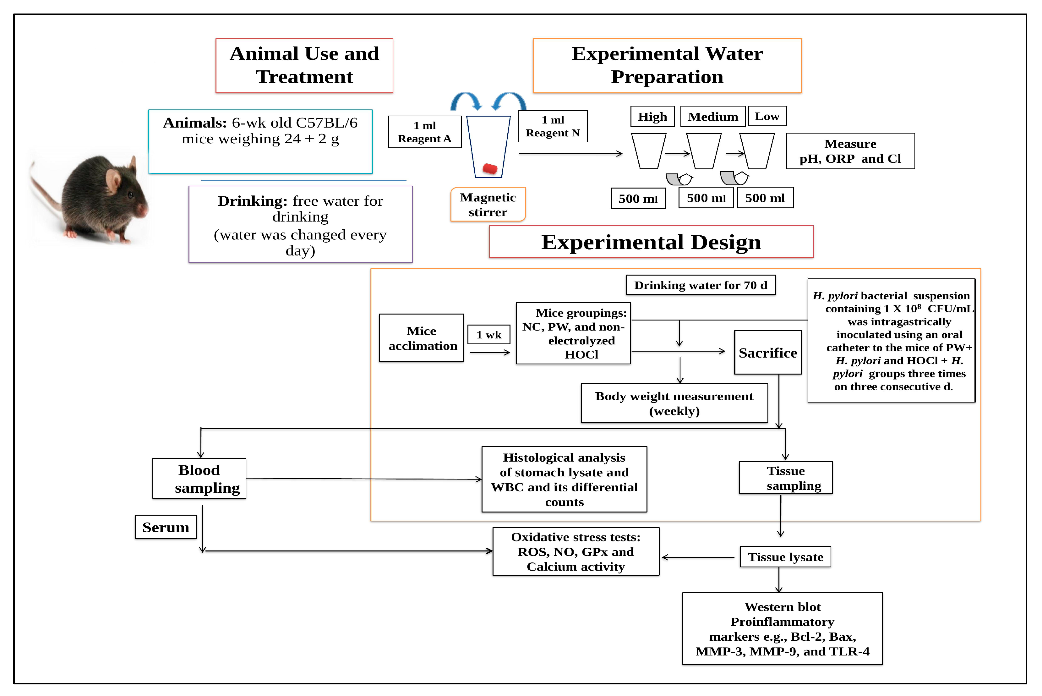
2.1. Animal Groupings and Treatment Procedure

C57BL/6 mice that were six weeks old and weighed an average of 25 ± 1.2 g were acclimated for seven days in a controlled setting with a temperature of 22 ± 2 °C and a humidity level of 40–60% during a 12 h light–dark cycle. Orient Bio Inc. sold these rodents to us. (SEONGNAM, Republic of Korea). Fifteen mice were randomly divided into three different groups (n = 5 per group) as follows: normal control (NC) group supplied with purified water (PW) without infection, PW + H. pylori group supplied with PW after H. pylori infection, and HOCl + H. pylori group supplied with HOCl water after H. pylori infection. All mice were fasted for 6 h with unrestricted access to water before each inoculation. Consequently, 100 μL of H. pylori bacterial suspension containing 1 × 10⁸ CFU/mL was intragastrically inoculated using an oral catheter into the mice of PW + H. pylori and HOCl + H. pylori groups three times on three consecutive days. Additionally, experimental water was administered with free access to the mice, and it was exchanged with new water every day during the experimental period. Body weight was measured weekly. After 10 weeks of drinking water, all mice were sacrificed under anesthesia (Isoflurane, Hana Pharm Co., Ltd., Seoul, Republic of Korea) and blood and tissue samples were collected for hematological and biochemical examinations and Western blot analysis. The Institutional Animal Care and Use Committee (IACUC) Wonju College of Medicine, Yonsei University, Gangwon, Wonju, Republic of Korea, authorized the study protocol for this investigation (ethical approval no: YWC-180615-1). Figure 1 shows the experimental design.

2.2. Water Preparation and Their Properties

Non-electrolyzed HOCl water was supplied by Sungjin Farm Co., Ltd. (Gyeongju-si, Gyeongsangbuk-do, Republic of Korea) and was composed of reagents A and N. In brief, 1000 μL of reagent A and N (ratio 1:1) was mixed with 2 L of tap water (TW). A pH and ORP meter (DKK-TOA Corporation, Tokyo, Japan) was used to detect the pH and oxidation-reduction potential (ORP) values, and a chlorine meter was used to assess the available chlorine concentration (ACC) (Lutron Electronic Enterprise, Co., Ltd., Taipei, Taiwan). The properties of the water are listed in Table 1.

2.3. H. pylori Infection in Mice

The strains of H. pylori used in this research were purchased from the American Type Culture Collection (ATCC® 43504TM; P.O. Box, 1549, Manassas, VA, USA) and propagated in accordance with ATCC guidelines for each strain. The concentrations were therefore determined by a direct colony count following a 10-fold dilution using bacteria that were collected during the stationary phase. Microbiological specimens were collected from the surface of the bacterial culture plate using an inoculating loop and swab. H. pylori was routinely cultured on Columbia agar with 5% sheep blood (Becton Dickinson & Company, Franklin Lakes, NJ, USA; 18-03 NJ-208, Franklin Lakes, NJ, USA) at 37 °C for 3–5 days under anaerobic conditions. All mice, except for those in the NC group, were fasted for 6 h with free access to water before each inoculation. Therefore, 100 μL H. pylori bacterial solution containing 1 × 10⁸ CFU/mL intragastrically inoculated using an oral catheter into mice of the PW + H. pylori and HOCl + H. pylori groups three times on three consecutive days.

2.4. Serum and Stomach Tissue Lysates Sample Preparation

The serum samples were obtained by centrifuging the blood in a BD Microtainer tube (Lot 9305148, Becton, Dickinson and Company, Franklin Lakes, NJ, USA) at 14,000 rpm for 5 min at 4 °C. The serum samples were then kept at −80 °C until use. To prepare the stomach tissue sample, stomach tissue (2 × 2 mm in size) was cut and placed in ice-cold radioimmunoprecipitation assay (RIPA) buffer (Pierce Biotechnology Inc., Waltham, MA, USA) with protease inhibitor compounds (Sigma Chemical Co., St Louis, MO, USA). The stomach tissue was homogenized for 10 min at 14,000 rpm and centrifuged for 5 min, and the supernatant of the stomach lysate was used as a sample.

2.5. Measurement of Body Weight

Weekly body weight checks were conducted during the trial to get a baseline.

2.6. Histological Examination by Giemsa Staining

The part of the stomach (5 × 5 mm in size) was sectioned for histological investigation. After dehydration, clearing, and infiltration in accordance with the standard procedure, tissue samples were fixed with 10% formalin solution for 12 h and embedded in paraffin. The sections were deparaffinized, rehydrated, and sliced before being stained with Giemsa solution (Muto Pure Chemicals Co., Ltd., Tokyo, Japan). A light microscope (BX51; Olympus Corp., Tokyo, Japan) was used for examination.

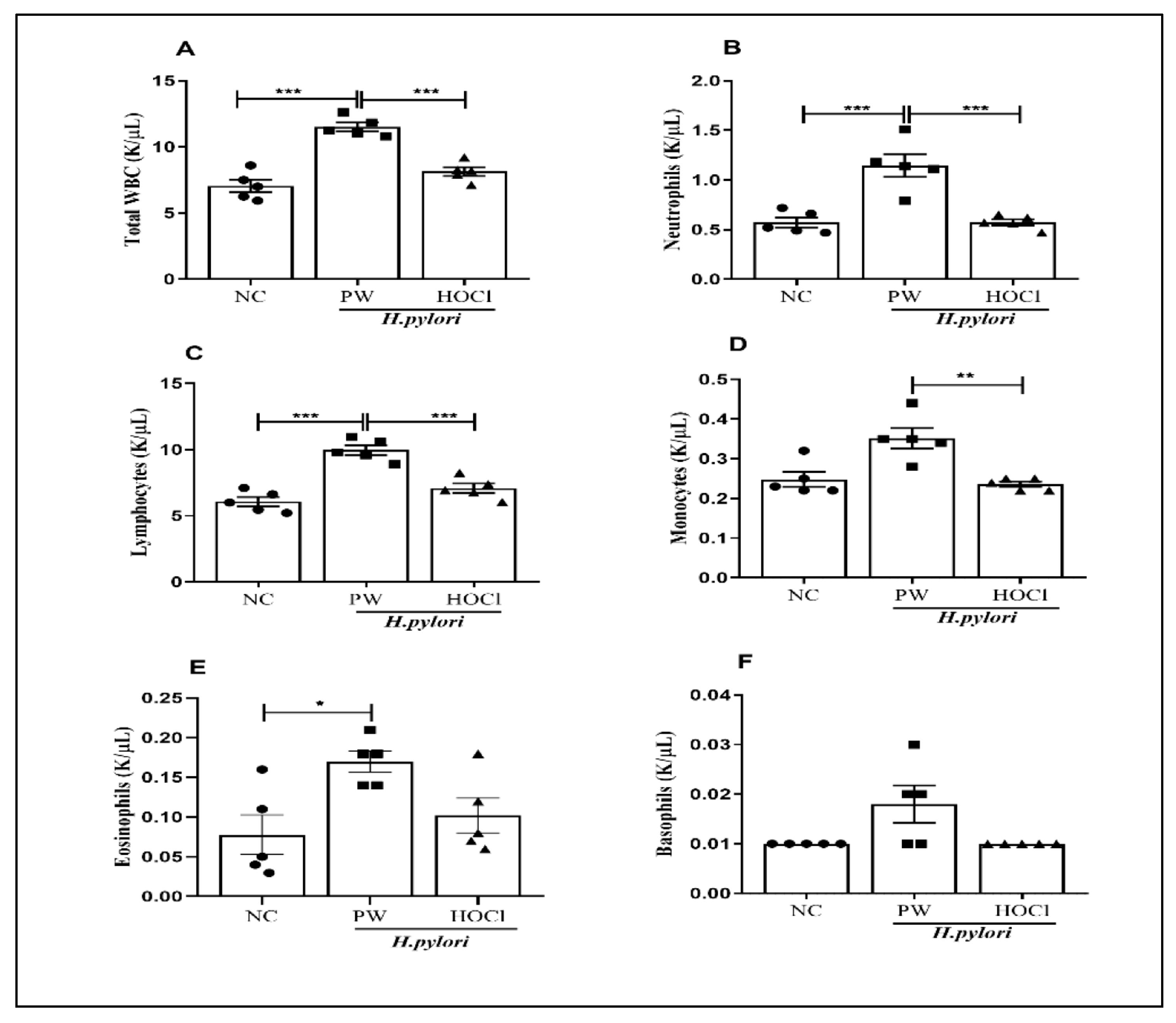
2.7. Total and Differential White Blood Cell (WBC) Counts

Retroorbital plexus blood was drawn and put into tubes coated with ethylenediaminetetraacetic acid. An automatic blood analyzer HEMAVET HV950 FS, (Drew Scientific Inc., Boston, MA, USA) was used to measure the total WBC and the differential counts of lymphocytes, monocytes, neutrophils, eosinophils, and basophils.

2.8. Measurement of Total Reactive Oxygen Species (ROS)

Total ROS formation in serum and stomach tissue lysates was determined by the oxidation of 2-4-dichlorodihydrofluorescein diacetate (DCFH-DA) (Abcam, Cambridge, MA, USA) using a procedure previously described [29].

2.9. Measurement of Nitric Oxide (NO) Levels

To assess NO production in the blood and stomach tissue lysates, Griess reagent (Promega Corp., Madison, WI, USA) was used according to a procedure previously described [29].

2.10. Measurement of Glutathione Peroxidase (GPx) Activity

The BioVision kit (Milpitas, CA, USA) was used to determine GPx activity in serum and stomach tissue lysates. As indicated by the manufacturer’s guidelines, GPx activity was determined using normalized protein samples. Briefly, assay standards were prepared, and the reagent assays were mixed in a 96-well microplate. SpectraMax® ABS Plus (Molecular Devices, San Jose, CA, USA) was used to measure the absorbance at 340 nm.

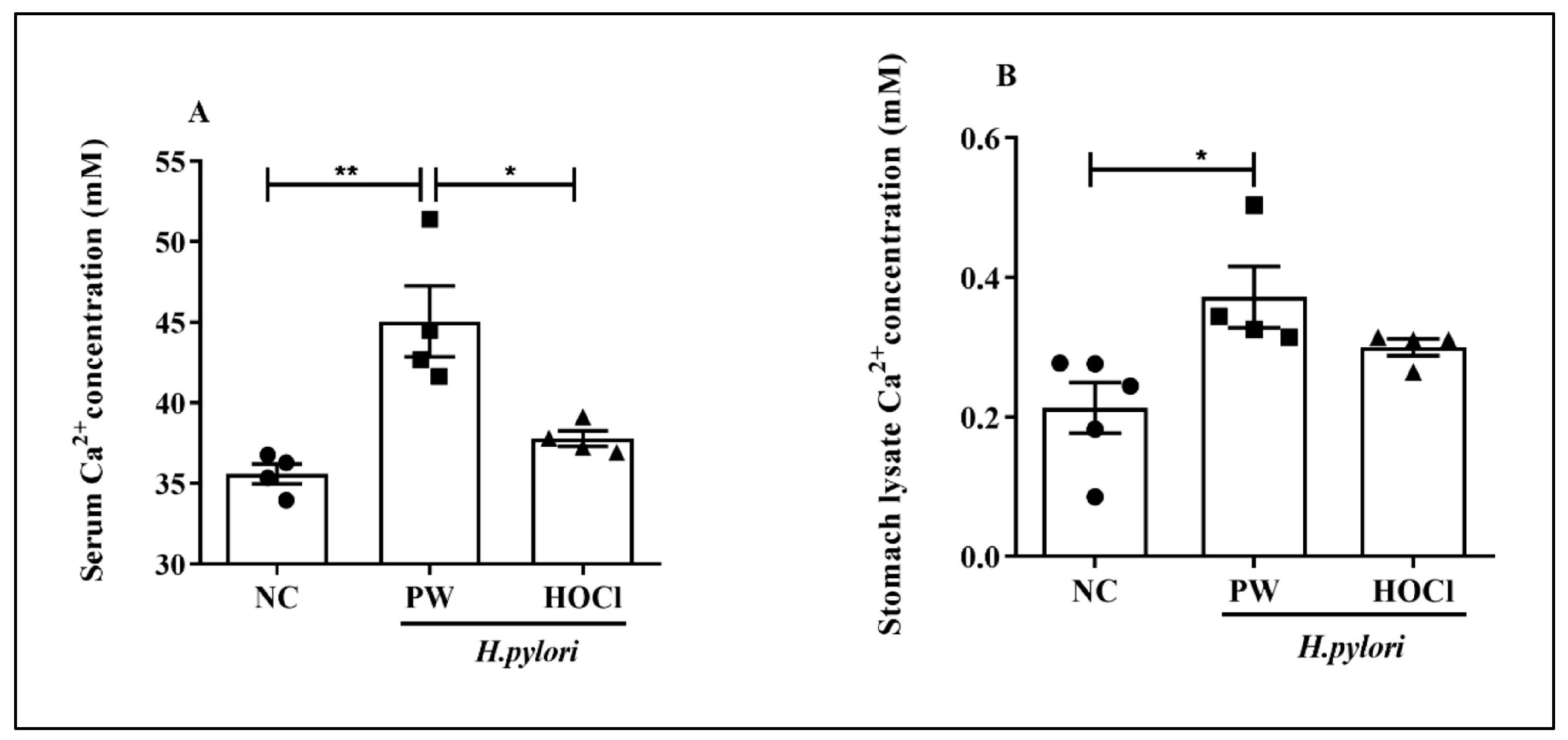
2.11. Detection of Intracellular Ca2+ Activity

A colorimetric calcium assay kit (Abcam, Cambridge, MA, USA) was used to determine total intracellular Ca2+ activity according to the manufacturer’s instructions for blood and stomach tissue lysates using a procedure previously described [29].

2.12. Measurement of Proinflammatory Cytokines

The Milliplex® MAP Mouse Cytokine/Chemokine Magnetic Bead Panel 96-well plate test (Millipore Corporation, Billerica, MA, USA) is a Luminex-based multiplex technique used for cytokine and chemokine profiling. Following the manufacturer’s instructions, a multiplex immunoassay was used to measure G-CSF, IL-1β, and TNF-α. To prepare serial dilutions of the standard, each standard concentration was resuspended in standard diluents. The standard, serum, and stomach lysates were combined with the bead combination. The plate underwent a washing procedure before being incubated for an additional 18 h at 4 °C. After adding the detection antibody, the plate was left to sit at room temperature for 1 h. The plate was then mixed with streptavidin and phycoerythrin, and left to sit at room temperature for 30 min. The plate was then examined using the Luminex 200 Bio-Plex instrument after the washing process, after which an assay buffer was applied.

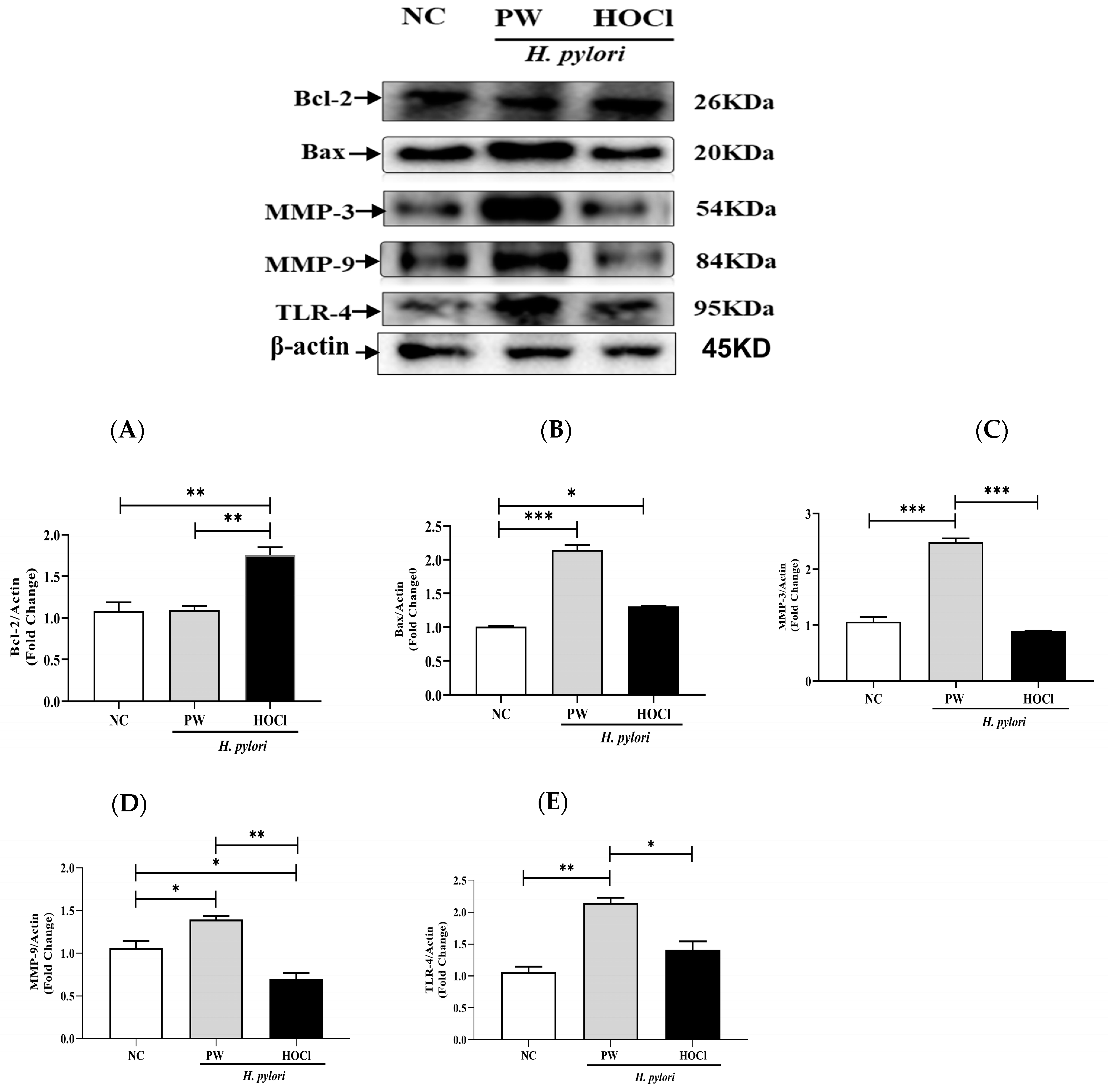
2.13. Western Blot Analysis

Protein extraction (tissue lysates) and Western blot were carried out as described previously [29]. Antibodies against the following proteins were used in this study: β-actin, MMP-3, MMP-9, Bcl-2, Bax, and TLR-4 (Cell Signaling Technology, Danvers, MA, USA).

2.14. Data Management and Statistical Analysis

The analysis of variance (ANOVA) and multiple comparison tests (Tukey’s post hoc test) were performed using the GraphPad Prism version 8.0 software tools (GraphPad Software, La Jolla, CA, USA). The standard error of the mean (SEM) is used to describe data; at p < 0.05, differences were deemed statistically significant.

3. Results

3.1. Effects of Non-Electrolyzed HOCl Water on the Body Weight of H. pylori-Infected C57BL/6 Mice

To study the effects of consuming HOCl water, the mice’s body weights were recorded on a weekly basis. In comparison to the NC group, our findings indicated that H. pylori infection caused a minor loss in body weight in both the PW- and HOCl-treated groups, particularly from the 3rd to the 10th week; however, the body weight difference was not statistically significant, as shown in Supplemental Figure S1.

3.2. Histological Observation of the Stomach of H. pylori-Infected C57BL/6 Mice

The stomach was cut 10 weeks post-infection, and the sections were observed under a light microscope. By looking for H. pylori bacteria in the gastric pits and surface mucous layer of the stomach in both the PW + H. pylori and HOCl + H. pylori groups in Figure 2, it was determined that all mice had the infection, indicated by arrow signs (→). NC mice partially showed inflammatory cells in the stomach tissue, but the level was negligible. Mice in the PW + H. pylori and HOCl + H. pylori groups showed mild inflammatory cell infiltration in the mucosal layer; however, there was no specific gastric mucosal injury, such as erosion or ulcer.

3.3. Effects of Non-Electrolyzed HOCl Water in H. pylori-Infected C57BL/6 Mice on Total WBC and Its Differential Counts

WBCs are important for both the body’s innate and adaptive immune responses. Any foreign invasion into the body causes the number of WBCs to increase and the recruited WBCs to perform certain functions. In the total and differential WBC counts, the HOCl + H. pylori group showed a significant decrease in the total WBC count (p < 0.001), neutrophils (p < 0.001), lymphocytes (p < 0.001), and monocytes (p < 0.01) compared to the PW + H. pylori group (Figure 3).

3.4. Effects of Non-Electrolyzed HOCl Water on OS Markers in H. pylori-Infected

We evaluated ROS and NO levels as well as GPx activity as OS indicators in the serum and stomach lysates to examine the effects of HOCl in C57BL/6 mice with H. pylori infection. When compared to the PW + H. pylori group, ROS levels in the HOCl + H. pylori group significantly decreased in both the serum and stomach lysates (p < 0.05) (Figure 4A,B).
NO levels also showed a declining tendency in comparison to the PW + H. pylori group, but the difference was not statistically significant (Figure 4C,D). GPx activity in the serum was also considerably higher in the HOCl + H. pylori group compared to the PW + H. pylori group (p < 0.001) and stomach lysate (p < 0.05) (Figure 4E,F).

3.5. Effects of Non-Electrolyzed HOCl Water on the Intracellular Ca2+ Activity in H. pylori-Infected C57BL/6 Mice

Intracellular Ca2+ activity was assessed in both the serum and stomach lysates to investigate the possible mechanisms of H. pylori infection in the stomach.
Comparing the PW + H. pylori group to the NC group (p < 0.01), the serum revealed a significantly higher level of Ca2+ activity; However, when compared to the PW + H. pylori group (p < 0.05), the HOCl + H. pylori group demonstrated a substantial reduction in Ca2+ activity (Figure 5A). Similarly, in the stomach lysate, the PW + H. pylori group showed significantly higher Ca2+ activity than the NC group (p < 0.05); however, the HOCl + H. pylori group tended to show higher Ca2+ activity than the PW + H. pylori group (p < 0.05) (Figure 5B).

3.6. Effects of Non-Electrolyzed HOCl on the Level of Inflammatory Cytokines in H. pylori-Infected C57BL/6 Mice

We looked at inflammatory cytokines including GM-CSF, IL-1β, and TNF-α in both blood and stomach lysates to learn how HOCl affected the inflammatory response (Figure 6). The HOCl + H. pylori infection group showed significant differences in GM-CSF (p < 0.05), IL-1β (p < 0.01), and TNF-α (p < 0.001) levels in stomach lysates compared to those in the PW + H. pylori group; however, there was no significant difference in serum.

3.7. Effects of Non-Electrolyzed HOCl on Bcl-2, Bax, MMP-3, MMP-9, and TLR-4 in Stomach Lysates

Using Western blot analysis, we investigated the underlying molecular mechanisms of HOCl water therapy against H. pylori infection and evaluated the protein expression levels of Bcl-2, Bax, MMP-3, MMP-9, and TLR-4. We discovered that the PW + H. pylori infection group’s expression level of Bcl-2 was substantially lower than that of the HOCl + H. pylori infection group’s (p < 0.01) (Figure 7A). However, with HOCl therapy, the expression of Bax was reduced in comparison to that in the PW + H. pylori infection group. In contrast, we found that the level of Bax expression was significantly raised in the PW + H. pylori infection group (p < 0.001) compared to that in the NC group (Figure 7B). Additionally, we discovered that the expression levels of MMP-3 were considerably greater in the PW + H. pylori infection group (p < 0.001) than in the HOCl + H. pylori infection group (Figure 7C). The expression level of MMP-9 was also considerably higher in the PW + H. pylori infection group than in the HOCl + H. pylori infection group (p < 0.01) (Figure 7D). Similarly, we found that TLR-4 expression levels in the PW group were significantly higher (p < 0.05) than in the HOCl + H. pylori infection group (Figure 7E). These findings suggest that oral gavage of HOCl administration in the stomach reduces H. pylori-related infection in C57BL/6 mice.

4. Discussion

The C57BL/6 mouse model for H. pylori infection was used in the current study to assess the anti-oxidative, anti-apoptotic, and anti-inflammatory effects of non-electrolyzed HOCl water treatment. Consequently, drinking non-electrolyzed HOCl water significantly suppressed the oxidative and inflammatory responses in H. pylori-infected C57BL/6 mice through redox regulation and immune responses [30]. H. pylori can induce a long-term inflammatory response that promotes carcinogenesis [31]. However, H. pylori can potentially defend itself from the host immunological response by triggering macrophage death. Therefore, non-antibiotic agents, such as non-electrolyzed acidic water, might be highly effective and safe for the treatment of both antibiotic susceptibility and resistance to H. pylori bacteria [32]. Several studies on HOCl have shown its numerous effects on various pathogenic bacteria, including H. pylori infection [33,34].
To this end, we investigated the bactericidal effect of the oral administration of non-electrolyzed HOCl against H. pylori. In our histological observation, the mice in both the PW + H. pylori and HOCl + H. pylori groups showed mild inflammatory cell infiltration in the mucosal layer; however, there was no specific gastric mucosal injury, such as erosion or ulcer. This suggests that drinking HOCl water is safe for histological damage of the mouse stomach compared to drinking PW. However, WBC count can be used as an inflammatory indicator to assess the severity of various gastric diseases, such as gastric cancer and H. pylori infection [35]. In our study, the total number of WBC, neutrophils, lymphocytes, and monocytes were significantly lower in the HOCl + H. pylori group compared to the PW + H. pylori group. This finding suggested that mice infected with H. pylori may experience systemic immune responses differently after consuming HOCl water. In contrast, neutrophils in tissue are unable to eliminate the established H. pylori colony via paracrine secretion, allowing H. pylori to persist for an extended period. Furthermore, it causes active chronic inflammation of the gastric mucosa. Inducible nitric oxide synthase (iNOS), a crucial enzyme that catalyzes the synthesis of NO and then converts O2 to peroxynitrite, has been discovered in host neutrophils and epithelial cells. This process promotes the generation of more ROS and RNS, aggravating the OS response [36]. Compared with untreated mice, iNOS gene-deficient mice showed a substantial reduction in the incidence of stomach cancer after H. pylori infection. It has been consequently asserted that iNOS-induced OS is directly related to the development of stomach cancer [37]. These investigations demonstrated an increase in genomic alterations, indicating that OS develops soon after infection [38]. Our study also revealed that H. pylori-infected mice produced more ROS, which could be one of the mechanisms underlying infection-related apoptosis. NO is a signaling molecule with strong immunomodulatory properties and is linked to a key host defense component in H. pylori infection. The current study discovered that NO produced by activated macrophages can destroy extracellular H. pylori. Several signaling pathways within the host gastric cell have been shown to be activated or used by H. pylori, which can result in ulcers or gastric cancer [39,40]. NO can inhibit 8-oxoguanine glycosylase by removing DNA mutations [41]. H. pylori infection, on the other hand, produces ROS that can damage DNA. H. pylori infection also increases intracellular ROS levels in gastric epithelial cells, according to these studies [42,43]. It has been demonstrated that antioxidants such as GPx reduce ROS production and prevent H. pylori-induced programmed cell death. Therefore, our results indicate that non-electrolyzed HOCl treatment attenuates OS by suppressing ROS and NO levels and increasing GPx levels. Additionally, Ca2+ levels were significantly decreased in the HOCl + H. pylori group in both the serum and stomach lysates compared to the PW + H. pylori group. However, the exact mechanism by which H. pylori infection causes calcium mobilization in gastric cancer cells remains unknown.
The signaling pathways involved in the damage by OS are very broad, stimulating not only the generation of inflammatory factors (IL-8, IL-6, and IL-1β) but also the activation of GM-CSF and TNF-α [44]. Various gene polymorphisms have been studied, with IL-1β, GM-CSF, and TNF-α being the most studied [45,46]. These toxins enable long-term H. pylori infection, which in turn encourages stomach epithelial cells to produce TNF-α [47]. IL-1β is a powerful inhibitor of stomach acid release [48,49]. In the presence of H. pylori, IL-1β is elevated and necessary for the initiation and progression of inflammatory responses to infection [50,51]. In our research, significant changes were noted in the production of some cytokines in both the serum and stomach lysate samples among the three groups. Due to H. pylori infection in C57BL/6 mice, inflammatory cytokines such as IL-1β, TNF-α, and GM-CSF were previously studied [25].
In H. pylori-infected patients, the Bax/Bcl-2 ratio offers the potential for screening and early cancer diagnosis. Infection with H. pylori adds to the buildup of molecular genetic anomalies. In individuals with stomach cancer, H. pylori infection increases the expression of the protein markers Bax and Bcl-2 [52]. According to previous studies, Bax expression was lower in gastric cancer than Bcl-2 expression, and the calculated Bax/Bcl-2 ratio in cancer was much lower than that in the nearby normal mucosa [53]. Our results showed that HOCl treatment significantly decreased Bax protein expression levels in H. pylori-infected mice, whereas HOCl treatment significantly increased Bcl-2 levels, which is consistent with the finding that HOCl treatment reduces apoptosis in stomach tissues.
According to one study, the production of MMP-9 and tissue inhibitor of metalloproteinases-1 (TIMP-1) was significantly correlated with the location of ulcer margins [54]. The expression of MMP-9, MMP-3, and TIMP-1 was higher in patients with stomach ulcers. Moreover, MMP-9 production has been linked to a higher risk of gastric ulcer recurrence [55]. Another study suggests that MMP-3 and MMP-9 may play a role in the development of gastric ulcers [56]. Our results revealed that treatment with non-electrolyzed HOCl significantly decreased MMP-3 and MMP-9 production by gastric epithelial cells in stomach lysates. Additionally, TLRs are believed to be essential for triggering adaptive immunity and regulating inflammation during the innate immune reaction to H. pylori. TLR signaling, however, influences gastric immunopathology, H. pylori-mediated T-cell responses, and in vivo colonization [57]. Our results demonstrated that treatment with non-electrolyzed HOCl significantly decreased TLR-4 production in gastric epithelial cells in stomach lysates. Overall, our results demonstrated that non-electrolyzed HOCl treatment ameliorated H. pylori infection from oxidative and inflammatory stress in C57BL/6 mice and might be a potential therapeutic agent against H. pylori-infection-related diseases, such as gastric cancer, stomach cancer, and peptic ulcer.

5. Conclusions

Overall, based on our findings, we showed that non-electrolyzed HOCl water has anti-oxidative, anti-inflammatory, and anti-apoptotic effects through redox and immune regulation pathways and may have promising therapeutic potential against diseases associated with H. pylori infection. However, the drinking safety of HOCl water has not been sufficiently confirmed, although external use of HOCl is known to be safe and effective for humans. Further studies are needed to fully understand the effects of genetic variants at the immune redox level as well as their roles and effects on H. pylori susceptibility.

Supplementary Materials

The following supporting information can be downloaded at: https://www.mdpi.com/article/10.3390/pr11051474/s1, Figure S1: Body weight measurement among three groups.

Author Contributions

Conceptualization, K.-J.L.; Methodology, C.-S.K., J.B. and M.H.R.; Software, C.-S.K. and M.H.R.; Validation, C.-S.K. Formal Analysis, J.B., M.H.R., A.F. and Y.C.; Investigation, C.-S.K., J.B., M.H.R., A.F., S.S., Y.J.J. and S.H.G.; Data Curation, M.H.R., J.B., A.F. and Y.J.J.; Writing—Original Draft Preparation, M.H.R.; Writing—Review & Editing, J.B., C.-S.K., Y.C., A.F., T.T.T., Y.K. and S.H.C.; Visualization, C.-S.K.; Supervision, K.-J.L.; Project Administration, K.-J.L. and C.-S.K.; Funding Acquisition, K.-J.L. All authors have read and agreed to the published version of the manuscript.

Funding

This study received no external funding.

Data Availability Statement

Not Applicable.

Conflicts of Interest

The authors declare that they have no competing interest.

References

  1. Young, S.B.; Setlow, P. Mechanisms of Killing of Bacillus Subtilis Spores by Hypochlorite and Chlorine Dioxide. J. Appl. Microbiol. 2003, 95, 54–67. [Google Scholar] [CrossRef]
  2. Hori, T.; Yamamoto, H.; Harada, H.; Yamamoto, M.; Yamada, M.; Yazawa, T.; Sasaki, B.; Tani, M.; Sato, A.; Katsura, H.; et al. Multiple Gastric Carcinomas Associated with Epstein-Barr Virus and Helicobacter Pylori: A Thought-Provoking Case. Am. J. Case Rep. 2021, 22, e931668. [Google Scholar] [CrossRef] [PubMed]
  3. Robinson, K.; Atherton, J.C. The Spectrum of Helicobacter—Mediated Diseases. Annu. Rev. Pathol. Mech. Dis. 2021, 16, 123–144. [Google Scholar] [CrossRef] [PubMed]
  4. Zhang, L.; Zhang, L.; Deng, H.; Li, H.; Tang, W.; Guan, L.; Qiu, Y.; Donovan, M.J.; Chen, Z.; Tan, W. In Vivo Activation of PH-Responsive Oxidase-like Graphitic Nanozymes for Selective Killing of Helicobacter Pylori. Nat. Commun. 2021, 12, 2002. [Google Scholar] [CrossRef]
  5. Suzuki, T.; Kato, K.; Ohara, S.; Noguchi, K.; Sekine, H.; Nagura, H.; Shimosegawa, T. Localization of Antigen-Presenting Cells in Helicobacter Pylori-Infected Gastric Mucosa. Pathol. Int. 2002, 52, 265–271. [Google Scholar] [CrossRef] [PubMed]
  6. Sipowicz, M.A.; Chomarat, P.; Diwan, B.A.; Anver, M.A.; Awasthi, Y.C.; Ward, J.M.; Rice, J.M.; Kasprzak, K.S.; Wild, C.P.; Anderson, L.M. Increased Oxidative DNA Damage and Hepatocyte Overexpression of Specific Cytochrome P450 Isoforms in Hepatitis of Mice Infected with Helicobacter Hepaticus. Am. J. Pathol. 1997, 151, 933–941. [Google Scholar] [PubMed]
  7. Ding, S.Z.; Minohara, Y.; Xue, J.F.; Wang, J.; Reyes, V.E.; Patel, J.; Dirden-Kramer, B.; Boldogh, I.; Ernst, P.B.; Crowe, S.E. Helicobacter Pylori Infection Induces Oxidative Stress and Programmed Cell Death in Human Gastric Epithelial Cells. Infect. Immun. 2007, 75, 4030–4039. [Google Scholar] [CrossRef]
  8. Farinati, F.; Cardin, R.; Degan, P.; Rugge, M.; Di Mario, F.; Bonvicini, P.; Naccarato, R. Oxidative DNA Damage Accumulation in Gastric Carcinogenesis. Gut 1998, 42, 351–356. [Google Scholar] [CrossRef] [PubMed]
  9. Drake, I.M.; Mapstone, N.P.; Schorah, C.J.; White, K.L.M.; Chalmers, D.M.; Dixon, M.F.; Axon, A.T.R. Reactive Oxygen Species Activity and Lipid Peroxidation in Helicobacter Pylori Associated Gastritis: Relation to Gastric Mucosal Ascorbic Acid Concentrations and Effect of H Pylori Eradication. Gut 1998, 42, 768–771. [Google Scholar] [CrossRef]
  10. Davies, G.R.; Simmonds, N.J.; Stevens, T.R.J.; Sheaff, M.T.; Banatvala, N.; Laurenson, I.F.; Blake, D.R.; Rampton, D.S. Helicobacter Pylori Stimulates Antral Mucosal Reactive Oxygen Metabolite Production in vivo. Gut 1994, 35, 179–185. [Google Scholar] [CrossRef]
  11. Zhang, Q.B.; Dawodu, J.B.; Husain, A.; Etolhi, G.; Gemmell, C.G.; Russell, R.I. Association of Antral Mucosal Levels of Interleukin 8 and Reactive Oxygen Radicals in Patients Infected with Helicobacter Pylori. Clin. Sci. 1997, 92, 69–73. [Google Scholar] [CrossRef] [PubMed]
  12. Robinson, K.; Argent, R.H.; Atherton, J.C. The Inflammatory and Immune Response to Helicobacter Pylori Infection. Best Pract. Res. Clin. Gastroenterol. 2007, 21, 237–259. [Google Scholar] [CrossRef]
  13. Petersen, O.H. Localization and Regulation of Ca2+ Entry and Exit Pathways in Exocrine Gland Cells. Cell Calcium 2003, 33, 337–344. [Google Scholar] [CrossRef]
  14. Pallagi, P.; Madácsy, T.; Varga, Á.; Maléth, J. Intracellular Ca2+ Signalling in the Pathogenesis of Acute Pancreatitis: Recent Advances and Translational Perspectives. Int. J. Mol. Sci 2020, 21, 4005. [Google Scholar] [CrossRef] [PubMed]
  15. Yang, I.H.; Wong, J.H.; Chang, C.M.; Chen, B.K.; Tsai, Y.T.; Chen, W.C.; Wang, E.T.; Hsu, W.L.; Chang, W.C. Involvement of Intracellular Calcium Mobilization in IL-8 Activation in Human Retinal Pigment Epithelial Cells. Investig. Ophthalmol. Vis. Sci. 2015, 56, 761–769. [Google Scholar] [CrossRef] [PubMed]
  16. Konturek, P.C.; Pierzchalski, P.; Konturek, S.J.; Meixner, H.; Faller, G.; Kirchner, T.; Hahn, E.G. Helicobacter Pylori Induces Apoptosis in Gastric Mucosa through an Upregulation of Bax Expression in Humans. Scand. J. Gastroenterol. 1999, 34, 375–383. [Google Scholar] [CrossRef]
  17. Konturek, P.C.; Konturek, S.J.; Brzozowski, T. Helicobacter Pylori Infection in Gastric Cancerogenesis. J. Physiol. Pharmacol. 2009, 60, 3–21. [Google Scholar]
  18. Lima, V.P.; de Lima, M.A.P.; André, A.R.; Ferreira, M.V.P.; Barros, M.A.P.; Rabenhorst, S.H.B. H Pylori (CagA) and Epstein-Barr Virus Infection in Gastric Carcinomas: Correlation With P53 Mutation and c-Myc, Bcl-2 and Bax Expression. World J. Gastroenterol. 2008, 14, 884–891. [Google Scholar] [CrossRef]
  19. Sokolova, O.; Naumann, M. Matrix Metalloproteinases in Helicobacter Pylori—Associated Gastritis and Gastric Cancer. Int. J. Mol. Sci. 2022, 23, 1883. [Google Scholar] [CrossRef]
  20. Smith, S.M. Role of Toll-like Receptors in Helicobacter Pylori Infection and Immunity. World J. Gastrointest. Pathophysiol. 2014, 5, 133–146. [Google Scholar] [CrossRef]
  21. O’Connor, A.; Molina-Infante, J.; Gisbert, J.P.; O’Morain, C. Treatment of Helicobacter Pylori Infection 2013. Helicobacter 2013, 18, 58–65. [Google Scholar] [CrossRef] [PubMed]
  22. Medina-Tamayo, J.; Sánchez-Miranda, E.; Balleza-Tapia, H.; Ambriz, X.; Cid, M.E.; González-Espinosa, D.; Gutiérrez, A.A.; González-Espinosa, C. Super-Oxidized Solution Inhibits IgE-Antigen-Induced Degranulation and Cytokine Release in Mast Cells. Int. Immunopharmacol. 2007, 7, 1013–1024. [Google Scholar] [CrossRef] [PubMed]
  23. Del Rosso, J.Q.; Bhatia, N. Status Report on Topical Hypochlorous Acid: Clinical Relevance of Specific Formulations, Potential Modes of Action, and Study Outcomes. J. Clin. Aesthetic Dermatol. 2018, 11, 36–39. [Google Scholar]
  24. De-Oliveira, R.B.B.; Kato, E.E.; Soares de Lima, T.; Alba-Loureiro, T.C.; Curi, R.; Cirillo, M.C.; Sampaio, S.C. Crotoxin-Treated Macrophages Stimulate ROS Production and Killing Activity in Co-Cultured Neutrophils Article Information. J. Appl. Microbiol. Res. 2018, 1, 2018050322. [Google Scholar]
  25. Mentis, A.F.A.; Boziki, M.; Grigoriadis, N.; Papavassiliou, A.G. Helicobacter Pylori Infection and Gastric Cancer Biology: Tempering a Double-Edged Sword. Cell. Mol. Life Sci. 2019, 76, 2477–2486. [Google Scholar] [CrossRef] [PubMed]
  26. Kubota, A.; Goda, T.; Tsuru, T.; Yonekura, T.; Yagi, M.; Kawahara, H.; Yoneda, A.; Tazuke, Y.; Tani, G.; Ishii, T.; et al. Efficacy and Safety of Strong Acid Electrolyzed Water for Peritoneal Lavage to Prevent Surgical Site Infection in Patients with Perforated Appendicitis. Surg. Today 2015, 45, 876–879. [Google Scholar] [CrossRef]
  27. Wang, L.; Bassiri, M.; Najafi, R.; Najafi, K.; Yang, J.; Khosrovi, B.; Hwong, W.; Barati, E.; Belisle, B.; Celeri, C.; et al. Hypochlorous Acid as a Potential Wound Care Agent: Part I. Stabilized Hypochlorous Acid: A Component of the Inorganic Armamentarium of Innate Immunity. J. Burn. Wounds 2007, 6, e5. [Google Scholar]
  28. Takeda, Y.; Matsuda, S.; Jamsransuren, D.; Ogawa, H. Comparison of the SARS-CoV-2-Inactivating Activities of the Differently Manufactured Hypochlorous Acid Water Products with Various PH. J. Water Health 2021, 19, 448–456. [Google Scholar] [CrossRef] [PubMed]
  29. Jeong, E.S.; Bajgai, J.; You, I.S.; Rahman, M.H.; Fadriquela, A.; Sharma, S.; Kwon, H.U.; Lee, S.Y.; Kim, C.S.; Lee, K.J. Therapeutic Effects of Hydrogen Gas Inhalation on Trimethyltin-Induced Neurotoxicity and Cognitive Impairment in the C57BL/6 Mice Model. Int. J. Mol. Sci. 2021, 22, 13313. [Google Scholar] [CrossRef]
  30. Jiménez, F.T.; Bayona, C.T. Molecular Pathophysiology in Infection by Helicobacter Pylori. Salud Uninorte 2021, 32, 500–512. [Google Scholar] [CrossRef]
  31. Peek, R.M.; Fiske, C.; Wilson, K.T. Role of Innate Immunity in Helicobacter Pylori-Induced Gastric Malignancy. Physiol Rev. 2010, 90, 831–858. [Google Scholar] [CrossRef]
  32. Iram, A.; Wang, X.; Demirci, A. Electrolyzed Oxidizing Water and Its Applications as Sanitation and Cleaning Agent. Food Eng. Rev. 2021, 13, 411–427. [Google Scholar] [CrossRef]
  33. Zhao, C.; Chen, Y.; Gao, L.; Huang, J.; Yang, X.; Pei, L.; Ye, Z.; Zhu, L. Acidic Electrolyzed Water Inhibits the Viability of Gardnerella Spp. via Oxidative Stress Response. Front. Med. 2022, 9, 817957. [Google Scholar] [CrossRef]
  34. Lucio-Sauceda, D.G.; Urrutia-Baca, V.H.; Gomez-Flores, R.; De La Garza-Ramos, M.A.; Tamez-Guerra, P.; Orozco-Flores, A. Antimicrobial and Anti-Biofilm Effect of an Electrolyzed Superoxidized Solution at Neutral-PH against Helicobacter pylori. BioMed Res. Int. 2019, 2019, 6154867. [Google Scholar] [CrossRef]
  35. Liu, C.; Li, X. Stage-Dependent Changes in Albumin, NLR, PLR, and AFR Are Correlated with Shorter Survival in Patients with Gastric Cancer. Clin. Lab. 2019, 65. [Google Scholar] [CrossRef] [PubMed]
  36. Tsugawa, H.; Suzuki, H.; Saya, H.; Hatakeyama, M.; Hirayama, T.; Hirata, K.; Nagano, O.; Matsuzaki, J.; Hibi, T. Reactive Oxygen Species-Induced Autophagic Degradation of Helicobacter Pylori CagA Is Specifically Suppressed in Cancer Stem-like Cells. Cell Host Microbe 2012, 12, 764–777. [Google Scholar] [CrossRef] [PubMed]
  37. Gmünder, H.; Dröge, W. Differential Effects of Glutathione Depletion on T Cell Subsets. Cell. Immunol. 1991, 138, 229–237. [Google Scholar] [CrossRef] [PubMed]
  38. Sheh, A.; Lee, C.W.; Masumura, K.; Rickman, B.H.; Nohmi, T.; Wogan, G.N.; Fox, J.G.; Schauer, D.B. Mutagenic Potency of Helicobacter Pylori in the Gastric Mucosa of Mice Is Determined by Sex and Duration of Infection. Proc. Natl. Acad. Sci. USA 2010, 107, 15217–15222. [Google Scholar] [CrossRef]
  39. Peek, R.M.; Blaser, M.J. Helicobacter Pylori and Gastrointestinal Tract Adenocarcinomas. Nat. Rev. Cancer 2002, 2, 28–37. [Google Scholar] [CrossRef]
  40. Jabeen, R.; Jami, A.; Shahab, A.; Shahab, A. Relationship of Halitosis with Gastric Helicobacter Pylori Infection. Pak. J. Med. Health Sci. 2021, 15, 2285–2288. [Google Scholar] [CrossRef]
  41. Kidane, D.; Murphy, D.L.; Sweasy, J.B. Accumulation of Abasic Sites Induces Genomic Instability in Normal Human Gastric Epithelial Cells during Helicobacter Pylori Infection. Oncogenesis 2014, 3, e128. [Google Scholar] [CrossRef] [PubMed]
  42. Maeda, S.; Yoshida, H.; Mitsuno, Y.; Hirata, Y.; Ogura, K.; Shiratori, Y.; Omata, M. Analysis of Apoptotic and Antiapoptotic Signalling Pathways Induced by Helicobacter Pylori. Gut 2002, 50, 771–778. [Google Scholar] [CrossRef] [PubMed]
  43. Bagchi, D.; Bhattacharya, G.; Stohs, S.J. Production of Reactive Oxygen Species by Gastric Cells in Association with Helicobacter Pylori. Free Radic. Res. 1996, 24, 439–450. [Google Scholar] [CrossRef] [PubMed]
  44. Beswick, E.J.; Pinchuk, I.V.; Suarez, G.; Sierra, J.C.; Reyes, V.E. Helicobacter Pylori CagA-Dependent Macrophage Migration Inhibitory Factor Produced by Gastric Epithelial Cells Binds to CD74 and Stimulates Procarcinogenic Events. J. Immunol. 2006, 176, 6794–6801. [Google Scholar] [CrossRef]
  45. El-Omar, E.M.; Oien, K.; El-Nujumi, A.; Gillen, D.; Wirz, A.; Dahill, S.; Williams, C.; Ardill, J.E.S.; McColl, K.E.L. Helicobacter Pylori Infection and Chronic Gastric Acid Hyposecretion. Gastroenterology 1997, 113, 15–24. [Google Scholar] [CrossRef] [PubMed]
  46. Graham, D.Y. Helicobacter Pylori Infection in the Pathogenesis of Duodenal Ulcer and Gastric Cancer: A Model. Gastroenterology 1997, 113, 1983–1991. [Google Scholar] [CrossRef]
  47. Crabtree, J.E.; Court, M.; Aboshkiwa, M.A.; Jeremy, A.H.T.; Dixon, M.F.; Robinson, P.A. Gastric Mucosal Cytokine and Epithelial Cell Responses to Helicobacter Pylori Infection in Mongolian Gerbils. J. Pathol. 2004, 202, 197–207. [Google Scholar] [CrossRef]
  48. Rébé, C.; Ghiringhelli, F. Interleukin-1β and Cancer. Cancers 2020, 12, 1791. [Google Scholar] [CrossRef]
  49. Beales, I.L.P.; Calam, J. Interleukin 1β and Tumour Necrosis Factor α Inhibit Acid Secretion in Cultured Rabbit Parietal Cells by Multiple Pathways. Gut 1998, 42, 227–234. [Google Scholar] [CrossRef]
  50. Outlioua, A.; Badre, W.; Desterke, C.; Echarki, Z.; El Hammani, N.; Rabhi, M.; Riyad, M.; Karkouri, M.; Arnoult, D.; Khalil, A.; et al. Gastric IL-1β, IL-8, and IL-17A Expression in Moroccan Patients Infected with Helicobacter Pylori May Be a Predictive Signature of Severe Pathological Stages. Cytokine 2020, 126, 154893. [Google Scholar] [CrossRef]
  51. Basso, D.; Scrigner, M.; Toma, A.; Navaglia, F.; Di Mario, F.; Rugge, M.; Plebani, M. Helicobacter Pylori Infection Enhances Mucosal Interleukin-1β, Interleukin-6, and the Soluble Receptor of Interleukin-2. Int. J. Clin. Lab. Res. 1996, 26, 207–210. [Google Scholar] [CrossRef] [PubMed]
  52. Zhang, H.; Fang, D.C.; Wang, R.Q.; Yang, S.M.; Liu, H.F.; Luo, Y.H. Effect of Helicobacter Pylori Infection on Expression of Bcl-2 Family Members in Gastric Adenocarcinoma. World J. Gastroenterol. 2004, 10, 227–230. [Google Scholar] [CrossRef] [PubMed]
  53. Konturek, P.C.; Konturek, S.J.; Pierzchalski, P.; Bielański, W.; Duda, A.; Marlicz, K.; Starzyńska, T.; Hahn, E.G. Cancerogenesis in Helicobacter Pylori Infected Stomach—Role of Growth Factors, Apoptosis and Cyclooxygenases. Med. Sci. Monit. 2001, 7, 1092–1107. [Google Scholar]
  54. Li, S.L.; Zhao, J.R.; Ren, X.Y.; Xie, J.P.; Ma, Q.Z.; Rong, Q.H. Increased Expression of Matrix Metalloproteinase-9 Associated with Gastric Ulcer Recurrence. World J. Gastroenterol. 2013, 19, 4590–4595. [Google Scholar] [CrossRef]
  55. Minyaylo, O.; Ponomarenko, I.; Reshetnikov, E.; Dvornyk, V.; Churnosov, M. Functionally Significant Polymorphisms of the MMP-9 Gene Are Associated with Peptic Ulcer Disease in the Caucasian Population of Central Russia. Sci. Rep. 2021, 11, 13515. [Google Scholar] [CrossRef] [PubMed]
  56. Bergin, P.J.; Raghavan, S.; Svensson, H.; Starckx, S.; Van Aelst, I.; Gjertsson, I.; Opdenakker, G.; Quiding-Järbrink, M. Gastric Gelatinase B/Matrix Metalloproteinase-9 Is Rapidly Increased in Helicobacter Felis-Induced Gastritis. FEMS Immunol. Med. Microbiol. 2008, 52, 88–98. [Google Scholar] [CrossRef]
  57. Chichirau, B.E.; Scheidt, T.; Diechler, S.; Neuper, T.; Horejs-Hoeck, J.; Huber, C.G.; Posselt, G.; Wessler, S. Dissecting the Helicobacter Pylori-Regulated Transcriptome of b Cells. Pathog. Dis. 2020, 78, ftaa049. [Google Scholar] [CrossRef]
Figure 1. Shows the experimental design.
Processes 11 01474 g001
Figure 2. Representative histological images of the stomach after 10 weeks. (A) NC: normal mouse supplied with purified water (PW) without infection, (B) PW + H. pylori: mouse supplied with PW after H. pylori infection, (C) HOCl + H. pylori: mouse supplied with HOCl water after H. pylori infection. Histological sections were stained with Giemsa solution. Scale bar = 20 μm. The arrow sign indicates the presence of H. pylori.
Processes 11 01474 g002
Figure 3. Effects of non-electrolyzed HOCl water on the number of total WBCs and differential WBC counts in H. pylori-infected mice. (A) Total WBCs, (B) neutrophils, (C) lymphocytes, (D) monocytes, (E) eosinophils, (F) basophils. NC: normal control, PW: purified water, HOCl: hypochlorous acid water. Data are shown as the mean ± SEM (n = 5). * p < 0.05, ** p < 0.01, *** p < 0.001.
Processes 11 01474 g003
Figure 4. Effects of non-electrolyzed HOCl on the oxidative stress markers in H. pylori-infected C57BL/6 mice after 10 weeks. (A) ROS level in serum, (B) ROS level in stomach lysates, (C) NO level in serum, (D) NO level in stomach lysates, (E) GPx activity in serum, and (F) GPx activity in stomach lysates. NC: normal control, PW: purified water, HOCl: hypochlorous acid water. Data are shown as the mean ± SEM (n = 5). * p < 0.05, ** p < 0.01, *** p < 0.001.
Processes 11 01474 g004
Figure 5. Effects of non-electrolyzed HOCl water on intracellular Ca2+ activity in H. pylori-infected C57BL/6 mice. (A) Ca2+ activity in serum, (B) Ca2+ activity in stomach lysates. NC: normal control, PW: purified water, HOCl: hypochlorous acid water. Data are shown as the mean ± SEM (n = 5). * p < 0.05, ** p < 0.01.
Processes 11 01474 g005
Figure 6. Effects of non-electrolyzed HOCl on the inflammatory response in serum and stomach lysates of C57BL/6 mice. GM-CSF in serum (A) and stomach lysate (B), IL-1β in serum (C) and stomach lysate (D), TNF-α in serum (E) and stomach lysate (F). NC: normal control, PW: purified water, HOCl: hypochlorous acid water. The data are presented as the mean ± SEM. ANOVA Tukey’s test (p < 0.05) was used to determine the significant difference. * p < 0.05, ** p < 0.01, *** p < 0.001.
Processes 11 01474 g006
Figure 7. Effects of non-electrolyzed HOCl treatment on Bcl-2, Bax, MMP-3, MMP-9, and TLR-4 protein levels in the stomach lysate of H. pylori-infected C57BL/6 mice. (A) Bcl-2 protein, (B) Bax protein (C) MMP-3 protein (D) MMP-9 protein (E) TLR-4 protein. Band intensity of each protein marker is normalized to the total in the bar graphs. NC: normal control, PW: purified water, HOCl: hypochlorous acid water. The data are presented as the mean ± SEM. ANOVA Tukey’s test (p < 0.05) was used to determine the significant difference. * p < 0.05, ** p < 0.01, *** p < 0.001.
Processes 11 01474 g007
Table 1. Properties of experimental water.
ParametersPWHOCl
pH7.56.1
ORP (mV)310931
ACC (ppm)051
Disclaimer/Publisher’s Note: The statements, opinions and data contained in all publications are solely those of the individual author(s) and contributor(s) and not of MDPI and/or the editor(s). MDPI and/or the editor(s) disclaim responsibility for any injury to people or property resulting from any ideas, methods, instructions or products referred to in the content.

