Article

Preparation of Polyvinyl Alcohol–Chitosan Nanocellulose–Biochar Nanosilver Composite Hydrogel and Its Antibacterial Property and Dye Removal Capacity

by
Licheng Xie
1,
Zhichao Zhang
1,2,3,
Yucai He
2,* and
Yan Jiang
4,*
1
Changzhou University Huaide College, Jingjiang 214500, China
2
School of Pharmacy & Biological and Food Engineering, Changzhou University, Changzhou 213164, China
3
School of Chemistry and Chemical Engineering, Guangxi University, Nanning 530004, China
4
School of Petrochemical Engineering, Changzhou University, Changzhou 213164, China
*
Authors to whom correspondence should be addressed.
Processes 2024, 12(10), 2277; https://doi.org/10.3390/pr12102277
Submission received: 5 September 2024 / Revised: 27 September 2024 / Accepted: 15 October 2024 / Published: 18 October 2024
(This article belongs to the Special Issue 2nd Edition of Innovation in Chemical Plant Design)

Abstract

:
In this research, silver-loaded biochar (C-Ag) was acquired from a waste fish scale, and nanocellulose (CNF) was prepared from the waste wheat stalk. Then C-Ag was loaded into chitosan-polyvinyl alcohol hydrogel (CTS-PVA) with CNC as a reinforcement agent, and a novel nanocomposite material was acquired, which could be efficiently applied for antibacterial and dye removal. By plate diffusion analysis, the inhibition areas of C-Ag-CTS-PVA-CNF (C/CTS/PVA/CNF) hydrogel against E. coli ATCC25922, S. aureus ATCC6538, and P. aeruginosa ATCC9027 could reach 22.5 mm, 22.0 mm, and 24.0 mm, respectively. It was found that the antibacterial rate was 100% in the water antibacterial experiment for 2 h, and the antibacterial activity was more than 90% within 35 days after preparation, and the antibacterial rate was more than 90% after repeated antibacterial tests for five times. Through swelling, water adsorption, water loss rate, and water content tests, the hydrogel manifested good moisturizing properties and could effectually block the loss of water and improve the stability of the C/CTS/PVA/CNF hydrogel. The pseudo-first-order and pseudo-second-order models were built, and the adsorption capacity of hydrogel to dye was analyzed, and the dye removal was more consistent with the pseudo-first-order kinetic model. The best removal effect for Congo red was 96.3 mg/g. The C/CTS/PVA/CNF hydrogel had a remarkable removal efficacy on Malachite green, Methyl orange, Congo red, and Methylene blue. As a result, the C/CTS/PVA/CNF hydrogels had robust antibacterial properties and reusability. In addition, the present research developed a facile strategy for effectual dyes removal from the aqueous medium.

1. Introduction

To date, the rapid modern industrial development and the complexity of human life activities have accelerated the pollution of water resources, resulting in a series of problems such as bacteria, heavy metal ions, and dye pollution [1,2]. Polluted wastewater has become a carrier for the spread of diseases and cause harm to human health [3]. Accordingly, bacteriostatic materials have attracted the attention of researchers. Considering the tolerance of different bacteria and the safety of antibacterial materials, it is urgent to acquire a biocompatible and effectual antibacterial material with a wide application [4,5]. As a renewable resource, biochar is cheap and easy to acquire. The porous structure of biochar is extensively utilized as a carrier of nano-metal ions, which is often utilized in the field of adsorption [6,7]. Aquaculture and fish slaughter produce a large amount of fish scale waste [8,9]. They are commonly dumped into the sea or shipped to landfills because they have little commercial value. It is known that fish scale biochar (FSB) has a dense and porous structure [10], so making full use of the porous characteristics of FSB and reusing fish scale waste has become a hot topic.
Silver nanomaterials are extensively used in life because of their broad antibacterial properties and durability [11]. Silver nanoparticles (AgNPs) have a small particle size, which can be attached to the surface of materials and have the merits of a wide application range and low biological toxicity [12]. AgNPs may promote bacterial resistance because nanoparticles can contact directly with bacterial cell walls, altering charges around the cell membrane, resulting in cell membrane damage, leading to the leakage of intracellular compounds and bacterial death [13]. Although the antibacterial properties of silver-carrying biochar (C-Ag) are effectual and durable [14,15], the small particle size of C-Ag is not easy to recover and recycle, resulting in material loss [16]. Consequently, environment-friendly AgNP antibacterial materials with fast recovery and easy preparation have been extensively considered.
Chitosan (CTS) and its derivatives are natural polysaccharides extracted from chitin [17]. They have excellent antibacterial, biocompatible, biodegradable, and adsorption properties [18]. Polyvinyl alcohol (PVA) is a biocompatible and hydrophilic biomaterial [19,20]. PVA-based materials find extensive applications in drug delivery, fruit packaging, pollutant removal, etc. [21]. PVA evidences excellent hydrophilicity and is often utilized as a support for immobilizing antimicrobial agents [22], whereas the water-soluble degradation of PVA-based composite materials limits their applications. Wheat straw is a plentiful and renewable material with potential value and can be utilized for preparing biodegradable composite materials [23]. A large amount of wheat straw is acquired from agricultural production [24]. However, this renewable agricultural residue is often wasted through disposal or incineration [25]. Nanocellulose (CNF) can be extracted from wheat straw [26]. Cellulose is a plentiful and renewable carbohydrate. The good mechanical properties and biodegradability of CNF make it an ideal material for preparing polymer nanocomposites [27]. CNF is compatible with both PVA and CTS, and it substantially enhances the mechanical strength of PVA materials [28,29]. Research has showcased that CTS/PVA hydrogels exhibit a relatively low internal porosity [30]. The introduction of C-Ag and CNF into the hydrogel alters its pore structure and improves hydrophobicity.
In this research, CTS-PVA hydrogel was used as the matrix, C-Ag as the filler, and CNF as the enhancer to acquire C/CTS/PVA/CNF hydrogels. The incorporation of CNF enhanced the mechanical strength of hydrogel, while the loading of carbon biochar contributed to recycling and environmental pollution issues. The antibacterial properties of hydrogel were testified, along with the antibacterial mechanisms of hydrogel. The experimental data on dye removal were analyzed using pseudo-first-order and pseudo-second-order kinetic equations. Additionally, the stability and antibacterial efficacy of the hydrogel were examined after regular preparation. Milk-simulation experiments and cytotoxicity tests were conducted to assess the antibacterial capability of the hydrogel. The influence of CTS on the swelling ratio, water-loss rate, water content, water-absorption rate, and degradation performance of the hydrogel were testified. By establishing kinetic models, the removal capacity of C/CTS/PVA/CNF hydrogel was assessed. The hydrogel material manifested excellent antibacterial efficiency, outstanding physical properties, and removal ability, holding significant promising prospects for applications in antimicrobial and dye removal.

2. Materials and Methods

2.1. Materials

Silver nitrate, dimethyl sulfoxide (DMSO), chitosan (CTS) (molecular weight 1526.45), polyvinyl alcohol (PVA ≥ 98.5%), glutaraldehyde (C5H8O2: 50% in water), and other chemicals were from Shanghai Lingfeng Reagents Co., (Shanghai, China). Wheat straw and discarded fish scales are both sourced from the agricultural market in Changzhou (China).

2.2. Fish Scale Carbon Composite Material (C-Ag) Preparation

C-Ag was prepared as described previously [6]. Fish scales were soaked and rinsed in water, followed by boiling in water to further eliminate impurities. After drying, the fish scales were placed in a muffle furnace and calcined for 120 min at 300 °C. After cooling, the calcined fish scales were retrieved and ground into powder. Afterwards, 10 g powders were supplemented with a stirring solution of 200 mL of AgNO3 (500 mg/L) and mixed for 120 min. A certain amount of sodium citrate (400 mg) was supplemented to the mixture to reduce Ag+. The solution was then stirred magnetically for 1 day. After rinsing, filtering, and drying the solution, the biochar was loaded into a crucible and placed in a muffle furnace, heated to a predetermined temperature of 250 °C, and calcined for 120 min. Eventually, the fish scale carbon composite material (C-Ag) was ground into powder for further use.

2.3. Nanocellulose Extraction

Wheat straw was subjected to multiple washing cycles with water to remove soil or dust particles. The washed wheat straw was oven-dried (60 °C) for 12 h and further pulverized into 60-mesh of powders. Afterwards, dewaxing treatment was performed by soaking the pulverized wheat straw in ethanol liquid-to-solid mass ratio 10:1 for 6 h. The treated wheat straw fibers were rinsed several times with DI water and then dried to eliminate moisture from the fibers. A 3.0 g sample of dewaxed fibers was soaked with 90 mL of DI water, and a suspension was prepared by adding acetic acid (0.75 m) and sodium chlorite (4.5 g) buffer solution. The suspension was then subjected to leaching at 80 °C for 240 min, with additional replenishment of the acetic acid (0.75 mL) and sodium chlorite (0.75 g) buffer solution every hour. This step was repeated three times. The fibers turned whiter with longer bleaching time and were washed multiple times with DI water, maintaining a pH value of 7.0, followed by another round of drying. The dried fibers were soaked with 17.5 wt% of sodium hydroxide solution for 1 h, rinsed several times with DI water until neutral, and then dried entirely. After the alkali treatment, the fibers were milled to acquire uniformly sized cellulose nanofibers (CNF) (Figure S1, see Supplementary File).

2.4. Preparation of C/CTS/PVA/CNF Hydrogel

At room temperature, 1 g of chitosan (CTS) powder was dissolved in a 2 wt% solution of 50 mL ice acetic acid. Simultaneously, 1 g of polyvinyl alcohol (PVA) was added to DI water and mixed, followed by stirring in a water bath (90 °C) until entirely dissolved. Afterwards, 1.0 g/L of C-Ag and 0.05 g of cellulose nanofiber (CNF) were uniformly mixed in the CTS-PVA solution for 1 h. Then, 1 mL of glutaraldehyde solution was supplemented for gelation, and the mixture was stirred until hydrogel formation occurred. Eventually, the C/CTS/PVA/CNF hydrogel was acquired by allowing it to stand overnight in a refrigerator at 4 °C and stored for later use.

2.5. C/CTS/PVA/CNF Hydrogel Characteristics

The C/CTS/PVA/CNF hydrogel was dried in an oven (60 °C) to constant weight. FT-IR (Thermo Fisher Scientific, Waltham, MA, USA) was employed for qualitative and quantitative analysis of the dried C/CTS/PVA/CNF hydrogel within the range of 500–4000 cm−1. SEM (SUPRA-55, Zeiss, Oberkochen, Germany) was applied to observe the surface morphology and structural features of the dried hydrogel composite material.

2.6. Antibacterial Testing

The antibacterial performance of the antimicrobial material was tested against P. aeruginosa ATCC9027, S. aureus ATCC6538, and E. coli ATCC25922. The antibacterial activity of the material was determined through the plate-counting analysis, and the antibacterial efficacy was assessed through the agar-diffusion method. A suspension of the bacteria (0.10 mL) was evenly spread on solid agar LB culture medium, forming holes with a diameter of 9 mm [31]. The C/CTS/PVA/CNF hydrogel (wet weight) at a quantity of 0.3 g was supplemented to the holes. The plates were then incubated at 37 °C for 1 day. Experimental data were recorded, and the antibacterial rate was given as below:
Antibacterial   rate   % = N 0 N 1 N 0 × 100
where N0 represents the colony number without inhibition N1 represents the colony number after inhibition.

2.7. Optimization C/CTS/PVA/CNF Hydrogel Composite and Its Antibacterial Ability

The hydrogel prepared by incorporating cellulose nanofiber (CNF) into the chitosan-polyvinyl alcohol (CTS-PVA) matrix was named C/P/F. The hydrogel prepared by combining CTS-PVA with C-Ag was named C/P/A. The hydrogel synthesized using CTS, C-Ag, and CNF was denoted as C/A/F. The hydrogel composed of PVA, C-Ag, CNF, and CTS was named C/P/A/F. The effects of C-Ag content and CTS solution concentration on the antibacterial efficacy of the hydrogels and the influencing factors were testified. The impact of different hydrogel combinations on the antimicrobial activity was observed using the agar-diffusion method. The optimal material combination was assayed by varying the C-Ag content (A content: 0.01, 0.05, 0.1, 0.2, 0.3, 0.4, 0.5, 0.6 wt%) and CTS concentration (C content: 1.0, 1.25, 1.5, 1.75, 2.0, 2.5 wt%).
The optimization of hydrogel dosage and incubation duration was conducted using P. aeruginosa ATCC9027, S. aureus ATCC6538, and E. coli ATCC25922 as test strains. The initial concentration of bacterial suspension for the plate-counting method was 108 CFU/mL [32]. The hydrogel (wet weight) was supplemented to the bacterial suspension at dosages of 0.5, 1.0, 1.5, 2.0, and 2.5 g/L. The antimicrobial hydrogel was entirely mixed with the bacterial suspension at 37 °C. Antimicrobial efficacy was assessed through the plate counting analysis.
Reusability is one of the important factors for assessing the practical application of antimicrobial materials [33]. The antimicrobial activity of the C/CTS/PVA/CNF hydrogel against P. aeruginosa ATCC9027, S. aureus ATCC6538, and E. coli ATCC25922 in water was assayed. Wet weight of 0.3 g of the C/CTS/PVA/CNF hydrogel was supplemented with a bacterial suspension (108 CFU/mL, 50 mL), and the bacterial count in the suspension was determined every 0.5 h after incubation. After each antimicrobial test, the hydrogel was washed and used for the next test. The antimicrobial efficacy of the hydrogel was assayed after eight cycles of antimicrobial experiments. The stability of antimicrobial materials is another important characteristic of their wide application [34]. C/CTS/PVA/CNF hydrogel was stored indoors, and its antimicrobial performance was tested at different storage durations (5–35 days). Antimicrobial effectiveness was recorded and analyzed for each test, evaluating the stability of the antimicrobial material.

2.8. Swelling Ratio, Moisture Content, Moisture Adsorption, and Water Loss of C/CTS/PVA/CNF Hydrogel

One of the important indicators for evaluating the performance of antibacterial gel materials is the swelling ratio in water. Different CTS contents (1.0–2.5 wt%) of C/CTS/PVA/CNF hydrogels were separately weighed at 100.0 mg. The hydrogels were immersed in DI water and retrieved every 60 min. The surface moisture was blotted dry with filter paper, and the mass was determined. The expansion rate was calculated as below:
Expansion   rate   % = W 2 W 1 W 1 × 100
where W2 represents the pellet weight after treatment, and W1 represents pellet weight before treatment.
Water content is a key indicator for evaluating their hydrogel moisturizing performance. The hydrogel water content with different CTS concentrations (1.0–2.5 wt%) was measured to assess their moisturizing performance.
The water uptake capacity is one of the important indicators for evaluating the daily storage performance of hydrogels [35]. Hydrogels with different CTS concentrations (1.0–2.5 wt%) were prepared and dried for subsequent use. Each sample (0.1 g) was placed in a light-protected corner. The samples were weighed every 24 h, and the morphological changes of hydrogels with different CTS concentrations were observed, along with the measurement of their water content. Data were then processed to analyze the impact of CTS concentration on the water absorption performance of hydrogels.
Hydrogels with different CTS concentrations (1.0–2.5 wt%) were prepared and left undisturbed. Water was allowed to evaporate in a 60 °C oven, and samples were withdrawn at 1–60 min after placement. The samples were weighed to record the mass loss, investigating the effect of CTS on the water loss rate of the hydrogels as below equation:
Water   loss   rate   % = M 1 M 2 M 1 × 100
where M2 indicates the gel bead weight after treatment, and M1 represents the gel bead weight before treatment.

2.9. Dye Removal by C/CTS/PVA/CNF Hydrogel

Methyl orange (0.5 g/L), Methylene blue (1 g/L), Congo red (2.5 g/L), and Malachite green (2.5 g/L) solutions were individually prepared. The prepared dye solution (5 mL) was added into 25 mL conical flasks, and 0.3 g of C/CTS/PVA/CNF hydrogel (wet weight) was added to this dye solution. This mixture was withdrawn once an hour for 9 h of oscillating with a thermostatic oscillator (150 rpm, 30 °C). The extracted samples were diluted and separated in a centrifuge at a speed of 8000 rpm for 2 min. The supernatant was determined by a UV-visible spectrophotometer.
The removal kinetics were modeled using pseudo-first- and pseudo-second-order model [36]. The adsorption (mg/g) of C/CTS/PVA/CNF hydrogel under balance was defined as below:
q e = C 0 C e V M
where C0 (mg/L) is the initial dye concentration. Ce (mg/L) is the equilibrium dye content. V (L) represents the dye solution volume. M (g) represents the mass of carbon aerogel. qe (mg/g) represents the capacity of equilibrium adsorption.
The equations representing the pseudo-first-order kinetic model and pseudo-second-order kinetic model were denoted below [36]:
q t = q e 1 exp k 1 t
q t = k 2 q e 2 t 1 + k 2 q e t
where qt (mg/g) is the adsorption amount when the removal time is t (h). qe (mg/g) is the adsorption amount when the adsorption equilibrium is reached. k1 and k2 are the rates of the pseudo-first-order and pseudo-second-order dynamics model, respectively.

3. Results and Discussion

3.1. Characterization of C/CTS/PVA/CNF Hydrogel Composite

Fourier Transform Infrared Spectroscopy (FTIR) is often implemented to measure specific chemical functional groups in materials and analyze molecular-level interactions between CTS, PVA, C-Ag, and CNF (Figure 1). The enhanced peak in the vicinity of 3448 cm−1 was associated with the hydrogen bonding formed between CTS, C-Ag, and PVA moieties [37]. The peaks near 2927 cm−1 and 1383 cm−1 were ascribed to the stretching and bending vibrations of C-H [38]. C/P/A/F, C/P/A, and C/A/F manifested clear adsorption near 1575 cm−1, which was ascribed to the stretching vibration of C=C in PVA. The significant adsorption around 1263 cm−1 was found in C/P/A/F and C/A/F, which was attributed to the C-O stretching vibration produced by C-Ag and CNF [39]. The range from 1078 cm−1 to 1024 cm−1 indicated the interaction between C-O and Ag [14]. The weak absorption peaks near 841 cm−1 and 642 cm−1 were ascribed to C-H bending vibrations and hydroxyl group rotations [10].
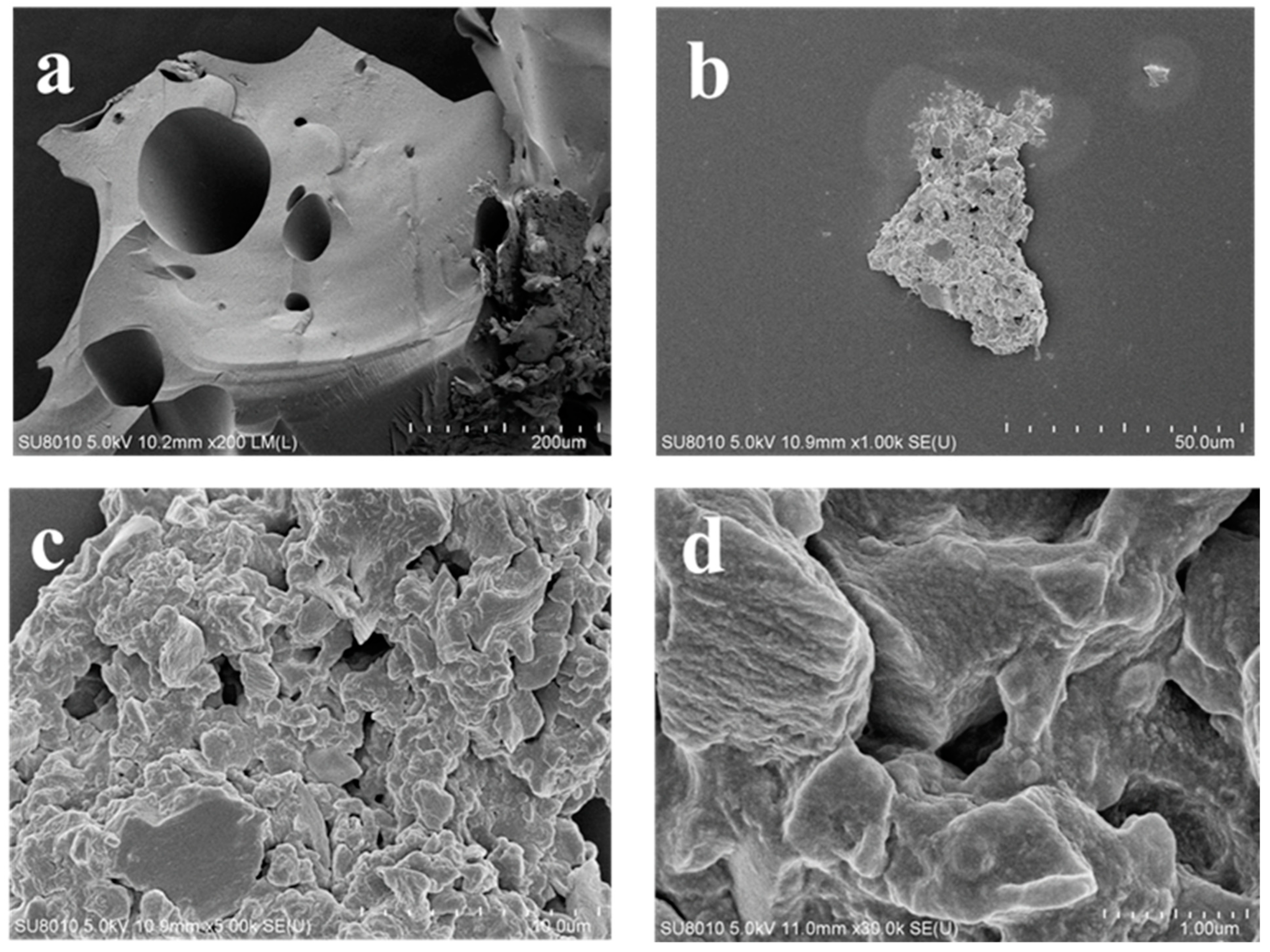
The C/CTS/PVA/CNF hydrogel was characterized by SEM (Figure 2). Its surface exhibited a porous structure with embedded cellulose nanofibers within the network structure constructed by CST and PVA. At a magnification of 200 times, the dried hydrogel surface revealed the presence of a certain number of pores. As the magnification increased to 1000 times and 5000 times, the hydrogel exhibited a complex porous structure, which was advantageous for water transport and the release of antibacterial agents. At a magnification of 30,000 times, some AgNPs were found to be embedded in C/CTS/PVA/CNF hydrogel.

3.2. Antimicrobial Property

3.2.1. Effect of CST and C-Ag

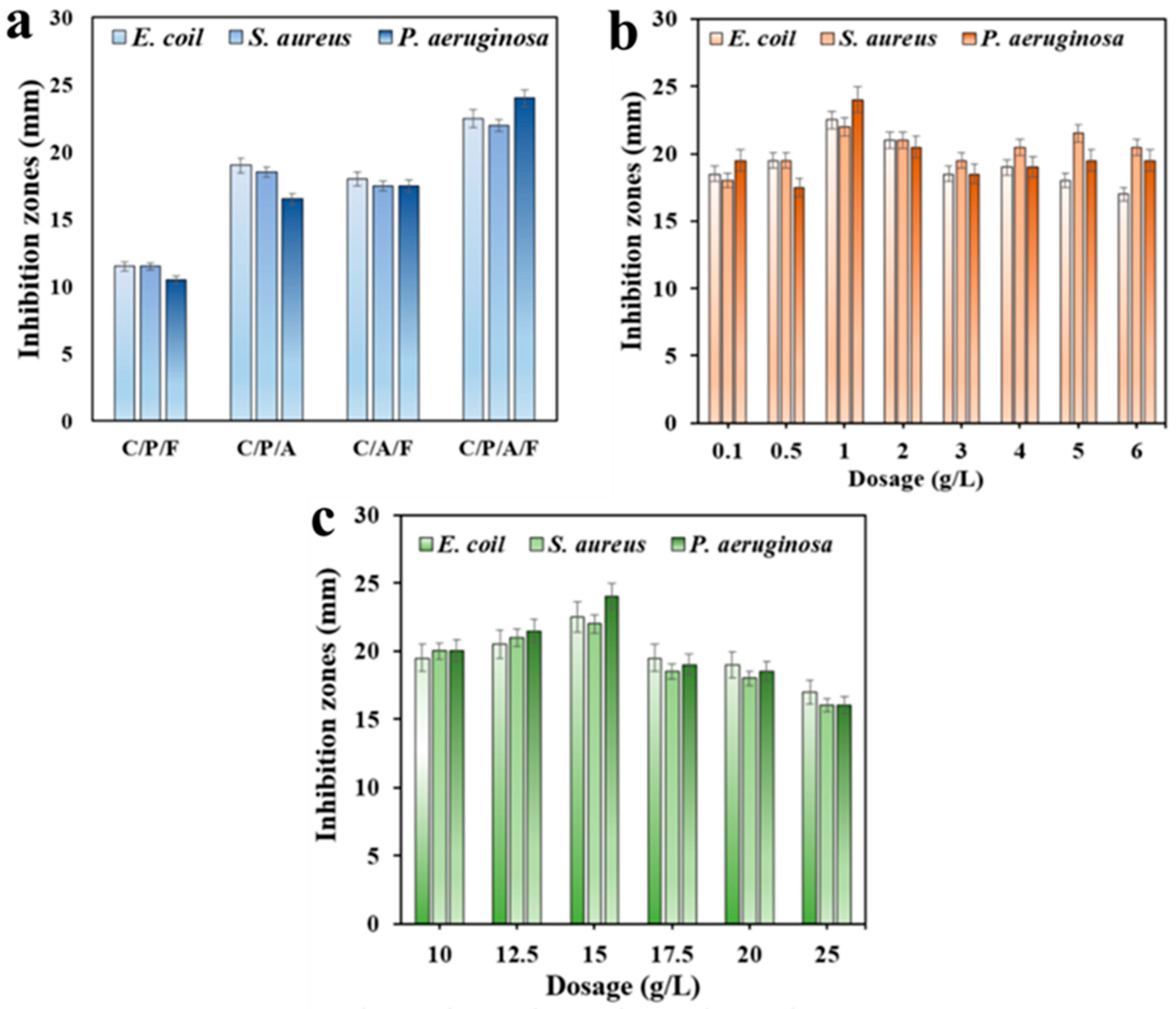
The composition of materials and the dosage of antimicrobial substances in the preparation of hydrogels can affect their antimicrobial efficacy [40]. Figure 3a showcased that hydrogel without the supplementary of C-Ag acquired weak antibacterial performance. Hydrogels without the addition of PVA and CNF demonstrated similar antibacterial performance. The C/CTS/PVA/CNF hydrogel exhibited a significant inhibitory effect on bacteria. The positively charged amino groups on CTS interacted with the negatively charged components of microbial cell membranes, leading to the leakage of bacterial proteins and other cell components [41]. AgNPs could also affect the charge difference inside and outside of cells, causing cell leakage and inducing microbial oxidative stress (ROS) [42].
Figure 3b revealed that the inhibitory capacity of bacteria by C-Ag increased with the increase in dosage at low loading concentrations (1 g/L). However, when the loading amount was too high, AgNPs aggregated within the gel, which hindered the liberation of silver ions and weakened the antibacterial effect [43]. Figure 3c showcased that the gel exhibited maximum antibacterial activity when the CTS content was 15 g/L. The inhibition areas of hydrogel against E. coli, S. aureus, and P. aeruginosa could reach 22.5 mm, 22.0 mm, and 24.0 mm, respectively. When the CTS content was excessively high, the tightly cross-linked structure formed by PVC and CTS could affect the release of AgNPs and amino groups, substantially impacting the antibacterial efficacy [44]. PVA/starch hydrogel containing turmeric and glutaraldehyde could inhibit E. coli and S. aureus, with an inhibition zone of 9.9–11.3 mm [19]. The royal jelly/chitosan/polyvinyl alcohol gel could inhibit P. aeruginosa with an inhibition zone of 13.0 mm [20]. The inhibition zones against P. aeruginosa ATCC9027, S. aureus ATCC6538, and E. coli ATCC25922 were over 20 mm when CST dosage was 15 g/L. Mesoporous Zn2+/Ag+ doped hydroxyapatite nanoparticles were utilized to inhibit P. aeruginosa with an inhibition zone of 7–14 mm [5]. Incorporating silver nanoparticles into solvent-free synthesized PEG-based hydrogels was used for antibacterial, acquiring the maximum antibacterial zone of inhibition against E. coli and S. aureus were 17.4 and 15.9 mm [4], respectively. In our work, the prepared C/CTS/PVA/CNF hydrogel had high antibacterial activity.

3.2.2. Effect of C/CTS/PVA/CNF Hydrogel Dosage and Incubation Time

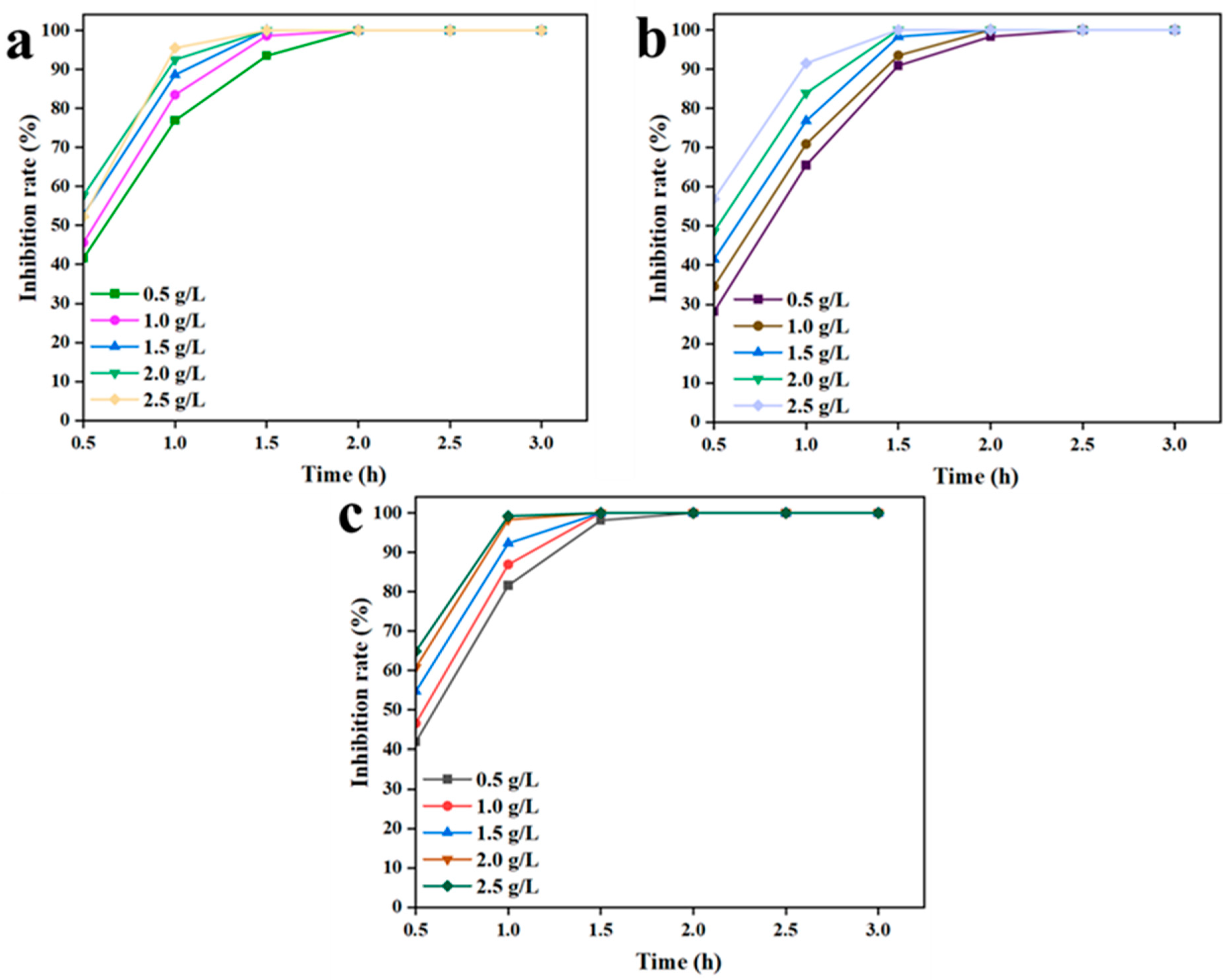
By increasing the material dosage and incubation time, the antibacterial efficiency of C/CTS/PVA/CNF hydrogel could be elevated (Figure 4). Figure 4a indicated that increasing the dosage of the C/CTS/PVA/CNF hydrogel led to a noticeable enhancement in the antibacterial effect on E. coli within 1 h. The difference in antibacterial effect decreased by 1.5 h, and the antibacterial rate was 100% by 2 h. However, increasing the gel dosage did not substantially improve the antibacterial efficiency. In the previous research, a concentration of 0.6 g/L of the antibacterial material acquired a 90% antibacterial effect within 5 h [14]. In our work, a C/CTS/PVA/CNF hydrogel dosage of 0.50 g/L acquired a 100% antibacterial rate within 2 h.
Inhibitors of S. aureus have been extensively studied, with some studies reporting inhibition rates as high as 95% for antibacterial materials [15]. In our research, the C/CTS/PVA/CNF hydrogel load was 0.50 g/L. After an incubation period of 1.5 h, the antibacterial rate approached 90% (Figure 4b), manifesting that the C/CTS/PVA/CNF hydrogel exhibited a good antibacterial effect against S. aureus. P. aeruginosa is a naturally occurring strain with strong drug resistance and good adaptability [45]. After 1 h of antibacterial treatment with the gel (1.5 g/L), the inhibition rate against P. aeruginosa could exceed 90%. By increasing the dosage of C/CTS/PVA/CNF hydrogel, the antibacterial rate also elevated (Figure 4c). For antibacterial time exceeding 90 min, the minimum C/CTS/PVA/CNF hydrogel dosage acquired a 100% inhibition rate. However, as the antibacterial duration increased, the inhibitory effect on bacteria no longer manifested significant enhancement. The antibacterial tests testified that the C/CTS/PVA/CNF hydrogel exhibited substantial and rapid bactericidal effects against both Gram-positive and Gram-negative bacteria. Even at low dosages, the hydrogel manifested excellent antibacterial activity during prolonged antibacterial processes.

3.3. Stability and Reusability of C/CTS/PVA/CNF Hydrogels

C/CTS/PVA/CNF hydrogel manifested strong stability and high inhibition rates, which might decrease performance costs and minimize potential pollution. Figure 5a shows that the C/CTS/PVA/CNF hydrogel maintained long-term stability in its antibacterial performance. In addition, it could be repeatedly used. The antibacterial rate of C/CTS/PVA/CNF hydrogel was 100% within 10 days and remained above 90% within 30 days after storage. In some stability studies of antibacterial gels, the inhibition rate declined to 90% after 15 days of preparation [46]. As showcased in Figure 5b, when the fifth batch of reuse experiment was over, the antibacterial rate was over 85%. Meanwhile, the antibacterial rate began to weaken apparently after the sixth run, which resulted from the loss of antibacterial compounds. The antibacterial durability of natural antibacterial materials might be relatively weak, and the antibacterial effect of biofiber material was less than 30% after the fifth test [47]. These results implied that the C/CTS/PVA/CNF hydrogel possessed good stability and reusability.

3.4. Synergistic Antibacterial Mechanism of C/CTS/PVA/CNF Hydrogels

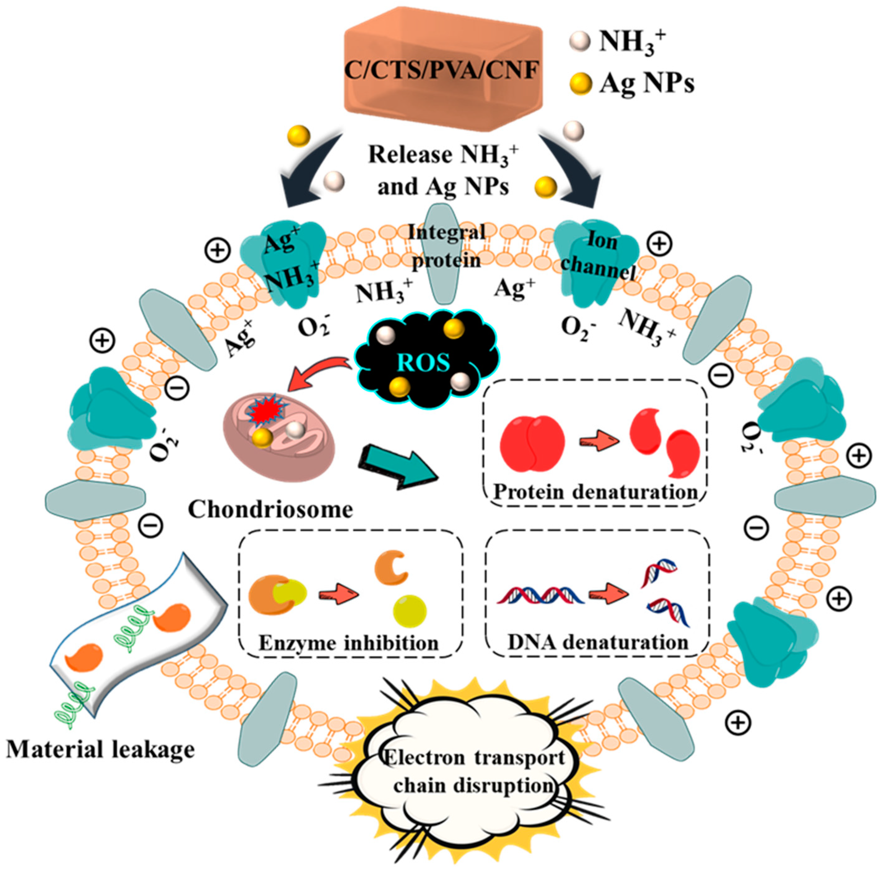
Biochar was combined with silver nanoparticles to form C-Ag, which exhibited high bactericidal activity. CTS is a natural antibacterial material capable of inhibiting the proliferation of many bacteria. PVA is a cross-network structure of polymer, which might be used as a carrier for dispersing C-Ag and CTS in C/CTS/PVA/CNF hydrogels. CTS, PVA, C-Ag, and CNF can cross-link to build a gel network structure for antibacterial assistance. In addition, CTS and PVA are hydrophilic biomaterials, and the prepared C/CTS/PVA/CNF hydrogels might absorb water and further expand. C-Ag will be released through its dispersion in C/CTS/PVA/CNF hydrogels, which have bacteriostatic action. The positively charged amino group (−NH2) in CTS and C-Ag contained in the C/CTS/PVA/CNF hydrogels can combine with the negatively charged cell membrane, resulting in the cell membrane structural alteration and damage [48,49,50]. Afterwards, bacteriostatic chemicals and materials will inhibit the formation of a cell wall, block the synthesis of protein, destroy ion transport channel, affect the function of the membrane, inhibit the biosynthesis of nucleic acid, and influence specific metabolic pathways [51,52,53], causing the death of the bacteria (Figure 6). The utilization of bio-based waste and natural antibacterial agents reduced the antibacterial cost. Improving the eco-friendly and sustainable aspects of antibacterial agents or materials holds great promising prospects for their application in antimicrobial and dye removal. For the antibacterial mechanism, in-depth exploration needs to be implemented in the future.

3.5. Swelling Ratio, Moisture Adsorption, Water Loss, and Moisture Content of C/CTS/PVA/CNF Hydrogels

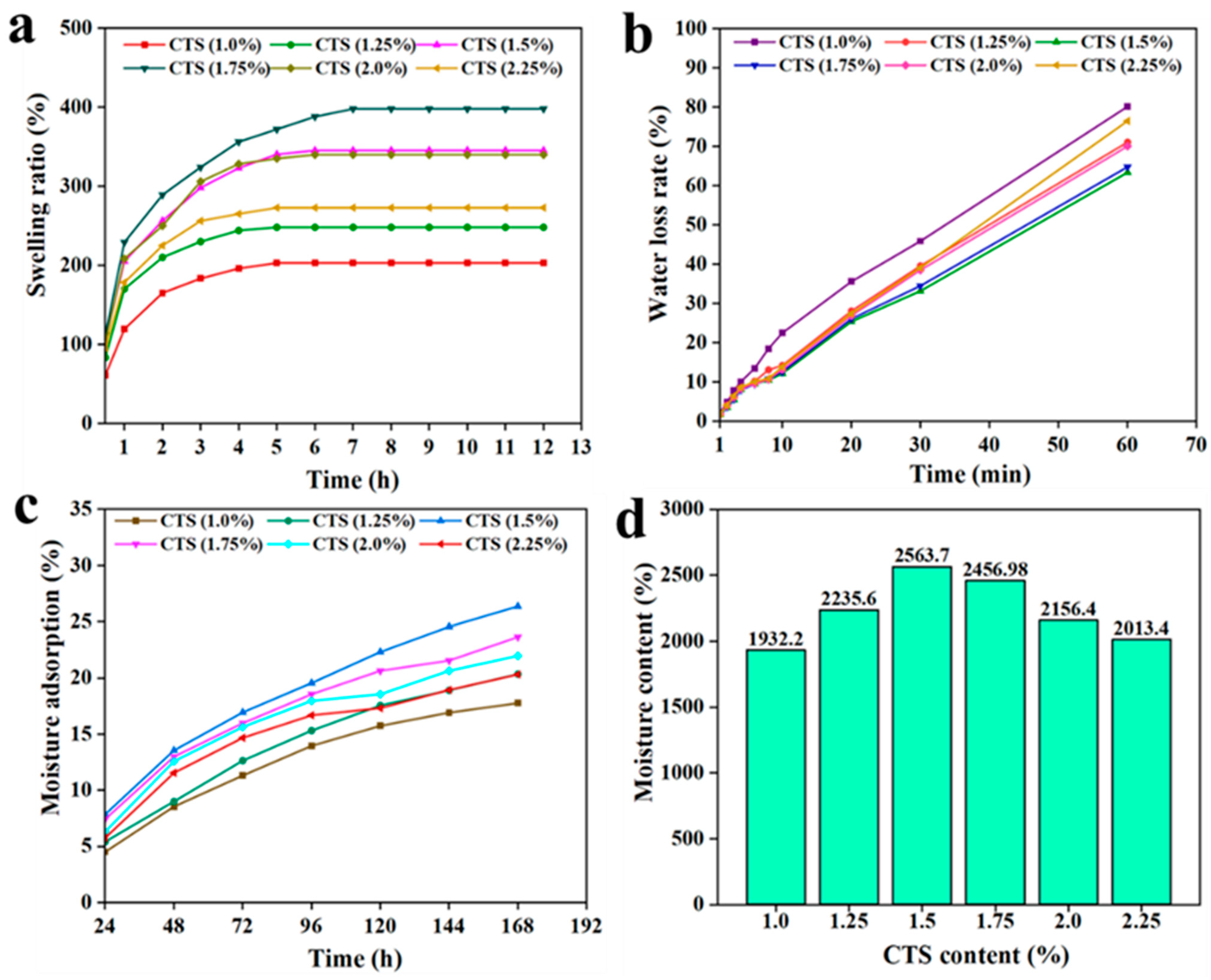
Swelling rate is one of the most important properties of hydrogels [53]. Within a certain period of time, the swelling ratio of hydrogel materials increased with the prolongation of immersion time in water (Figure 7a). When the CTS content was 1.0, 1.25, 2.0, and 2.25 wt%, the gel reached equilibrium swelling within 200 min. For CTS contents of 1.5 and 1.75 wt%, the gel reached equilibrium swelling around 240 min. Within a certain CTS concentration range, the swelling capacity of the hydrogel increased with increasing concentration. This was ascribed to the sufficient cross-linking of CTS and PVA, which enlarged the pore structure of the hydrogel, promoting swelling [54]. The highest swelling ratio was observed at approximately 400% when the CTS concentration was 1.75 wt%.
In the dehydration test experiment, as the hydrogel is subjected to increasing time in the oven, the water content within the hydrogel starts to diminish. The intermolecular interactions between CTS and PVA affected the water loss in the gel [55]. Figure 7b shows the influence of CTS dosage on the dehydration rate of the hydrogel. Within a certain CTS concentration range, the water loss rate of the gel gradually declined with increasing CTS dosage. However, when the CTS concentration exceeded 1.5 wt%, the dehydration rate of hydrogel started to increase. When the CTS concentration was 1.5 wt%, the water loss rate reached 63.4% after drying for 60 min. As CTS concentration reached 1.0 wt% and 2.25 wt%, the water loss rate approached ~80%. These results manifested that an appropriate CTS dosage substantially promoted the water retention capability of the hydrogel.
Moisture absorption is represented by the water ratio, which is the ratio of the weight of water contained in the material over the weight of the material in the absolute dry state [56]. It was manifested that the CTS content affected the moisture absorption rate and moisture content of C/CTS/PVA/CNF hydrogel (Figure 7c,d). As CTS was 1.5 wt%, this prepared gel continued to exhibit an increasing trend in moisture absorption even after 168 h, surpassing the gels with lower CTS content. This implied that the hydrogel manifested superior moisture absorption performance within a certain CTS concentration range. In the moisture content test, C/CTS/PVA/CNF hydrogel displayed strong water-holding capacity, with a moisture content of 2563.7% when the CTS concentration was 1.5 wt%. For CTS concentrations below 1.5 wt%, both the MA and MC of the hydrogel increased with increasing CTS dosage. This was attributed to the influence of CTS concentration on the gel’s structural morphology, which facilitated the diffusion of water within the network-like adhesive structure, a typical scenario of water diffusion within a reticular adhesive structure [57]. The incorporation of C-Ag effectually enhanced the frictional force within the hydrogel, possibly because -OH and -NH2 groups might interact with AgNPs, as well as the cross-linking of polymer chains, which would weaken the water permeability of the prepared material [58]. The addition of CNF also strengthened the internal network structure of this prepared hydrogel to some extent.
Overall, C/CTS/PVA/CNF hydrogel exhibited excellent physical properties, and its complex and diverse internal network structure effectually controlled water permeation.

3.6. C/CTS/PVA/CNF Hydrogel Dye-Removal Properties

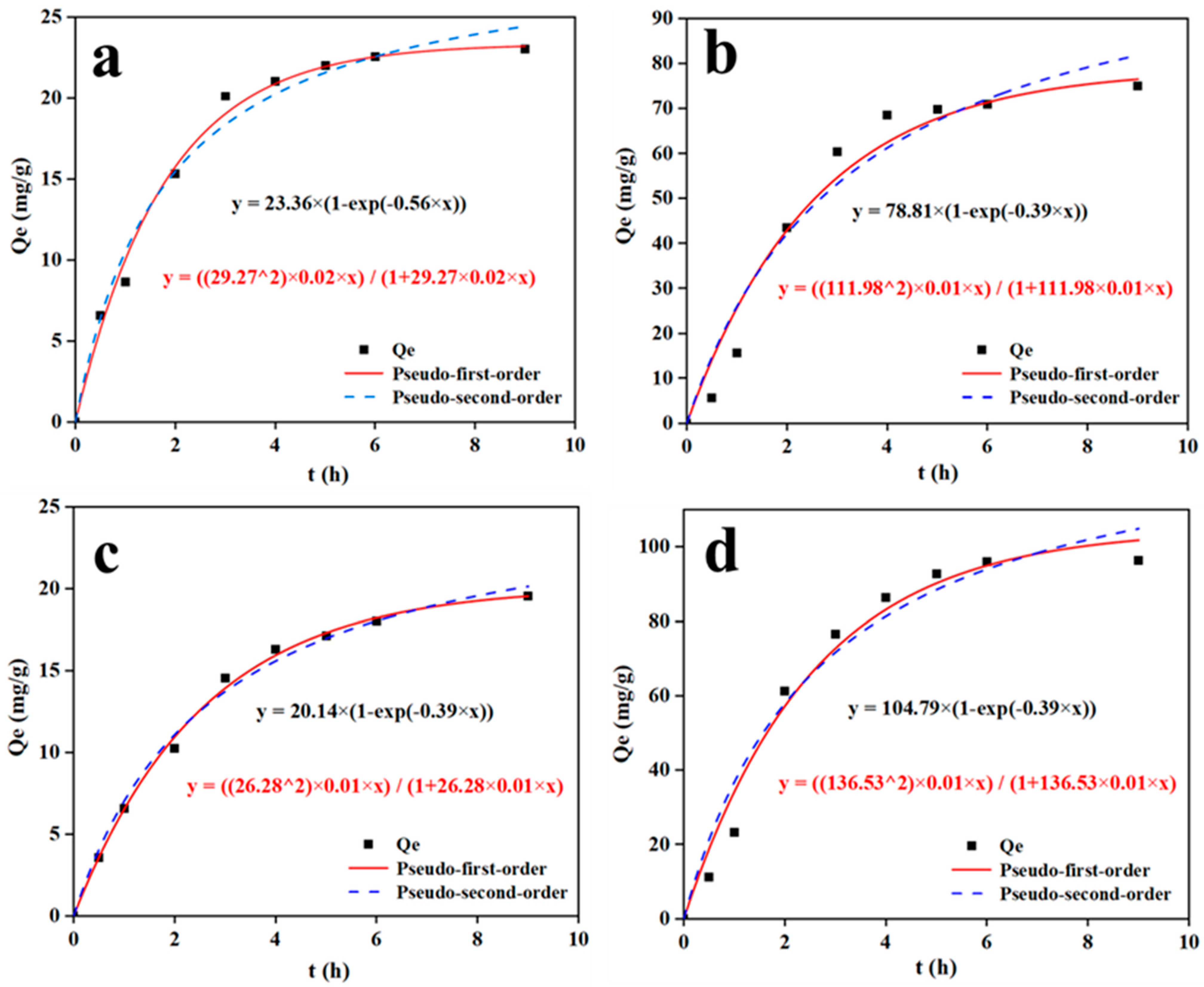
In order to test the ability and property of dye removal by the C/CTS/PVA/CNF hydrogel, the dye-removal kinetics were studied (Figure 8). The removal results of dyes indicated that the removal rate of MB, MG, and MO reached equilibrium at around 5 h, while the removal rate of CR reached equilibrium at around 6 h. High adsorption rate of MB and MO occurred within 1/2 h, while high adsorption rate of MG and CR happened within 2 h. This result implied that a large amount of dye was eliminated around these time points. Through data fitting, it was found that the hydrogel manifested outstanding affinity for CR removal, with a maximum removal capacity of 96.32 mg/g. This was comparable to the removal capacity of some nanomaterial [59]. Table 1 illustrated that the removal kinetics of C/CTS/PVA/CNF hydrogel well fitted the pseudo-first-order kinetics, with a correlation coefficient (R2 ≥ 0.99) for MB, MO and CR, while the correlation coefficient (R2) for the pseudo-second-order kinetic equation was less than 0.99 for three sets (MB, MG and CR).
By fitting the pseudo-first-order kinetic equation, the equilibrium-removal capacities of MB, MG, MO, and CR were calculated to be 23.21, 76.54, 19.55, and 101.81 mg/g. According to the pseudo-second-order kinetic equation, the capacities of equilibrium adsorption (qe cal) were 24.38, 81.31, 20.14, and 104.91 mg/g for MB, MG, MO, and CR, respectively. The experimental maximum values (qe exp) were observed to be 23.01, 75.01, 19.56, and 96.32 mg/g for MB, MG, MO, and CR, respectively. Accordingly, the fitting curves better matched the pseudo-first-order kinetic equation: R2, with a value close to 1 [60]. Consequently, the pseudo-first-order kinetic equation might effectually describe the entire adsorption process. The adsorption experimental results showcased the excellent dye adsorption capacity of the C/CTS/PVA/CNF hydrogel. The acid-promoted UiO-66 had the adsorption capacities of MO (84.8 mg/g) and MB (13.2 mg/g) [61]. The maximum adsorption capacity of ZnTPA-O/Tea with MB dye was 31.81 mg/g [62]. The C/CTS/PVA/CNF hydrogels, with good antibacterial and adsorption properties, have a certain application value in dye adsorption.
Biochar, C-Ag, CTS, and PVA had good adsorption properties [63,64,65,66,67,68]. The electrostatic interaction between the hydroxyl group (-OH) and the amino group (-NH2) in CTS molecules caused the dye adsorption on the surface of gels [63]. PVA has a cross-network structure [66], rendering it as a support for dispersive C-Ag and CTS in C/CTS/PVA/CNF hydrogels. The formed network adhesive structure fabricated by CTS, C-Ag, and PVA enhanced swelling properties, improved water retention capability, and improved moisture content of C/CTS/PVA/CNF hydrogels, which would favor expanding the adsorption area of dyes.
Hydrogels have shown potential in antibacterial and waste adsorption [69]. In this research, the C/CTS/PVA/CNF hydrogel has a high potential for antibacterial and dye-removal control. However, the involved mechanism is not clear yet. In-depth exploration of this subject needs to be well implemented through the characterization of prepared C/CTS/PVA/CNF hydrogel (e.g., Brunner−Emmet−Teller (BET), X-ray Diffractometer (XRD), Atomic Force Microscope (AFM), Differential Scanning Calorimeter (DSC), Thermal Gravimetric Analyzer (TGA), Transmission Electron Microscope (TEM), Energy Dispersive Spectroscopy (EDS), Elemental Mapping (EM), etc.). In addition, optimal antibacterial and dye removal control processes need to be well developed in the future.

4. Conclusions

This study adhered to the principle of waste utilization by employing discarded fish scales to prepare C-Ag and utilizing waste wheat straw to extract CNF for the preparation of a C/CTS/PVA/CNF hydrogel with antibacterial properties and dye-removal capacity. The hydrogel’s porous hydrophobic structure enhanced stability and degradation ability, while also strengthening the antibacterial effect by facilitating the liberation of silver ions and amino groups to promote synergistic antibacterial activity. Additionally, the hydrogel manifested a maximum removal capacity of 96.3 mg of Congo red dye/g. In summary, the C/CTS/PVA/CNF hydrogel was an environmentally sustainable antibacterial material with significant potential in sewage treatment.

Supplementary Materials

The following supporting information can be downloaded at: https://www.mdpi.com/article/10.3390/pr12102277/s1, Figure S1. SEM image of CNF.

Author Contributions

Conceptualization, methodology, formal analysis, investigation, resources, data curation, writing—original draft: L.X.; Conceptualization, methodology, software, formal analysis, investigation, data curation, writing—original draft: Z.Z.; Supervision, writing—review and editing: Y.H. and Y.J. All authors have read and agreed to the published version of the manuscript.

Funding

This research received no external funding.

Data Availability Statement

Data are contained within the article.

Acknowledgments

The authors thank the Analysis and Testing Center (Changzhou University) for the analysis of samples with FT-IR and SEM. All individuals included in this section have consented to the acknowledgment.

Conflicts of Interest

The authors declare no conflict of interest to this research, and the paper has not been submitted to any other journal simultaneously.

References

  1. He, Q.; Zhang, H.; Ma, M.; He, Y.; Jia, J.; Hu, Q.; Gong, Y. Critical assessment of protozoa contamination and control measures in mass culture of the diatom Phaeodactylum tricornutum. Bioresour. Technol. 2022, 359, 127460. [Google Scholar] [CrossRef] [PubMed]
  2. Jiang, S.; Meng, L.; Lou, Y.; Yan, Z.; Xi, J.; Bian, H.; Wu, W.; Xiao, H. Fabrication of multifunctional air filter paper with flame retardant, antibacterial and hydrophobic properties. J. Environ. Chem. Eng. 2023, 11, 111540. [Google Scholar] [CrossRef]
  3. Hezma, A.M.; Shaltout, W.A.; Kabary, H.A.; El-Bahy, G.S.; Abdelrazzak, A.B. Fabrication, characterization and adsorption investigation of nano zinc oxide–sodium alginate beads for effective removal of chromium (VI) from aqueous solution. J. Inorg. Organomet. Polym. Mater. 2023, 33, 1400–1408. [Google Scholar] [CrossRef]
  4. Kazeminava, F.; Arsalani, N.; Ahmadi, R.; Kafil, H.S.; Geckeler, K.E. A facile approach to incorporate silver nanoparticles into solvent-free synthesized PEG-based hydrogels for antibacterial and catalytical applications. Polym. Test. 2021, 101, 106906. [Google Scholar] [CrossRef]
  5. Phatai, P.; Prachumrak, N.; Kamonwannasit, S.; Kamcharoen, A.; Roschat, W.; Phewphong, S.; Futalan, C.M.; Khemthong, P.; Butburee, T.; Youngjan, S.; et al. Zinc-Silver doped mesoporous hydroxyapatite synthesized via ultrasonic in combination with sol-gel method for increased antibacterial activity. Sustainability 2022, 14, 11756. [Google Scholar] [CrossRef]
  6. Zhang, Z.; He, Y. Synthesis and characteristics of a fish scale-based biochar–nanosilver antibacterial material. Processes 2023, 11, 1992. [Google Scholar] [CrossRef]
  7. Jiang, W.; Cai, Y.; Liu, D.; Yu, X.; Wang, Q. Enhanced adsorption performance of oxytetracycline in aqueous solutions by Mg-Fe modified suaeda-based magnetic biochar. Environ. Res. 2024, 241, 117662. [Google Scholar] [CrossRef]
  8. Cai, Q.; Bai, X.; Pu, J. Author Correction: Adaptive VAlCN-Ag composite and VAlCN/VN-Ag multilayer coatings intended for applications at elevated temperature. J. Mater. Sci. 2022, 57, 15674–15675. [Google Scholar] [CrossRef]
  9. Oliveira, Í.M.; de Jesus, R.A.; Nascimento, V.R.S.; Bilal, M.; Iqbal, H.M.N.; Ferreira, L.F.R.; Cestari, A.R. Bioremediation potential of Dicentrarchus labrax fish scales for dye-based emerging contaminants by ANN–GA hybrid modeling. Bioprocess Biosyst. Eng. 2022, 45, 1189–1200. [Google Scholar] [CrossRef]
  10. Xia, D.; Liu, Y.; Cheng, X.; Gu, P.; Chen, Q.; Zhang, Z. Temperature-tuned fish-scale biochar with two-dimensional homogeneous porous structure: A promising uranium extractant. Appl. Surf. Sci. 2022, 591, 153136. [Google Scholar] [CrossRef]
  11. Kesarwani, V.; Rai, V.K. Impact of AgNPs on the optical thermometry and stability of bismuth modified tellurium-tungstate upconverting glass. J. Non-Cryst. Solids 2023, 603, 122129. [Google Scholar] [CrossRef]
  12. Yang, D.; Fan, B.; Sun, G.; He, Y.-C.; Ma, C. Ultraviolet blocking ability, antioxidant and antibacterial properties of newly prepared polyvinyl alcohol-nanocellulose-silver nanoparticles-ChunJian peel extract composite film. Int. J. Biol. Macromol. 2023, 252, 126427. [Google Scholar] [CrossRef] [PubMed]
  13. Hariram, M.; Vivekanandhan, S.; Ganesan, V.; Muthuramkumar, S.; Rodriguez-uribe, A.; Mohanty, A.K.; Misra, M. Tecoma stans flower extract assisted biogenic synthesis of functional Ag-Talc nanostructures for antimicrobial applications. Bioresour. Technol. Rep. 2019, 7, 100298. [Google Scholar] [CrossRef]
  14. Hu, Z.; Zhang, L.; Zhong, L.; Zhou, Y.; Xue, J.; Li, Y. Preparation of an antibacterial chitosan-coated biochar-nanosilver composite for drinking water purification. Carbohydr. Polym. 2019, 219, 290–297. [Google Scholar] [CrossRef]
  15. Cui, J.; Liang, Y.; Yang, D.; Liu, Y. Facile fabrication of rice husk based silicon dioxide nanospheres loaded with silver nanoparticles as a rice antibacterial agent. Sci. Rep. 2016, 6, 21423. [Google Scholar] [CrossRef]
  16. Zhou, X.; Fu, Y.; Chen, L.; Wang, R.; Wang, X.; Miao, Y.; Ji, X.; Bian, H.; Dai, H. Diisocyanate modifiable commercial filter paper with tunable hydrophobicity, enhanced wet tensile strength and antibacterial activity. Carbohydr. Polym. 2020, 248, 116791. [Google Scholar] [CrossRef]
  17. Potaś, J.; Szymańska, E.; Winnicka, K. Challenges in developing of chitosan—Based polyelectrolyte complexes as a platform for mucosal and skin drug delivery. Eur. Polym. J. 2020, 140, 110020. [Google Scholar] [CrossRef]
  18. Singh, S.; Arputharaj, E.; Dahms, H.U.; Patel, A.K.; Huang, Y.L. Chitosan-based nanocomposites for removal of Cr(VI) and synthetic food colorants from wastewater. Bioresour. Technol. 2022, 351, 127018. [Google Scholar] [CrossRef]
  19. Sekhavat Pour, Z.; Ghaemy, M. Removal of dyes and heavy metal ions from water by magnetic hydrogel beads based on poly(vinyl alcohol)/carboxymethyl starch-g-poly(vinyl imidazole). RSC Adv. 2015, 5, 64106–64118. [Google Scholar] [CrossRef]
  20. Su, J.F.; Li, G.Q.; Wen, Q.; Xue, L.; Chen, C.L.; Huang, T.L. Highly efficient nitrate and phosphorus removal and adsorption of tetracycline by precipitation in a chitosan/polyvinyl alcohol immobilized bioreactor. Bioprocess Biosyst. Eng. 2020, 43, 1761–1771. [Google Scholar] [CrossRef]
  21. Bandyopadhyay, S.; Saha, N.; Brodnjak, U.V.; Sáha, P. Bacterial cellulose and guar gum based modified PVP-CMC hydrogel films: Characterized for packaging fresh berries. Food Packag. Shelf Life 2019, 22, 100402. [Google Scholar] [CrossRef]
  22. Ma, S.; Shi, W.; Li, H.; Zhang, Y. Biomimetic mineralization of nacre-inspired multiple crosslinked PVA/CaAlg/SiO2 membrane with simultaneously enhanced mechanical and separation properties. Int. J. Biol. Macromol. 2023, 234, 123650. [Google Scholar] [CrossRef] [PubMed]
  23. Alemdar, A.; Sain, M. Isolation and characterization of nanofibers from agricultural residues—Wheat straw and soy hulls. Bioresour. Technol. 2008, 99, 1664–1671. [Google Scholar] [CrossRef] [PubMed]
  24. Froese, A.G.; Sparling, R. Cross-feeding and wheat straw extractives enhance growth of Clostridium thermocellum-containing co-cultures for consolidated bioprocessing. Bioprocess Biosyst. Eng. 2021, 44, 819–830. [Google Scholar] [CrossRef] [PubMed]
  25. Zhang, C.-W.; Li, F.-Y.; Li, J.-F.; Li, Y.-L.; Xu, J.; Xie, Q.; Chen, S.; Guo, A.-F. Novel treatments for compatibility of plant fiber and starch by forming new hydrogen bonds. J. Clean. Prod. 2018, 185, 357–365. [Google Scholar] [CrossRef]
  26. Kumar Trivedi, A.; Kumar, A.; Gupta, M.K. Extraction of nanocellulose from wheat straw and its characterization. Mater. Today Proc. 2023, 78, 48–54. [Google Scholar] [CrossRef]
  27. Wang, Y.; Xu, T.; Liu, K.; Zhang, M.; Zhao, Q.; Liang, Q.; Si, C. Nanocellulose-based advanced materials for flexible supercapacitor electrodes. Ind. Crops Prod. 2023, 204, 117378. [Google Scholar] [CrossRef]
  28. Srivastava, K.R.; Dixit, S.; Pal, D.B.; Mishra, P.K.; Srivastava, P.; Srivastava, N.; Hashem, A.; Alqarawi, A.A.; Abd_Allah, E.F. Effect of nanocellulose on mechanical and barrier properties of PVA–banana pseudostem fiber composite films. Environ. Technol. Innov. 2021, 21, 101312. [Google Scholar] [CrossRef]
  29. Bai, H.; Liang, Z.; Wang, D.; Guo, J.; Zhang, S.; Ma, P.; Dong, W. Biopolymer nanocomposites with customized mechanical property and exceptionally antibacterial performance. Compos. Sci. Technol. 2020, 199, 108338. [Google Scholar] [CrossRef]
  30. Luo, C.; Guo, A.; Zhao, Y.; Sun, X. A high strength, low friction, and biocompatible hydrogel from PVA, chitosan and sodium alginate for articular cartilage. Carbohydr. Polym. 2022, 286, 119268. [Google Scholar] [CrossRef]
  31. Balouiri, M.; Sadiki, M.; Ibnsouda, S.K. Methods for in vitro evaluating antimicrobial activity: A review. J. Pharm. Anal. 2016, 6, 71–79. [Google Scholar] [CrossRef] [PubMed]
  32. Parmar, A.; Kapil, S.; Sachar, S.; Sharma, S. Design of experiment based methodical optimization and green syntheses of hybrid patchouli oil coated silver nanoparticles for enhanced antibacterial activity. Curr. Res. Green Sustain. Chem. 2020, 3, 100016. [Google Scholar] [CrossRef]
  33. Kumar, A.; Negi, Y.S.; Bhardwaj, N.K.; Choudhary, V. Synthesis and characterization of methylcellulose/PVA based porous composite. Carbohydr. Polym. 2012, 88, 1364–1372. [Google Scholar] [CrossRef]
  34. Lin, D.; Kelly, A.L.; Maidannyk, V.; Miao, S. Effect of concentrations of alginate, soy protein isolate and sunflower oil on water loss, shrinkage, elastic and structural properties of alginate-based emulsion gel beads during gelation. Food Hydrocoll. 2020, 108, 105998. [Google Scholar] [CrossRef]
  35. Xie, L.; Zhang, Z.; He, Y. Antibacterial effect of polyvinyl alcohol/biochar–nano silver/sodium alginate gel beads. Processes 2023, 11, 2330. [Google Scholar] [CrossRef]
  36. Feng, Q.; Fan, B.; He, Y. Antibacterial, antioxidant, Cr (VI) adsorption and dye adsorption effects of biochar-based silver nanoparticles-sodium alginate-tannic acid composite gel beads. Int. J. Biol. Macromol. 2024, 271, 132453. [Google Scholar] [CrossRef]
  37. Xiang, X.; Yi, X.; Zheng, W.; Li, Y.; Zhang, C.; Wang, X.; Chen, Z.; Huang, M.; Ying, G.G. Enhanced biodegradation of thiamethoxam with a novel polyvinyl alcohol (PVA)/sodium alginate (SA)/biochar immobilized Chryseobacterium sp. H5. J. Hazard. Mater. 2023, 443, 130247. [Google Scholar] [CrossRef]
  38. Salunke, A.S.; Salunke, S.T.; Deokate, R.J.; Kale, B.B. Tuning of photoluminescence behavior of gold coated chitosan-polyvinyl alcohol binding with graphene quantum dots. Mater. Today Proc. 2022, 62, 1752–1757. [Google Scholar] [CrossRef]
  39. Bian, H.; Chen, L.; Dong, M.; Wang, L.; Wang, R.; Zhou, X.; Wu, C.; Wang, X.; Ji, X.; Dai, H. Natural lignocellulosic nanofibril film with excellent ultraviolet blocking performance and robust environment resistance. Int. J. Biol. Macromol. 2021, 166, 1578–1585. [Google Scholar] [CrossRef]
  40. Abbaszadegan, A.; Ghahramani, Y.; Gholami, A.; Hemmateenejad, B.; Dorostkar, S.; Nabavizadeh, M.; Sharghi, H. The effect of charge at the surface of silver nanoparticles on antimicrobial activity against Gram-positive and Gram-negative bacteria: A preliminary study. J. Nanomater. 2015, 2015, 720654. [Google Scholar] [CrossRef]
  41. Dhandapani, P.; Santhoshkumar, M.; Narenkumar, J.; AlSalhi, M.S.; Kumar, P.A.; Devanesan, S.; Kokilaramani, S.; Rajasekar, A. Bio-approach: Preparation of RGO-AgNPs on cotton fabric and interface with sweat environment for antibacterial activity. Bioprocess Biosyst. Eng. 2022, 45, 1825–1837. [Google Scholar] [CrossRef] [PubMed]
  42. Wang, D.; Zhao, L.; Ma, H.; Zhang, H.; Guo, L.H. Quantitative analysis of reactive oxygen species photogenerated on metal oxide nanoparticles and their bacteria toxicity: The role of superoxide radicals. Environ. Sci. Technol. 2017, 51, 10137–10145. [Google Scholar] [CrossRef] [PubMed]
  43. Yang, D.; Fan, B.; He, Y.C. UV-blocking, antibacterial, corrosion resistance, antioxidant, and fruit packaging ability of lignin-rich alkaline black liquor composite film. Int. J. Biol. Macromol. 2024, 275, 133344. [Google Scholar] [CrossRef] [PubMed]
  44. Janani, B.; Okla, M.K.; Abdel-Maksoud, M.A.; AbdElgawad, H.; Thomas, A.M.; Raju, L.L.; Al-Qahtani, W.H.; Khan, S.S. CuO loaded ZnS nanoflower entrapped on PVA-chitosan matrix for boosted visible light photocatalysis for tetracycline degradation and anti-bacterial application. J. Environ. Manag. 2022, 306, 114396. [Google Scholar] [CrossRef] [PubMed]
  45. Masilan, K.; Neethiselvan, N.; Shakila, R.J.; Muralidharan, N.; Karthy, A.; Ravikumar, T.; Parthiban, F. Investigation on the coacervation of fish scale gelatin hydrogel with seafood waste hydrolysates for the development of artificial fish bait: Physico-chemical, thermodynamic, and morpho-structural properties. J. Indian Chem. Soc. 2022, 99, 100783. [Google Scholar] [CrossRef]
  46. Zhao, H.; Li, X.; Zhang, L.; Hu, Z.; Zhong, L.; Xue, J. Preparation and bacteriostatic research of porous polyvinyl alcohol/biochar/nanosilver polymer gel for drinking water treatment. Sci. Rep. 2021, 11, 12205. [Google Scholar] [CrossRef]
  47. Thakur, K.; Kalia, S.; Kaith, B.S.; Pathania, D.; Kumar, A.; Thakur, P.; Knittel, C.E.; Schauer, C.L.; Totaro, G. The development of antibacterial and hydrophobic functionalities in natural fibers for fiber-reinforced composite materials. J. Environ. Chem. Eng. 2016, 4, 1743–1752. [Google Scholar] [CrossRef]
  48. Qi, D.; Zhao, S.; Zhang, H.; Liu, B.; She, P.; Yue, X. Development of high-strength porous polyetheretherketone foam/nanosilver antibacterial composites for the prevention of postoperative infections in bone repair. Compos. Commun. 2022, 31, 101127. [Google Scholar] [CrossRef]
  49. Velidandi, A.; Pabbathi NP, P.; Dahariya, S.; Baadhe, R.R. Green synthesis of novel Ag–Cu and Ag–Znbimetallic nanoparticles and their in vitro biological, eco-toxicity and catalytic studies. Nano-Struct. Nano-Objects 2021, 26, 100687. [Google Scholar] [CrossRef]
  50. Ahmed, B.; Hashmi, A.; Khan, M.S.; Musarrat, J. ROS mediated destruction of cell membrane, growth and biofilms of human bacterial pathogens by stable metallic AgNPs functionalized from bell pepper extract and quercetin. Adv. Powder Technol. 2018, 29, 1601–1616. [Google Scholar] [CrossRef]
  51. Jiang, B.; Tian, C.; Song, G.; Pan, Q.; Wang, Z.; Shi, L.; Qiao, Y.; Fu, H. A green route to synthesize novel Ag/C antibacterial agent. Mater. Res. Bull. 2012, 47, 458–463. [Google Scholar] [CrossRef]
  52. Aryan, N.; Behpour, M.; Benvidi, A.; Jookar Kashi, F.; Azimzadeh, M.; Zare, H.R. Evaluation of sodium alendronate drug released from TiO2 nanoparticle doped with hydroxyapatite and silver–strontium for enhancing antibacterial effect and osteoinductivity. Mater. Chem. Phys. 2023, 295, 126934. [Google Scholar] [CrossRef]
  53. Vityazev, F.V.; Khramova, D.S.; Saveliev, N.Y.; Ipatova, E.A.; Burkov, A.A.; Beloserov, V.S.; Belyi, V.A.; Kononov, L.O.; Martinson, E.A.; Litvinets, S.G.; et al. Pectin-glycerol gel beads: Preparation, characterization and swelling behaviour. Carbohydr. Polym. 2020, 238, 116166. [Google Scholar] [CrossRef] [PubMed]
  54. Sethi, V.; Kaur, M.; Thakur, A.; Rishi, P.; Kaushik, A. Unravelling the role of hemp straw derived cellulose in CMC/PVA hydrogel for sustained release of fluoroquinolone antibiotic. Int. J. Biol. Macromol. 2022, 222, 844–855. [Google Scholar] [CrossRef] [PubMed]
  55. Rezagholizade-shirvan, A.; Fathi Najafi, M.; Behmadi, H.; Masrournia, M. Preparation of nano-composites based on curcumin/chitosan-PVA-alginate to improve stability, antioxidant, antibacterial and anticancer activity of curcumin. Inorg. Chem. Commun. 2022, 145, 110022. [Google Scholar] [CrossRef]
  56. Bhat, V.G.; Masti, S.P.; Narasagoudr, S.S.; Chougale, R.B.; Kumar, P.; Vantamuri, A.B. Development and characterization of Chitosan/Guar gum/Gum ghatti bionanocomposites with in situ silver nanoparticles. Chem. Data Collect. 2023, 44, 101009. [Google Scholar] [CrossRef]
  57. Mathew, S.; Jayakumar, A.; Kumar, V.P.; Mathew, J.; Radhakrishnan, E.K. One-step synthesis of eco-friendly boiled rice starch blended polyvinyl alcohol bionanocomposite films decorated with in situ generated silver nanoparticles for food packaging purpose. Int. J. Biol. Macromol. 2019, 139, 475–485. [Google Scholar] [CrossRef]
  58. Li, Q.; Lu, F.; Zhou, G.; Yu, K.; Lu, B.; Xiao, Y.; Dai, F.; Wu, D.; Lan, G. Silver inlaid with gold nanoparticle/chitosan wound dressing enhances antibacterial activity and porosity, and promotes wound healing. Biomacromolecules 2017, 18, 3766–3775. [Google Scholar] [CrossRef]
  59. Aghaei, F.; Tangestaninejad, S.; Bahadori, M.; Moghadam, M.; Mirkhani, V.; Mohammadpoor-Baltork, I.; Khalaji, M.; Asadi, V. Green synthesize of nano-MOF-ethylcellulose composite fibers for efficient adsorption of Congo red from water. J. Colloid Interface Sci. 2023, 648, 78–89. [Google Scholar] [CrossRef]
  60. Chen, L.; Mi, B.; He, J.; Li, Y.; Zhou, Z.; Wu, F. Functionalized biochars with highly-efficient malachite green adsorption property produced from banana peels via microwave-assisted pyrolysis. Bioresour. Technol. 2023, 376, 128840. [Google Scholar] [CrossRef]
  61. Qiu, J.; Feng, Y.; Zhang, X.; Jia, M.; Yao, J. Acid-promoted synthesis of UiO-66 for highly selective adsorption of anionic dyes: Adsorption performance and mechanisms. J. Colloid Interface Sci. 2017, 499, 151–158. [Google Scholar] [CrossRef] [PubMed]
  62. Arunkumar, G.; Pannipara, M.; Deviga, G.; Mariappan, M.; Al-Sehemi, A.G.; Anthony, S.P. Natural tea extract coated porous MOF nano/microparticles for highly enhanced and selective adsorption of cationic dyes from aqueous medium. J. Mol. Liq. 2024, 394, 123747. [Google Scholar] [CrossRef]
  63. Liu, W.; Lou, T.; Wang, X. Enhanced dye adsorption with conductive polyaniline doped chitosan nanofibrous membranes. Int. J. Biol. Macromol. 2023, 242, 124711. [Google Scholar] [CrossRef] [PubMed]
  64. Liu, Y.; Wu, Y.; He, Y.C.; Ma, C. Synthesis of furoic acid from biomasses by sequential catalysis with fish scale-rice husk-based heterogeneous chemocatalyst and dehydrogenase biocatalyst. Ind. Crops Prod. 2023, 202, 117033. [Google Scholar] [CrossRef]
  65. Zhang, H.; Li, R.; Zhang, Z. A versatile EDTA and chitosan bi-functionalized magnetic bamboo biochar for simultaneous removal of methyl orange and heavy metals from complex wastewater. Environ. Pollut. 2022, 293, 118517. [Google Scholar] [CrossRef]
  66. Wang, J.; Liang, J.; Sun, L.; Li, G.; Temmink, H.; Rijnaarts, H.H.M. Granule-based immobilization and activity enhancement of anammox biomass via PVA/CS and PVA/CS/Fe gel beads. Bioresour. Technol. 2020, 309, 123448. [Google Scholar] [CrossRef]
  67. Rodwihok, C.; Suwannakeaw, M.; Charoensri, K.; Wongratanaphisan, D.; Woon Woo, S.; Kim, H.S. Alkali/zinc-activated fly ash nanocomposites for dye removal and antibacterial applications. Bioresour. Technol. 2021, 331, 125060. [Google Scholar] [CrossRef]
  68. Tomczyk, A.; Sokołowska, Z.; Boguta, P. Biomass type effect on biochar surface characteristic and adsorption capacity relative to silver and copper. Fuel 2020, 278, 118168. [Google Scholar] [CrossRef]
  69. Bhuyan, M.M.; Jophous, M.; Jeong, J.H. Preparation of pectin–acrylamide–(vinyl phosphonic acid) hydrogel and its selective adsorption of metal ions. Polym. Bull. 2023, 80, 4625–4641. [Google Scholar] [CrossRef]
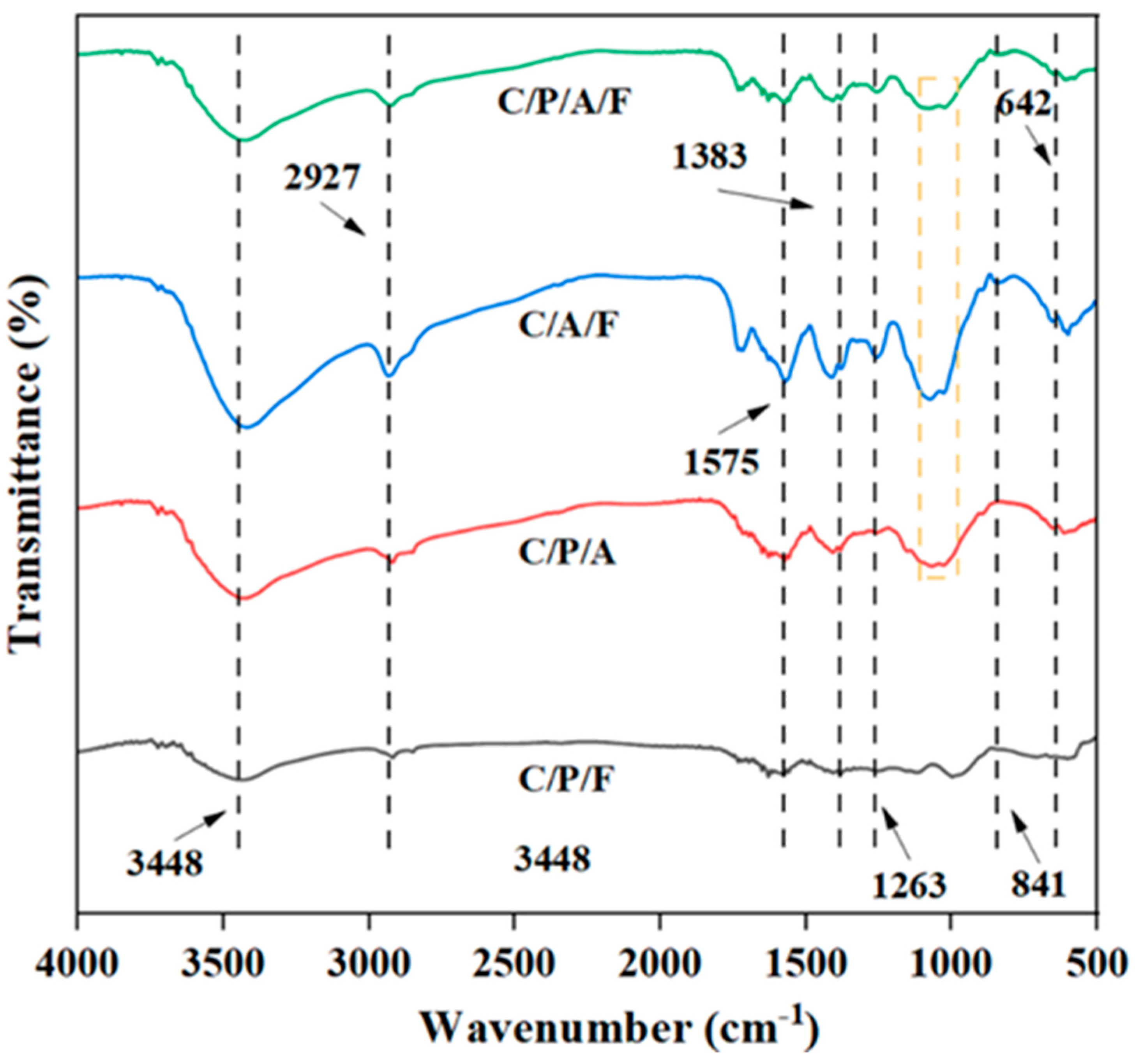
Figure 1. FT-IR spectra of C/P/F hydrogels, C/P/A hydrogels, C/A/F hydrogels, and C/P/A/F hydrogels.
Processes 12 02277 g001
Figure 2. SEM images C/CTS/PVA/CNF hydrogels ×200 (a), ×1000 (b), ×5000 (c), ×30,000 (d).
Processes 12 02277 g002
Figure 3. Antibacterial effect of gels prepared from different materials (C/P/F, C/P/A, C/P/F, and C/P/A/F) (a), C–Ag dosage (0.1, 0.5, 1, 2, 3, 4, 5, and 6 g/L) (b), chitosan dosage (10, 12.5, 15, 17.5, 20, and 25 g/L) (c).
Processes 12 02277 g003
Figure 4. Antibacterial activity against E. coli (a), S. aureus (b), and P. aeruginosa (c) was observed in C/CTS/PVA/CNF hydrogel composite gel at different dosages (0.5, 1.0, 1.5, 2.0, and 2.5 g/L) and time (0.5, 1.0, 1.5, 2.0, 2.5, and 3.0 h).
Processes 12 02277 g004
Figure 5. Antibacterial effect images of C/CTS/PVA/CNF hydrogels prepared on different days (5, 10, 15, 20, 25, 30, and 35 days) (a), repetitive antibacterial properties (1, 2, 3, 4, 5, 6, 7, and 8 times) (b).
Processes 12 02277 g005
Figure 6. Synergistic antibacterial mechanism of C/CTS/PVA/CNF hydrogels.
Processes 12 02277 g006
Figure 7. Swelling effect (a), water loss (b), moisture adsorption (c), and moisture content (d) images of different CTS dosage (1.0, 1.25, 1.5, 1.75, 2.0, and 2.25 wt%).
Processes 12 02277 g007
Figure 8. Kinetic fitting results for methylene blue (MB: 1 g/L) (a), Malachite green (MG: 2.5 g/L) (b), Methyl orange (MO: 1 g/L) (c), and Congo red (CR: 2.5 g/L) (d) adsorption onto C/CTS/PVA/CNF hydrogels.
Processes 12 02277 g008
Table 1. Kinetic parameters for the adsorption of dyes on C/CTS/PVA gel.
Pseudo-First-Order ModelPseudo-Second-Order Model
qe exp (mg/g)qe cal (mg/g)k1 (min−1)R2qe cal (mg/g)k2 (g/mg·min)R2
MB23.0123.210.560.9924.380.020.98
MG75.0176.540.390.9781.310.010.95
MO19.5619.550.160.9920.140.010.99
CR96.32101.810.390.99104.910.010.98
Disclaimer/Publisher’s Note: The statements, opinions and data contained in all publications are solely those of the individual author(s) and contributor(s) and not of MDPI and/or the editor(s). MDPI and/or the editor(s) disclaim responsibility for any injury to people or property resulting from any ideas, methods, instructions or products referred to in the content.