Article

Carboxymethyl-Cellulose-Based Hydrogels Incorporated with Cellulose Nanocrystals Loaded with Vitamin D for Controlled Drug Delivery

by
Nathália da Cunha Silva
,
Carla Jeany Teixeira Silva
,
Max Pereira Gonçalves
and
Fernanda G. L. Medeiros Borsagli
*
Institute of Engineering, Science and Technology, Universidade Federal dos Vales do Jequitinhonha e Mucuri/UFVJM, Av. 01, 4050 Cidade Universitária, Janaúba 39440-039, MG, Brazil
*
Author to whom correspondence should be addressed.
Processes 2024, 12(7), 1437; https://doi.org/10.3390/pr12071437
Submission received: 17 May 2024 / Revised: 14 June 2024 / Accepted: 23 June 2024 / Published: 9 July 2024
(This article belongs to the Special Issue Drug Carriers Production Processes for Innovative Human Applications)

Abstract

:
Currently, the development of innovative materials for the treatment of various diseases is highly interesting and effective. Additionally, in recent years, environmental changes, including the search for a sustainable world, have become the main goal behind developing sustainable and suitable materials. In this context, this research produced innovative hydrogels that incorporate cellulose nanocrystals and nanofibres from underutilised fibres from a semiarid region of Brazil; the hydrogels were loaded with vitamin D to evaluate controlled drug release for the treatment of diverse diseases. Spectroscopic (FTIR, Raman, UV–VIS), X-ray diffraction, zeta potential and morphology (SEM, TEM) analyses were used to characterise these hydrogels. In addition, biocompatibility was assessed using a resazurin assay, and the in vitro kinetic accumulative release of vitamin D was measured. The results showed that nanocrystals and nanofibres changed the structure and crystallinity of the hydrogels. In addition, the chemical groups of the hydrogels were red- and blueshifted in the FTIR spectra when the nanocrystals, nanofibres and vitamin D were incorporated. Moreover, the nanocrystals and nanofibres were homogeneously spread into the hydrogel when vitamin D was loaded into the hydrogel matrix. Furthermore, the cytotoxicity was greater than 90%. Additionally, the in vitro accumulative kinetic data of vitamin D release were robust (close to 40 ng·mL−1), with equilibrium being reached in the first 30 min. These results confirm the potential of using these hydrogels as therapeutic biomaterials for diverse diseases and problems in humans, mainly in women, who are the most harmed by vitamin D deficiency.

1. Introduction

Currently, climate change, the rapid growth of the population and animal production, energy consumption and various other transformations have garnered human research on diverse frontiers, here with the goal of searching for a more sustainable, economic and social world. Along these lines, the UN has promoted 17 goals for reaching the sustainable development of the planet. Among these 17 goals, it is important to highlight some that are linked to human health: zero hunger and sustainable agriculture, health and wellness, poor eradication, affordable and clean energy, industry, innovation and infrastructure, sustainable cities and communities, responsible production and consumption, decent work and economic growth [1]. Brazil can be characterised as a semiarid region, a poor region with sparse rain during the year and a climate that is difficult for agriculture. Additionally, there are unique plant yields that produce a large amount of fibre in the spring, namely Ceiba speciosa. Ceiba speciosa produces fibre that is underutilised and discharged into the environment every year; however, this fibre could be modified to produce an innovative nanomaterial that can have social and economic benefits [2]. Indeed, these nanomaterials are incredible materials for diverse areas, such as pharmaceuticals, energy, biomaterials and agriculture.
These nanomaterials have been highlighted for their use in pharmaceutical products, such as systems used to transport molecules of therapeutic interest. The nanoparticles that mediate this transport can be solid materials or stable colloidal suspensions, or they can be used to transport substances and control their release in cells and tissues because of their small size (10−9 m) [3]. In this context, different types of nanoparticles have been produced, including polymeric nanoparticles, which have attracted increasing attention because of their therapeutic potential and better stability in biological fluids, and magnetic nanoparticles; these nanoparticles have a wide range of functions, such as hyperthermia and disease diagnosis, and the application of these nanomaterials is immense, ranging from agriculture to medical applications [4].
Currently, investments in nanotechnology are enormous, and more than 60 countries possess national and/or private initiatives aimed at developing nanoscale materials, with investments exceeding USD 15 billion [5]. This nanotechnology has been applied in diverse sectors and has improved material properties, reduced costs and improved living standards. In this context, several studies have been conducted in academic and industrial contexts to implement this technology in everyday life [5]. Furthermore, because of its range of applications, nanotechnology presents a multidisciplinary approach because it involves physics, chemistry, biology, mathematics and engineering [5].
Based on this, investments in different sectors have become promising in nanotechnology, and interest in this class of materials has been exponentially increasing [6]. Considering this, over the past two decades, nanotechnology has played a crucial role in the medical sector, leading to significant improvements in diagnosis, monitoring, control, prevention and disease treatment. This intersection between nanotechnology and medicine has resulted in the rise of an innovative disciplinary field known as nanomedicine. The application of nanomedicine has had a significant impact on areas such as drug delivery, therapy, diagnosis and biomaterials [7].
One of the most promising areas of nanomedicine is controlled drug delivery. Nanoparticles can be designed to transport drugs in a targeted and controlled manner to specific locations in the body, such as tumours. This allows for a gradual and prolonged release of the medication, increasing its effectiveness and reducing its side effects. Furthermore, nanoparticles can be functionalised by targeting molecules that bind to specific receptors on target cells, thereby increasing treatment selectivity [8].
Nanoparticles loaded into hydrogel systems have great potential for minimising pharmacotherapeutic problems, increasing the therapeutic efficacy of chemotherapeutic agents and reducing their side effects and toxicity. Furthermore, these systems present high drug encapsulation efficiency, protecting them against degradation and reducing the possibility of allergies in patients because of their polymeric coating [9]. In drug release, these materials and technologies have been used to construct nanodelivery systems [10].
These hydrogels can be synthesised from a variety of polymers, including polyacrylates, polyacrylamides, alginates, collagen and gelatine. Each type of polymer has specific characteristics that determine the properties of the resulting hydrogel, such as its adsorption capacity, mechanical resistance and biocompatibility [9]. Because of their properties, hydrogels have been widely used in various applications, such as wound dressings, controlled drug release systems and supports for tissue engineering [11]. They are also applied in cosmetic products, such as moisturising creams and facial masks, because of their ability to retain moisture and promote skin hydration [12].
In summary, hydrogels are often used as wound dressings because of their ability to maintain a moist environment and promote healing. They can adsorb wound exudate, providing a sterile environment and promoting tissue regeneration [13]. They are also excellent materials for use in the controlled release of bioactive molecules, such as medicines or growth factors. The porous structure of hydrogels allows molecules to be incorporated into their matrix and released gradually over time, providing a sustained and controlled release [14]. Ghorbanzadeh et al. [15] developed hydrogels with vitamin D incorporated within as a way to treat psoriasis, a hard disease that affects the skin. Their research promoted a nanocarrier for vitamin D, a hydrophobic drug, using chitosan as a polymeric hydrogel matrix, with promising results for psoriasis treatment. In addition, these hydrogels have been used in biomedical implants such as contact lenses, intraocular devices and dermal fillers. Their ability to retain water and their biocompatibility make them suitable for applications where a soft and flexible material is required [16]. Furthermore, they can be functionalised with specific biomolecules for detecting analytes or biological markers. These functional hydrogels can be used in biosensors to monitor the presence or concentration of substances in biological samples [17].
In this analysis, diverse studies on controlled drug release systems are constantly growing because they aim to improve and prolong the control of drug administration and avoid uncontrolled use. These hydrogels offer a versatile and effective platform for controlled drug delivery, providing sustained and controlled release over time [18]. This is particularly useful for medications that require constant or prolonged dosing to obtain the desired therapeutic effect [19].
In addition, vitamin D, or cholecalciferol, is a steroid hormone whose main function is to regulate calcium homeostasis, bone formation and reabsorption through its interaction with the parathyroid glands, kidneys and intestines, and it can be incorporated into the hydrogel matrix. However, studies have shown that it is difficult to incorporate this hormone into hydrogels because of its high hydrophobicity [15]. Moreover, evidence obtained in the laboratory indicates that vitamin D3 induces a series of extraskeletal biological responses, including the regulation of skin cell proliferation, effects on the cardiovascular system, and protection against several autoimmune diseases, multiple sclerosis, cancer, obesity and chronic intestinal inflammation [20,21].
Vitamin D deficiency can cause a range of health problems, including rickets in children, osteoporosis in adults and autoimmune diseases. Furthermore, a lack of vitamin D may also be associated with a greater risk of developing chronic diseases such as diabetes, cancer and heart disease. Based on this, many people resort to vitamin D supplementation to ensure adequate levels of vitamin D in the body [22]. Additionally, vitamin D plays a crucial role in calcium adsorption and bone health [22]. Some studies have also suggested that vitamin D deficiency may be associated with irregularities in the menstrual cycle, such as longer or irregular cycles. Vitamin D plays a role in hormonal regulation, and a lack of vitamin D can negatively affect female hormonal balance [23]. Moreover, vitamin D plays an important role in female fertility, and vitamin D deficiency may be associated with ovulation problems and pregnancy difficulties. Furthermore, studies have shown a possible link between vitamin D deficiency and reduced success rates in assisted fertility treatments; for example, research has indicated that vitamin D deficiency is linked to the development and worsening of polycystic ovarian syndrome [24]. Furthermore, a deficiency of this steroid hormone has been linked to an increased risk of ovarian cancer, with reduced vitamin D levels being observed in ovarian cancer patients [25].
Thus, considering how critical it is to produce innovative materials for sustainable development—mainly in the area of new materials for controlled drug release with the potential for tissue regeneration—and for promoting poor Brazilian regions, the present research synthesised new nanocrystals and nanofibres from underutilised fibres from a semiarid region of Brazil and incorporated them into a hydrogel matrix modified with vitamin D3 for use in controlled drug delivery systems. These hydrogels were intensively characterised using different techniques. Biological analyses, including in vitro accumulative kinetic drug release, were subsequently performed to determine the biocompatibility and potential of the novel controlled drug delivery system. Similar systems have been developed in the literature [6,10,12,15,16], but to the best of the authors’ knowledge, no systems using nanocellulose with different morphologies of this fibre from the Brazilian region that successfully incorporated vitamin D3, a common hormone with a hydrophobic character, have been evaluated. Thus, this is the first time that these systems have been proposed and evaluated for their high potential as drug delivery systems in diverse treatments where a lack of vitamin D is a major problem.

2. Materials and Methods

2.1. Materials

Sodium salt of carboxymethyl cellulose (CMC) (≥99.5%, Synth, Brazil, DS = 0.7, Mw = 250,000 g·mol−1), citric acid (C6H8O7, ≥99.5%, Synth, Brazil), sodium hydroxide (Neon, Brazil, ≥99%, NaOH), (CH3)2CHOH), acetic acid (Synth, Brazil, 99.8%, MM = 60.05 g·mol−1, CH3CO2H), hydrochloric acid (Neon, Brazil, 36.5–38.0%, HCl), sulfuric acid (Synth, Brazil, 99.8%, H2SO4) and sodium chlorate (Synth, Brazil, 99.9%, NaCl) were used. Deionised water (DI water) (Millipore Simplicity, Merck, Darmstadt, Germany) with a resistivity of 18 MΩ·cm was used. Fibres from Ceiba speciosa (a typical plant of the semiarid/Brazilian Cerrado) were used as feedstocks to produce nanocrystals and nanofibres (CNCs). The fibre collected from Ceiba speciosa was the ‘seed hair’ that spreads throughout the seeds in the spring in Brazil. The fibres were collected in the northern region of Minas Gerais (a semiarid region), Brazil, in the municipality of Janaúba at 15°48′10″ S 43°18′32″ O during the spring (a unique time at which the fibres appeared). Cholecalciferol 50,000 UI (50,000 ng·mL−1) from Myralis pharmaceutics (Brazil) was used.

2.2. Carboxymethyl Cellulose Hydrogel Conjugation with Cellulose Nanocrystals and Vitamin D3

The hydrogel was produced using sodium salt carboxymethyl cellulose (CMC) and citric acid (AC); this was done by incorporating cellulose nanocrystals and nanofibres (CNCs) extracted from Ceiba speciosa via two different chemical routes, as described in our previous research [2], and using cholecalciferol (vitamin D3) for controlled drug delivery. First, 2% CMC was dissolved in deionised water for 24 h under moderate stirring (CMC_CNC solution). Then, 1 mL of CNCs was added to the CMC solution and left under moderate stirring for 24 h to homogenise the system. Subsequently, 100,000 UI of cholecalciferol (vitamin D3) was added to the CMC_CNC solution and left for 48 h under moderate stirring for full homogenisation. Then, 10% (m/m of polymer) of citric acid was added to the solution and left under moderate stirring for 20 min. Finally, the solution was put into a polystyrene Petri dish and placed in an oven at 40 °C for 24 h to remove the water. The temperature was increased to 80 °C for cross-linking among the citric acid and CMC chains, producing the hydrogel. The procedure was performed on both types of CNCs extracted by diverse chemical routes in the same way.

2.3. Carboxymethyl Cellulose Hydrogel Conjugated with CNCs and Vitamin D3 Characterisation

The hydrogels were characterised by Fourier transform infrared spectroscopy (FTIR) on a Nicolet 6700 device (Thermo Fisher, Waltham, MA, USA), with attenuated total reflectance (ATR) in the wavelength range of 675 to 4000 cm−1 and 48 scans. Additionally, SEM analysis was used to characterise the hydrogels. Images were recorded on a Zeiss Ultra Plus system with accelerating voltages of 2–6 kV at a working distance of 4–5 mm and an in-lens detector. EDX spectra were acquired at 15 kV on an Oxford Inca EDX detector. Transmission electron microscopy (TEM) analysis was performed using an FEI Titan G2 80-300FEG S/TEM instrument with a Schottky-type electron gun operated at 300 kV and a Bruker XFlash 6T-30 detector (resolution: 129 eV). In addition, X-ray diffraction (XRD) analysis was carried out using a PANalytical Empyrean (UK) diffractometer (Cu-Kα radiation with λ = 1.5418° Å). Measurements were performed in the 2θ range from 15.0021 to 69.9941° with steps of 0.04° at 2 °C/min. Moreover, the absorbance wavelength was determined by ultraviolet visible spectroscopy with a BEL UV-Visible spectrometer (BEL UV-MX, Italy) in the range of 190 to 800 nm.
To determine the physiological adsorption in tissue and the kinetic dissolution of the hydrogel, the swelling (SD) and gel fraction (GF) procedures were used. Then, all hydrogels were then put into a phosphate-buffered saline (PBS) solution (pH = 7.0). The kinetic intervals were 1, 2, 3, 4, 6, 8, 24 and 48 h to reach equilibrium for both procedures. Altogether, 14 samples were used for each system (n = 14, with 7 samples in two different syntheses). The results were statistically equivalent (ANOVA, one-way included Tukey’s test, p < 0.05, software Origin v.8.1, OriginLab Corporation, Northampton, MA, USA).

2.4. Biological Analyses of Carboxymethyl Cellulose Hydrogels Conjugated with CNCs and Vitamin D3

2.4.1. Cell Culture

Human dermofibroblast (HDF) cells were used as a cell line to evaluate drug delivery in tissue regeneration. The cells were cultured in Dulbecco’s modified Eagle medium (DMEM) supplemented with 10% (v/v) foetal bovine serum (FBS), streptomycin sulphate (10 mg/mL), sodium penicillin G (10 units/mL) and amphotericin-b (0.025 mg/mL), all of which were supplied by Gibco BRL (New York, NY, USA), in an oven with 5% CO2 and at a temperature of 37 °C.

2.4.2. Cell Viability Assay with Resazurin

HDFs were trypsinised and plated (3 × 10⁴ cells/well) in 24-well plates. As a reference control, cells plus culture medium (DMEM + 10% SFB) were used, and as a positive control, Triton X-100 (2% v/v in phosphate-buffered saline) and PBS (Gibco BRL, New York, NY, USA) were used. A total of 500 μL of hydrogel was added to each well, and the culture medium was added. After 24 h of treatment, the medium was aspirated and replaced with culture medium (DMEM + 10% SFB) supplemented with 10% resazurin solution in PBS at a proportion of 5%, then incubated for 3 h in an oven with 5% CO2 at 37 °C. Then, 100 μL of this solution was transferred to a flat 96-well plate and subjected to spectrophotometric analysis at wavelengths of 530 nm and 590 nm using Varioskan equipment (Thermo Fisher). The cell viability was measured using Equation (1).
C e l l   v i a b i l i t y   ( % ) = ( A b s   o f   s a m p l e   a n d   c e l l s / A b s   o f   c o n t r o l ) × 100
Additionally, LIVE/DEAD® Viability/Cytotoxicity was used. The HDF cells at passages 13 and 8 were synchronised in serum-free medium for 24 h. Sequentially, the cells were trypsinised, seeded (3 × 10⁵ cells/well) on hydrogels (1 mg of hydrogel and 200 µL of medium, w/v) and placed in a 96-well plate. The reference controls were cultured in DMEM supplemented with 10% FBS. Subsequently, all the media were aspirated, and the cells were washed twice with 10 mL of PBS (Gibco BRL, NY, USA). The cells were treated with the LIVE/DEAD® kit (Life Technologies of Brazil Ltd., São Paulo, Brazil) for 30 min according to the manufacturer’s specifications. Images of fluorescent emissions were acquired with an inverted optical microscope (Leica DMIL LED, Wetzlar, Germany) and revealed calcein at 530 ± 12 nm and EthD-1 (ethidium homodimer-1) at 645 ± 20 nm.

2.5. In Vitro Accumulative Kinetic Drug Delivery in Carboxymethyl Cellulose Hydrogels Conjugated with CNCs and Vitamin D3

Kinetic experiments were performed to determine the time to equilibrium for vitamin D3 delivery in tissue. The accumulative kinetics of drug delivery were determined in triplicate at 37.0 ± 0.1 °C in ethanol (pH 7.4). Hydrogels with an area of 1 cm2 were placed inside a plastic basket immersed in 15 mL of ethanol under magnetic stirring, and drug release was monitored for 96 h (n = 3). At each time interval, 1 mL of ethanol medium was collected and analysed by UV-VIS to determine the vitamin D3 concentration based on the Beer–Lambert correlation curve (λ = 246 nm).

3. Results and Discussion

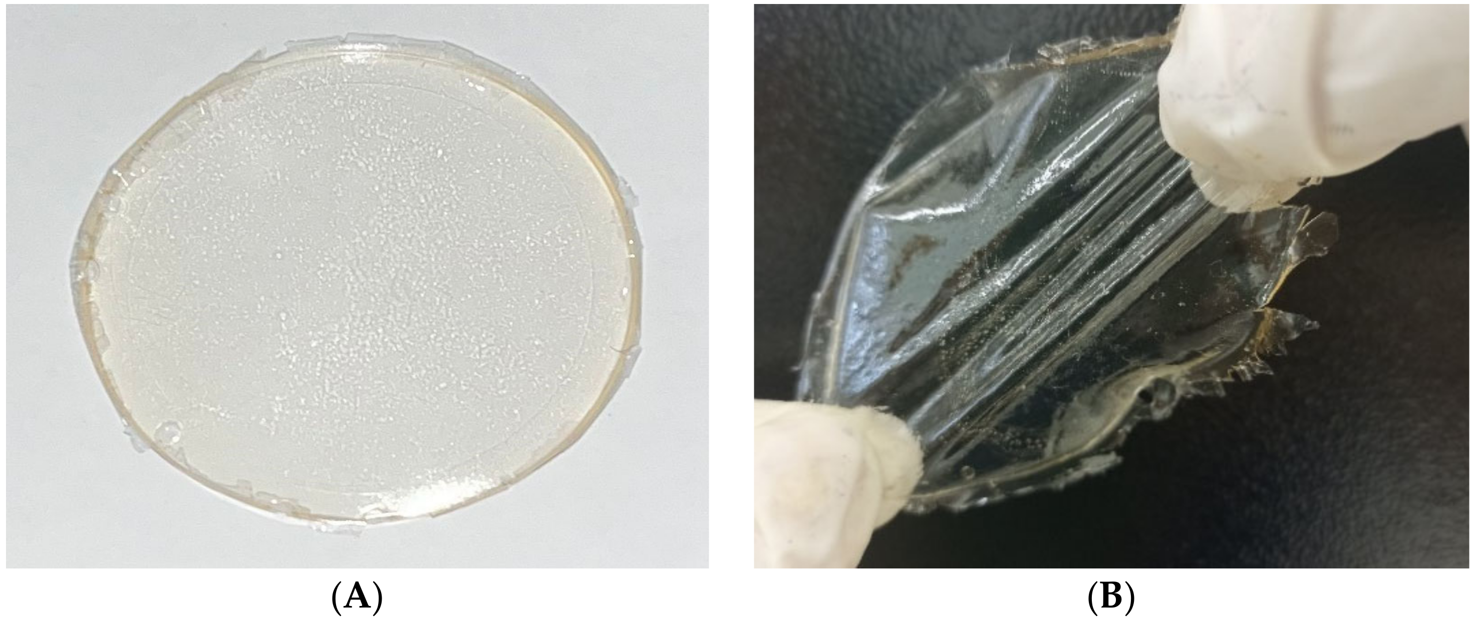
3.1. Visual Characterisation of Hydrogels

A visual qualitative evaluation was performed on the hydrogels with incorporated nanocrystals and nanofibres. In this analysis, complete solubilisation of carboxymethylcellulose occurred, as characterised by the clear and transparent appearance of the hydrogels (Figure 1A). After the hydrogels were dried, a smooth, homogeneous and transparent hydrogel was observed (Figure 1). In the case of hydrogels with vitamin D, a more flexible film was observed, which was also translucent and homogeneous (Figure 1B). This can be justified by the fact that vitamin D also cross-linked the hydrogel, enabling greater elasticity of the hydrogel, although future mechanical analysis is necessary to evaluate this elasticity.

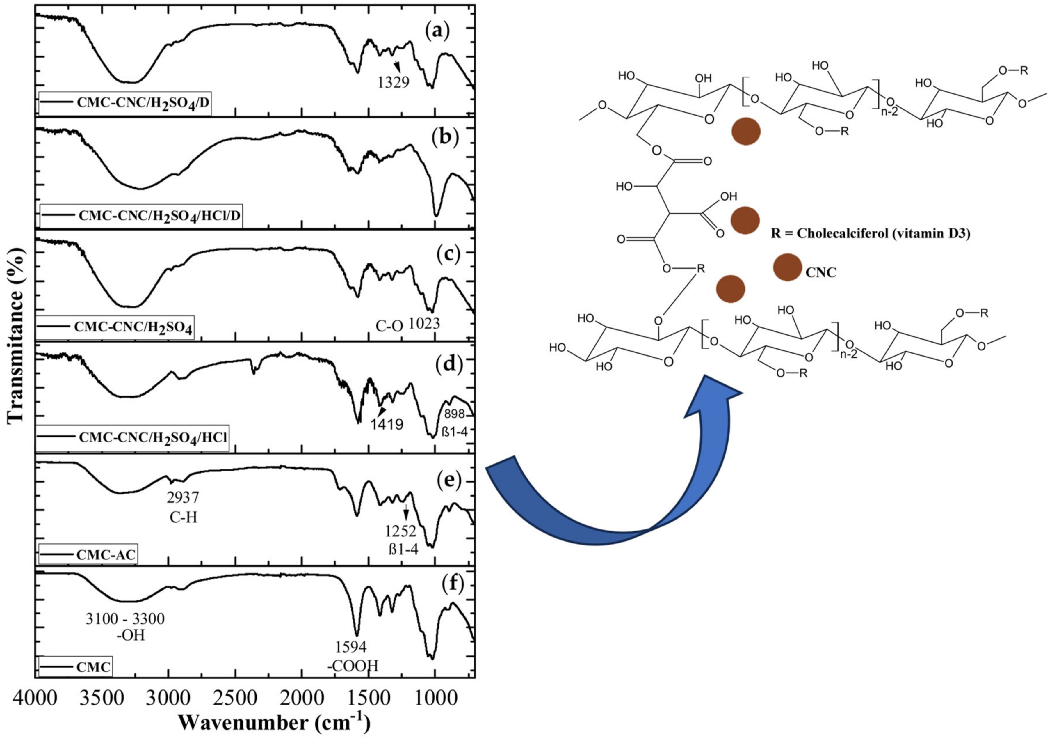
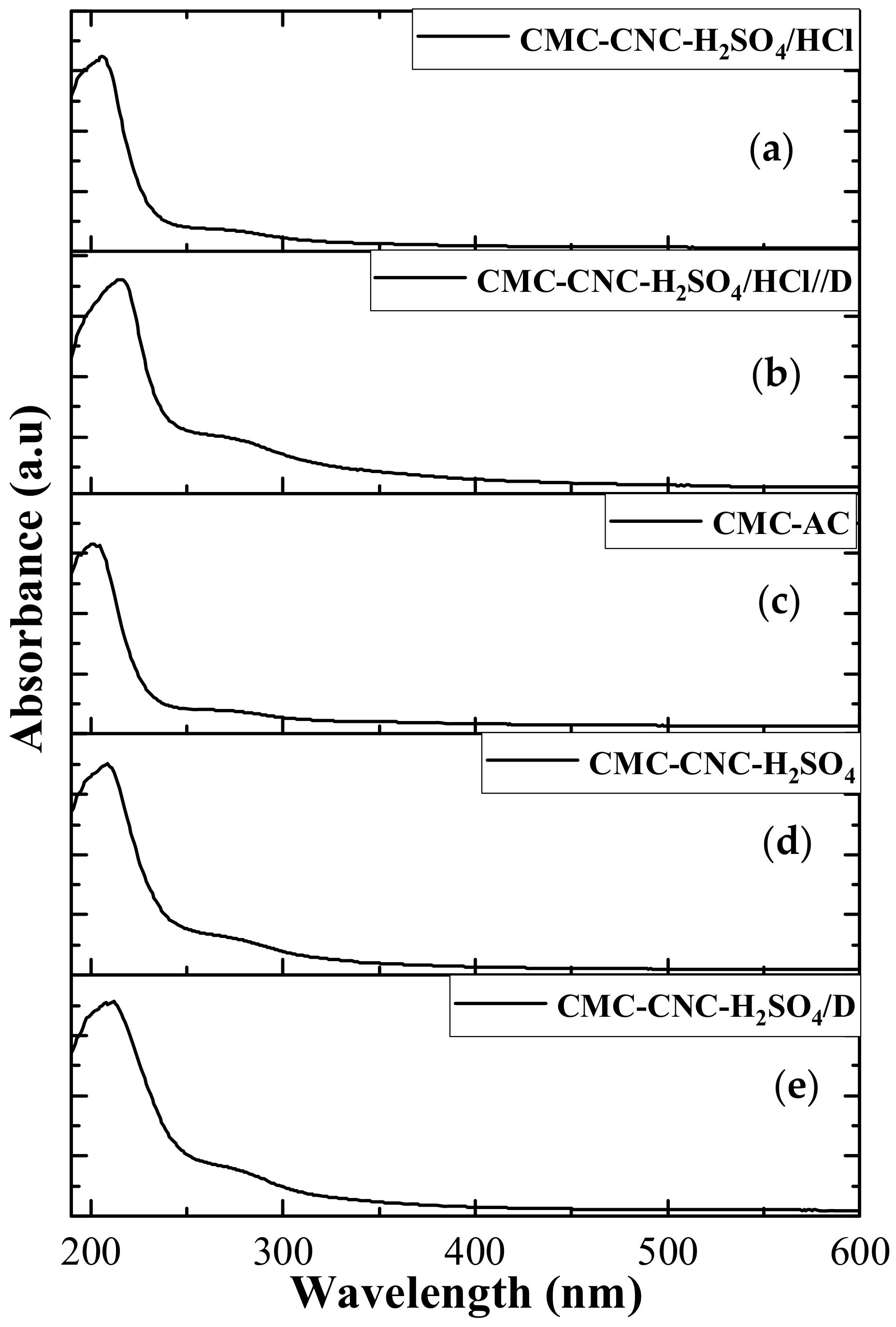
3.2. Spectroscopy Analyses

In our previous research [2], the FTIR spectra of pure Ceiba speciosa showed bands at 3100 cm−1, 2982 cm−1, 1282 cm−1 and 1109 cm−1 related to OH, -C-H, -CH2 stretching, -C-O-C and -C-H vibrations, respectively, associated with cellulose chemical groups [2,26,27]. In the case of the CNCs obtained by two different chemical routes, slight differences were detected at 3010 cm−1, 2975 cm−1, 2987 cm−1, 1382 cm−1, 1119 or 1109 cm−1 and 898 cm−1, associated with -OH, -C-H, -CH2 stretching, -C-O-C and -C-H vibrations, respectively [2]; for example, the disappearance of hydroxyl groups may be caused by the formation of acid sulphate (O-SO3H) groups in the hydrolysis step [2,28]. This formation should help with the incorporation of vitamin D because it may provide a more hydrophobic character to the hydrogel [29].
Additionally, in the CMC, bands related to OH groups at 3100–3300 cm−1 were observed. Moreover, for all hydrogels (CMC_AC, CMC_CNC_H2SO4, CMC_CNC_H2SO4/HCl, CMC_CNC_H2SO4_D, and CMC_CNC_H2SO4/HCl_D), this band increased in intensity compared with that of pure CMC because of the interaction between citric acid and CMC (Figure 2). Moreover, in the case of hydrogels with vitamin D (CMC_CNC_H2SO4_D, CMC_CNC_H2SO4/HCl_D), a slight increase was observed, probably because of the incorporation of vitamin D (Figure 2 inset) [2,30]. Furthermore, the intensity of the band at 2916 cm−1 decreased for hydrogels with vitamin D and almost disappeared, probably because of the incorporation of vitamin D [30,31]. In addition, bands at 1651 and 1719 cm−1 related to C=O groups were more intense in hydrogels with vitamin D associated with fatty acids [31]. The band related to the (-N+ (CH2)3) group of vitamin D at 1060 cm−1 was blueshifted in the hydrogels (CMC_CNC_H2SO4_D, CMC_CNC_H2SO4/HCl_D) [32,33].
UV-VIS spectroscopic analysis was performed for both CNCs in our previous research [2], which showed that bands at 230 and 280 nm were linked with cellulose I and n–π* (surface functional groups), which are associated with the C=C, carbonyl and hydroxyl groups in CNC structures [2,34]. For the hydrogels (CMC-AC, CMC-CNC-H2SO4, CMC-CNC-H2SO4/HCl, CMC-CNC-H2SO4/D, CMC-CNC-H2SO4/D and CMC-CNC-H2SO4/HCl/D), the same bands were observed but with slight differences in intensity (Figure 3), mainly in the band at 280 nm, which was more intense for the hydrogels with vitamin D; this is probably associated with chromophores in the chemical structure of vitamin D. Additionally, the opacity of all the hydrogels was calculated based on Equation (2) to evaluate the passage of light through the hydrogels, which can affect the chemical structure of vitamin D in the presence of sunlight [34,35]. The values of transparency found based on Equation (2) for all hydrogels were very close: 80 ± 1%, 81 ± 1%, 78 ± 1%, 79 ± 1% and 74 ± 1% for CMC-AC, CMC-CNC-H2SO4, CMC-CNC-H2SO4/HCl, CMC-CNC-H2SO4/D and CMC-CNC-H2SO4/HCl-D, respectively. These results indicate good transparency for the passage of sunlight through hydrogels for skin applications.
T r a n s p a r e n c y   ( % ) = l o g   ( % T 600 ) x  
here, % T 600 is the percentage of transmittance of light at 600 nm and x is the thickness of the hydrogel [36].

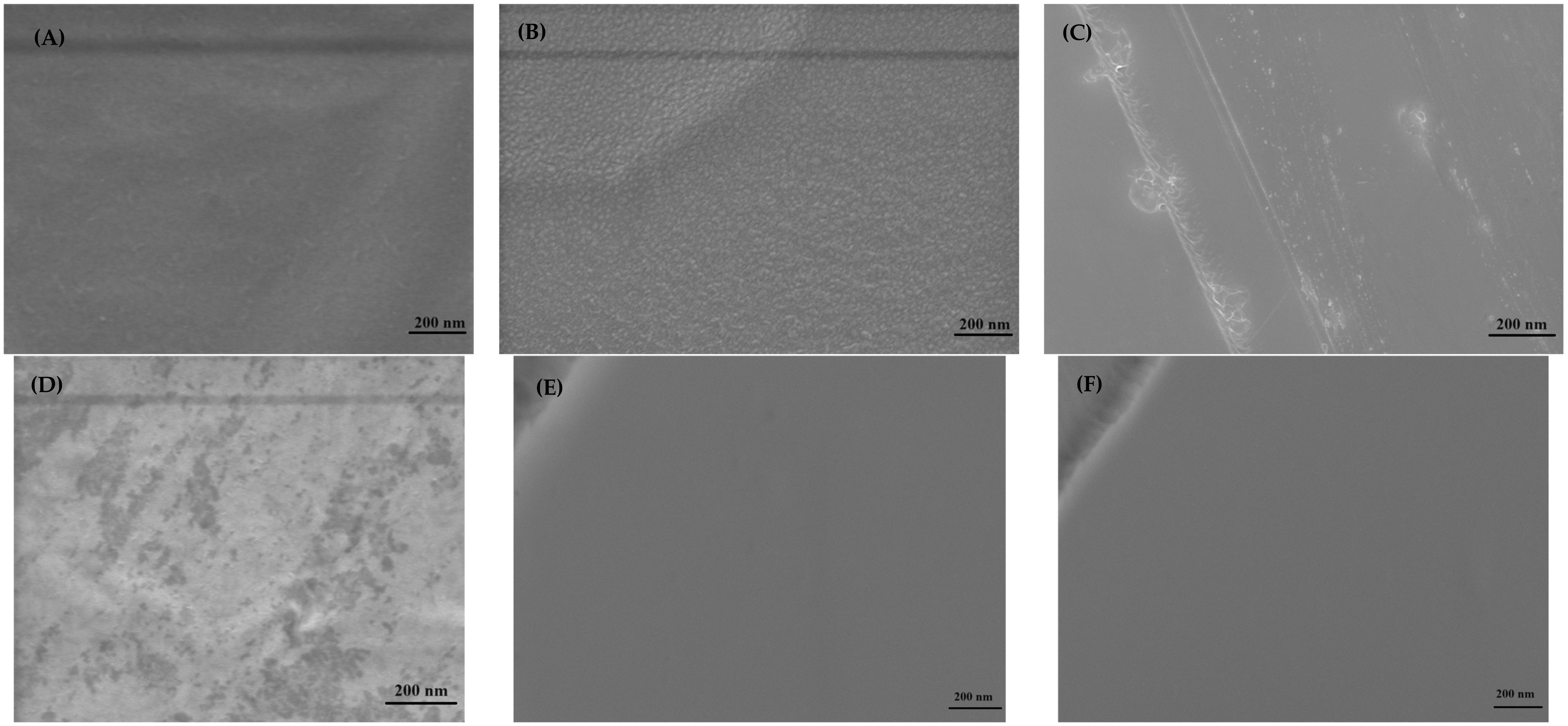
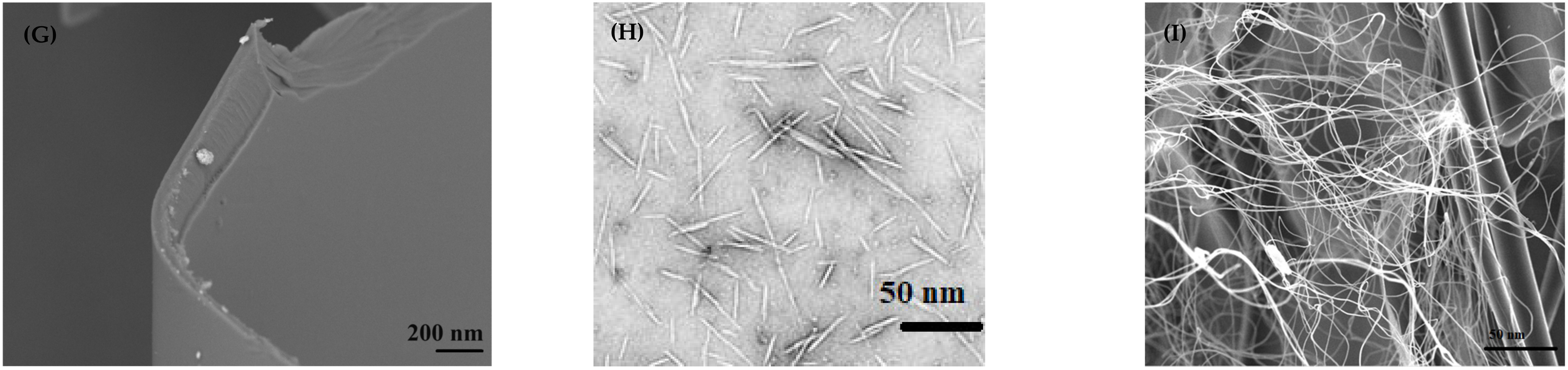
3.3. Microscopy Analysis

SEM analyses were performed on the CNCs and all the hydrogels (CMC, CMC-AC, CMC-CNC-H2SO4, CMC-CNC-H2SO4/HCl, CMC-CNC-H2SO4/D and CMC-CNC-H2SO4/HCl/D). In the case of the CNCs, as previously shown in our research [2], SEM images did not reveal the real morphology of the CNCs, and TEM analysis was subsequently performed, which revealed that the presence of nanocrystals and nanofibres depended on the chemical route used (Figure 4G,H). The SEM images of the hydrogels revealed a homogenous hydrogel (Figure 4), even though slight differences were detected between the hydrogels with citric acid (Figure 4B) and those with CNCs (Figure 4C,D). In the hydrogel with citric acid (chemical cross-link) (Figure 4B), some roughness was not detected in the hydrogel with pure CMC (Figure 4A); this morphology is likely because of the cross-linking process performed with citric acid, which slowly evaporates (48 h). In the hydrogels with nanocrystals (Figure 4C) and nanofibres (Figure 4D), slight differences in morphological agglomeration with different colours were observed between the two hydrogels, which may be caused by the clustering of nanocrystals and nanofibres in the hydrogel during cross-linking. Additionally, TEM images of the nanocrystals and nanofibres showed that, in the case of the CNCs produced by the chemical route using H2SO4, the nanocrystals were similar to those reported in the literature (Figure 4H), but in the case of the chemical route with the introduction of HCl, nanofibres were predominant (Figure 4I). Moreover, in the hydrogels with vitamin D3 (Figure 4E,F), no difference was observed, indicating that vitamin incorporation promoted a more homogeneous spread of CNCs, independent of the type of morphology.

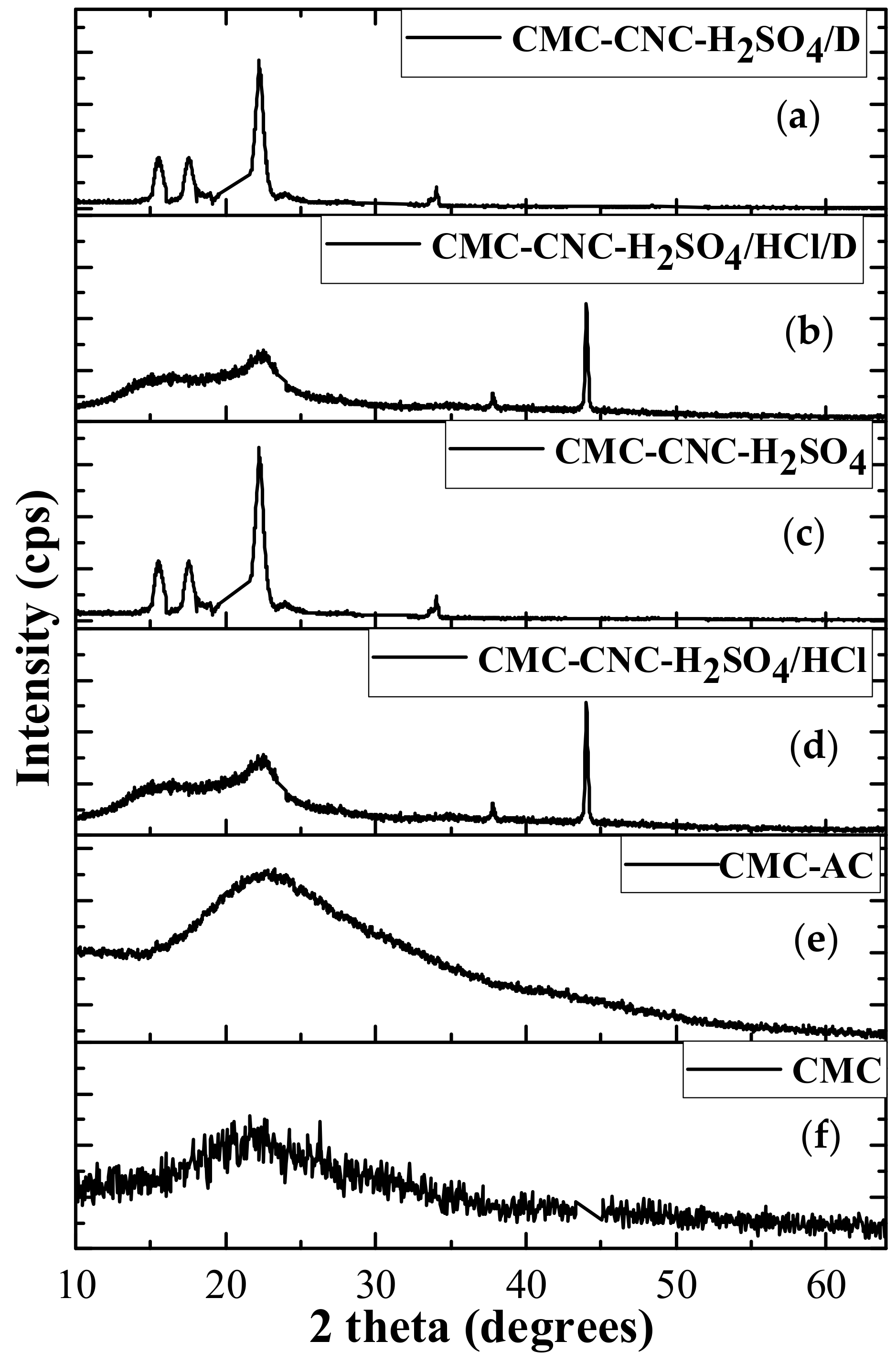
3.4. XRD Analysis of Hydrogels Loaded with Nanocrystals and Nanofibres Modified with Vitamin D

An XRD analysis of the CNCs revealed peaks at 15°, 17° and 34.3° (Figure 5), which were associated with cellulose I [2,39,40], and a prominent peak at 22.6° linked to the pattern (0 0 2) [2,40,41,42]. Additionally, the sizes found according to Equation (3) were 10 nm and 20 nm for CNC_H2SO4 and CNC_H2SO4/HCl, respectively, which were very close to the sizes determined via TEM analysis. Moreover, the crystallinity based on the total area and the amorphous area was determined, and the values were 81% and 77% for CNC_H2SO4 and CNC_H2SO4/HCl, respectively.
C r y s t a l l i n i t y   ( % ) = ( c r y s t a l l i n e   a r e a / t o t a l   a r e a ) × 100  
A slight difference was detected between the hydrogels containing vitamin D and the other hydrogels (Figure 5). In these hydrogels, a slight amorphisation process was observed, mainly in the samples with CNCs extracted with H2SO4 and HCl because a small shift in the peaks associated with cellulose in the nanocrystals (CNC_H2SO4) and nanofibres (CNC_H2SO4/HCl) was observed, probably because of the incorporation of vitamin D, which promoted a decrease in the crystallinity of the hydrogels. Moreover, a peak at 44° was observed, which was attributed to the NaOH that remained in the CNCs after extraction, even after dialysis. This accumulation in hydrogels with vitamin D was expected considering the visual analysis described in Section 3.1, where greater elasticity was visualised in these hydrogels than in the other hydrogels. However, as mentioned before, future mechanical analyses should be performed.

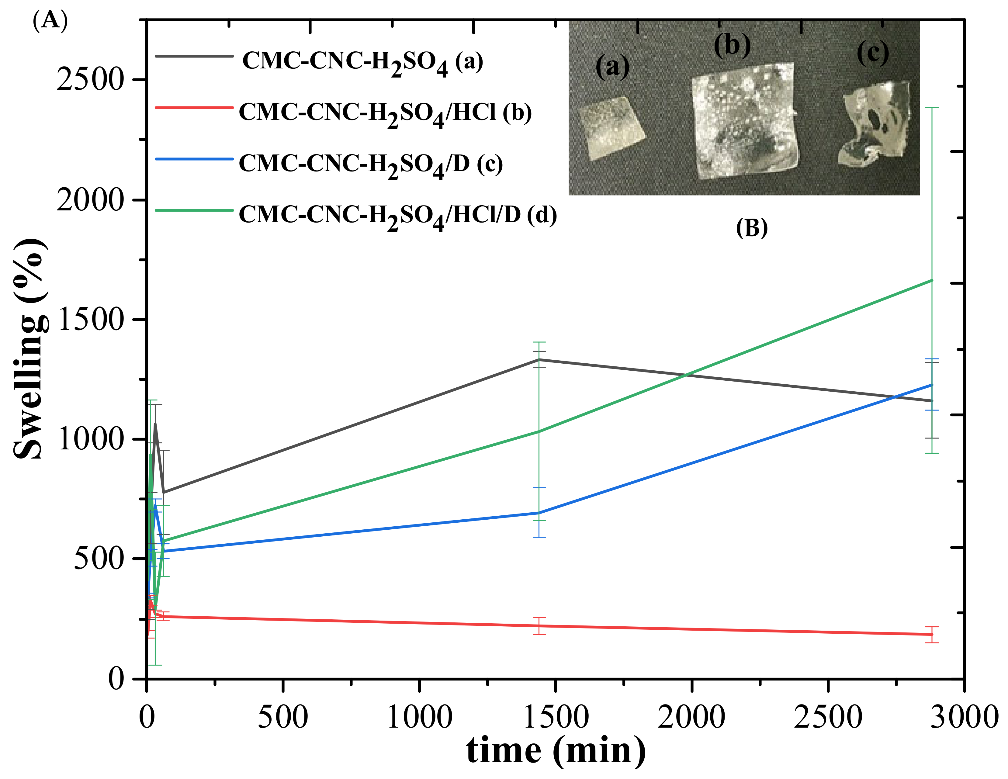
3.5. Swelling and Gel Fraction of Hydrogels Loaded with Nanocrystals and Nanofibres Modified with Vitamin D

An important parameter for biomaterials is physiological fluid adsorption and degradation/dissolution in the physiological environment. Thus, swelling (SD) and GF (solvent dissolution) tests were performed on the hydrogels. The results are presented in Figure 6. In the case of GF, the behaviours of the hydrogels without vitamin D were very similar, showing good chemical stability when compared with the hydrogels in the literature [43,44,45]. In the case of hydrogels with vitamin D, greater swelling was observed, mainly in the hydrogels with nanofibres (CMC-CNC-H2SO4/HCl/D), probably because of the presence of CNCs with a nanofibre morphology, which may enhance the entanglement of vitamin D3 and promote faster fluid release compared to the other hydrogels (CMC-CNC-H2SO4/D). However, the dissolution of fluid in the medium indicated by GF was greater after the first few hours, which may be a good indicator of the controlled release of the drug.

3.6. In Vitro Kinetic Studies of Vitamin D Release

Vitamin D3—or cholecalciferol—is a precursor of the steroid calcitriol, a regulator of calcium and phosphate metabolism in the body. This prohormone is soluble in lipids and plays a fundamental role in the development of bone tissue, skeletal muscles and several other areas of the body [46]. A lack of vitamin D in the body causes several problems, including low bone density, bone fractures, osteopenia, osteoporosis and muscle injuries, and some studies have indicated infertility problems in women [47]. Furthermore, studies have indicated that vitamin D has anti-inflammatory effects on immune system cells and can affect several processes [46,47,48]. These anti-inflammatory effects have been linked to the regulation of proinflammatory cytokines. Additionally, the skin has been shown to play an important role in the modification of 7-dehydrocholesterol in cholecalciferol when exposed to ultraviolet radiation [49]. Furthermore, studies have proposed a strong role for vitamin D3 in protecting against colon and breast cancer, diabetes and other health problems [15].
In this context, promoting biomaterials to control vitamin D3 release is a great idea for diverse biomedical applications. Thus, one of the main proposals of the present research was to evaluate the potential of incorporating CNCs and vitamin D3 into hydrogels produced from carboxymethyl cellulose and citric acid via a friendlier chemical route using a raw material from a semiarid region in Brazil. For this purpose, hydrogels with innovative cellulose nanocrystals and nanofibres conjugated with vitamin D3 were evaluated for their in vitro kinetics of vitamin D3 release, determining the cumulative amount of vitamin D3 released over time, here using 96 h of analysis (Figure 7).
The results of vitamin D3 release showed a great discharge in the first 30 min, but there was a slight difference between the two hydrogels (inset Figure 7). Additionally, these releases reached nearly 40 ng·mL−1 of vitamin D3 accumulated in the medium, a value slightly higher than that recommended by the National Institutes of Health (NIH) (≥20 ng·mL−1) but lower than an excess daily intake of the vitamin (>50 ng·mL−1) [50]. Moreover, the release of vitamin D3 from the CMC_CNC_H2SO4/HCl_D hydrogel was greater than that from the CMC_CNC_H2SO4_D hydrogel and occurred faster in the first 15 min (Figure 7 inset). Most research [45,46,47,48,49,50] has shown slower vitamin D release, over a period close to 2 days, at which point the release equilibrium of the vitamin is reached. Although hydrogels are not suitable for conjugation with hydrophobic drugs, as is the case for vitamin D [48,51], the present research showed strong results because the release of vitamin D reached its equilibrium in hours, not days, and the daily value of equilibrium approached that recommended by the NIH. Additionally, there was a slight difference between the hydrogels (38.0 ± 0.5 ng·mL−1 and 39.0 ± 0.5 ng·mL−1 for CMC-CNC-H2SO4/D and CMC-CNC-H2SO4/HCl/D, respectively), even though the equilibrium results were very close. This slight difference in vitamin release may be related to the CNC morphology in the hydrogel with nanofibres (CMC-CNC-H2SO4/HCl/D), which promoted better vitamin D release.

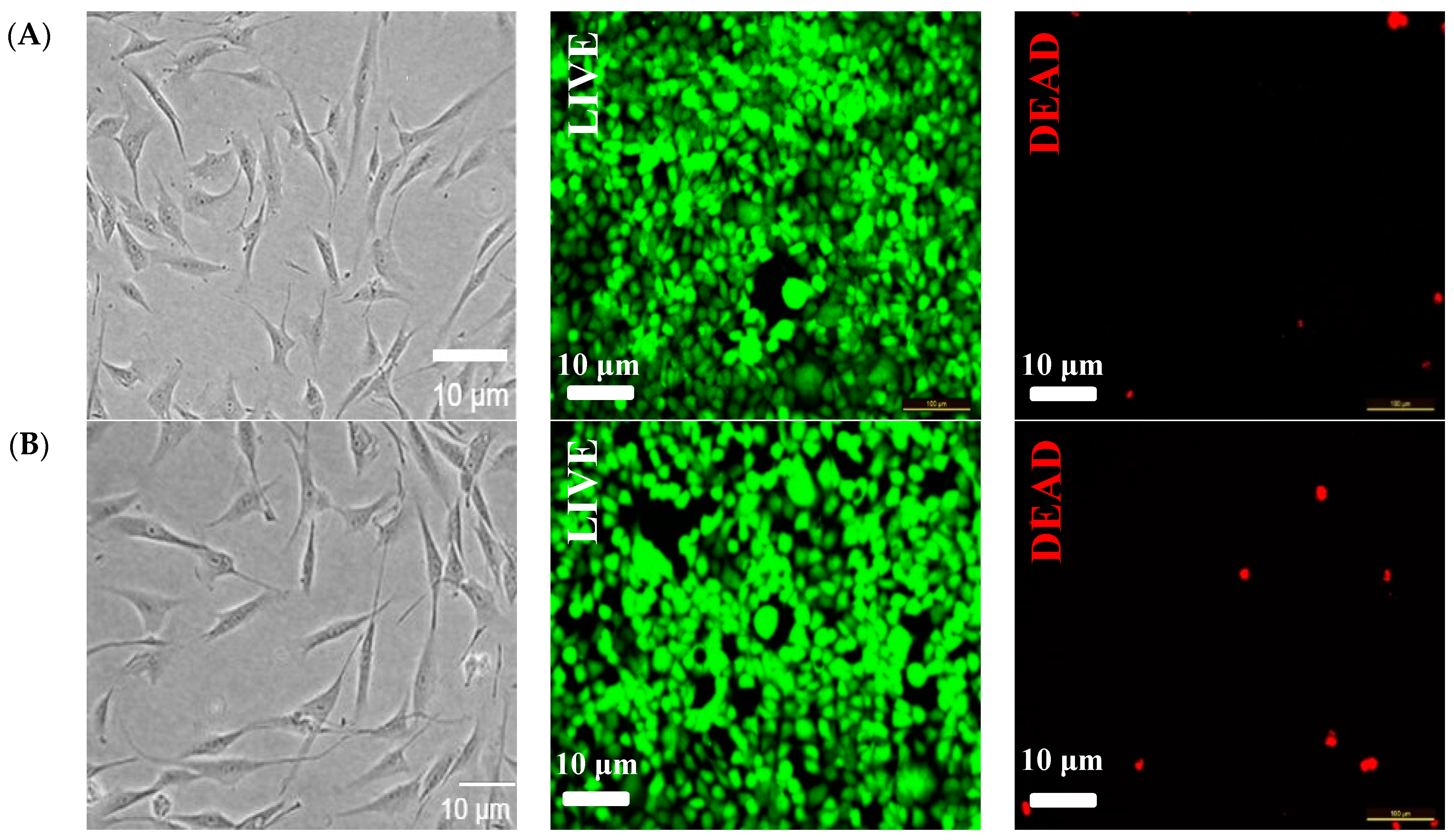
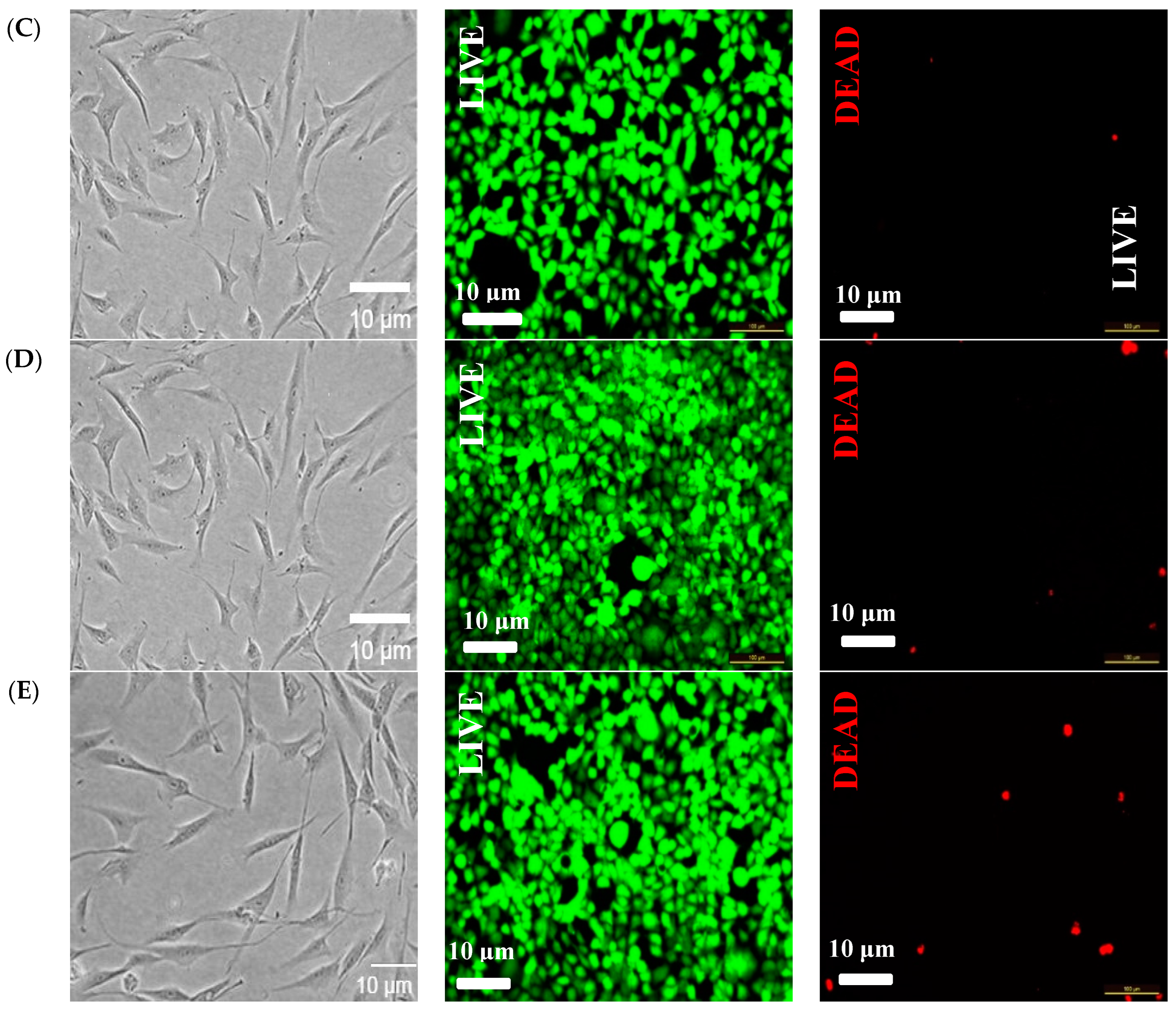
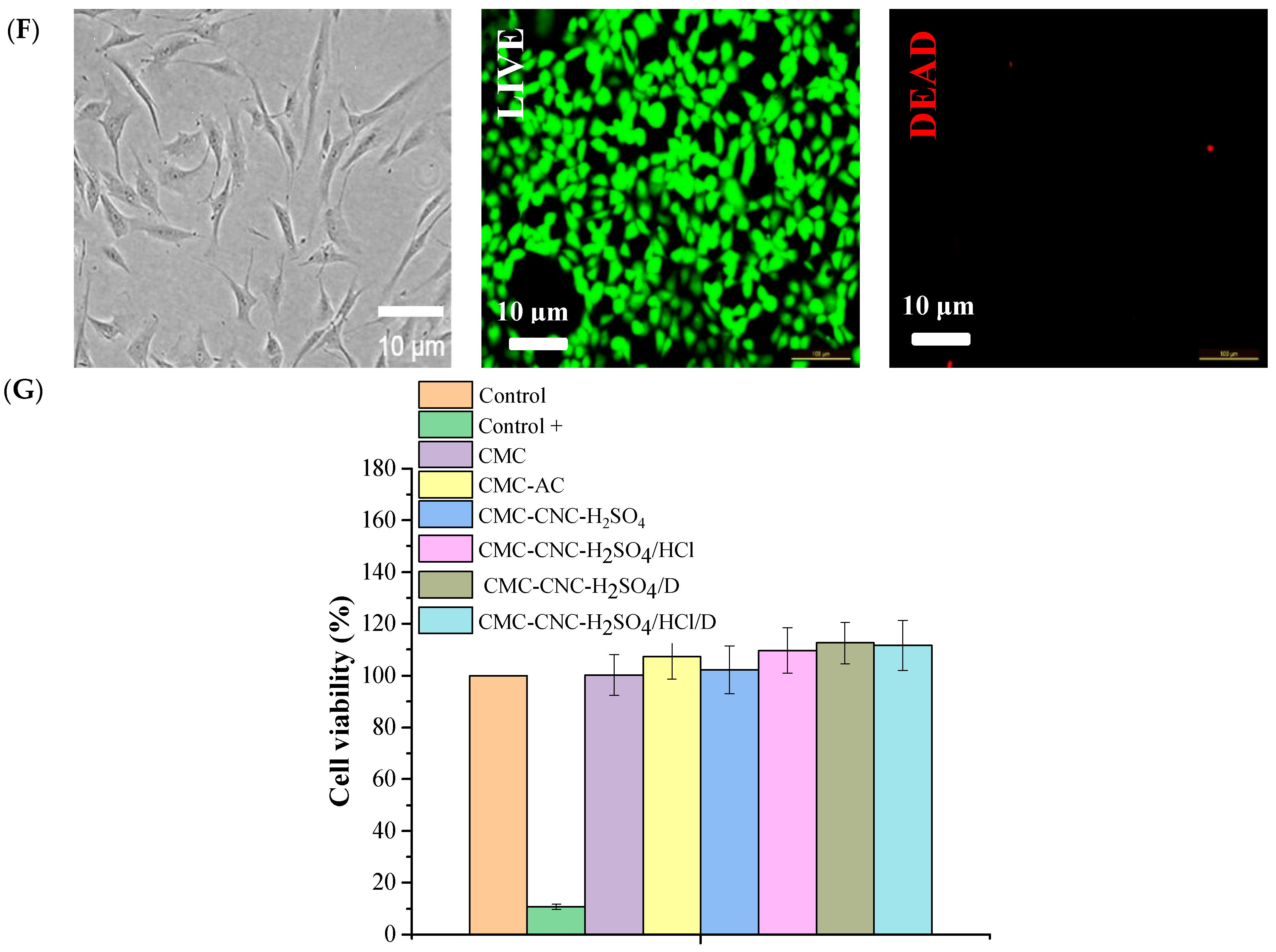
3.7. Cytotoxicity Evaluation of Hydrogels Loaded with Nanocrystals and Nanofibres Modified with Vitamin D

In our previous research [2], it was shown that different concentrations of CNCs were nontoxic when the nanocrystals and nanofibres were formed via two chemical routes. However, because the present research focused on hydrogels loaded with nanocrystals and nanofibres modified with vitamin D, it was important to evaluate the cytotoxicity of the hydrogels. Therefore, for this evaluation, HDF cells were used. As described above, vitamin D is crucial to the health of many organs in the body, including effects on fertility and some cancers that affect reproductive organs in women [21,24,25,46,47,48,52].
Various studies have indicated that hydrogels have promising features, such as mechanical performance, biodegradability, chemical stability and tissue compatibility [53], most of which are associated with their polymeric matrices, which can also improve drug delivery linked with physiochemical characteristics, allowing for better controlled release and pharmacokinetic behaviour in biomedical areas [53,54]. In the current study, the cellular viability of hydrogels with and without vitamin D3 was assessed using an assay with resazurin and LIVE/DEAD in HDFs to evaluate the cytotoxicity of the nanomaterial (Figure 8). Figure 8G shows that the positive control performed with Triton X-100 (a nonionic surfactant for cell lysis) was the only one that showed a significant difference when compared with the reference control. All hydrogels, when in contact with HDFs, did not show toxicity when compared with the control group. Furthermore, the incorporation of CNCs and vitamin D3 into the hydrogel matrix did not cause toxicity, as indicated by the LIVE/DEAD ratio and cell viability, indicating that these hydrogels can be used for controlled drug release applications.

4. Conclusions

The present research described the production of new CMC hydrogels with the incorporation of different CNCs (nanocrystals and nanofibres) obtained from a new Brazilian semiarid biomass source and loaded with vitamin D3 for controlled drug release. These innovative hydrogels produced with the incorporation of CNCs from Ceiba speciosa fibres are a potential sustainable alternative for improving the semiarid Brazilian region both socially and economically; thus, they can promote poverty eradication, zero hunger and sustainable agriculture in this area. Additionally, the results for these hydrogels showed that the different physiochemical characteristics and morphologies of the CNCs changed the vitamin D3 release performance. Moreover, the swelling and GF behaviour of the hydrogels treated with vitamin D3 differed, probably because of the incorporation of vitamin D3, as shown by spectroscopy analysis. Furthermore, no differences in cytotoxicity were detected, indicating the high potential of applying these hydrogels for controlled vitamin D3 release in diverse biomedical applications.

Supplementary Materials

The following supporting information can be downloaded at https://www.mdpi.com/article/10.3390/pr12071437/s1. Figure S1: Calibration curve of vitamin D3.

Author Contributions

N.d.C.S.: conceptualisation, methodology, investigation. C.J.T.S.: conceptualisation, methodology, investigation. M.P.G.: writing—original draft, validation, writing—review and editing. F.G.L.M.B.: conceptualisation, supervision, project administration, writing—original draft, validation, writing—review and editing, funding acquisition. All authors have read and agreed to the published version of the manuscript.

Funding

This research was funded by BIOSEM-LESMA from the Universidade Federal dos Vales do Jequitinhonha e Mucuri (UFVJM) for all chemical experiments and by the FAPEMIG (APQ-02565-21), FINEP/MCTI (0 1 22 0528 00), CAPES, and CNPq.

Data Availability Statement

The authors confirm that the data supporting the findings of this study are available within the article and can be shared if necessary. Calibration curve of vitamin D3 is shown in Figure S1.

Conflicts of Interest

The authors declare no conflicts of interest.

References

  1. UN. The ODS in Brazil. 2024. Available online: https://brasil.un.org/pt-br/sdgs (accessed on 29 April 2024).
  2. Silva, N.C.; Rodrigues, J.S.; Bernardes, M.S.; Gonçalves, M.P.; Medeiros Borsagli, F.G.L. Cellulose nanocrystals and nano-fiber from sub-wear out Brazilian semiarid source for biological applications. J. Cluster Sci. 2024, 1, 1–10. [Google Scholar]
  3. Junges, A.; Denti, A.F.; Bernardi, J.L.; Polina, C.C.; Meregalli, M.M.; Vanz, J.B.; Mignoni, M.L. Application of nanotechnology in food engineering: A review. Res. Soc. Dev. 2022, 11, 1–10. [Google Scholar]
  4. Borsagli, F.G.L.M.; Borsagli, A. Luminescent carbon dots for environmental photocatalytic. In Inorganic-Organic Composites for Water and Wastewater Treatment; Lichtfouse, E., Muthu, S.S., Khadir, A., Eds.; Springer: Singapore, 2021; pp. 201–228. [Google Scholar]
  5. Paiva, A.E.; Guin, J.P.; Vasquez, J.F.B.; Thampi, K.R.; Sullivan, J.A.; Borsagli, F.G.L.M.; Morris, M.A. Degradation of an antibiotic via a photo-Fenton process on block copolymernanopatterned iron oxide coated substrates. Chem. Eng. J. 2023, 466, 142925. [Google Scholar] [CrossRef]
  6. Rodrigues, J.S.; Silva, C.J.T.; Silva, N.C.; Aguiar, R.A.; Bernardes, M.S.; Soares, C.M.; Madureira, J.A.; Borsagli, F.G.L.M. Carbohydrate polymer hydrogels: An environmental and eco-friendly choice for biomedical applications? J. Polym. Res. 2023, 30, 382. [Google Scholar] [CrossRef]
  7. Thomas, P.; Duolikun, T.; Rumjit, N.P.; Moosavi, S.; Lai, C.W.; Johan, M.R.B.; Fen, L.B. Comprehensive review on nanocellulose: Recent developments, challenges and future prospects. J. Mech. Behav. Biomed. Mater. 2020, 110, 103884. [Google Scholar] [CrossRef]
  8. Newman, L.; Jasim, D.A.; Prestat, E.; Lozano, N.; Lazaro, I.; Nam, Y.; Assas, B.M.; Pennock, J.; Haigh, S.J.; Bussy, C.; et al. Splenic capture and in vivo intracellular biodegradation of biological-grade graphene oxide sheets. ACS Nano 2020, 14, 10168–10186. [Google Scholar] [CrossRef] [PubMed]
  9. Park, K.; Choi, H.; Kang, K.; Shin, M.; Son, D. Soft stretchable conductive carboxymethylcellulose hydrogels for wearable sensors. Gels 2022, 8, 92. [Google Scholar] [CrossRef] [PubMed]
  10. Barhoum, A.; Sadak, O.; Ramirez, I.A.; Iverson, N. Stimuli-bioresponsive hydrogels as new generation materials for implantable, wearable, and disposable biosensors for medical diagnostics: Principles, opportunities, and challenges. Adv. Colloid Interface Sci. 2023, 317, 102920. [Google Scholar] [CrossRef] [PubMed]
  11. Fadl, F.I.A.E.; Hegazy, D.E.; Maziad, N.A.; Ghobashy, M.M. Effect of nano-metal oxides (TiO2, MgO, CaO, and ZnO) on antibacterial property of (PEO/PEC-co-AAm) hydrogel synthesized by gamma irradiation. Int. J. Biol. Macromol. 2023, 250, 126248. [Google Scholar] [CrossRef] [PubMed]
  12. Borsagli, F.G.L.M.; Souza, A.J.M.; Paiva, A.E. Ecofriendly multifunctional thiolated carboxymethyl chitosan-based 3D scaffolds with luminescent properties for skin repair and theragnostic of tissue regeneration. Int. J. Biol. Macromol. 2020, 165, 3051–3064. [Google Scholar] [CrossRef] [PubMed]
  13. Parfenyuk, E.V.; Dolinina, E.S. Silica hydrogel composites as a platform for soft drug formulations and cosmetic compositions. Mater. Chem. Phys. 2022, 287, 126160. [Google Scholar] [CrossRef]
  14. Ijaz, F.; Tahir, H.M.; Ali, S.; Ali, A.; Khan, H.A.; Muzamil, A.; Manzoor, H.H.; Qayyum, K.A. Biomolecules based hydrogels and their potential biomedical applications: A comprehensive review. Int. J. Biol. Macromol. 2023, 253, 127362. [Google Scholar] [CrossRef]
  15. Ghorbanzadeh, M.; Golmohammadzadeh, S.; Karimi, M.; Farhadian, N. Evaluation of vitamin D3 serum level of microemulsion based hydrogel containing Calcipotriol drug. Mater. Todays Commun. 2022, 33, 104409. [Google Scholar] [CrossRef]
  16. Franco, P.; Marco, I. Contact Lenses as Ophthalmic Drug Delivery Systems: A review. Polymers 2021, 17, 1102. [Google Scholar] [CrossRef] [PubMed]
  17. Zheng, C.; Lu, K.; Lu, Y.; Zhu, S.; Yue, Y.; Xu, X.; Mei, C.; Xiao, H.; Wu, Q.; Han, J. A stretchable, self-healing conductive hydrogels based on nanocellulose supported graphene towards wearable monitoring of human motion. Carbohyd. Polym. 2020, 250, 116905. [Google Scholar] [CrossRef] [PubMed]
  18. Cai, Y.; Xin, L.; Li, H.; Sun, P.; Liu, C.; Fang, L. Mussel-inspired controllable drug release hydrogel for transdermal drug delivery: Hydrogen bond and ion-dipole interactions. J. Control. Release 2024, 365, 161–175. [Google Scholar] [CrossRef] [PubMed]
  19. Narayanaswamy, R.; Torchilin, V.P. Hydrogels and Their Applications in Targeted Drug Delivery. Molecules 2019, 24, 603. [Google Scholar] [CrossRef] [PubMed]
  20. Lee, W.K.; Chng, A.L.B.; Tan, K.H.; Shub, A.; Tan, T.; Hian, T.K.; Lee, R.W.K.; Ling, L.S.; Kuma, K.; Siew, C.Y. Clinical practice of vitamin D screening and supplementation in pregnancy in Asia-pacific countries: A cross-sectional study. Heliyon 2023, 9, 21186. [Google Scholar] [CrossRef] [PubMed]
  21. Qi, D.; Nie, X.-L.; Wu, S.; Cai, J. Vitamin D and hypertension: Prospective study and meta-analysis. PLoS ONE 2017, 12, e0174298. [Google Scholar] [CrossRef]
  22. Haitchi, S.; Moliterno, P.; Widhalm, K. Prevalence of vitamin D deficiency in seniors—A retrospective study. Clin. Nutr. ESPEN 2023, 57, 691–696. [Google Scholar] [CrossRef] [PubMed]
  23. Holick, M. The vitamin D deficiency pandemic: Approaches for diagnosis, treatment and prevention. Rev. Endocr. Metab. Disord. 2017, 18, 153–165. [Google Scholar] [CrossRef] [PubMed]
  24. Berry, S.; Seidler, K.; Neil, J. Vitamin D deficiency and female infertility: A mechanism review examining the role of vitamin D in ovulatory dysfunction as a symptom of polycystic ovary syndrome. J. Reprod. Immunol. 2022, 151, 103633. [Google Scholar] [CrossRef] [PubMed]
  25. Seraphin, G.; Reiger, S.; Hewison, M.; Capobianco, E.; Lisse, T.S. The impact of vitamin D on cancer: A mini review. J. Steroid Biochem. Mol. Biol. 2023, 231, 106308. [Google Scholar] [CrossRef] [PubMed]
  26. Mousa, H.M.; Fahmy, H.S.; Abouzeid, R.; Abdel-Jaber, G.T.; Ali, W.Y. Polyvinylidene fluoride-cellulose nanocrystals hybrid nanofiber membrane for energy harvesting and oil-water separation applications. Mater. Lett. 2022, 306, 130965. [Google Scholar] [CrossRef]
  27. Sobrinho, M.S.; Tabatinga, G.M.; Machado, I.C.; Lopes, A.V. Pollination biology and mating system of Angelonia hirta (Scrophulariaceae), a neotropical shrub with uncommon floral traits. Acta Bot. Bras. 2013, 27, 456. [Google Scholar] [CrossRef]
  28. Costa, L.A.S.; Fonsêca, A.F.; Pereira, F.V.; Druzian, J.I. Biodegradability of nanocomposites based on cassava starch and clay. Cellul. Chem. Technol. 2015, 49, 127–134. [Google Scholar]
  29. Medeiros Borsagli, F.G.L.; Rodrigues, J.S.; Aguiar, R.A.; Paiva, A.E.; Vasquez, J.F.B.; Ramos, W.T.S.; Allibrandini, P.; Rocha, E.P.A.; Gonçalves, M.P.; Souza, F.E. Low-cost luminescent scaffolds-based on thiol chitosans by microwave radiation for vertebral disc repair/theragnostic. Int. J. Biol. Macromol. 2022, 209, 2109–2118. [Google Scholar] [CrossRef] [PubMed]
  30. Bampidis, V.; Azimonti, G.; Bastos, M.L.; Christensen, H.; Dusemund, B.; Durjava, M.K.; Kouba, M.; Lopez-Afonso, M.; Puente, S.L.; Marcon, F.; et al. Safety and efficacy of sodium carboxymethyl cellulose for all animal species. EFSA J. 2020, 18, 6211. [Google Scholar]
  31. Paiva, A.E.; Medeiros Borsagli, F.G.L. Ecofriendly multiphase aqueous colloidal based on carboxymethylcellulose nanoconjugates with luminescence properties for potential bioimaging cancer cells. J. Polym. Environ. 2020, 23, 3076–3096. [Google Scholar] [CrossRef]
  32. Molaveisi, M.; Shahidi-Noghabi, M.; Naji-Tabasi, S. Vitamin D3-loaded nanophytosomes for enrichment purposes: Formulation, structure optimization, and controlled release. Food Process Eng. 2020, 2020, 13560. [Google Scholar] [CrossRef]
  33. Semalty, A.; Semalty, M.; Rawat, M.S.M.; Franceschi, F. Supramolecular phospholipids–polyphenolics interactions: The PHYTOSOME® strategy to improve the bioavailability of phytochemicals. Fitoterapia 2010, 81, 306–314. [Google Scholar] [CrossRef] [PubMed]
  34. Zhang, Y.; Gao, Z.; Zhang, W.; Wang, W.; Chang, J.; Kai, J. Fluorescent carbon dots as nanoprobe for determination of lidocaine hydrochloride. Sens. Actuators B Chem. 2018, 262, 928. [Google Scholar] [CrossRef]
  35. Zhang, W.; Zhang, Y.; Cao, J.; Jiang, W. Improving the performance of edible food packaging films by using nanocellulose as an additive. Int. J. Biol. Macromol. 2020, 166, 288–296. [Google Scholar] [CrossRef] [PubMed]
  36. Zhao, J.; Wang, Y.; Liu, C. Film transparency and opacity measurements. Food Anal. Methods 2022, 15, 2840–2846. [Google Scholar] [CrossRef]
  37. Kirtane, A.R.; Verma, M.; Karandikar, P.; Furin, J.; Langer, R.; Traverso, G. Nanotechnology approaches for global infectious diseases. Nat. Nanotechnol. 2021, 16, 369–384. [Google Scholar] [CrossRef] [PubMed]
  38. Raza, H.Z.; Shah, A.A.; Noreen, Z.; Usman, S.; Zafar, S.; Yasin, N.A.; Sayed, S.R.M.; Al-Mana, R.A.; Elansary, H.O.; Ahmad, A.; et al. Calcium oxide nanoparticles mitigate lead stress in Abelmoschus esculentus through improving the key antioxidative enzymes, nutritional content and modulation of stress markers. Plant Physiol. Biochem. 2024, 206, 108171. [Google Scholar] [CrossRef]
  39. Fekete, T.; Borsa, J.; Takacs, E.; Wojnarovitis, L. Synthesis and characterization of superabsorbent hydrogels based on hydroxyethylcellulose and acrylic acid. Carbohyd. Polym. 2017, 166, 300–308. [Google Scholar] [CrossRef] [PubMed]
  40. French, A.D. Correction to: Increment in evolution of cellulose crystallinity analysis. Cellulose 2020, 27, 5445–5448. [Google Scholar] [CrossRef]
  41. French, A.D.; Santiago Cintrón, M. Cellulose polymorphy, crystallite size, and the Segal Crystallinity Index. Cellulose 2020, 20, 583–588. [Google Scholar] [CrossRef]
  42. Liu, Y.Y.; Yu, N.Y.; Fang, W.D.; Tan, Q.-G.; Ji, R.; Yang, L.-Y.; Wei, S.; Zhang, X.-W.; Miao, A.-J. Photodegradation of carbon dots cause cytotoxicity. Nat. Commun. 2021, 12, 812. [Google Scholar] [CrossRef] [PubMed]
  43. Schmitz, T.; Grabovac, V.; Palmberger, T.F.; Hoffer, M.H.; Bernkop-Schnurch, A. Synthesis and characterization of a chitosan-N-acetyl cysteine conjugate. Int. J. Pharm. 2008, 347, 79–85. [Google Scholar] [CrossRef] [PubMed]
  44. Chen, Y.; Zhang, Y.; Mensaha, A.; Li, D.; Wang, Q.; Wei, Q. A plant-inspired long-lasting adhesive bilayer nanocomposite hydrogel based on redox-active Ag/Tannic acid-Cellulose nanofibers. Carbohydr. Polym. 2021, 255, 117508. [Google Scholar] [CrossRef] [PubMed]
  45. Alizadeh, R.; Zarrintaj, P.; Kamrava, S.K.; Bagher, Z.; Farhadi, M.; Heidari, F.; Komeili, A.; Gutierrez, T.J.; Saeb, M.R. Conductive hydrogels based on agarose/alginate/chitosan for neural disorder therapy. Carbohydr. Polym. 2019, 224, 115161. [Google Scholar] [CrossRef] [PubMed]
  46. Kim, D.-S.; Kim, J.H.; Baek, S.-W.; Lee, J.-K.; Park, S.-Y.; Choi, B.; Kim, T.-H.; Min, K.; Han, D.K. Controlled vitamin D delivery with injectable hyaluronic acid-based hydrogel for restoration of tendinopathy. J. Tissue Eng. 2022, 13, 1–15. [Google Scholar] [CrossRef] [PubMed]
  47. Chu, C.; Tsuprykov, O.; Chen, X.; Elitok, S.; Krämer, B.K.; Hocher, B. Relationship between vitamin d and hormones important for human fertility in reproductive-aged women. Front. Endocrinol. 2021, 12, 666667. [Google Scholar] [CrossRef] [PubMed]
  48. Simioni, Y.R.; Ricatti, F.; Salvay, A.G.; Jerez, H.E.; Schilrreff, P.; Romero, E.L.; Morilla, M.J. Activity of hydrogel-vitamin D3/bacterioruberin nanoparticles on imiquimod-induced fibroblasts-keratinocytes spheroids. J. Drug Deliv. Sci. Technol. 2024, 97, 105738. [Google Scholar] [CrossRef]
  49. Eslami, M.; Shahedi, M.; Fath, M. Development of hydrogels for entrapment of vitamin D3: Physicochemical characterization and release study. Food Biophys. 2018, 13, 284–291. [Google Scholar] [CrossRef]
  50. National Institutes of Health (NIH). Vitamin D. Available online: https://ods.od.nih.gov/factsheets/VitaminD-HealthProfessional/ (accessed on 30 April 2024).
  51. Desfrançois, C.; Auzély, R.; Texier, I. Lipid nanoparticles and their hydrogel composites for drug delivery: A review. Pharmaceuticals 2018, 11, 118. [Google Scholar] [CrossRef] [PubMed]
  52. Goodwin, E.C.; Yang, E.; Lee, C.-J.; Lee, H.-W.; DiMaio, D.; Hwang, E.-S. Rapid induction of senescence in human cervical carcinoma cells. Proc. Natl. Acad. Sci. USA 2000, 97, 10978–10983. [Google Scholar] [CrossRef] [PubMed]
  53. Yuan, M.; Ruibo, Z.; Gao, H.; Li, W.; Yun, X.; Liu, J.; Zhao, X.; Zhao, G.; Zhang, F. One-step, green, and economic synthesis of water-soluble photoluminescent carbon dots by hydrothermal treatment of wheat straw and their bio-applications in labeling, imaging, and sensing. Appl. Surf. Sci. 2015, 355, 1136–1144. [Google Scholar] [CrossRef]
  54. Thankam, F.G.; Muthu, J.; Sankar, V.; Gopal, R.K. Growth and survival of cells in biosynthetic poly vinyl alcohol–alginate IPN hydrogels for cardiac applications. Colloids Surf. B Biointerfaces 2013, 107, 137–145. [Google Scholar] [CrossRef]
Figure 1. (A) Carboxymethyl cellulose hydrogel with CNCs without vitamin D3 and (B) carboxymethyl cellulose hydrogel with CNCs and vitamin D.
Processes 12 01437 g001
Figure 2. FTIR spectra of the (a) CMC, (b) CMC-AC, (c) CMC-CNC-H2SO4, (d) CMC-CNC_H2SO4/HCl, (e) CMC-CNC-H2SO4/D and (f) CMC-CNC-H2SO4/HCl/D hydrogels (inset, schematic of the chemical linkage in the hydrogel with vitamin D).
Processes 12 01437 g002
Figure 3. UV-VIS spectra of the (a) CMC-CNC-H2SO4/HCl, (b) CMC-CNC-H2SO4/HCl/D, (c) CMC-AC, (d) CMC-CNC-H2SO4 and (e) CMC-CNC-H2SO4/D hydrogels.
Processes 12 01437 g003
Figure 4. SEM images of hydrogels: (A) CMC, (B) CMC-AC, (C) CMC-CNC-H2SO4, (D) CMC-CNC-H2SO4/HCl, (E) CMC-CNC-H2SO4/D and (F) CMC-CNC-H2SO4/HCl/D. (G) Thickness of hydrogels and TEM images of hydrogels with CNCs (copyright 5801880110187 by Springer [2]), (H) CMC-CNC_H2SO4 and (I) CMC-CNC-H2SO4/HCl. In biomaterial applications, promoting the cell adhesion and biocompatibility of materials is critical. In controlled drug delivery, adequate adhesion of the biomaterials to membrane cells is the first step in targeted drug release [37,38]. Moreover, homogeneous spreading of nanoparticles in the hydrogel matrix should improve the properties of the hydrogel [39]. Thus, when vitamin D3 was incorporated into hydrogels with nanocrystals and nanofibres, a more homogeneous spread was perceived, indicating that the incorporation of the drug improved the hydrogel. As shown in Figure 4, SEM images revealed a homogeneous spread of nanocrystals and nanofibres because no clusters were observed in hydrogels with vitamin D3. Furthermore, roughness was visualised only in the hydrogels with citric acid without CNCs and vitamin D3, showing that the incorporation of these two compounds improved the hydrogels. Additionally, the thickness of the hydrogels was homogeneous, as shown in Figure 3.
Processes 12 01437 g004aProcesses 12 01437 g004b
Figure 5. XRD diffractograms of the (a) CMC, (b) CMC-AC, (c) CMC-CNC-H2SO4/HCl, (d) CMC-CNC-H2SO4, (e) CMC-CNC-H2SO4/HCl/D and (f) CMC-CNC-H2SO4/D hydrogels.
Processes 12 01437 g005
Figure 6. (A) Swelling of hydrogels (a) CMC-CNC-H2SO4, (b) CMC-CNC-H2SO4/HCl, (c) CMC-CNC-H2SO4/D and (d) CMC-CNC-H2SO4/HCl/D. (B) Photography of hydrogels with vitamin D (a) before swelling, (b) after swelling and (c) after gel fraction. (C) Gel fractions of hydrogels (a) CMC-CNC-H2SO4, (b) CMC-CNC-H2SO4/HCl, (c) CMC-CNC-H2SO4/D and (d) CMC-CNC-H2SO4/HCl/D.
Processes 12 01437 g006aProcesses 12 01437 g006b
Figure 7. (A) Kinetics of vitamin D3 release in vitro over time in (a) CMC-CNC-H2SO4/D and (b) CMC-CNC-H2SO4/HCl/D hydrogels; (B) cumulative vitamin D released at 48, 72 and 96 h in both hydrogels with vitamin D (CMC-CNC-H2SO4/D and CMC-CNC-H2SO4/HCl/D) (inset shows the vitamin D3 release from the two hydrogels in the first 30 min).
Processes 12 01437 g007
Figure 8. (A) Micrographs and LIVE/DEAD® assays of HDFs after 168 h of incubation (live cells (green) and dead cells (red) in the control group at 200× magnification) with (A) CMC, (B) CMC-AC, (C) CMC-CNC-H2SO4, (D) CMC-CNC-H2SO4/HCl, (E) CMC-CNC-H2SO4/D and (F) CMC-CNC-H2SO4/HCl/D. (G) Histogram of the viability of HDFs in the presence of all hydrogels and in the control groups.
Processes 12 01437 g008aProcesses 12 01437 g008bProcesses 12 01437 g008c
Disclaimer/Publisher’s Note: The statements, opinions and data contained in all publications are solely those of the individual author(s) and contributor(s) and not of MDPI and/or the editor(s). MDPI and/or the editor(s) disclaim responsibility for any injury to people or property resulting from any ideas, methods, instructions or products referred to in the content.

