Article

Computational Insight of Oleracone L, Portulacatone B, and Portulacatal from Portulaca oleracea L. as Potential Anticholinesterase Inhibitors for Alzheimer’s

by
Shifaa O. Alshammari
Department of Biology, College of Science, University of Hafr Al Batin, P.O. Box 1803, Hafr Al Batin 31991, Saudi Arabia
Processes 2024, 12(7), 1456; https://doi.org/10.3390/pr12071456
Submission received: 19 June 2024 / Revised: 8 July 2024 / Accepted: 9 July 2024 / Published: 12 July 2024

Abstract

:
Alzheimer’s disease, characterized by a decline in cognitive functions, is frequently associated with decreased levels of acetylcholine due to the overactivity of acetylcholinesterase (AChE). Inhibiting AChE has been a key therapeutic strategy in treating Alzheimer’s disease, yet the search for effective inhibitors, particularly from natural sources, continues due to their potential for fewer side effects. In this context, three new alkaloids—oleracone L, portulacatone B, and portulacatal—extracted from Portulaca oleracea L., have recently shown promising anticholinesterase activity in vitro. However, no experimental or computational studies have yet explored their binding potential. This study represents the first comprehensive in silico analysis of these compounds, employing ADME prediction, molecular docking, molecular dynamics simulations, and MM-PBSA calculations to assess their therapeutic potential. The drug-likeness was evaluated based on Lipinski, Pfizer, Golden Triangle, and GSK rules, with all three alkaloids meeting these criteria. The ADME profiles suggested that these alkaloids can effectively cross the blood–brain barrier, a critical requirement for Alzheimer’s treatment. Molecular docking studies revealed that oleracone L had the highest binding affinity (−10.75 kcal/mol) towards AChE, followed by portulacatal and portulacatone B, demonstrating significant interactions with crucial enzyme residues. Molecular dynamics simulations over 200 ns confirmed the stability of these interactions, with RMSD values below 2 Å for all complexes, indicating stable binding throughout the simulation period. RMSF and the radius of gyration analyses further corroborated the minimal impact of these alkaloids on the enzyme’s overall flexibility and compactness. Moreover, MM-PBSA calculations provided additional support for the binding efficacy, showing that oleracone L, with the most favorable binding energy, could be a superior inhibitor, potentially due to its stronger and more consistent hydrogen bonding and favorable electrostatic interactions compared to the other studied alkaloids. These computational findings highlight the binding efficiency and potential therapeutic viability of these alkaloids as AChE inhibitors, suggesting they could be promising candidates for Alzheimer’s disease treatment. The study underscores the importance of further validation through in vitro and in vivo experiments to confirm these predictions.

1. Introduction

Alzheimer’s disease (AD) is a devastating progressive neurodegenerative disorder characterized by significant cognitive decline and memory loss [1,2,3,4,5]. This condition is primarily associated with the accumulation of amyloid-beta plaques and neurofibrillary tangles in the brain, which disrupt neuronal function and lead to cell death [1,2,3,4,5]. A critical enzyme involved in the pathophysiology of AD is acetylcholinesterase (AChE), which is responsible for the hydrolysis of acetylcholine, a neurotransmitter essential for memory and learning processes [1,2,3,4,5]. Inhibiting AChE has become a key therapeutic strategy for alleviating the symptoms of AD as it helps to enhance cholinergic transmission by increasing the levels of acetylcholine in the brain [1,2,3,4,5]. Despite the development of several cholinesterase inhibitors (ChEIs) such as donepezil, tacrine, rivastigmine, and galantamine, the treatment options for AD remain limited [6,7,8]. These drugs often come with adverse drug reactions, and their efficacy is sometimes insufficient to significantly alter disease progression [9,10,11].
There is indeed a rising interest in investigating natural products as alternative therapeutic agents for AD [12,13]. Natural products derived from plants, such as flavonoids, terpenes, phenolic acids, and alkaloids, have shown promising anti-AD activities by targeting mechanisms like antioxidant, anti-inflammatory, and mitochondrial function improvement [14]. Researchers have explored the potential of natural bioactive compounds in preventing and treating neurodegenerative disorders like AD, emphasizing the importance of these compounds as drug candidates [3,4,15,16]. The unique advantages of natural products, including their ability to interact with multiple AD-related targets and undergo structural modifications, make them attractive for further study and development as potential multi-target drugs for AD treatment [17].
Ayurveda and Siddha, traditional systems of medicine, have extensively utilized herbs and minerals for managing neurological conditions [18,19,20]. For instance, medicinal plants like Bacopa monnieri, Withania somnifera, and Cannabis sativa have been historically employed in treating CNS disorders [21,22]. Additionally, mineral–herb preparations, a common practice in Ayurveda and Chinese traditional medicine, involve minerals like Bhasma, Zuotai, and realgar, which are processed to alter their properties for therapeutic use [23]. These traditional medicinal approaches not only offer alternative treatments for neurological disorders but also highlight the importance of combining herbs in specific ratios (polyherbalism) to enhance therapeutic effects and reduce toxicity [20].
Among the various medicinal plants investigated, Portulaca oleracea L. (commonly known as purslane) has garnered significant attention for its rich phytochemical profile [24,25,26,27]. This plant is known to contain a variety of bioactive compounds, including alkaloids, flavonoids, and omega-3 fatty acids, which possess significant antioxidant and anti-inflammatory properties [24,25,26,27]. These properties are crucial in combating oxidative stress and inflammation, which are major contributors to neurodegeneration in AD [24,25,26,27]. In a recent study, three new alkaloids, named oleracone L, portulacatone B, and portulacatal (Figure 1), were isolated from Portulaca oleracea L. [28]. These alkaloids were structurally characterized using advanced techniques such as UV, IR, 1D and 2D NMR spectroscopy, and UHPLC-ESI-QTOF/MS [28]. The study revealed that these compounds exhibited significant anticholinesterase activities in a dose-dependent manner, highlighting their potential as therapeutic agents in AD management [28].
The chemical structures of oleracone L, portulacatone B, and portulacatal are of particular interest due to their unique functional groups and molecular frameworks. These structural features are crucial for their interaction with the AChE enzyme. Specifically, oleracone L contains multiple hydroxyl groups, which can form hydrogen bonds with active site residues of AChE [29]. Portulacatone B features a tricyclic structure, potentially fitting well within the enzyme’s active site and thereby enhancing binding affinity [30]. Portulacatal possesses both hydroxyl and formyl groups, likely to interact with AChE through hydrogen bonding and electrostatic interactions [31]. Understanding these structural aspects is essential for elucidating their mechanisms of action as AChE inhibitors.
However, while the initial findings on these alkaloids are promising, the molecular-binding potential of oleracone L, portulacatone B, and portulacatal on the acetylcholinesterase enzyme remains unexplored through experimental or computational studies. Understanding the molecular interactions and binding affinities of these compounds with AChE is critical for developing effective inhibitors. To address this gap, this study proposes to explore the potential affinity and molecular interaction stability of oleracone L, portulacatone B, and portulacatal against the key enzyme in AD using in silico approaches such as molecular docking and molecular dynamics (MD). These computational techniques will allow for the detailed analysis of the binding modes, interaction energies, and stability of the alkaloid–enzyme complexes, providing insights into their therapeutic potential and guiding future experimental validations.

2. Methodology

2.1. ADME and Physicochemical Predictions

To evaluate the ADME properties of the three new alkaloids—oleracone L, portulacatone B, and portulacatal—isolated from Portulaca oleracea L. [28], the SwissADME web service was utilized, available at http://www.swissadme.ch (accessed on 10 April 2024). This computational tool allows for the calculation of a wide range of physicochemical descriptors and the estimation of ADME parameters, pharmacokinetic profiles, drug-likeness, and medicinal chemistry compatibility. The two-dimensional chemical structures of the alkaloids were converted into SMILES (Simplified Molecular Input Line Entry System) format and subsequently entered into the web server for predictive analysis.

2.2. Molecular Docking Simulation

In this research, the focal point was the human acetylcholinesterase (AChE) enzyme, analyzed in relation to its interaction with new alkaloids—oleracone L, portulacatone B, and portulacatal—extracted from Portulaca oleracea L. [28], with huprine W serving as the co-crystallized reference ligand. The selection of AChE as the subject of my study is due to its critical role in neurotransmission and its inhibition’s therapeutic potential in treating neurological disorders such as Alzheimer’s Disease [32,33]. Crystallographic details of the AChE enzyme in association with huprine W were accessed from the Protein Data Bank (PDB), cataloged under the accession number 4BDT [34], with the dataset procured as of 10 April 2024. This PDB entry, which has a resolution of 3.1 Å [34], was selected because it represents the human form of AChE without any mutations and is complexed with a co-crystallized cyclic ligand [34], making it particularly relevant for my analysis. Additionally, 4BDT is widely reported and cited in the literature [35,36,37,38], providing a reliable basis for my computational study. For comparative analysis, huprine W was employed as the reference compound.
To facilitate molecular docking, the protein target underwent a series of preprocessing steps to ensure its suitability for analysis. This process included the removal of non-essential water molecules and heteroatoms using Biovia Discovery Studio Visualizer (Biovia, 2020), resulting in a refined protein structure in PDB format. The ionization states of titratable amino acids were calculated at a neutral pH of 7.4 via the H++ web server [39]. Polar hydrogen atoms and Kollman charges were added to convert the structure to PDBQT format for subsequent analysis using AutoDock Tools version 1.5.6 [40,41].
This investigation also focused on the preparation and optimization of the new alkaloids for molecular docking simulations to enhance the precision and reliability of the results. The ChemDraw JS web server facilitated the creation of the chemical structures of oleracone L, portulacatone B, and portulacatal, which were saved in structural data file (SDF) format [42,43]. The alkaloids underwent energy minimization using the universal force field and a conjugate gradient optimization algorithm over a thousand iterations, performed using Open Babel 3.1.1 software [44], with the resulting structures preserved in PDB format. Gasteiger charges were assigned using AutoDock Tools version 1.5.6, preparing the structures for docking simulations in PDBQT format [3,4,41,45,46,47,48]. The molecular docking simulations were conducted using AutoDock 4.2 [49]. The grid box dimensions were set to 40 × 40 × 40 (xyz), with the grid center coordinates at x = −2.31, y = −36.072, and z = −50.837. The docking simulations were conducted using the Lamarckian genetic algorithm [40,50], with 100 runs and a population size of 150. Medium complexity was set with 2,500,000 evaluations and a maximum of 27,000 generations. All remaining parameters were set to default values in the docking parameter files (DPFs).

2.3. Molecular Dynamic Simulation

This research primarily explored the interaction and stability of the new alkaloids—oleracone L, portulacatone B, and portulacatal—within the active site of AChE, employing molecular dynamics (MD) simulations over a span of 200 ns. The foundational structure for these simulations was the human crystallographic form of AChE complexed with the inhibitor huprine W. Utilizing the GROMACS 2016.3 software package and the Gromos96 54a7 force field [51], I embarked on a detailed analysis of the ligand–enzyme interactions. Over the course of 200 ns, these simulations provided insightful data on the binding dynamics and molecular stability within the enzyme’s active site.
To initiate the simulations, topology files were prepared for both the ligands and the protein. The ligand topology utilized the GROMACS “pdb2gmx” function, while the protein topology was generated with the assistance of the PRODRG server (http://davapc1.bioch.dundee.ac.uk/cgi-bin/prodrg, accessed on 10 April 2024). The next phase involved solvating the simulation systems in a TIP3P water model and neutralizing them with counterions, creating a balanced environment for interaction studies. Energy minimization followed, employing the steepest descent method for 50,000 steps, each quantified at 0.01 energy units, to optimize system stability [42,43]. System equilibration was conducted in two phases: initially under the NVT ensemble for 100 ps at a temperature of 310 Kelvin using the v-rescale thermostat, followed by the NPT ensemble for another 100 ps at 1.0 bar, utilizing the Berendsen pressure-coupling technique [52,53]. These preparatory steps ensured the systems were accurately stabilized for the ensuing MD simulations.
The simulations proceeded for 200 ns at a constant temperature of 310 K and pressure of 1 bar. Short-range non-bonded interactions were defined with a 1 nm cut-off, and the Particle Mesh Ewald method addressed long-range electrostatics [54]. The LINCS algorithm constrained bonds to hydrogen atoms, ensuring structural integrity [55]. With a timestep of 2 fs and coordinate resets every 5000 steps (10 ps), this approach fostered the precision and reliability of the simulation outcomes [42,43]. To assess the fidelity and stability of the ligand–enzyme interactions, my analysis incorporated a comprehensive suite of tools, including root mean square deviation and fluctuation (RMSD and RMSF), radius of gyration (Rg), and hydrogen bond profile, applied to the MD trajectory data. This methodology underscores my commitment to deriving accurate, insightful conclusions about the molecular behavior under study.

2.4. MM-PBSA Calculation

In this research, the Molecular Mechanics Poisson–Boltzmann Surface Area (MM-PBSA) method was applied to calculate the binding free energies from molecular dynamics (MD) trajectory snapshots [56]. Known for its precision in determining binding energies, this method facilitated an in-depth analysis of the energetic interactions between the new alkaloids (oleracone L, portulacatone B, and portulacatal) and the co-crystallized ligand huprine W within the active site of the AChE enzyme. The MM-PBSA calculations were conducted during the production phase of the MD simulations, with snapshots taken at 100 ps intervals from 180 to 200 ns. These calculations were performed using the g_mmpbsa tool within the GROMACS software package (version 2016.3) [57,58], allowing for a thorough evaluation of interaction strengths. This method provided valuable insights into the binding efficiencies of oleracone L, portulacatone B, and portulacatal relative to huprine W and elucidated the molecular features that could affect their therapeutic potential. The equations for calculating the binding free energy of the ligand–enzyme complex in solvents, which are central to the MM-PBSA method, have been thoroughly described in the existing literature [42,43].

3. Results

3.1. ADME and Physicochemical Predictions

ADME properties and drug-likeness of the new alkaloids—oleracone L, portulacatone B, and portulacatal—were predicted using the ADMETlab 2.0 web service [59]. The analysis included key parameters such as drug-likeness (Lipinski Rule, Pfizer Rule, Golden Triangle, GSK Rule), absorption (Papp, Caco-2 Permeability, MDCK Permeability, Human Intestinal Absorption), distribution (Plasma–Protein Binding, Blood–Brain Barrier permeability, Volume Distribution, fraction unbound in plasma), metabolism (inhibition of CYP1A2, CYP2C19, CYP2C9, CYP2D6, and CYP3A4 enzymes), and excretion (Clearance Rate and Half-Life). The outcomes, detailed in Table 1, provide comprehensive insights into the compounds’ pharmacokinetic profiles, which are critical for evaluating their potential as CNS-active therapeutic agents.

3.2. Molecular Docking Simulation

In this study, molecular docking simulations were conducted for the new alkaloids extracted from Portulaca oleracea L. (oleracone L, portulacatone B, portulacatal), along with the co-crystallized ligand (huprine W), targeting the human acetylcholinesterase enzyme (AChE) (PDB ID: 4BDT). The results are summarized in Figure 2 and Table 2.

3.3. Molecular Dynamic Simulation

Using the GROMACS 2016 software suite, a 200 ns molecular dynamics (MD) simulation was conducted to examine the interactions between the new alkaloids from Portulaca oleracea L. (oleracone L, portulacatone B, portulacatal) and the human acetylcholinesterase enzyme (AChE), compared to the co-crystallized ligand huprine W. Critical metrics, including root mean square deviation (RMSD), root mean square fluctuation (RMSF), radius of gyration (Rg), and hydrogen bond profiles, were analyzed, along with MM-PBSA calculations to assess the interactions of the ligands with the enzyme’s backbone atoms. Detailed results are presented in Figure 3, Figure 4, Figure 5 and Figure 6 and summarized in Table 3.

4. Discussion

4.1. Drug Likeness and ADME Predictions

Table 1 demonstrates the comprehensive evaluation of the ADME properties and drug-likeness of the alkaloids—oleracone L, portulacatone B, and portulacatal—extracted from Portulaca oleracea L. [28], using the ADMETlab 2.0 web service [61]. The drug-likeness properties, assessed through established criteria such as the Lipinski Rule [62], Pfizer Rule [63], Golden Triangle [64], and GSK Rule [62], show that all three compounds adhere to these rules. This indicates that they possess favorable drug-like characteristics, enhancing their potential as effective therapeutic agents [59,60].
The absorption parameters, including Papp (Caco-2 Permeability), MDCK permeability, and human intestinal absorption (HIA), reveal that all three alkaloids exhibit high permeability and absorption potential. Specifically, the Caco-2 permeability values exceed the threshold for high permeability (−5.15 cm/s), and the MDCK permeability values fall within the medium range (2–20 × 10−6 cm/s) [59,60], suggesting moderate permeability. Furthermore, the HIA values, well below the threshold for high absorption (<0.30) [59], indicate robust intestinal absorption for these compounds, which is essential for oral bioavailability.
In terms of distribution, the plasma protein binding (PPB) percentages for oleracone L, portulacatone B, and portulacatal are 88.12%, 86.15%, and 79.55%, respectively. These values fall within the optimal range (<90%) [59], indicating a high therapeutic index and suggesting that these compounds are less likely to be sequestered by plasma proteins, thus remaining pharmacologically active [59,60]. The blood–brain barrier (BBB) permeability values of 0.39, 0.51, and 0.139 for the respective compounds indicate their potential to cross the BBB (≥0.1), which is crucial for treating CNS disorders [59,60]. Additionally, the volume distribution (VD) values, which lie within the optimal range of 0.04–20 L/kg, along with the fraction unbound in plasma (Fu) values of 5.62%, 8.71%, and 11.55%, further support their favorable distribution profiles [59,60].
The metabolism of these alkaloids was evaluated by their ability to inhibit key cytochrome P450 (CYP) enzymes [59,60], including CYP1A2, CYP2C19, CYP2C9, CYP2D6, and CYP3A4. All three compounds were found to inhibit CYP1A2, but not the other CYP enzymes, suggesting a selective inhibition profile [59,60]. This selective inhibition is advantageous as it reduces the likelihood of adverse drug–drug interactions associated with CYP450 metabolism, enhancing their safety profiles [59,60]. Excretion properties, including clearance rate (CL) and half-life (T ½), indicate that the compounds have moderate clearance rates of 10.59, 13.78, and 13.16 mL/min/kg, respectively. The relatively short half-lives of 0.85, 0.76, and 0.88 h suggest that these alkaloids are cleared from the body relatively quickly, which may necessitate frequent dosing but also reduces the risk of accumulation and potential toxicity [59,60].
These findings highlight the potential of the alkaloids to cross the BBB and their favorable ADME profiles, making them promising candidates for further investigation as AChE inhibitors. Due to their predicted CNS activity, it is hypothesized that these compounds could be a good candidate to inhibit AChE, thereby ameliorating the symptoms of Alzheimer’s Disease by enhancing cholinergic neurotransmission. Previous studies have underscored the importance of BBB permeability and favorable pharmacokinetic profiles in the development of effective AChE inhibitors for Alzheimer’s disease [10,65,66,67]. Based on the drug-likeness and ADME predictions, it is recommended to proceed with molecular docking studies to further elucidate the binding interactions of these alkaloids with the key AD enzyme (AChE enzyme). Docking studies are expected to provide insights into the binding affinities and interaction mechanisms [68,69,70,71], thereby supporting the potential therapeutic application of these compounds.

4.2. Molecular Docking Simulation

Molecular docking remains a cornerstone computational technique, invaluable in drug discovery, for predicting interactions between a small molecule, often a ligand, and a protein/enzyme, leading to the formation of a stable complex [68,69,70,71]. This approach provides insights into binding orientations and affinities, essential for evaluating potential inhibitory effects on specific target enzymes. Redocking the co-crystallized ligand serves as a vital validation step [72,73], ensuring that the computational model can accurately replicate the experimentally observed orientation of the ligand within the enzyme’s active site. Figure S1 illustrates the precision of this process of huprine W within the active site of the AChE enzyme. Figure S1a shows the superimposition of the co-crystallized ligand against the re-docked ligand, demonstrating a close conformational similarity, as indicated by the root mean square deviation (RMSD) of 0.34 Å. This metric suggests a high degree of accuracy in reproducing the ligand’s orientation within the binding pocket and falls within the accepted range for high-quality docking simulations [3,46,48,74,75,76]. In addition, the free binding energy of the co-crystallized ligand post-redocking, reported at −9.49 kcal/mol (Table 2), indicates a strong and favorable binding affinity, underscoring the efficacy of the docking process. Figure S1b,c further dissect the interaction profiles of the co-crystallized and re-docked ligands, respectively. Key interactions that stabilize the ligand within the enzymatic cleft include hydrogen bonds, pi-sigma, pi-pi T-shaped, alkyl, and pi–alkyl interactions. Noteworthy among these are a hydrogen bond with SER203, maintained at a distance of 2.33 Å, and pi–sigma interactions with TYR337. The pi-pi T-shaped interaction involving PRO446 and alkyl interactions with GLY122, TRP86, MET443, and TYR449 are critical for the ligand’s positional stability within the binding pocket.
Building on the redocking analysis of huprine W, which demonstrated the reliability and accuracy of molecular docking protocols, the study extended to explore the binding affinity and molecular interactions of three new alkaloids—oleracone L, portulacatone B, and portulacatal—extracted from Portulaca oleracea L. against the AChE enzyme. Table 2 and Figure 2 provide a comprehensive account of these interactions, highlighting not only the free binding energies but also the nature and strength of molecular interactions within the AChE active site. The docking scores reveal that oleracone L exhibits the strongest binding affinity to the AChE active site with a free binding energy of −10.75 kcal/mol (Table 2). This alkaloid forms a dense network of hydrogen bonds with ASN87, GLY122, SER203, and HIS447, featuring bond distances of 1.96 Å, 2.51 Å, 2.04 Å, and 1.89 Å, respectively, suggesting potent and stable interactions (Figure 2b). Additionally, pi–sigma interactions with ASP74 and TRP439 and hydrophobic contacts with TRP86 and TYR337 further enhance its binding stability, making it a particularly promising candidate for further investigation. Portulacatone B, also demonstrating a strong binding affinity with a free binding energy of −8.89 kcal/mol (Table 2), engages effectively with the enzyme primarily through strong hydrogen bonds with HIS447 at exceptionally close distances of 1.68 Å and 1.89 Å (Figure 2c). Although it lacks pi–sigma interactions, its extensive hydrophobic interactions with residues such as TRP86, TYR337, TRP439, and TYR449 provide a substantial basis for its inhibitory potential against AChE. Portulacatal shows a higher binding affinity than huprine W, with a free binding energy of −9.95 kcal/mol (Table 2). It forms robust hydrogen bonds with GLY122, SER203, TRP439, and HIS447 at distances of 2.07 Å, 2.30 Å, 2.49 Å, 1.75 Å, and 1.82 Å, respectively (Figure 2d). Supported by substantial hydrophobic interactions, this profile marks portulacatal as another viable candidate for Alzheimer’s Disease therapy, warranting detailed dynamic investigations to understand its full potential.
The binding pose similarity among the three alkaloids—oleracone L, portulacatone B, and portulacatal—and the reference ligand huprine W was thoroughly analyzed. All three alkaloids demonstrated a significant degree of overlap in their binding poses with huprine W within the AChE active site. This alignment was evident in the binding orientations observed in Figure 2. Specifically, oleracone L and portulacatal showed a remarkable structural alignment with huprine W, interacting with key residues such as GLY122, SER203, and TYR337, which are critical for the stabilization of the ligand within the binding pocket [77]. The ability of oleracone L and portulacatal to mimic the binding mode of huprine W suggests that these compounds can engage the same active site residues effectively, thereby enhancing their inhibitory potential against AChE. This similarity in binding underscores the potential of these natural alkaloids to serve as viable alternatives to synthetic inhibitors [78], offering the possibility of reduced side effects and enhanced biocompatibility. To further substantiate these findings, molecular dynamics simulations are performed to provide deeper insights into the dynamic stability of these interactions over time, offering crucial data on the physiological viability of these alkaloids as AChE inhibitors.

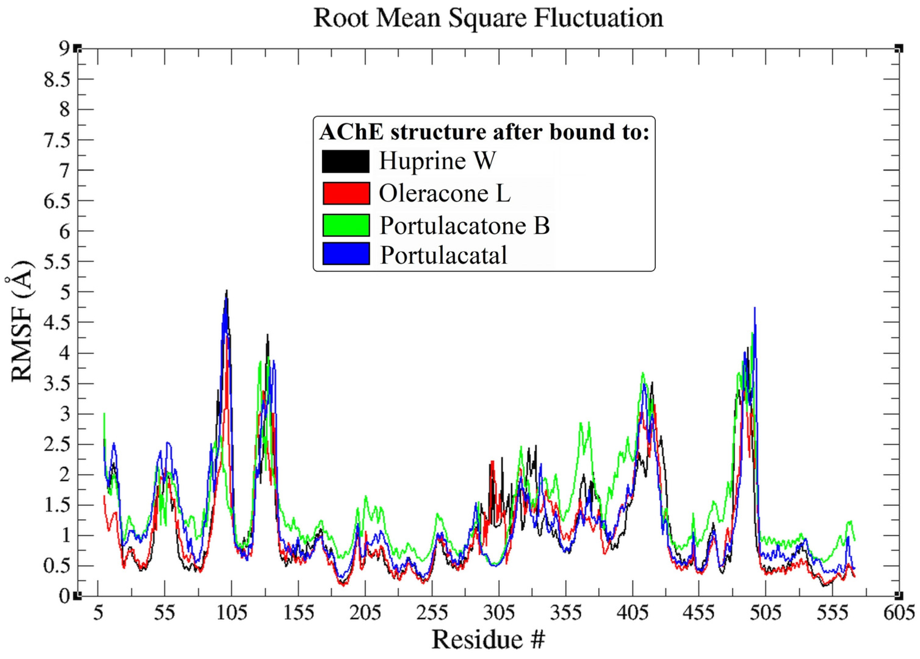
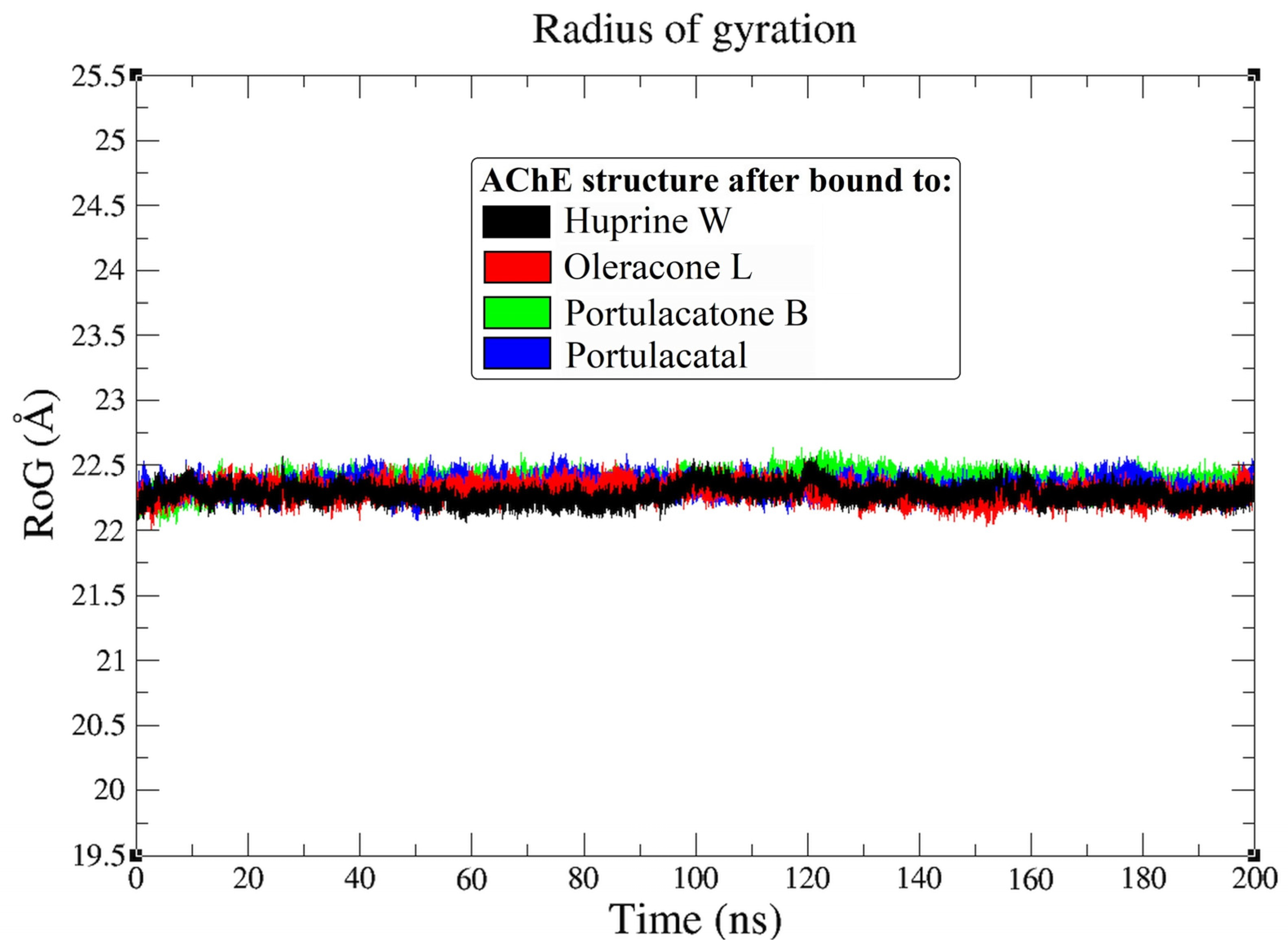
4.3. Molecular Dynamic Simulation

The stability and conformational behaviors of AChE enzyme complexes with ligands (new alkaloids and huprine W) were investigated over a 200 ns duration time. Root mean square deviation (RMSD) analyses were performed to evaluate the structural dynamics of these protein–ligand complexes [42,43], with a specific focus on how each ligand influenced the conformational stability of AChE. The RMSD trajectories for the AChE enzyme backbone reveal distinct stability patterns across different ligands (Figure 3). The complex with the co-crystallized ligand huprine W maintained an average RMSD value of approximately 2.3 Å, indicating stable enzyme integrity throughout the simulation. This consistency sets a benchmark for comparing the stability of investigational ligands. Oleracone L bound to AChE demonstrated superior stability with a narrower RMSD fluctuation between 1.4 Å and 2.6 Å, averaging at 1.9 Å, suggesting a potentially more enhanced structural alignment within the active site. Conversely, portulacatone B exhibited RMSD fluctuations similar to the control, ranging from 1.3 Å to 3.2 Å with an average of 2.5 Å, indicating comparable stability. Portulacatal, while still maintaining fluctuations within a stable range, showed a slightly wider RMSD range from 1.4 Å to 2.7 Å, averaging approximately 2.3 Å. This broader fluctuation may suggest a dynamic interaction within the active site, which could impact its binding affinity and efficacy [45,79].
The RMSD values of the ligands themselves provide further insights into their conformational stability within the AChE binding site. Huprine W, as a control, showed high stability with minimal conformational changes, as evidenced by an RMSD fluctuation averaging ~0.8 Å. In contrast, oleracone L exhibited a highly consistent RMSD value averaging 1.2 Å with minimal deviation, which underscores its potential fit and effectiveness in the active site. Both portulacatone B and portulacatal also demonstrated stability in the binding site, with average RMSD values of approximately 1.4 Å, though the deviations of about 1 Å suggest slight variations in their binding conformations. These RMSD data highlight the differential stability profiles of the AChE–ligand complexes. Oleracone L emerges as potentially the most conformationally stable within the AChE binding site, suggesting an enhanced ability to maintain consistent interactions critical for effective inhibition. The stability observed with huprine W and the comparable stability of portulacatone B and portulacatal also reinforce their potential as AChE inhibitors.
The root-mean-square fluctuation (RMSF) analysis provides essential insights into the flexibility of specific amino acid residues within AChE upon binding with various ligands [80]. The RMSF plots, Figure 4, are instrumental in identifying regions within the enzyme that demonstrate significant mobility, which is vital for understanding the enzyme’s function and its interaction with ligands. Upon binding with the new alkaloids, all complexes exhibited RMSF fluctuations below 2 Å, indicating that the new alkaloids induce minimal disturbances to the residues of AChE [76,79]. This suggests that the binding of these alkaloids does not significantly alter the inherent flexibility of the enzyme, which is crucial for maintaining its functional integrity. Notably, the AChE structure in complex with portulacatone B demonstrated slightly higher mobility in some regions compared to other complexes, though the increase was less than 1 Å. This heightened mobility could be attributed to the active site’s groove accommodating the ligand, potentially positioning it more effectively within the binding site [81]. The minimal RMSF values across all complexes imply that while the new alkaloids engage the active site effectively [82], they do so without disrupting the essential dynamics of the AChE enzyme. Such stability in the face of ligand binding is indicative of the compounds’ compatibility with the enzyme, maintaining the structural and functional integrity of AChE while potentially modulating its activity.
Continuing the detailed analysis of MD, the radius of gyration (RoG), which serves as an indicator of the compactness of the enzyme structure upon ligand binding was calculated [83]. The Rg plots (Figure 5), depicted for the AChE backbone atoms across the simulation timeframe of 0–200 ns, provide insights into the structural integrity and compactness of the protein–ligand complexes. Remarkably, all complexes demonstrated very similar average Rg values around 22.4 Å, with minimal variations of less than 0.3 Å between them. This consistency suggests that the binding of the new alkaloids—oleracone L, portulacatone B, and portulacatal—preserves the compactness of the AChE protein. Such uniformity in Rg values indicates that these ligands do not significantly affect the structural stability or integrity of the enzyme. To further elucidate the nature of these interactions, especially in terms of their stability, the hydrogen bond (H-bond) profiles were examined over the course of the 200 ns MD simulations.
Figure 6 displays the number of H-bonds formed between the ligands and AChE throughout the simulation, providing a quantitative assessment of interaction strength and stability, critical for evaluating the efficacy of these compounds as inhibitors. The AChE complex with the co-crystallized ligand huprine W consistently maintained at least three H-bonds throughout the simulation. This observation supports the binding pose as predicted and illustrates a stable interaction, consistent with expectations based on the initial docking results shown in Figure 2. Notably, oleracone L demonstrated higher consistency in forming H-bonds, reaching up to five and averaging four throughout the 200 ns MD simulation. This indicates a potentially more effective interaction with AChE, suggesting that oleracone L may offer enhanced inhibitory effects due to its strong and stable hydrogen bonding. In contrast, portulacatone B showed the fewest number of H-bonds, with an average of approximately two bonds. This lower number of H-bonds, as anticipated from the molecular docking analysis and depicted in Figure 2c, may account for the increased movement observed in the RMSF profiles of this complex. The fewer H-bonds could lead to a less stable interaction within the active site [84], potentially affecting the ligand’s efficacy as an AChE inhibitor.
Interestingly, portulacatal managed to consistently form at least three H-bonds, positioning it well within the active site. This consistent formation of H-bonds suggests enhanced stability of the portulacatal–AChE complex, potentially translating to more effective inhibitory action due to better positional integrity within the enzyme. Overall, the H-bond profiles from the MD simulations underscore the varying degrees of interaction strength and stability across the different ligand–AChE complexes. Oleracone L stands out with its higher number of consistent H-bonds, suggesting it is a particularly promising candidate for further development. Meanwhile, the differences observed with portulacatone B highlight the importance of optimizing H-bond interactions to enhance ligand efficacy. Portulacatal, with its ability to maintain a solid number of H-bonds, also indicates potential for effective AChE inhibition. These findings suggest avenues for further optimization and testing to improve the therapeutic profiles of these compounds for potential use in treating Alzheimer’s Disease.
Complementing the findings from earlier analyses, Molecular Mechanics Poisson-Boltzmann Surface Area (MM-PBSA) calculations were conducted to provide deeper insights into the binding free energies of the AChE complexes with the new alkaloids. These calculations are crucial for reinforcing the significance of the hydrogen bond interactions observed and for understanding the energetic contributions at the active binding site of AChE [56]. Table 3 presents the MM-PBSA binding energies for the co-crystallized ligand huprine W and the investigational compounds oleracone L, portulacatone B, and portulacatal. These energies provide a quantitative window into the various energetic contributions to the overall binding affinity at the active site of the AChE enzyme. The binding free energy (ΔGbind) for the AChE–huprine W complex is measured at −28.43 ± 0.11 kcal/mol, which sets a baseline for comparison with the other ligands. Notably, the electrostatic contributions for huprine W are significant at −17.22 ± 0.12 kcal/mol, suggesting that electrostatic forces are a major contributor to the binding affinity. These are balanced by van der Waals interactions at −14.64 ± 0.14 kcal/mol and a polar solvation energy, which is unfavorable at 18.28 ± 0.11 kcal/mol, counteracted by a beneficial non-polar solvation energy of −14.85 ± 0.12 kcal/mol. The non-polar solvation energy plays a critical role in stabilizing the ligand within the hydrophobic pockets of the enzyme.
Oleracone L exhibits a more favorable binding free energy of −31.74 ± 0.14 kcal/mol, suggesting a stronger binding affinity compared to huprine W. This can be attributed to higher electrostatic interactions at −19.42 ± 0.12 kcal/mol and stronger van der Waals forces at −17.64 ± 0.13 kcal/mol. The increased electrostatic interaction scores indicate a better alignment within the active site [85], enhancing the ligand’s interaction with key amino acid residues. Although the polar solvation penalty is slightly higher at 19.40 ± 0.12 kcal/mol, the non-polar solvation energy remains favorable at −14.08 ± 0.13 kcal/mol, indicating effective accommodation within the enzyme’s non-polar regions. In contrast, portulacatone B shows a ΔGbind of −25.21 ± 0.13 kcal/mol, with the least favorable binding energy among the compounds. Its electrostatic interactions at −14.60 ± 0.11 kcal/mol and van der Waals interactions closely match its electrostatic contributions, suggesting less optimal interaction dynamics within the active site. The slightly less unfavorable polar solvation energy and comparable non-polar solvation energy suggest a weaker overall binding interaction. Portulacatal demonstrates a ΔGbind of −29.85 ± 0.11 kcal/mol, with substantial electrostatic and van der Waals contributions, suggesting that it balances both interaction types effectively. The polar and non-polar solvation energies for portulacatal indicate a balanced energetic profile conducive to stable binding, contributing to a strong overall binding affinity.
Overall, the MM-PBSA binding energy analysis indicates that while all ligands exhibit potential as AChE inhibitors, oleracone L stands out with the highest binding affinity, closely followed by portulacatal. These investigational ligands demonstrate the potential to serve as effective AChE inhibitors, as suggested by their favorable binding free energies and significant electrostatic contributions. However, while these computational predictions are promising, it is crucial to corroborate the in silico findings with in vitro and in vivo studies to confirm the therapeutic efficacy and safety profile of these compounds. Such studies will validate the investigational ligands’ inhibitory action on AChE and determine their suitability for clinical development.

5. Conclusions

This study conducted the first comprehensive in silico evaluation of the binding potential of three newly identified alkaloids from Portulaca oleracea L.—oleracone L, portulacatone B, and portulacatal—against the AChE enzyme, a key target in Alzheimer’s disease treatment. The investigation employed a series of computational techniques, including ADME prediction using ADMETlab 2.0, molecular docking, molecular dynamics simulations, and MM-PBSA calculations, to assess the therapeutic potential of these compounds. The ADME analysis predicted favorable profiles for these alkaloids, indicating their potential to cross the blood–brain barrier effectively. The drug-likeness assessment, absorption, distribution, metabolism, and excretion profiles collectively suggest these compounds as promising candidates for CNS-active agents. Molecular docking results showed that oleracone L had the highest binding affinity to AChE with a score of −10.75 kcal/mol, followed by portulacatal at −9.95 kcal/mol and portulacatone B at −8.89 kcal/mol. These findings suggest that oleracone L might offer the strongest inhibitory action against AChE due to its superior molecular interactions within the active site.
Molecular dynamics simulations reinforced these findings, where all three compounds maintained stable complexes with AChE, as evidenced by RMSD values consistently below 2 Å. Specifically, the RMSD values for the AChE–oleracone L complex fluctuated narrowly between 1.4 Å and 2.6 Å, averaging at 1.9 Å, indicating superior stability. The RMSD values for portulacatone B ranged from 1.3 Å to 3.2 Å, averaging 2.5 Å, and for portulacatal ranged from 1.4 Å to 2.7 Å, averaging 2.3 Å. RMSF and radius of gyration analyses indicated minimal disruption to the enzyme’s overall structure and dynamics, essential for maintaining its biological functionality. Specifically, the RMSF values for all complexes were below 2 Å, indicating minimal disturbances to AChE residues. The average radius of gyration (RoG) for all complexes was around 22.4 Å, with variations less than 0.3 Å, suggesting maintained compactness of the enzyme structure upon ligand binding. The hydrogen bond analysis revealed that oleracone L consistently formed a higher number of stable H-bonds with AChE, with up to five H-bonds and an average of four H-bonds throughout the 200 ns MD simulation. Portulacatone B showed fewer H-bonds, averaging approximately two, and portulacatal maintained at least three H-bonds, indicating variable interaction strengths. Furthermore, MM-PBSA calculations corroborated the docking and dynamics results, confirming that oleracone L possesses the most favorable binding energy (−31.74 ± 0.14 kcal/mol), enhanced by significant electrostatic (−19.42 ± 0.12 kcal/mol) and van der Waals (−17.64 ± 0.13 kcal/mol) interactions. The binding free energies of portulacatone B (−25.21 ± 0.13 kcal/mol) and portulacatal (−29.85 ± 0.11 kcal/mol) also demonstrated their potential as AChE inhibitors, albeit to a lesser extent compared to oleracone L. This study highlights the promising inhibitory potential of these alkaloids, particularly oleracone L, against AChE, suggesting that they could be valuable leads in the development of new therapeutic agents for Alzheimer’s Disease. Future research should focus on experimental validation through in vitro and in vivo testing to confirm these computational predictions and fully evaluate the therapeutic efficacy of these compounds.

Supplementary Materials

The following supporting information can be downloaded at: https://www.mdpi.com/article/10.3390/pr12071456/s1, Figure S1. Superimposition (a) and 2D interactions analysis of the co-crystallized ligand (green C, red O, and blue N) (b) and re-docked ligand (lime C, red O, and blue N) (c). The crystal structure of human acetylcholinesterase in complex with huprine W (4BDT.pdb) (RMSD is 0.34 Å).

Funding

This research received no external funding.

Data Availability Statement

Data are contained within the article and Supplementary Materials.

Acknowledgments

The author wishes to express her gratitude to the Deanship of Scientific Research at the University of Hafr Al Batin in Saudi Arabia for their unending support of this research.

Conflicts of Interest

The author declares no conflicts of interest.

References

  1. Amin, F.; Shamsi, A.; Asghar, M.N.; Khaki, P.S.S.; Khan, M.S.; Tabrez, S.; Zaidi, S.K.; Khan, W.; Bano, B. Alzheimer’s: A Progressive Brain Disease: Causes, Symptoms, and Prevention. In Biological, Diagnostic and Therapeutic Advances in Alzheimer’s Disease: Non-Pharmacological Therapies for Alzheimer’s Disease; Springer: Berlin/Heidelberg, Germany, 2019; pp. 31–51. [Google Scholar]
  2. Bhavani, K.; Kuriyakose, J.; Sindhuja, R.; Chandrashekhar, U.; Ashab, A. A review on neurodegenerative disorder--alzheimer’s disease. Drug Invent. Today 2020, 13, 954. [Google Scholar]
  3. Alhawarri, M.B.; Al-Thiabat, M.G.; Dubey, A.; Tufail, A.; Fouad, D.; Alrimawi, B.H.; Dayoob, M. ADME profiling, molecular docking, DFT, and MEP analysis reveal cissamaline, cissamanine, and cissamdine from Cissampelos capensis Lf as potential anti-Alzheimer’s agents. RSC Adv. 2024, 14, 9878–9891. [Google Scholar] [CrossRef]
  4. Alhawarri, M.B.; Dianita, R.; Rawa, M.S.A.; Nogawa, T.; Wahab, H.A. Potential Anti-Cholinesterase Activity of Bioactive Compounds Extracted from Cassia grandis Lf and Cassia timoriensis DC. Plants 2023, 12, 344. [Google Scholar] [CrossRef]
  5. Alhawarri, M.B.; Dianita, R.; Razak, K.N.A.; Mohamad, S.; Nogawa, T.; Wahab, H.A. Antioxidant, anti-inflammatory, and inhibition of acetylcholinesterase potentials of Cassia timoriensis DC. flowers. Molecules 2021, 26, 2594. [Google Scholar] [CrossRef]
  6. Moreira, N.C.d.S.; Lima, J.E.B.d.F.; Marchiori, M.F.; Carvalho, I.; Sakamoto-Hojo, E.T. Neuroprotective effects of cholinesterase inhibitors: Current scenario in therapies for Alzheimer’s disease and future perspectives. J. Alzheimer’s Dis. Rep. 2022, 6, 177–193. [Google Scholar] [CrossRef]
  7. Vecchio, I.; Sorrentino, L.; Paoletti, A.; Marra, R.; Arbitrio, M. The state of the art on acetylcholinesterase inhibitors in the treatment of Alzheimer’s disease. J. Cent. Nerv. Syst. Dis. 2021, 13, 11795735211029113. [Google Scholar] [CrossRef]
  8. Ozdemir, Z.; Ozcelik, A.; Uysal, M. Approaches based on cholinergic hypothesis and cholinesterase inhibitors in the treatment of alzheimer’s disease. Front. Clin. Drug Res.-Alzheimer Disord. 2019, 8, 154–190. [Google Scholar]
  9. Hansen, R.A.; Gartlehner, G.; Webb, A.P.; Morgan, L.C.; Moore, C.G.; Jonas, D.E. Efficacy and safety of donepezil, galantamine, and rivastigmine for the treatment of Alzheimer’s disease: A systematic review and meta-analysis. Clin. Interv. Aging 2008, 3, 211–225. [Google Scholar]
  10. Marucci, G.; Buccioni, M.; Dal Ben, D.; Lambertucci, C.; Volpini, R.; Amenta, F. Efficacy of acetylcholinesterase inhibitors in Alzheimer’s disease. Neuropharmacology 2021, 190, 108352. [Google Scholar] [CrossRef]
  11. Kabir, M.T.; Uddin, M.S.; Begum, M.; Thangapandiyan, S.; Rahman, M.S.; Aleya, L.; Mathew, B.; Ahmed, M.; Barreto, G.E.; Ashraf, G.M. Cholinesterase inhibitors for Alzheimer’s disease: Multitargeting strategy based on anti-Alzheimer’s drugs repositioning. Curr. Pharm. Des. 2019, 25, 3519–3535. [Google Scholar] [CrossRef]
  12. Li, W.; Rang, Y.; Liu, H.; Liu, C. Update on new trends and progress of natural active ingredients in the intervention of Alzheimer’s disease, based on understanding of traditional Chinese and Western relevant theories: A review. Phytother. Res. 2023, 37, 3744–3764. [Google Scholar] [CrossRef]
  13. Kim, S.W.; Lee, J.H.; Kim, B.; Yang, G.; Kim, J.U. Natural products as the potential to improve Alzheimer’s and Parkinson’s disease. Int. J. Mol. Sci. 2023, 24, 8827. [Google Scholar] [CrossRef] [PubMed]
  14. Liu, J.-Y.; Guo, H.-Y.; Quan, Z.-S.; Shen, Q.-K.; Cui, H.; Li, X. Research progress of natural products and their derivatives against Alzheimer’s disease. J. Enzym. Inhib. Med. Chem. 2023, 38, 2171026. [Google Scholar] [CrossRef]
  15. Deng, M.; Yan, W.; Gu, Z.; Li, Y.; Chen, L.; He, B. Anti-neuroinflammatory potential of natural products in the treatment of Alzheimer’s disease. Molecules 2023, 28, 1486. [Google Scholar] [CrossRef]
  16. Puri, V.; Kanojia, N.; Sharma, A.; Huanbutta, K.; Dheer, D.; Sangnim, T. Natural product-based pharmacological studies for neurological disorders. Front. Pharmacol. 2022, 13, 1011740. [Google Scholar] [CrossRef]
  17. Iqbal, D.; Rehman, M.T.; Alajmi, M.F.; Alsaweed, M.; Jamal, Q.M.S.; Alasiry, S.M.; Albaker, A.B.; Hamed, M.; Kamal, M.; Albadrani, H.M. Multitargeted virtual screening and molecular simulation of natural product-like compounds against GSK3β, NMDA-receptor, and BACE-1 for the management of Alzheimer’s disease. Pharmaceuticals 2023, 16, 622. [Google Scholar] [CrossRef] [PubMed]
  18. Tung, B.T.; Hue, N.T.; Long, N.V.; Ngoc, N.T. Medicinal Herbs Against Central Nervous System Disorders. In Pharmacological Benefits of Natural Agents; IGI Global: Hershey, PA, USA, 2023; pp. 85–103. [Google Scholar]
  19. Awuchi, C.G. Plants, phytochemicals, and natural practices in complementary and alternative system of medicine for treatment of central nervous system disorders. Int. J. Food Prop. 2023, 26, 1190–1213. [Google Scholar] [CrossRef]
  20. Islam, A.; Mishra, A.; Ahsan, R.; Fareha, S. Phytopharmaceuticals and Herbal Approaches to Target Neurodegenerative Disorders. Drug Res. 2023, 73, 388–407. [Google Scholar] [CrossRef]
  21. Srivastava, A.; Srivastava, P.; Pandey, A.; Khanna, V.; Pant, A. Phytomedicine: A potential alternative medicine in controlling neurological disorders. In New Look to Phytomedicine; Elsevier: Amsterdam, The Netherlands, 2019; pp. 625–655. [Google Scholar]
  22. Kakooza-Mwesige, A. The importance of botanical treatments in traditional societies and challenges in developing countries. Epilepsy Behav. 2015, 52, 297–307. [Google Scholar] [CrossRef]
  23. Perera, P.K.; Dahanayake, J.M.; Diddeniya, J.I.D. Pharmacology, Toxicology, and Therapeutic Effects of Metals and Minerals Used in Traditional Medicine. In Medical Geology: En Route to One Health; John and Wiley and Sons: Hoboken, NJ, USA, 2023; pp. 303–313. [Google Scholar]
  24. Zhou, Y.-X.; Xin, H.-L.; Rahman, K.; Wang, S.-J.; Peng, C.; Zhang, H. Portulaca oleracea L.: A review of phytochemistry and pharmacological effects. BioMed Res. Int. 2015, 2015, 925631. [Google Scholar] [CrossRef]
  25. Iranshahy, M.; Javadi, B.; Iranshahi, M.; Jahanbakhsh, S.P.; Mahyari, S.; Hassani, F.V.; Karimi, G. A review of traditional uses, phytochemistry and pharmacology of Portulaca oleracea L. J. Ethnopharmacol. 2017, 205, 158–172. [Google Scholar] [CrossRef]
  26. Montoya-García, C.O.; García-Mateos, R.; Becerra-Martínez, E.; Toledo-Aguilar, R.; Volke-Haller, V.H.; Magdaleno-Villar, J.J. Bioactive compounds of purslane (Portulaca oleracea L.) according to the production system: A review. Sci. Hortic. 2023, 308, 111584. [Google Scholar] [CrossRef]
  27. Masoodi, M.H.; Ahmad, B.; Mir, S.R.; Zargar, B.A.; Tabasum, N. Portulaca oleracea L. a review. J. Pharm. Res. 2011, 4, 3044–3048. [Google Scholar]
  28. Cui, X.; Ying, Z.; Ying, X.; Jia, L.; Yang, G. Three new alkaloids from Portulaca oleracea L. and their bioactivities. Fitoterapia 2021, 154, 105020. [Google Scholar] [CrossRef]
  29. Xie, Y.; Yang, W.; Chen, X.; Xiao, J. Inhibition of flavonoids on acetylcholine esterase: Binding and structure–activity relationship. Food Funct. 2014, 5, 2582–2589. [Google Scholar] [CrossRef]
  30. Tanoli, S.T.; Ramzan, M.; Hassan, A.; Sadiq, A.; Jan, M.S.; Khan, F.A.; Ullah, F.; Ahmad, H.; Bibi, M.; Mahmood, T. Design, synthesis and bioevaluation of tricyclic fused ring system as dual binding site acetylcholinesterase inhibitors. Bioorganic Chem. 2019, 83, 336–347. [Google Scholar] [CrossRef]
  31. Silvia Matos, K.; FF da Cunha, E.; Abagyan, R.; C Ramalho, T. Computational evidence for the reactivation process of human acetylcholinesterase inhibited by carbamates. Comb. Chem. High Throughput Screen. 2014, 17, 554–564. [Google Scholar] [CrossRef]
  32. Carmo Carreiras, M.; Mendes, E.; Jesus Perry, M.; Paula Francisco, A.; Marco-Contelles, J. The multifactorial nature of Alzheimer’s disease for developing potential therapeutics. Curr. Top. Med. Chem. 2013, 13, 1745–1770. [Google Scholar] [CrossRef]
  33. Jankowska, A.; Wesolowska, A.; Pawlowski, M.; Chlon-Rzepa, G. Multi-target-directed ligands affecting serotonergic neurotransmission for Alzheimer’s disease therapy: Advances in chemical and biological research. Curr. Med. Chem. 2018, 25, 2045–2067. [Google Scholar] [CrossRef]
  34. Nachon, F.; Carletti, E.; Ronco, C.; Trovaslet, M.; Nicolet, Y.; Jean, L.; Renard, P.-Y. Crystal structures of human cholinesterases in complex with huprine W and tacrine: Elements of specificity for anti-Alzheimer’s drugs targeting acetyl-and butyryl-cholinesterase. Biochem. J. 2013, 453, 393–399. [Google Scholar] [CrossRef]
  35. Tariq, S.; Mutahir, S.; Khan, M.A.; Mutahir, Z.; Hussain, S.; Ashraf, M.; Bao, X.; Zhou, B.; Stark, C.B.; Khan, I.U. Synthesis, in vitro cholinesterase inhibition, molecular docking, DFT, and ADME studies of novel 1, 3, 4-oxadiazole-2-thiol derivatives. Chem. Biodivers. 2022, 19, e202200157. [Google Scholar] [CrossRef]
  36. Borges, N.M.; Sartori, G.R.; Ribeiro, J.F.; Rocha, J.R.; Martins, J.B.; Montanari, C.A.; Gargano, R. Similarity search combined with docking and molecular dynamics for novel hAChE inhibitor scaffolds. J. Mol. Model. 2018, 24, 41. [Google Scholar] [CrossRef]
  37. Khan, I.; Bakht, S.M.; Ibrar, A.; Abbas, S.; Hameed, S.; White, J.M.; Rana, U.A.; Zaib, S.; Shahid, M.; Iqbal, J. Exploration of a library of triazolothiadiazole and triazolothiadiazine compounds as a highly potent and selective family of cholinesterase and monoamine oxidase inhibitors: Design, synthesis, X-ray diffraction analysis and molecular docking studies. RSC Adv. 2015, 5, 21249–21267. [Google Scholar] [CrossRef]
  38. Ejaz, S.A.; Fayyaz, A.; Mahmood, H.M.K.; Aziz, M.; Ejaz, S.R.; Alkhuriji, A.F.; Al-Megrin, W.A.I.; Aborode, A.T.; Batiha, G.E.-S. 4-Phthalimidobenzenesulfonamide Derivatives as Acetylcholinesterase and Butyrylcholinesterase Inhibitors: DFTs, 3D-QSAR, ADMET, and Molecular Dynamics Simulation. Neurodegener. Dis. 2023, 22, 122–138. [Google Scholar] [CrossRef]
  39. Gordon, J.C.; Myers, J.B.; Folta, T.; Shoja, V.; Heath, L.S.; Onufriev, A. H++: A server for estimating p K as and adding missing hydrogens to macromolecules. Nucleic. Acids Res. 2005, 33, W368–W371. [Google Scholar] [CrossRef]
  40. Morris, G.M.; Huey, R.; Lindstrom, W.; Sanner, M.F.; Belew, R.K.; Goodsell, D.S.; Olson, A.J. AutoDock4 and AutoDockTools4: Automated docking with selective receptor flexibility. J. Comput. Chem. 2009, 30, 2785–2791. [Google Scholar] [CrossRef]
  41. Yamari, I.; Abchir, O.; Mali, S.N.; Errougui, A.; Talbi, M.; El Kouali, M.; Chtita, S. The anti-SARS-CoV-2 activity of novel 9, 10-dihydrophenanthrene derivatives: An insight into molecular docking, ADMET analysis, and molecular dynamics simulation. Sci. Afr. 2023, 21, e01754. [Google Scholar] [CrossRef]
  42. Shalayel, M.H.F.; Al-Mazaideh, G.M.; Alanezi, A.A.; Almuqati, A.F.; Alotaibi, M. Diosgenin and Monohydroxy Spirostanol from Prunus amygdalus var amara Seeds as Potential Suppressors of EGFR and HER2 Tyrosine Kinases: A Computational Approach. Pharmaceuticals 2023, 16, 704. [Google Scholar] [CrossRef]
  43. Shalayel, M.H.F.; Al-Mazaideh, G.M.; Alanezi, A.A.; Almuqati, A.F.; Alotaibi, M. The Potential Anti-Cancerous Activity of Prunus amygdalus var. amara Extract. Processes 2023, 11, 1277. [Google Scholar] [CrossRef]
  44. O’Boyle, N.M.; Banck, M.; James, C.A.; Morley, C.; Vandermeersch, T.; Hutchison, G.R. Open Babel: An open chemical toolbox. J. Cheminform. 2011, 3, 1–14. [Google Scholar] [CrossRef]
  45. Al-Thiabat, M.G.; Saqallah, F.G.; Gazzali, A.M.; Mohtar, N.; Yap, B.K.; Choong, Y.S.; Wahab, H.A. Heterocyclic substitutions greatly improve affinity and stability of folic acid towards FRα. An in silico insight. Molecules 2021, 26, 1079. [Google Scholar] [CrossRef]
  46. Alidmat, M.M.; Khairuddean, M.; Kamal, N.N.S.N.M.; Muhammad, M.; Wahab, H.A.; Althiabat, M.G.; Alhawarri, M.B. Synthesis, Characterization, Molecular Docking and Cytotoxicity Evaluation of New Thienyl Chalcone Derivatives against Breast Cancer Cells. Syst. Rev. Pharm. 2022, 13, 1. [Google Scholar]
  47. Alhawarri, M.B.; Olimat, S. Potential Serotonin 5-HT2A Receptor Agonist of Psychoactive Components of Silene undulata Aiton: LC-MS/MS, ADMET, and Molecular Docking Studies. In Current Pharmaceutical Biotechnology; Bentham Science Publishers: Sharjah, United Arab Emirates, 2024. [Google Scholar]
  48. Yunos, N.M.; Al-Thiabat, M.G.; Sallehudin, N.J. Quassinoids from Eurycoma longifolia as Potential Dihydrofolate Reductase Inhibitors: A Computational Study. In Current Pharmaceutical Biotechnology; Bentham Science Publishers: Sharjah, United Arab Emirates, 2024. [Google Scholar]
  49. Forli, W.; Halliday, S.; Belew, R.; Olson, A.J. AutoDock Version 4.2. J. Med. Chem. 2012, 55, 623–638. [Google Scholar] [CrossRef]
  50. Ross, B.J. A Lamarckian evolution strategy for genetic algorithms. In Practical Handbook of Genetic Algorithms; CRC Press: Boca Raton, FL, USA, 2019; pp. 1–16. [Google Scholar]
  51. Schmid, N.; Eichenberger, A.P.; Choutko, A.; Riniker, S.; Winger, M.; Mark, A.E.; Van Gunsteren, W.F. Definition and testing of the GROMOS force-field versions 54A7 and 54B7. Eur. Biophys. J. 2011, 40, 843–856. [Google Scholar] [CrossRef]
  52. Rühle, V. Pressure Coupling/Barostats. Journal Club. 2008; pp. 1–5. Available online: https://www2.mpip-mainz.mpg.de/~andrienk/journal_club/barostats.pdf (accessed on 29 April 2024).
  53. Berendsen, H.J.; Postma, J.v.; Van Gunsteren, W.F.; DiNola, A.; Haak, J.R. Molecular dynamics with coupling to an external bath. J. Chem. Phys. 1984, 81, 3684–3690. [Google Scholar] [CrossRef]
  54. Petersen, H.G. Accuracy and efficiency of the particle mesh Ewald method. J. Chem. Phys. 1995, 103, 3668–3679. [Google Scholar] [CrossRef]
  55. Hess, B.; Bekker, H.; Berendsen, H.J.; Fraaije, J.G. LINCS: A linear constraint solver for molecular simulations. J. Comput. Chem. 1997, 18, 1463–1472. [Google Scholar] [CrossRef]
  56. Ben-Shalom, I.Y.; Pfeiffer-Marek, S.; Baringhaus, K.-H.; Gohlke, H. Efficient approximation of ligand rotational and translational entropy changes upon binding for use in MM-PBSA calculations. J. Chem. Inf. Model. 2017, 57, 170–189. [Google Scholar] [CrossRef]
  57. Kumari, R.; Kumar, R.; Consortium, O.S.D.D.; Lynn, A. g_mmpbsa—A GROMACS tool for high-throughput MM-PBSA calculations. J. Chem. Inf. Model. 2014, 54, 1951–1962. [Google Scholar] [CrossRef]
  58. Verma, S.; Grover, S.; Tyagi, C.; Goyal, S.; Jamal, S.; Singh, A.; Grover, A. Hydrophobic interactions are a key to MDM2 inhibition by polyphenols as revealed by molecular dynamics simulations and MM/PBSA free energy calculations. PLoS ONE 2016, 11, e0149014. [Google Scholar] [CrossRef]
  59. Xiong, G.; Wu, Z.; Yi, J.; Fu, L.; Yang, Z.; Hsieh, C.; Yin, M.; Zeng, X.; Wu, C.; Lu, A. ADMETlab 2.0: An integrated online platform for accurate and comprehensive predictions of ADMET properties. Nucleic Acids Res. 2021, 49, W5–W14. [Google Scholar] [CrossRef]
  60. Van De Waterbeemd, H.; Gifford, E. ADMET in silico modelling: Towards prediction paradise? Nat. Rev. Drug Discov. 2003, 2, 192–204. [Google Scholar] [CrossRef]
  61. Yang, H.; Lou, C.; Sun, L.; Li, J.; Cai, Y.; Wang, Z.; Li, W.; Liu, G.; Tang, Y. admetSAR 2.0: Web-service for prediction and optimization of chemical ADMET properties. Bioinformatics 2019, 35, 1067–1069. [Google Scholar] [CrossRef]
  62. Walters, W.P. Going further than Lipinski’s rule in drug design. Expert Opin. Drug Discov. 2012, 7, 99–107. [Google Scholar] [CrossRef]
  63. Gehlhaar, D.K.; Luty, B.A.; Cheung, P.P.; Litman, A.H.; Owen, R.M.; Rose, P.W. The Pfizer Crystal Structure Database: An essential tool for structure-based design at Pfizer. J. Comput. Chem. 2022, 43, 1053–1062. [Google Scholar] [CrossRef]
  64. Johnson, T.W.; Dress, K.R.; Edwards, M. Using the Golden Triangle to optimize clearance and oral absorption. Bioorganic Med. Chem. Lett. 2009, 19, 5560–5564. [Google Scholar] [CrossRef]
  65. Zhou, Y.; Fu, Y.; Yin, W.; Li, J.; Wang, W.; Bai, F.; Xu, S.; Gong, Q.; Peng, T.; Hong, Y. Kinetics-driven drug design strategy for next-generation acetylcholinesterase inhibitors to clinical candidate. J. Med. Chem. 2021, 64, 1844–1855. [Google Scholar] [CrossRef]
  66. Bortolami, M.; Rocco, D.; Messore, A.; Di Santo, R.; Costi, R.; Madia, V.N.; Scipione, L.; Pandolfi, F. Acetylcholinesterase inhibitors for the treatment of Alzheimer’s disease–a patent review (2016–present). Expert Opin. Ther. Pat. 2021, 31, 399–420. [Google Scholar] [CrossRef]
  67. Mathew, B.; Parambi, D.G.; Mathew, G.E.; Uddin, M.S.; Inasu, S.T.; Kim, H.; Marathakam, A.; Unnikrishnan, M.K.; Carradori, S. Emerging therapeutic potentials of dual-acting MAO and AChE inhibitors in Alzheimer’s and Parkinson’s diseases. Arch. Pharm. 2019, 352, 1900177. [Google Scholar] [CrossRef]
  68. Agu, P.; Afiukwa, C.; Orji, O.; Ezeh, E.; Ofoke, I.; Ogbu, C.; Ugwuja, E.; Aja, P. Molecular docking as a tool for the discovery of molecular targets of nutraceuticals in diseases management. Sci. Rep. 2023, 13, 13398. [Google Scholar] [CrossRef]
  69. Anwar, T.; Kumar, P.; Khan, A.U. Modern tools and techniques in computer-aided drug design. In Molecular Docking for Computer-Aided Drug Design; Elsevier: Amsterdam, The Netherlands, 2021; pp. 1–30. [Google Scholar]
  70. De Ruyck, J.; Brysbaert, G.; Blossey, R.; Lensink, M.F. Molecular docking as a popular tool in drug design, an in silico travel. Adv. Appl. Bioinform. Chem. 2016, 9, 1–11. [Google Scholar] [CrossRef]
  71. Ferreira, L.G.; Dos Santos, R.N.; Oliva, G.; Andricopulo, A.D. Molecular docking and structure-based drug design strategies. Molecules 2015, 20, 13384–13421. [Google Scholar] [CrossRef]
  72. Acúrcio, R.C.; Leonardo-Sousa, C.; García-Sosa, A.T.; Salvador, J.A.; Florindo, H.F.; Guedes, R.C. Structural insights and binding analysis for determining the molecular bases for programmed cell death protein ligand-1 inhibition. Medchemcomm 2019, 10, 1810–1818. [Google Scholar] [CrossRef]
  73. Alves, M.J.; Froufe, H.J.; Costa, A.F.; Santos, A.F.; Oliveira, L.G.; Osório, S.R.; Abreu, R.M.; Pintado, M.; Ferreira, I.C. Docking studies in target proteins involved in antibacterial action mechanisms: Extending the knowledge on standard antibiotics to antimicrobial mushroom compounds. Molecules 2014, 19, 1672–1684. [Google Scholar] [CrossRef]
  74. Amir Rawa, M.S.; Al-Thiabat, M.G.; Nogawa, T.; Futamura, Y.; Okano, A.; Wahab, H.A. Naturally Occurring 8ß, 13ß-kaur-15-en-17-al and Anti-Malarial Activity from Podocarpus polystachyus Leaves. Pharmaceuticals 2022, 15, 902. [Google Scholar] [CrossRef]
  75. Yunos, N.M.; Wahab, H.A.; Al-Thiabat, M.G.; Sallehudin, N.J.; Jauri, M.H. In Vitro and In Silico Analysis of the Anticancer Effects of Eurycomanone and Eurycomalactone from Eurycoma longifolia. Plants 2023, 12, 2827. [Google Scholar] [CrossRef]
  76. Larue, L.; Kenzhebayeva, B.; Al-Thiabat, M.G.; Jouan–Hureaux, V.; Mohd–Gazzali, A.; Wahab, H.A.; Boura, C.; Yeligbayeva, G.; Nakan, U.; Frochot, C. tLyp–1: A peptide suitable to target NRP–1 receptor. Bioorganic Chem. 2023, 130, 106200. [Google Scholar] [CrossRef]
  77. Chadha, N.; Tiwari, A.K.; Kumar, V.; Lal, S.; Milton, M.D.; Mishra, A.K. Oxime-dipeptides as anticholinesterase, reactivator of phosphonylated-serine of AChE catalytic triad: Probing the mechanistic insight by MM-GBSA, dynamics simulations and DFT analysis. J. Biomol. Struct. Dyn. 2015, 33, 978–990. [Google Scholar] [CrossRef]
  78. Almeida, M.C.; Resende, D.I.; da Costa, P.M.; Pinto, M.M.; Sousa, E. Tryptophan derived natural marine alkaloids and synthetic derivatives as promising antimicrobial agents. Eur. J. Med. Chem. 2021, 209, 112945. [Google Scholar] [CrossRef]
  79. Al-Thiabat; Gazzali, A.; Mohtar, N.; Murugaiyah, V.; Kamarulzaman, E.E.; Yap, B.K.; Rahman, N.A.; Othman, R.; Wahab, H.A. Conjugated β-cyclodextrin enhances the affinity of folic acid towards FRα: Molecular dynamics study. Molecules 2021, 26, 5304. [Google Scholar] [CrossRef]
  80. Hernández-Rodríguez, M.; C Rosales-Hernández, M.; E Mendieta-Wejebe, J.; Martínez-Archundia, M.; Correa Basurto, J. Current tools and methods in molecular dynamics (MD) simulations for drug design. Curr. Med. Chem. 2016, 23, 3909–3924. [Google Scholar] [CrossRef]
  81. Rosenberry, T.L.; Brazzolotto, X.; Macdonald, I.R.; Wandhammer, M.; Trovaslet-Leroy, M.; Darvesh, S.; Nachon, F. Comparison of the binding of reversible inhibitors to human butyrylcholinesterase and acetylcholinesterase: A crystallographic, kinetic and calorimetric study. Molecules 2017, 22, 2098. [Google Scholar] [CrossRef]
  82. Da Fonseca, A.M.; Caluaco, B.J.; Madureira, J.M.C.; Cabongo, S.Q.; Gaieta, E.M.; Djata, F.; Colares, R.P.; Neto, M.M.; Fernandes, C.F.C.; Marinho, G.S. Screening of potential inhibitors targeting the main protease structure of SARS-CoV-2 via molecular docking, and approach with molecular dynamics, RMSD, RMSF, H-bond, SASA and MMGBSA. Mol. Biotechnol. 2023, 1–15. [Google Scholar] [CrossRef]
  83. Janati-Fard, F.; Housaindokht, M.R.; Monhemi, H. Investigation of structural stability and enzymatic activity of glucose oxidase and its subunits. J. Mol. Catal. B Enzym. 2016, 134, 16–24. [Google Scholar] [CrossRef]
  84. Goodman, M.F. Hydrogen bonding revisited: Geometric selection as a principal determinant of DNA replication fidelity. Proc. Natl. Acad. Sci. USA 1997, 94, 10493–10495. [Google Scholar] [CrossRef]
  85. Chakraborty, S.; Minda, R.; Salaye, L.; Bhattacharjee, S.K.; Rao, B.J. Active site detection by spatial conformity and electrostatic analysis—Unravelling a proteolytic function in shrimp alkaline phosphatase. PLoS ONE 2011, 6, e28470. [Google Scholar] [CrossRef]
Figure 1. Chemical structure of oleracone L (a), portulacatone B (b), and portulacatal (c).
Processes 12 01456 g001
Figure 2. 3D binding poses interactions between the original co-crystallized ligand, huprine W (green C, red O, pale blue N), oleracone L (blue C, red O, pale blue N), portulacatone B (cyan C, red O, pale blue N), and portulacatal (pink C, red O, pale blue N) and human acetylcholinesterase enzyme (AChE) (a). 2D interaction analysis of oleracone L (b), portulacatone B (c), and portulacatal (d) docked in the active binding site of AChE, represented by a solid surface rendering with pale yellow color-coding. AChE (PDB ID: 4BDT) is shown as a gray ribbon. This figure illustrates that the three alkaloids are able to reposition in the same active binding site of AChE as the co-crystallized ligand, demonstrating their potential binding affinity and interaction.
Processes 12 01456 g002
Figure 3. Root-mean-square deviation (RMSD) analyses for the molecular dynamics (MD) simulation trajectories were performed over a 200 ns duration. (A) RMSD plots for the AChE enzyme backbone, demonstrating molecular fluctuations following interaction with the co-crystallized ligand (huprine W) (in black), oleracone L (in red), portulacatone B (in green), and portulacatal (in blue). (B) The RMSD plots also reveal the conformational alterations of huprine W (in black), oleracone L (in red), portulacatone B (in green), and portulacatal (in blue) upon binding with the AChE enzyme.
Processes 12 01456 g003
Figure 4. The root mean square fluctuation (RMSF) plots of the backbone atoms in AChE during a 200 ns molecular dynamic simulation for all systems are depicted. The RMSF values reflect the remaining atomic fluctuations of each protein residue in their interaction with ligands throughout this progression path.
Processes 12 01456 g004
Figure 5. Radius of gyration (RoG) graphs for all systems’ AChE backbone atoms at molecular dynamics interval timeframes (0–200 ns); depicting interactions with the co-crystallized ligand (huprine W) (in black), oleracone L (in red), portulacatone B (in green), and portulacatal (in blue). The root mean square fluctuation (RMSF) plots of the backbone atoms in AChE during a 200 ns molecular dynamic simulation for all systems are depicted. The RMSF values reflect the remaining atomic fluctuations of each protein residue in their interaction with ligands throughout this progression path.
Processes 12 01456 g005
Figure 6. Hydrogen bond profile acquired from MD simulation over a time period of 0–200 ns for (a) AChE-huprine W, (b) AChE-oleracone L, (c) AChE-portulacatone B, and (d) AChE-portulacatal.
Processes 12 01456 g006
Table 1. Predicted drug-likeness and ADME properties of oleracone L, portulacatone B, and portulacatal using ADMET lab 2.0.
PropertyModel NamePredicted ValueComment
Oleracone LPortulacatone BPortulacatal
Drug-Likeness*Lipinski RuleAcceptedAcceptedAcceptedCompounds satisfying the Golden Triangle and GSK rules may have more favourable ADMET profiles [59,60].
*Pfizer RuleAcceptedAcceptedAccepted
*Golden TriangleAcceptedAcceptedAccepted
*GSK RuleAcceptedAcceptedAccepted
AbsorptionPapp (Caco-2 Permeability) cm/s−5.123−4.82−4.93Caco-2 Permeability: High: >−5.15 cm/s; low: <−5.15 cm/s [59,60].
MDCK Permeability
cm/s
7 × 10−614 × 10−69 × 10−6MDCK permeability (cm/s): Low: <2 × 10⁻⁶; Medium: 2–20 × 10−6; High: >20 × 10−6 [59,60].
HIA (Human Intestinal Absorption)0.0220.0410.031HIA: High: <0.30; Low: >0.30 [59,60].
DistributionPPB (Plasma Protein Binding) %88.1286.1579.55PPB Optimal: <90%. Drugs with lower 90% protein-bound may have a high therapeutic index [59,60].
BBB (Blood–Brain Barrier)0.390.510.139Cross BBB: ≥0.1; Cannot cross BBB: <0.1 [59,60].
VD (Volume Distribution)
L/kg
0.490.520.711VD Optimal: 0.04–20 L/kg [59,60].
Fu (The fraction unbound in plasms) %5.628.7111.55Fu: Low: 5%; Moderate: 5~20%; High: >20% [59,60].
MetabolismCYP1A2 inhibitorYesYesYes
CYP2C19 inhibitorNoNoNo
CYP2C9 inhibitorNoNoNo
CYP2D6 inhibitorNoNoNo
CYP3A4 inhibitorNoNoNo
Excretion*CL (Clearance Rate) mL/min/kg10.5913.7813.16CL: High: >15 mL/min/kg; Moderate: CL 5–15 mL/min/kg; Low: CL < 5 mL/min/kg [59,60].
T ½ (Half Lifetime) h0.850.760.88T ½: Long half-life: > 3 h; Short half-life: < 3 h [59,60].
*M.W: Molecular weight (g/mol), *logP: Partition coefficient (Lipophilicity), *Hacc: Hydrogen bond acceptor, *Hdon: Hydrogen bond donor, *logD: Distribution coefficient. *Lipinski Rule: *M.W ≤ 500; *logP ≤ 5; *Hacc ≤ 10; *Hdon ≤ 5, if two properties are out of range, a poor absorption or permeability is possible, one is acceptable. *Pfizer Rule: 200 ≤ *M.W ≤ 50; −2 ≤ *logD ≤ 5. *Golden Triangle: 200 ≤ *M.W ≤ 50; −2 ≤ *logD ≤ 5. *GSK Rule: *M.W ≤ 400; *logP ≤ 4.
Table 2. Molecular docking scores (free binding energy in kcal/mol) for the new alkaloids extracted from Portulaca oleracea L.: oleracone L, portulacatone B, and portulacatal, alongside the co-crystallized ligand huprine W, against the human acetylcholinesterase (AChE) therapeutic target. The table also includes an analysis of the 2D molecular interactions between these compounds and the residues within the AChE active site (PDB ID: 4BDT).
CompoundsFree Binding
Energy
(kcal/mol)
Molecular Interactions Analysis within the AChE Active Binding Site
H-BondDistance (Å)Pi-SigmaHydrophobic
Interaction
Oleracone L−10.75ASN87, ASN87, GLY122, SER203, and HIS4471.96, 2.51, 2.86, 2.04, and 1.89ASP74 and TRP439TRP86 and TYR337
Portulacatone B−8.89HIS4471.68 and 1.89----------------TRP86, TYR337, TRP439, and TYR449
Portulacatal−9.95GLY122, SER203, TRP439, HIS447, and HIS4472.07, 2.30, 2.49, 1.75, and 1.82----------------TRP86, TYR337, and TRP439
Co-crystalized
Ligand (huprine W, original pose)
−9.49GLY122 and SER2032.96 and 2.33----------------TRP86, TYR337, TRP439, MET443, PRO446, and TYR449
Table 3. MM-PBSA-binding energies (ΔGbind) of huprine W (control), oleracone L, portulacatone B, and portulacatal at the active binding site of AChE receptor (PDB ID: 4BDT). Energy units are expressed in kcal/mol.
SystemΔGbind
(kcal/mol)
Electrostatic (kcal/mol)Van der Waal (kcal/mol)Polar Salvation (kcal/mol)Non-Polar Salvation (kcal/mol)
AChE-Huprine W−28.43 ± 0.11−17.22 ± 0.12−14.64 ± 0.1418.28 ± 0.11−14.85 ± 0.12
AChE-Oleracone L−31.74 ± 0.14−19.42 ± 0.12−17.64 ± 0.1319.40 ± 0.12−14.08 ± 0.13
AChE-Portulacatone B−25.21 ± 0.13−14.60 ± 0.11−14.66 ± 0.1418.11 ± 0.12−14.06 ± 0.13
AChE-Portulacatal−29.85 ± 0.11−17.86 ± 0.12−16.75 ± 0.1419.21 ± 0.13−14.45 ± 0.12
Disclaimer/Publisher’s Note: The statements, opinions and data contained in all publications are solely those of the individual author(s) and contributor(s) and not of MDPI and/or the editor(s). MDPI and/or the editor(s) disclaim responsibility for any injury to people or property resulting from any ideas, methods, instructions or products referred to in the content.

