Article

Morphological and Structural Characterization of Encapsulated Arginine Systems for Dietary Inclusion in Ruminants

by
Germán Contreras-López
1,
Simón Morales-Rodríguez
2,
Alfredo R. Vilchis-Néstor
3,
Ana L. Rentería-Monterrubio
1,
Agustín Corral-Luna
1,
Ivan A. García-Galicia
4 and
Luis M. Carrillo-López
5,6,*
1
Facultad de Zootecnia y Ecología, Universidad Autónoma de Chihuahua, Periférico Francisco R. Almada Km. 1, Zootecnia 31453, Chihuahua, Mexico
2
Fitosanidad-Fitopatología, Colegio de Postgraduados en Ciencias Agrícolas, Carretera México Texcoco Km. 36.5, Montecillo, Texcoco 56100, Estado de México, Mexico
3
Centro Conjunto de Investigación en Química Sustentable (CCIQS), Universidad Autónoma del Estado de México-Universidad Nacional Autónoma de México, Toluca 50200, Estado de México, Mexico
4
C.E.I.E.G.T., Facultad de Medicina Veterinaria y Zootecnia, Universidad Nacional Autónoma de México, Km. 5.5 Carr. Fed, Martínez de la Torre-Tlapacoyan, Tlapacoyan 93600, Veracruz, Mexico
5
Consejo Nacional de Humanidades, Ciencias y Tecnologías, Facultad de Zootecnia y Ecología, Universidad Autónoma de Chihuahua, Periférico Francisco R. Almada Km. 1, Zootecnia 31453, Chihuahua, Mexico
6
Consejo Nacional de Humanidades, Ciencias y Tecnologías-Botánica, Colegio de Postgraduados en Ciencias Agrícolas, Carretera México Texcoco Km. 36.5, Montecillo, Texcoco 56100, Estado de México, Mexico
*
Author to whom correspondence should be addressed.
Processes 2024, 12(7), 1498; https://doi.org/10.3390/pr12071498
Submission received: 11 April 2024 / Revised: 14 June 2024 / Accepted: 24 June 2024 / Published: 17 July 2024
(This article belongs to the Special Issue Monitoring, Detection and Control of Food Contaminants)

Abstract

:
This research evaluated two methods of arginine encapsulation, melt emulsification and nanoprecipitation, using a lipid matrix of carnauba wax and commercial polymers (Eudragit®) as a protective material. The ratios of wax–arginine were 1:1, 2:1, 3:1, and 4:1, while those of Eudragit® RS:RL were 30:70 and 40:60 in proportions of 1:0.5 and 1:1 Eudragit®–arginine. The microcapsules were morphostructurally characterized by scanning electron microscopy, and a microelement analysis was performed via energy-dispersive X-ray spectroscopy and Fourier transform infrared spectroscopy. Additionally, in vitro digestibility was used to determine the protection efficiency. Both encapsulated systems presented regular (crystals) and spherical (microcapsules) polyhedral morphologies. Qualitative nitrogen decreased significantly as the wax ratio increased in the wax–arginine formulations. The formulations with a 1:1 Eudragit:–arginine ratio (1000 mg arginine) produced a higher nitrogen content in the encapsulated systems than the formulations containing 500 mg of arginine. The 2:1 and 3:1 wax–arginine formulations had the lowest degradability after 5 h of rumen fluid exposure (40.7 and 21.26%, respectively) in comparison with 100% unencapsulated arginine. The 3:1 wax–arginine formulation is an efficient encapsulating system which protects against rumen degradation. The more intense absorption bands at 1738 cm−1 and 1468 cm−1 associated with the C=O and C-H groups in carnauba wax indicate that arginine was more protected than in the other systems.

1. Introduction

Ruminants obtain amino acids from both microbially synthesized proteins and undegraded dietary rumen proteins; depending on their solubility and speed of transit toward the small intestine, the amount of undegraded dietary rumen protein changes depending on the foodstuff of origin [1]. Arginine is one of the most versatile amino acids; it is a precursor to the synthesis of urea, nitric oxide, and polyamines, and it also regulates metabolic pathways related to animal health, growth, reproduction, and homeostasis [2]. Although arginine can be synthesized by ruminants, it is normally considered an essential amino acid because de novo synthesis is not enough to meet their requirements, particularly during early growth phases or the production peak [3]. Dietary supplementation with essential amino acids increases the utilization efficiency of crude protein in the diet [4]. Conversely, an unbalanced essential amino acid composition causes greater urinary nitrogen loss, increases the release of ammonia, and deteriorates animal health, which ultimately leads to animal production failure [5]. Crystalline essential amino acid supplementation in a deficient diet improves the biological value of proteins [6]. However, the rapid absorption of crystalline essential amino acids can lead to a transient imbalance among amino acids in systemic circulation [7]. An amino acid imbalance can cause metabolic disorders, anorexia [8], and increased catabolism and amino acid oxidation [5].
Although it is still considered a relatively new method, microencapsulation has been used since the 1970s as a packaging technology in which thin polymeric coatings are applied to solid or liquid droplets or gaseous materials to form tiny particles called microcapsules which can release their contents over time or under certain conditions [9]. The microencapsulation of active ingredients in a matrix could allow for slow intestinal release [10] and prevent the production of ammonium and an imbalance in essential amino acids. The protective encapsulation of ingredients and feed to increase digestibility is a promising area of research. In ruminants, the encapsulation of amino acids and salts (calcium and ammonium nitrates; urea) for supplementation purposes has been reported [11,12,13]. Unfortunately, most research uses commercial products protected by international patents so that the encapsulation techniques, materials, and characteristics of the encapsulation are not fully described [14]. This applies for the encapsulation of some amino acids such as lysine, arginine, and methionine, for which the wall materials (fatty acids, glycerides, hydrogenate soybean oil, and lecithin) are described without including the encapsulation methodology [15,16]. Baggerman et al. [17] studied patented commercial products as supplements in animal production. Carvalho et al. [18] researched methionine encapsulation with carnauba wax using the melt-emulsification method; however, there was no evidence of microcapsule formation even though the micrographs showed arginine crystals embedded in a wax matrix, showing that encapsulated methionine has a lower in situ degree of degradability in comparison to pure methionine. Invariably, the supplementation of protected amino acids prevents degradation in the rumen, improving meat quality (greater muscle area and changes in fat thickness and tenderness). Therefore, new studies are subject to a lack of information for generating new knowledge in microencapsulation with potential applications in animal production. In this scenario, two encapsulation systems for arginine were designed as methods of protection from rumen degradation in vitro.

2. Materials and Methods

2.1. Design and Production of Microencapsulated Systems

Formulations of microencapsulated arginine systems were made using nanoprecipitation and melt emulsification. The protective or covering materials were carnauba wax (melt emulsification) and Eudragit® (RS and RL) (nanoprecipitation). Melt emulsification was performed according to Carvalho Neto et al. [18], with modifications. The wax–arginine ratios were 1:1, 2:1, 3:1, and 4:1. Briefly, the wax was melted at 90 °C and, once completely melted, soy lecithin was added at 7% of the weight of the wax; arginine was then added under constant agitation at 500 rpm for 3 min until a homogeneous emulsion was obtained. The nanoprecipitation technique was carried out according to Romero-Pérez et al. [19], with slight modifications. The organic phase was prepared by dissolving 1000 mg of Eudagrit® RL and RS (ratios of 30:70 and 40:60) in 10 mL of ethanol using sonication lasting 15 min (Elmasonic® S60H). Then, 500 or 1000 mg of arginine was added to 20 mL of an aqueous solution of Pluronic® F68 (0.5%, w/w). Considering 1000 mg of Eudagrit® RL and RS and 500 and 1000 mg of arginine, the Eudagrit® RL/RS–arginine ratios evaluated were 1:0.5 and 1:1, respectively. The microcapsules produced in both systems were characterized prior to the determination of their in vitro digestibility. Samples from both systems were ground in a mortar to a fine powder. The production of microcapsules was performed in triplicate for each encapsulation system, and for each system, the variables were assessed thrice (n = 9).

2.2. Morphological, Compositional, and Chemical–Structural Characterization

Powder samples of the encapsulated systems, the protective materials, and the arginine were visualized by scanning electron microscopy. They were mounted on nickel brackets with copper tape and then coated with a layer of gold–palladium to facilitate electronic conduction. The samples were observed using a scanning electron microscope (SEM-JSM-6390, Jeol, Tokyo, Japan) at 10 kV. Micrographs were obtained at magnifications of 100×, 500×, 1500×, and 5000×, and the size and morphology of the microencapsulated systems were analyzed using Image J software (version 1.53a, Wayne Rasband, National Institute of Health, Bethesda, MD, USA).
A microelement analysis was performed by energy-dispersive X-ray spectroscopy (using a JEOL JSM-6510LV coupled to an X-ray detector, with a resolution of 137 eV). The percentage by weight (qualitative evaluation) and mapping of the elements of interest (nitrogen) were determined in the encapsulated systems.
A small number of powdered samples were analyzed by Fourier transform infrared spectroscopy (FTIR model Tensor 27, Bruker®, Brooke, Germany) with a wave-number accuracy and spectral resolution of 500 to 4000 cm−1 and 1 cm−1, respectively. Spectral data from the encapsulated samples, protective materials, and arginine were compared to identify changes in their functional groups.

2.3. In Vitro Digestibility

The in vitro digestibility of the encapsulated wax–arginine systems was evaluated [20] 3 times (1, 3, and 5 h), with 3 repetitions per treatment at each time, using 3 digestion jars. F57 filter bags were rinsed in acetone for 5 min and dried completely prior to the experiment. Weighted filter bags and 0.5 g of each encapsulated system were registered. Two liters of ruminal fluid were collected and stored in a pre-heated thermal container at 30 °C. Buffer solutions were prepared as follows: solution A (KH2PO4 10 g/L, MgSO4·7H2O 0.5 g/L, NaCl 0.5 g/L, CaCl2·2H2O 0.1 g/L, and urea 0.5 g/L) and solution B (Na2CO3 15 g/L and Na2S·2H2O 1 g/L). Both solutions were preheated to 39 °C by adding ~266 mL of solution B to 1330 mL of solution A (ratio 1:5). An exact amount of A:B solution was adjusted to a final pH of 6.8 at 39 °C. Then, 1.6 L of the A:B mixture and 400 mL of ruminal fluid were added to each digestion jar. Each jar was purged using CO2, and the samples were incubated within filter bags for 1, 3, and 5 h in a DAISY incubator. The total in vitro digestibility was calculated according to the following expression:
%   I n   V i t r o   T r u e   D i g e s t i b i l i t y   a s r e c e i v e d   b a s i s = 100 ( W 3 W 1 × C 1 ) W 2 × 100
where W1 = bag tare weight, W2 = sample weight, W3 = final weight of the bag after in vitro digestion, and C1 = corrected blank bag.

2.4. Statistical Analysis

Data were analyzed using an ANOVA in SAS v.9.4. (SAS Institute Inc., Cary, NC, USA), a comparison of means was carried out by the Tukey test (p < 0.05), and the effects of the treatments on the variability of the response variables were evaluated. Values were reported as means ± standard deviations.

3. Results and Discussion

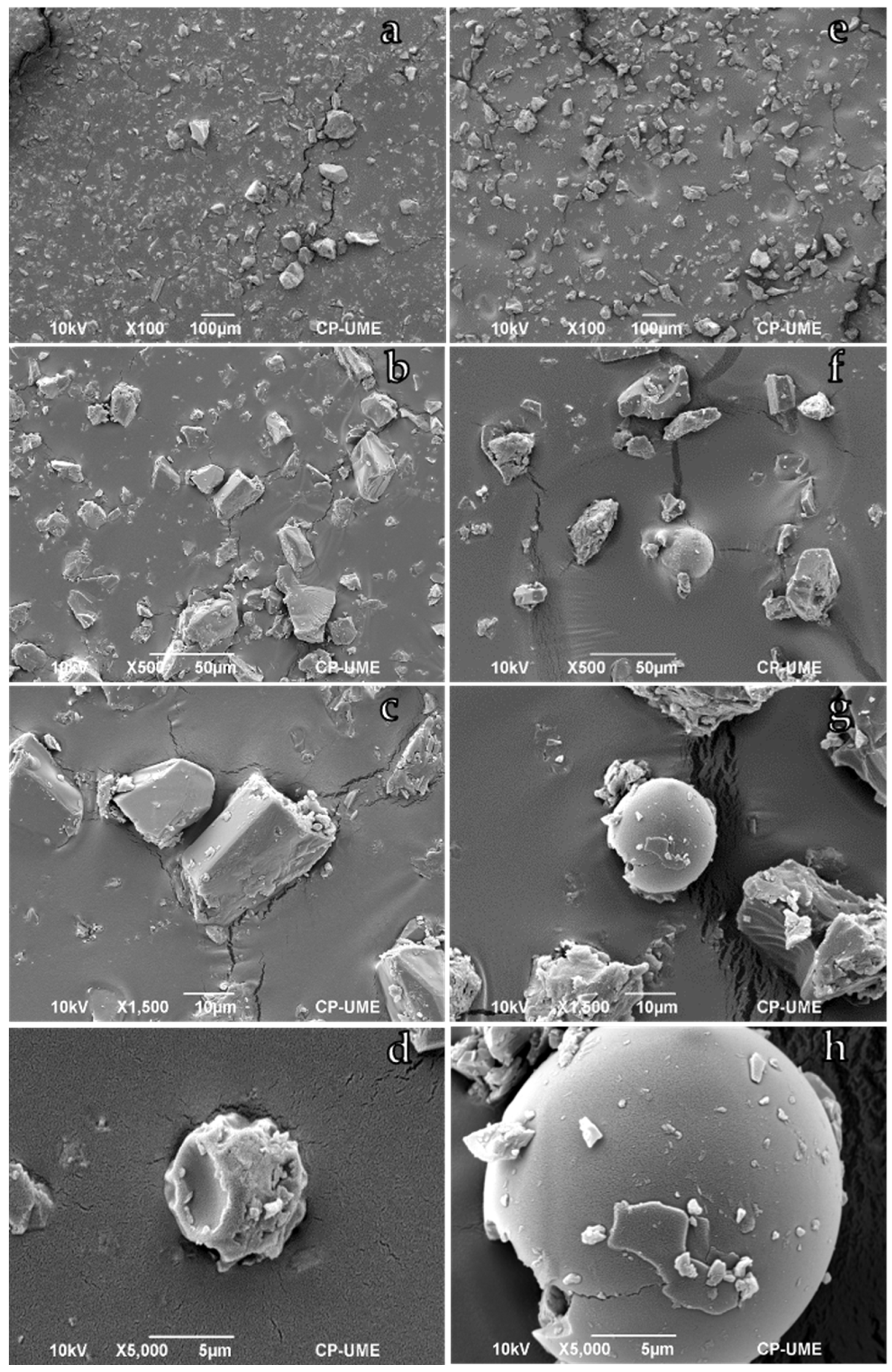
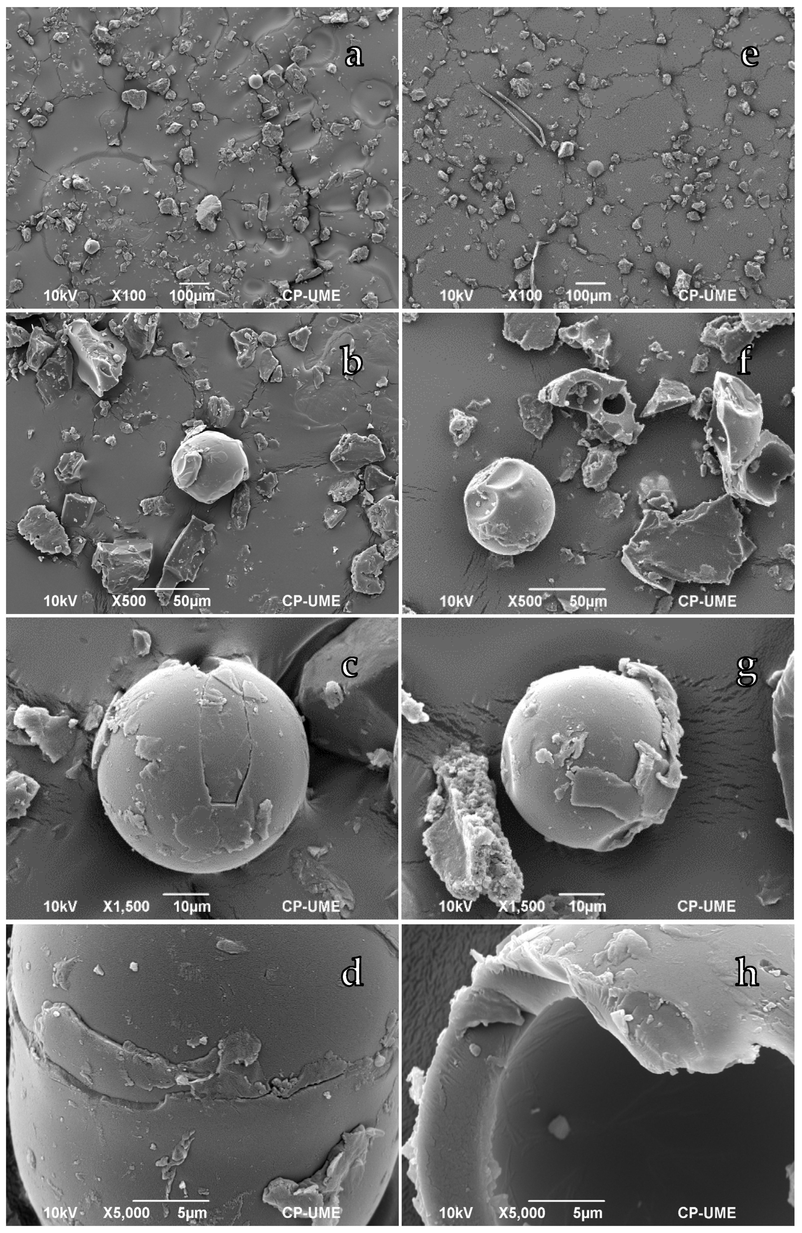
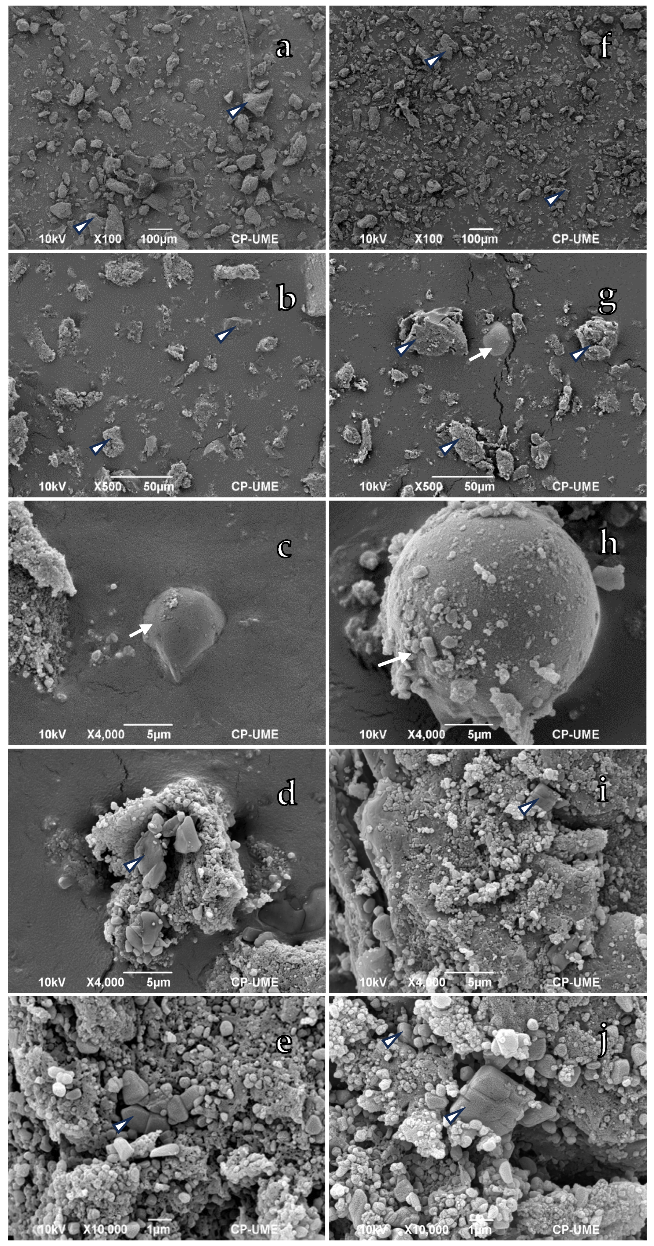
3.1. Morphological Characterization

Figure 1 and Figure 2 show the morphology and size of the encapsulated wax–arginine systems. Both microencapsulated systems had regular and irregular polyhedral morphologies (crystals) and spheres. As shown in Figure 3, the diameters of the spherical shapes increased significantly as the wax concentration increased. The wax–arginine formulations with 3:1 and 4:1 ratios had more microspheres than the wax–arginine formulations with 1:1 and 2:1 ratios, although they were larger and had fewer irregular and cracked polyhedral microcrystals. Conversely, the microcrystals in the 1:1 and 1:2 wax–arginine formulations were regular polyhedrons with smoother, flatter surfaces, while the microspheres had smooth surfaces, though some were cracked due to the crushing process. In any case, the micrographs show crystals coated with carnauba wax regardless of their size, like those obtained by other researchers who encapsulated crystalline amino acids (methionine) using carnauba wax without obtaining microspheres [18]. Carvalho et al. [21] also obtained irregular, filled, sealed, non-porous, and smooth microencapsulated systems whose surfaces resemble beeswax-like micrographs, indicating that urea was encapsulated within the microparticle; however, they also observed unencapsulated, exposed urea. In our study, the larger the microcapsule, the thicker the shell (Figure 1h and Figure 2h). Although crystalline arginine microencapsulation is a promising technique for weight gain in animals, the chemical compatibility of the active ingredient (arginine) with the conditions of the process and the chemistry of the shell restrict its efficiency. Another limitation is the exclusive use of foodstuffs. As a coating material, wax produces large particles without controlling the thickness of the coating wall [22]; microparticles with a heterogeneous size distribution (from 210 to more than 500 μm) have been generated with flow membranes or water-in-oil-in-water emulsions, allowing for better size control [23,24]. However, this applies to hydrophilic components, so the encapsulation of arginine in the present study requires further research to increase the number of microspheres, considering the bipolar nature of arginine. In arginine, the basic side chain is very polar (positively charged), and in a basic medium, during emulsion via melting with carnauba wax, only the carboxyl group is completely dissociated, increasing its solubility (hydrophilic). Similar results were observed by Milanovic et al. [25], who noted that ethyl vanilla encapsulated in carnauba wax produced microcapsules with a regular, spherical shape, smooth surface, and a diameter 300 μm above the sizes obtained in the present research. On the other hand [18], encapsulated crystalline methionine in carnauba wax that produced encapsulated systems like ours, showing only crystal-like structures with smooth, non-porous surfaces, without achieving the formation of microspheres. Although carnauba wax can protect amino acids due to its hydrophobic nature, the encapsulation process is challenging by itself in terms of the encapsulation materials and the standardization of the process [18,26]. Carvalho et al. [21] also obtained heterogeneous and amorphous systems using urea as a nitrogen source in the diet of sheep. Spray-drying is the most commonly used technique for microencapsulation. In research by Niu et al. [27], the use of gelatin and sodium alginate (in a 1:1 ratio) for the encapsulation of methionine resulted in a high yield and efficiency of microencapsulation (spherical shapes); the methionine was released in a controlled way under in vitro conditions and stimulated the absorption of other amino acids.
The morphology and sizes of the Eudragit®–arginine microcapsules are shown in Figure 4 and Figure 5. In general, few microcapsules with spherical shapes are observed and the proportion of polyhedrons, both regular and irregular, which represent polymer-coated crystals, is larger. In the wax–arginine encapsulation system, there are more microspheres than in the Eudragit®–arginine system. In systems containing 1000 mg of Eudragit® RS:RL 40:60 and 500 mg (Figure 4e) and 1000 mg (Figure 4j) of arginine, the crystals had sizes of about 1 μm, including nanometer-size crystals (1.15–1.73 μm, Figure 6). However, the Eudragit® RS:RL 70:30 produced significantly larger crystals (up to 5793 μm, Figure 6) as the ratio of arginine increased (Figure 5e,j). This behavior might be associated with the coating polymers and functionality under the encapsulation conditions. Low quantities of polymer in relation to the material to be encapsulated produce microcapsules of poor quality which are irregular in shape and dented. Khamanga et al. [28] recommend a 4:1 (polymer–drug) ratio to produce uniform, discrete, and slightly porous microcapsules. Among other factors, the stirring rate during the encapsulation process must be adequate to cause high shear rates and the dispersion of droplets of a smaller size. In addition, dispersing agents help to decrease interfacial forces, thus preventing coalescence. Results similar to ours were reported by Romero-Pérez et al. [19], who encapsulated sodium selenite with Eudragit® and produced spherical nanoparticles which were highly variable in size. The present research produced lower percentages of microspheres than crystals, ranging in size between 463 and 560 nm (Figure 6), without statistical differences among treatments (p > 0.05). Different morphologies could be associated with the solubility of the polymer in the solvent and the volatility of the solvent. According to Romero-Perez et al. [19], the Eudragit® polymer is more soluble in acetone than in ethanol. Apart from the relevance of the effect of the Eudragit® RS/RL polymer ratio on the size of the crystals, Quinten et al. [29] demonstrated that Eudragit® RL releases the active ingredient faster so that the substitution of Eudragit® RL for RS results in a slower release. These researchers showed that the form of the ingredient to be encapsulated (metoprolol salts) significantly influences the releasing properties. Although the use of triethyl citrate as a plasticizer improved the process due to the formation of H-bonds between Eudragit® and the ingredient to be encapsulated, its use may be detrimental to long-term stability.
Acrylic polymers emulsified via the simple emulsification evaporation method are widely used as gastro-resistant coatings. Briefly, a polymer solution is emulsified in an aqueous phase; then, the solvent is evaporated to precipitate the polymer in the form of nanospheres [30]. The success of the encapsulation of food ingredients and their application in animal production depends on the materials of the wall and the solvents. Lira-Casas et al. [31] reported that the encapsulation of urea using calcium silicate, urea, and Eudragit RS100® as wall materials had an encapsulation efficiency of 69%, while the use of activated carbon as a silicate substitute increased this efficiency to 71%. The unprotected urea reached its maximum peak after 6 h during release, while the silicate or activated charcoal took more than 24 h. Romero-Pérez et al. [19] showed that using acetone and Eudragit RL:RS® in 30:70 ratios produces larger (44.8 nm) sodium selenite polymeric nanoparticles and greater selenium entrapment (26%) in comparison with the use of ethanol and Eudragit RL:RS® in 40:60 ratios; the nanoparticles were spherical, amorphous, and homogeneous in size. At a pH below 4.0, the release of selenium was greater, with better availability in the small intestine.

3.2. Compositional Characterization

The results of the microelement analysis (nitrogen, N) for the encapsulated systems are presented in Table 1. In the wax–arginine formulations, the qualitative nitrogen (N) content decreased as the wax ratio increased, even though the amount of arginine was the same. As part of the analysis technique, an electron beam bombarded the microspheres. Consequently, this behavior may suggest that the N was not encapsulated because increasing the amount of carnauba wax diluted the presence of N in the system due to its nonpolar properties or that N was efficiently encapsulated but with a wall of carnauba wax thick enough to be detected. The second hypothesis might be more reliable because the micrographs (Figure 2h) show a thick wall of carnauba wax. The EDS technique quantifies qualitative nitrogen in a certain area of the surface of the capsules and/or crystals, meaning that the result, in the form of a percentage by weight, indicates a relative concentration of this element because the weight of the sample is not available [32]. On the other hand, the detection limit of minor elements is between 0.1 and 1% by weight of the minimum detectable mass fraction, so the EDS technique may present a limitation when the element of interest is present in an amount too low to be detected. Figure 7a shows that nitrogen (referring to arginine) is present inside and outside the microcapsules in the encapsulated wax–arginine system. Figure 7b corroborates the thick shell in the microcapsule and a greater presence of N inside. The microelement analysis also showed C, N, O, and Cl, all components of the carnauba wax and arginine, as other authors have shown [33,34]. As expected, the nitrogen content of the Eudragit formulation containing 1000 mg of arginine was higher than in the formulations containing 500 mg of arginine (Table 1. The mappings show that N was successfully encapsulated in the regular-shaped polyhedral crystals (Figure 7c), with N present inside and outside the microspheres (Figure 7d). With the use of other techniques such as that of Kjeldahl, Carvalho et al. [21] reported N content values of 14.6% and 14.8% in urea–beeswax capsules in a 1:2 ratio with sulfur and a 1:2 ratio without sulfur, respectively, values below those reported in the present research. The qualitative EDS technique could overestimate the N content, so results should be interpreted cautiously as they reveal a heterogeneous distribution of nitrogen in the sample. The data from the EDS analysis are complementary because factors such as characteristic X-rays, the probability of cross-section interaction, the volume of interaction, fluorescence, and absorption can impair the quality of the analysis. Carvalho Neto et al. [18] encapsulated methionine in carnauba wax in ratios of 1:2 and 1:4 and reported N content values of 3.09 and 1.82%, respectively. Similar results were reported by Medeiros et al. [26] for the encapsulation of urea in carnauba wax in ratios of 1:2, 1:3, and 1:4 (N content values of 14.8%, 11.1%, and 8.63%, respectively).

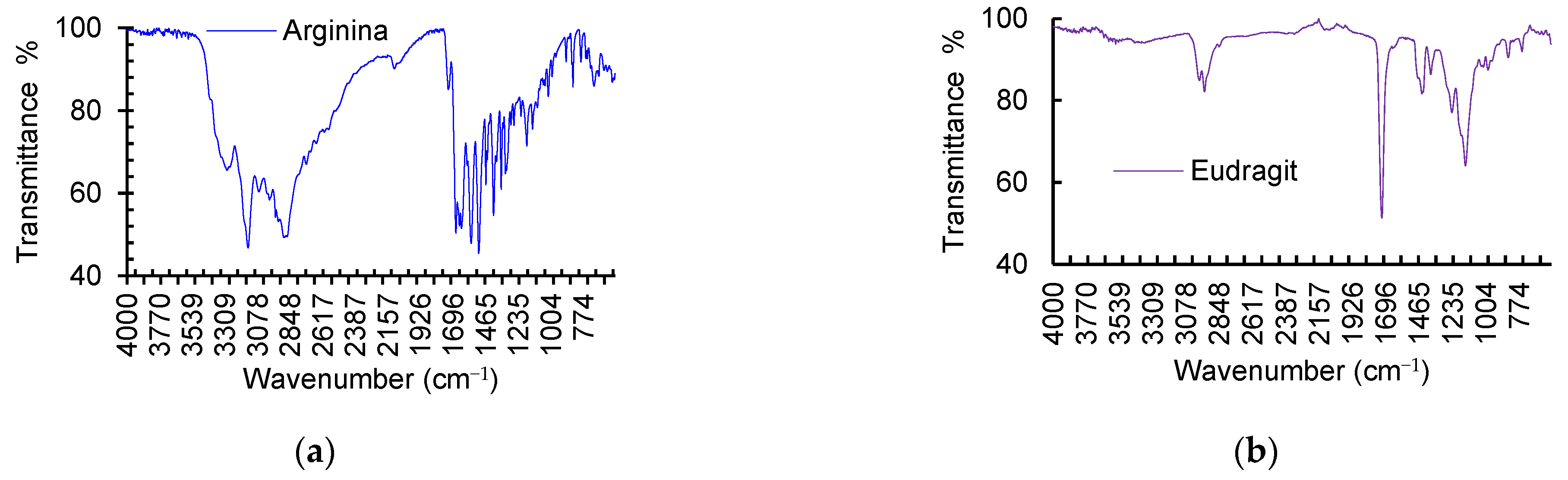
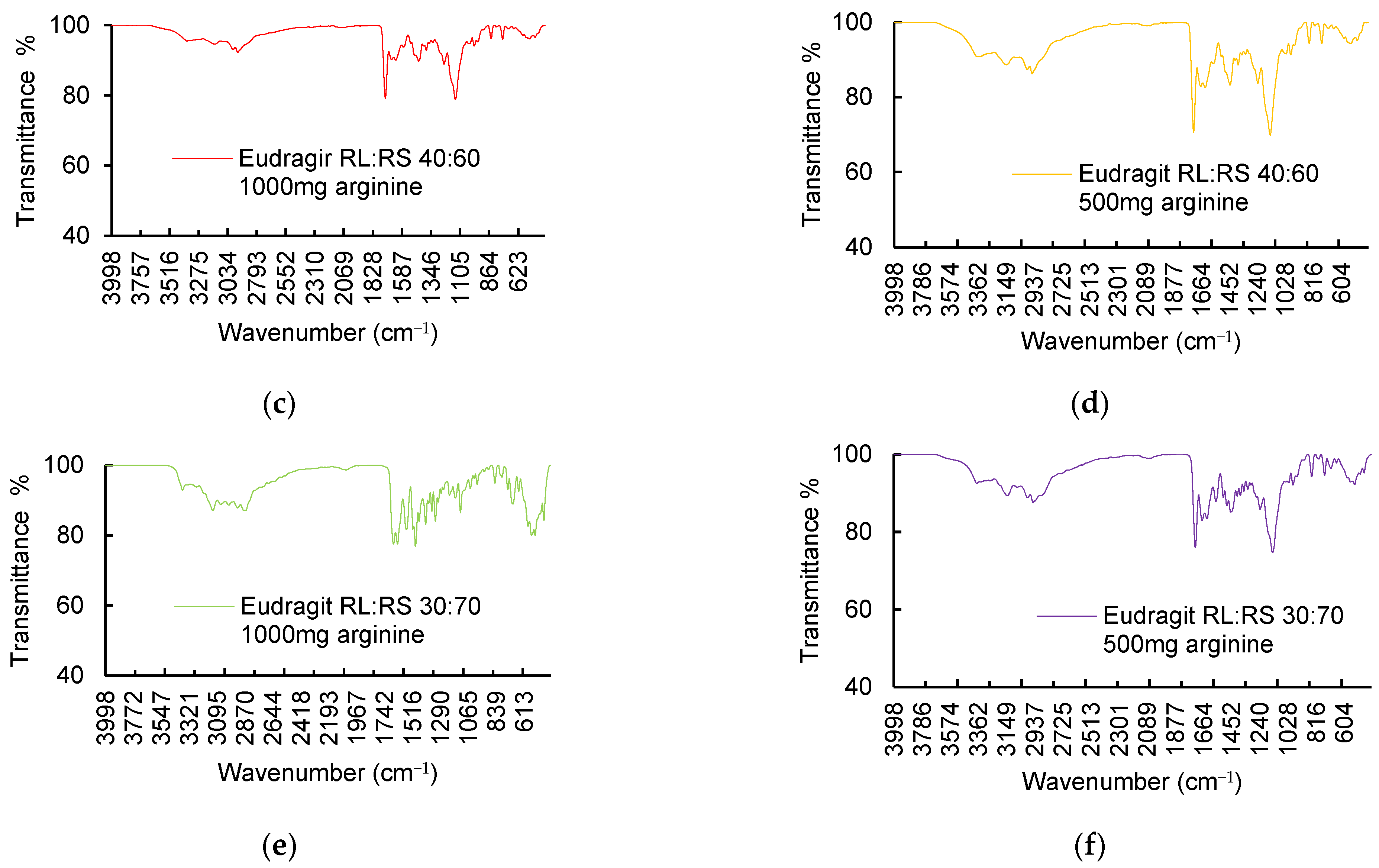
3.3. Chemical–Structural Characterization

Figure 8 shows the main absorption bands for the encapsulated wax–arginine and the Eudragit®–arginine systems. The main absorption bands of arginine are between 2600 and 3500 and 1634 and 1674 cm−1. In this case, the bands at 3238 cm−1, 3097 cm−1, and 2879 cm−1 are associated with the absorption of N-H, O-H, and C-H bonds, respectively, characteristics of the amino and carboxylic acid functional groups in amino acids. Arginine is a basic (positive) polar amino acid, so the guanidine side chain has N-H bonds. The wide shape of the band (between 2600 and 3500 cm−1) in the arginine spectrum corroborates the characteristic O-H bonds of the carboxyl groups of arginine. Absorption bands at 1634 cm−1, 1639 cm−1, and 1674 cm−1 are associated with the carbon oxygen bonds of the carbonyl group in the arginine, while those at 1518 cm−1 and 1568 cm−1 might be associated with C-C bonds. In this case, extremely polar bonds produce stronger bands, so the band for the carbonyl group is intense and narrow. The range between 600 cm−1 and 1400 cm−1 corresponds to the fingerprint region, a complex area with many bands overlapping and associated with the C-C, C-O, and C-N bonds. Carnauba wax displays two absorption regions; the one between 2700 and 3500 has strong absorption bands at 2913 cm−1 and 2846 cm−1, characteristic of C-H stretching vibrations in alkanes. Carnauba wax contains between 1 and 3% hydrocarbons, so the alkane functional group is associated with its composition. The absorption at 1734 cm−1 seems to be associated with the C=O bond, which is a functional group in fatty acid esters, the main components of carnauba wax. The bands at 1470 cm−1 and 1473 cm−1 are also associated with C-H flexions. The infrared spectra of the wax–arginine microcapsules (1:1 and 2:1 ratios) show strong attenuation in the absorption bands of arginine, suggesting that it is encapsulated and coated by the carnauba wax. Conversely, the 3:1 and 4:1 ratios of carnauba wax–arginine produced infrared spectra with mildly attenuated absorption bands. In this case, due to the high proportion of wax, less intense absorption bands for arginine would be expected. This behavior suggests less efficiency in arginine encapsulation. The results obtained in the microelement analysis corroborate this behavior (Table 1). However, in the spectrum of the 3:1 wax–arginine system, more intense bands can be seen at 1738 cm−1 and 1468 cm−1, associated with the C=O and C-H groups in carnauba wax, so arginine was more protected compared to the other systems. Similar results were obtained by Guimarães-Inácio et al. [35], who encapsulated chia oil in carnauba wax. Their infrared analyses also reported bands of the -CH groups characteristic of the composition of carnauba wax, observing that the characteristic bands of chia oil were attenuated in the microparticles loaded with the oil; this trend demonstrated that the oil was within the microparticles. Similar results were also obtained by Carvalho et al. [21], who developed two formulations for the microencapsulation of urea in beeswax using 1:2 ratios (w/w) between the core (urea) and the encapsulant (beeswax), with or without a sulfur source (MgSO4·7H2O). The absorption bands for urea were between 3428 cm−1 and 3330 cm−1 and were attributed to the vibration of the N-H bond, while for beeswax, the bands appeared between 2915 cm−1 and 2847 cm−1 due to the stretching of the C-H bond and at 1736 cm−1 for C-O. In the encapsulated system, the bands between 3429 cm−1 and 3332 cm−1 showed a shift with respect to the bands in urea; the lower region indicated the interaction of the N-H group with the H group as urea tends to form H-bridge interactions. Because of this low level of reactivity, the wax is suitable for encapsulating urea. Results similar to ours were also obtained by Huo et al. [36], who observed the same bands by encapsulating paraffin wax with a low melting point in a urea–formaldehyde resin to develop a novel microencapsulated phase-change material (Micro-P6). Absorption bands at 3360 cm−1 and 1660 cm−1 are produced by the N-H and C=O bonds in the pre-polymer, and the absorption bands at 2924 cm−1 and 2854 cm−1 are produced by the C-H bond in the paraffin wax. Therefore, both materials are present in the microencapsulation, and results indicated that the chemical structure of the encapsulation complies with the core–shell structure.
Eudragit®’s absorption spectrum reveals absorption bands at 2948 cm−1 and 2984 cm−1 which are associated with its C-H bonds, as well as at 1721 cm−1 and 1443 cm−1, which are associated with the carbonyl group C=O belonging to the carboxylic acids (Figure 9). As an acrylic polymer, Eudragit® contains these functional groups in its composition. The absorption spectra of the Eudragit® encapsulations demonstrated a similar behavior to the carnauba–arginine wax system. In this case, the most attenuated absorption bands were those of the system containing Eudragit® RL:RS 40:60 and 1000 mg of arginine, suggesting that arginine was efficiently protected by the polymer, as shown in Figure 9c. The system containing Eudragit® RL:RS 30:70 and 1000 mg of arginine had the lowest encapsulation efficiency, and the bands between 1634 cm−1 and 1674 cm−1 were associated with the C=O group of arginine, which was more exposed than in the other systems. Thus, for the system containing Eudragit® RL:RS 40:60 and 500 mg of arginine and the system containing Eudragit® RL:RS 30:70 and 500 mg of arginine, there was a medium encapsulation efficiency since the signals associated with the presence of the polymer were stronger than for the system containing Eudragit® RL:RS 40:60 and 1000 mg of arginine, but those associated with arginine were not sufficiently attenuated, indicating that part of the arginine was exposed. In this case, a tiny portion of the arginine was exposed and unencapsulated. Figure 7d corroborates the presence of N outside the system. Similar results were reported by Kashif et al. [37], who encapsulated ropirinol in Eudragit® RS 100. These researchers also reported absorption bands at 1725 cm−1 and 1145 cm−1 for the polymer and concluded that there was no interaction between ropirinol and Eudragit® in the microparticle formulations. Murillo-Creames et al. [38] also reported C=O vibrations at 1725 cm−1, as well as characteristic bands of asymmetrical and symmetrical alkyl stretching modes at 2990 and 2950 cm−1, respectively. However, the presence of the encapsulated drug was difficult to assess in the coated matrices due to its low percentage in the samples.

3.4. In Vitro Digestibility

The wax–arginine formulations and pure arginine were used as controls for assessing in vitro digestibility due to their higher encapsulation efficiency (the formation of microspheres). The 3:1 wax–arginine formulation showed significantly less degradability (21.3%) after 5 h of ruminal fluid incubation (Table 2), whereas unprotected arginine (crystalline arginine) was fully degraded (100%). These results confirmed the protection efficiency of the carnauba wax and the controlled release of the encapsulated systems. The other systems also had low percentages of degradability (close to 40%), so they protected the amino acid efficiently. The efficiency of the 3:1 wax–arginine system seems to be associated with the amount of wax (a 3:1 arginine–wax ratio) and its greater protection capacity, as indicated by the signal intensities in the infrared spectra, so that the system that provided the least protection was the 1:1 wax–arginine system (Table 2) as there was more degradability during exposure to ruminal fluid. On the other hand, the 3:1 wax–arginine system had fused spherical shapes (bi-spheres, Figure 2d) which could favor protection and prolonged release during in vitro digestion. Similar results were described by Carvalho et al. [21], who evidenced the controlled-release of urea encapsulated in beeswax in ratios of 1:2; digestibility was increased with the inclusion of microencapsulated urea without affecting N intake and balance. In another study, Carvalho Neto et al. [18] designed two methionine-encapsulated systems with carnauba wax–methionine ratios of 2:1 and 4:1, showing ruminal degradation of the systems by 17% and 16.3%, respectively, compared to pure unencapsulated methionine, which was degraded completely. These results are similar, showing greater efficiency and amino acid protection capacity in systems with a higher wax content. In animal nutrition, the encapsulation process protects amino acids from attack by ruminal microbes, regulating their transit throughout the gastrointestinal tract until their release in the small intestine [14]. Although not all encapsulation techniques are suitable, spray-drying, emulsion, and the coacervation method may modulate release in ruminants. The protection of food ingredients (e.g., arginine, methionine, lysine, urea, salts such as nitrates, probiotics such as Lactobacillus plantarum, eugenol-based essential oils, thymol, and vanillin) from rumen degradation has led to changes in animal metabolism related to feed intake and methane emissions [12,39]. The quality of animal products has also improved. Supplementation with protected urea increases fat and protein content in milk [13], while structural changes related to the length of the sarcomere have been evidenced in meat [40].

4. Conclusions

Arginine can be efficiently encapsulated by melt emulsification, producing spherical shapes with thick walls and regular polyhedral crystals. However, the encapsulation of crystalline amino acids requires further research considering the nature of the biomolecules involved. The present study produced arginine microspheres, whereas other studies only reported the production of crystals when crystalline amino acids were encapsulated. While the wax–arginine ratio significantly influences the shape, size, composition, and structure of the microcapsules and microcrystals, in general, carnauba wax as a wall material ensures arginine protection and slow release by simulating ruminal conditions in vitro. Attenuation in the absorption bands of the encapsulated ingredient and microelement analysis and mapping ensure encapsulation efficiency. The use of commercial polymers such as Eudragit® allows for the development of encapsulation systems using the nanoprecipitation technique, guaranteeing the production of crystals up to nanometric sizes. Subsequent studies should evaluate stability under ruminal conditions in vitro and in vivo.

Author Contributions

Conceptualization, L.M.C.-L. and G.C.-L.; methodology, G.C.-L., S.M.-R., A.R.V.-N., A.C.-L. and L.M.C.-L.; formal analysis, G.C.-L. and L.M.C.-L.; investigation, G.C.-L. and L.M.C.-L.; resources, A.R.V.-N., S.M.-R., A.C.-L., I.A.G.-G. and L.M.C.-L.; data curation, G.C.-L. and L.M.C.-L.; writing—original draft preparation, L.M.C.-L.; writing—review and editing, L.M.C.-L. and A.L.R.-M.; supervision, L.M.C.-L.; project administration, L.M.C.-L. All authors have read and agreed to the published version of the manuscript.

Funding

This research received no external funding.

Data Availability Statement

The data presented in this study are available on request from the corresponding author. The data are not publicly available due to privacy.

Conflicts of Interest

The authors declare no conflicts of interest.

References

  1. Kamalak, A.; Canbolat, Ö.; Gürbüz, Y.; Özay, O. Protected Protein and Amino Acids in Ruminant Nutrition. KMU J. Sci. Eng. 2005, 8, 84–88. [Google Scholar]
  2. Morris, S.M. Recent advances in arginine metabolism: Roles and regulation of the arginases. Br. J. Pharmacol. 2009, 157, 922–930. [Google Scholar] [CrossRef] [PubMed]
  3. The National Academies of Sciences, Engineering and Medicine. Nutrient Requirements of Dairy Cattle, 8th ed.; National Academic Press: Washington, DC, USA, 2001. [Google Scholar]
  4. Wang, W.W.; Qiao, S.Y.; Li, D.F. Amino acids and gut function. Amino Acids 2009, 37, 105–110. [Google Scholar] [CrossRef] [PubMed]
  5. Li, Y.; Lyu, Z.; Li, Z.; Liu, L.; Wang, F.; Li, D.; Lai, C. Effects of feeding level and dietary supplementation with crystalline amino acids on digestible, metabolizable and net energy values of corn in growing pigs. Anim. Feed Sci. Technol. 2018, 240, 197–205. [Google Scholar] [CrossRef]
  6. Pennings, B.; Boirie, Y.; Senden, J.M.G.; Gijsen, A.P.; Kuipers, H.; Van Loon, L.J.C. Whey protein stimulates postprandial muscle protein accretion more effectively than do casein and casein hydrolysate in older men. Am. J. Clin. Nutr. 2011, 93, 997–1005. [Google Scholar] [CrossRef] [PubMed]
  7. Wu, G. Amino acids: Metabolism, functions, and nutrition. Amino Acids 2009, 37, 1–17. [Google Scholar] [CrossRef] [PubMed]
  8. Gloaguen, M.; Le Floc’h, N.; Corrent, E.; Primot, Y.; van Milgen, J. Providing a diet deficient in valine but with excess leucine results in a rapid decrease in feed intake and modifies the postprandial plasma amino acid and α-keto acid concentrations in pigs. J. Anim. Sci. 2012, 90, 3135–3142. [Google Scholar] [CrossRef] [PubMed]
  9. Todd, R.D. Microencapsulation and the flavour industry. Flavour Ind. 1970, 1, 768–771. [Google Scholar]
  10. Piva, A.; Pizzamiglio, V.; Morlacchini, M.; Tedeschi, M.; Piva, G. Lipid microencapsulation allows slow release of organic acids and natural identical flavors along the swine intestine. J. Anim. Sci. 2007, 85, 486–493. [Google Scholar] [CrossRef]
  11. El-Zaiat, H.M.; Araujo, R.C.; Louvandini, H.; Patiño, H.O.; Abdalla, A.L. Effects of dietary replacement of urea with encapsulated nitrate and cashew nut shell liquid on nutrient digestibility, nitrogen balance, and carcass characteristics in growing lambs. Anim. Feed Sci. Technol. 2020, 266, 114515. [Google Scholar] [CrossRef]
  12. Alemu, A.W.; Romero-Pérez, A.; Araujo, R.C.; Beauchemin, K.A. Effect of encapsulated nitrate and microencapsulated blend of essential oils on growth performance and methane emissions from beef steers fed backgrounding diets. Animals 2019, 9, 21. [Google Scholar] [CrossRef] [PubMed]
  13. Highstreet, A.; Robinson, P.H.; Robison, J.; Garrett, J.G. Response of Holstein cows to replacing urea with with a slowly rumen released urea in a diet high in soluble crude protein. Livest. Sci. 2010, 129, 179–185. [Google Scholar] [CrossRef]
  14. Contreras-López, G.; Carrillo-López, L.M.; Vargas-Bello-Pérez, E.; García-Galicia, I.A. Microencapsulation of Feed Additives with Potential in Livestock and Poultry Production: A Systematic Review. Chil. J. Agric. Anim. Sci. 2024, 40, 229–249. [Google Scholar] [CrossRef]
  15. Meyer, A.M.; Klein, S.I.; Kapphahn, M.; Dhuyvetter, D.V.; Musser, R.E.; Caton, J.S. Effects of rumen-protected arginine supplementation and arginine-HCl injection on site and extent of digestion and small intestinal amino acid disappearance in forage-fed steers. Transí. Anim. Sci. 2018, 2, 205–215. [Google Scholar] [CrossRef]
  16. Teixeira, P.D.; Tekippe, J.A.; Rodrigues, L.M.; Ladeira, M.M.H.; Pukrop, J.R.; Brad Kim, Y.H.; Schoonmaker, J.P. Effect of ruminally protected arginine and lysine supplementation on serum amino acids, performance, and carcass traits of feedlot steers. J. Anim. Sci. 2019, 97, 3511–3522. [Google Scholar] [CrossRef] [PubMed]
  17. Baggerman, J.O.; Thompson, A.J.; Jennings, M.A.; Hergenreder, J.E.; Rounds, W.; Smith, Z.K.; Johnson, B.J. Effects of encapsulated methionine on skeletal muscle growth and development and subsequent feedlot performance and carcass characteristics in beef steers. Animals 2021, 11, 1627. [Google Scholar] [CrossRef] [PubMed]
  18. Carvalho Neto, J.P.; Bezerra, L.R.; da Silva, A.L.; de Moura, J.F.P.; Pereira Filho, J.M.; da Silva Filho, E.C.; Guedes, A.F.; Araújo, M.J.; Edvan, R.L.; Oliveira, R.L. Methionine microencapsulated with a carnauba (Copernicia prunifera) wax matrix for protection from degradation in the rumen. Livest. Sci. 2019, 228, 53–60. [Google Scholar] [CrossRef]
  19. Romero-Pérez, A.; García-García, E.; Zavaleta-Mancera, A.; Ramírez-Bribiesca, J.E.; Revilla-Vázquez, A.; Hernández-Calva, L.M.; López-Arellano, R.; Cruz-Monterrosa, R.G. Designing and evaluation of sodium selenite nanoparticles in vitro to improve selenium absorption in ruminants. Vet. Res. Commun. 2010, 34, 71–79. [Google Scholar] [CrossRef] [PubMed]
  20. Valentine, M.E.; Karayilanli, E.; Cherney, J.H.; Cherney, D.J. Comparison of In Vitro Long Digestion Methods and Digestion Rates for Diverse Forages. Crop Sci. 2019, 59, 422–435. [Google Scholar] [CrossRef]
  21. Carvalho, A.D.; da Silva, A.L.; Silva, A.D.; Netto, A.J.; de Medeiros, T.T.; Araújo Filho, J.M.; Agostini, D.D.; de Oliveira, D.L.; Mazzetto, S.E.; Kotzebue, L.R.; et al. Effect of slow-release urea microencapsulated in beeswax and its inclusion in ruminant diets. Small Rumin. Res. 2019, 179, 56–63. [Google Scholar] [CrossRef]
  22. Mellema, M.; van Benthum, W.A.J.; Boer, B.; von Harras, J.; Visser, A. Wax encapsulation of water-soluble compounds for application in foods. J. Microencapsul. 2006, 23, 729–740. [Google Scholar] [CrossRef] [PubMed]
  23. Williams, R.; Yuan, Q.; Collins, S. Membrane Emulsification. In Encyclopedia of Surface and Colloid Science, 3rd ed.; Somasundaran, P., Ed.; Taylor & Francis: London, UK, 2010; Volume 1, pp. 1–27. [Google Scholar]
  24. Collins, S.; York, D.W.; Kazmi, S.; Kaleem Mohammed, A. Formation of wax walled microcapsules via double emulsion using cross membrane emulsification at elevated temperatures. J. Food Eng. 2020, 269, 109739. [Google Scholar] [CrossRef]
  25. Milanovic, J.; Manojlovic, V.; Levic, S.; Rajic, N.; Nedovic, V.; Bugarski, B. Microencapsulation of flavors in carnauba wax. Sensors 2010, 10, 901–912. [Google Scholar] [CrossRef] [PubMed]
  26. Medeiros, T.T.B.; de Azevedo Silva, A.M.; da Silva, A.L.; Bezerra, L.R.; da Silva Agostini, D.L.; de Oliveira, D.L.V.; Mazzetto, S.E.; Kotzebue, L.R.V.; Oliveira, J.R.; Souto, G.S.B.; et al. Carnauba wax as a wall material for urea microencapsulation. J. Sci. Food Agric. 2019, 99, 1078–1087. [Google Scholar] [CrossRef] [PubMed]
  27. Niu, H.X.; Chang, J.; Jia, Y.D. Microencapsulation of crystalline-methionine enclosed with gelatine and sodium alginate by spray-drying. Mater. Rese. Innov. 2015, 19, S5257–S5262. [Google Scholar] [CrossRef]
  28. Khamanga, S.M.; Parfitt, N.; Nyamuzhiwa, T.; Haidula, H.; Walker, R.B. The Evaluation of Eudragit Microcapsules Manufactured by Solvent Evaporation Using USP Apparatus 1. Dissolut. Technol. 2009, 16, 15–22. [Google Scholar] [CrossRef]
  29. Quinten, T.; Andrews, G.P.; De Beer, T.; Saerens, L.; Bouquet, W.; Jones, D.S.; Hornsby, P.; Remon, J.P.; Vervaet, C. Preparation and evaluation of sustained-release matrix tablets based on metoprolol and an acrylic carrier using injection moulding. AAPS PharmSciTech 2012, 13, 1197–1211. [Google Scholar] [CrossRef] [PubMed]
  30. Ghaderi, S.; Ghanbarzadeh, S.; Mohammadhassani, Z.; Hamishehkar, H. Formulation of gammaoryzanol-loaded nanoparticles for potential application in fortifying food products. Adv. Pharm. Bull. 2014, 4, 549–554. [Google Scholar] [CrossRef] [PubMed]
  31. Lira-Casas, R.; Ramirez-Bribiesca, J.E.; Zavaleta-Mancera, H.A.; Hidalgo-Moreno, C.; Cruz-Monterrosa, R.G.; Crosby-Galvan, M.M.; Mendez-Rojas, M.A.; Domínguez-Vara, I.A. Designing and evaluation of urea microcapsules in vitro to improve nitrogen slow-release availability in rumen. J. Sci. Food Agric. 2019, 99, 2541–2547. [Google Scholar] [CrossRef] [PubMed]
  32. Wolfgong, W.J. Chemical Analysis Techniques for Failure Analysis: Part 1, Common Instrumental Methods; Elsevier: Amsterdam, The Netherlands, 2016. [Google Scholar] [CrossRef]
  33. Singh, S.; Sharma, N.; Arora, S.; Singla, Y. An overview of multifaceted significance of eudragit polymers in drug delivery systems. Asian J. Pharm. Clin. Res. 2015, 8, 1–6. [Google Scholar]
  34. Freitas, C.A.S.; de Sousa, P.H.M.; Soares, D.J.; da Silva, J.Y.G.; Benjamin, S.R.; Guedes, M.I.F. Carnauba wax uses in food—A review. Food Chem. 2019, 291, 38–48. [Google Scholar] [CrossRef] [PubMed]
  35. Guimarães-Inácio, A.; Francisco, C.R.; Rojas, V.M.; de Souza Leone, R.; Valderrama, P.; Bona, E.; Leimann, F.V.; Tanamati, A.A.; Gonçalves, O.H. Evaluation of the oxidative stability of chia oil-loaded microparticles by thermal, spectroscopic and chemometric methods. LWT Food Sci. Technol. 2018, 87, 498–506. [Google Scholar] [CrossRef]
  36. Huo, J.H.; Peng, Z.G.; Feng, Q. Synthesis and properties of microencapsulated phase change material with a urea–formaldehyde resin shell and paraffin wax core. J. Appl. Polym. Sci. 2020, 137, 48578. [Google Scholar] [CrossRef]
  37. Kashif, P.M.; Madni, A.; Ashfaq, M.; Rehman, M.; Mahmood, M.A.; Khan, M.I.; Tahir, N. Development of Eudragit RS 100 Microparticles Loaded with Ropinirole: Optimization and In Vitro Evaluation Studies. AAPS PharmSciTech 2017, 18, 1810–1822. [Google Scholar] [CrossRef] [PubMed]
  38. Murillo-Cremaes, N.; Subra-Paternault, P.; Saurina, J.; Roig, A.; Domingo, C. Compressed antisolvent process for polymer coating of drug-loaded aerogel nanoparticles and study of the release behavior. Colloid. Polym. Sci. 2014, 292, 2475–2484. [Google Scholar] [CrossRef]
  39. Abdelbagi, M.; Ridwan, R.; Fidriyanto, R.; Rohmatussolihat, R.; Nahrowi, N.; Jayanegara, A. Effects of probiotics and encapsulated probiotics on enteric methane emission and nutrient digestibility in vitro. In Proceedings of the 3rd International Conference of Animal Science and Technology, Makassar, Indonesia, 3–4 November 2020; Volume 788. [Google Scholar] [CrossRef]
  40. Monteschio, J.O.; Vargas-Junior, F.M.; Almeida, F.L.; Pinto, L.A.; Kaneko, I.N.; Almeida, A.A.; Freitas, L.W.; Alves, S.P.; Bessa, R.J.; Prado, I.N. The effect of encapsulated active principles (eugenol, thymol and vanillin) and clove and rosemary essential oils on the structure, collagen content, chemical composition and fatty acid profile of Nellore heifers muscle. Meat Sci. 2019, 155, 27–35. [Google Scholar] [CrossRef]
Figure 1. Scanning electron micrographs showing size and morphology of arginine microcapsules and microcrystals produced via melt emulsification; (ad) arginine–wax ratio of 1:1; (eh) arginine–wax ratio of 2:1 (n = 9).
Processes 12 01498 g001
Figure 2. Scanning electron micrographs showing size and morphology of arginine microcapsules and microcrystals produced via melt emulsification; (ad) arginine–wax ratio of 3:1; (eh) arginine–wax ratio of 4:1 (n = 9).
Processes 12 01498 g002
Figure 3. Diameter of arginine microspheres synthesized by melt emulsification. Data are expressed as mean ± SD values. a,b different letters in columns within the same graph indicate differences among treatments (Tukey’s multi-rank tests, assuming a significant difference at p < 0.05, n = 15).
Processes 12 01498 g003
Figure 4. Scanning electron micrographs showing dimension and morphology of arginine microcapsules (white arrows) and micro and nanocrystals (white triangles) produced from nanoprecipitation Eudragit® RS:RL ratio 40:60; (ae) Eudragit® arginine ratio 1:0.5; (fj) Eudragit®–arginine ratio 1:1 (n = 9).
Processes 12 01498 g004
Figure 5. Scanning electron micrographs showing dimension and morphology of arginine microcapsules (white arrows) and micro and nanocrystals (white triangles) produced from nanoprecipitation Eudragit® ratio RS:RL70:30; (ae) Eudragit®–arginine ratio 1:0.5; (fj) Eudragit®–arginine ratio 1:1 (n = 9).
Processes 12 01498 g005
Figure 6. Diameter of spheres and length of regular faces in crystals synthesized by nanoprecipitation method (n = 15) from Eudragit® RS:RL and arginine. Different letters indicate statistical difference (Tukey’s means, p < 0.05) (n = 9).
Processes 12 01498 g006
Figure 7. Nitrogen mapping (red dots) in arginine encapsulation systems; (a) microcapsule wax–arginine ratio 1:1, (b) microcapsule wax–arginine ratio 4:1; (c) Eudragit RS:RL 30:70 crystals, 500 mg arginine; (d) Eudragit RS:RL 40:60 microcapsules, 1000 mg arginine.
Processes 12 01498 g007
Figure 8. Infrared spectra of arginine (a), carnauba wax (b), and microcapsules of carnauba wax–arginine in ratios of 1:1 (c), 2:1 (d), 3:1 (e), and 4:1 (f), synthesized by the melt-emulsification method.
Processes 12 01498 g008aProcesses 12 01498 g008b
Figure 9. Infrared spectra of arginine (a), Eudragit® (b), microcapsules of Eudragit® RL:RS 40:60 1000 + 1000 arginine (c), Eudragit® RL:RS 40:60 + 500 mg arginine (d), Eudragit® RL:RS 30:70 + 1000 mg arginine (e), and Eudragit® RL:RS 30:70 + 500 mg arginine (f) synthesized by the nanoprecipitation method.
Processes 12 01498 g009aProcesses 12 01498 g009b
Table 1. Quantification of nitrogen (N) in encapsulation systems.
Sample% Nitrogen
Eudragit RL:RS 30:70 and 500 mg of arginine11–16
Eudragit RL: 30:70 and 1000 mg of arginine14–18
Eudragit RL:RS 40:60 and 500 mg of arginine26–32
Eudragit RL:RS 40:60 and 1000 mg of arginine25–35
Wax–arginine 1:121–24
Wax–arginine 2:118–23
Wax–arginine 3:112–16
Wax–arginine 4:118–21
Table 2. In vitro digestibility of microcapsules of wax–arginine and arginine formulations.
Treatment1 h 3 h5 h
Wax–arginine 1:142.74 ± 1.42 Ac34.5 ± 0.11 Ac47.12 ± 0.2 Ab
Wax–arginine 2:122.27 ± 0.05 Bd25.86 ± 0.41 Bd40.7 ± 0.12 Ac
Wax–arginine 3:115.94 ± 0.79 Be11.81 ± 0.07 Be21.26 ± 1.75 Ad
Wax–arginine 4:146.14 ± 0.09 Ab41.8 ± 12.94 Ab41.77 ± 4.25 Ac
Arginine97.77 ± 0.17 Aa97.9 ± 0.52 Aa100 Aa
a,b,c,d,e different letters within the same column indicate significant differences between treatments (Tukey’s multiple range tests, assuming a significant difference at p < 0.05, n = 18). A,B different letters within the same row indicate significant differences between hours (Tukey’s multiple range tests, assuming a significant difference at p < 0.05, n = 9).
Disclaimer/Publisher’s Note: The statements, opinions and data contained in all publications are solely those of the individual author(s) and contributor(s) and not of MDPI and/or the editor(s). MDPI and/or the editor(s) disclaim responsibility for any injury to people or property resulting from any ideas, methods, instructions or products referred to in the content.

