Article

Optimization of Sample Preparation Procedure for Determination of Fat-Soluble Vitamins in Milk and Infant Food by HPLC Technique

1
Teaching Institute of Public Health “Dr. Andrija Štampar”, Mirogojska 16, HR-10000 Zagreb, Croatia
2
University of Applied Health Sciences, HR-10000 Zagreb, Croatia
3
Faculty of Health Studies, University of Rijeka, Viktora Cara Emina 5, HR-51000 Rijeka, Croatia
4
Faculty of Science, University of Zagreb, Horvatovac 102a, HR-10000 Zagreb, Croatia
5
State Inspector Office, Republic of Croatia, Ulica Pavla Šubića 29, HR-10000 Zagreb, Croatia
*
Author to whom correspondence should be addressed.
Processes 2024, 12(7), 1530; https://doi.org/10.3390/pr12071530 (registering DOI)
Submission received: 4 July 2024 / Revised: 16 July 2024 / Accepted: 18 July 2024 / Published: 20 July 2024
(This article belongs to the Special Issue Emerging Technologies in Sustainable Dairy Processing)

Abstract

:
Background: The analysis of vitamins in baby food is a challenging task given the complexity of the food matrix, vitamin stability, and strict regulations of the European Union regarding permissible deviations from declared values. Vitamins in food exist in different concentrations and forms and have different stabilities; thus, the preparation of samples for a reliable analysis using the same procedure is not straightforward. Therefore, significant attention has been devoted to optimizing sample preparation in the analysis of vitamins. Methods: This study aims to determine which of the sample preparation and extraction methods is the most efficient for the simultaneous determination of vitamins A, D, E, and K in milk and baby food using high-performance liquid chromatography (HPLC). Different samples of baby food were prepared in seven different ways based on four methods (saponification, enzymatic hydrolysis, solvent extraction, and solid-phase extraction). Results and Conclusion: According to the validation parameters, the optimal preparation method proved to be solid-phase extraction with a C18 stationary phase, with recoveries of 97.4%, 96.1%, 98.3%, and 96.2% for vitamins A, D, E, and K, respectively, and HPLC with a UV–Vis detector was identified as a sufficiently sensitive technique for the identification and quantification of fat-soluble vitamins in milk and baby food.

1. Introduction

Vitamins are essential compounds, crucial for the normal functioning of the human body, facilitating numerous enzymatic and metabolic functions. They are categorized into two groups based on their solubility: fat-soluble vitamin A, D, E, and K, and water-soluble vitamin C and B complexes [1,2,3,4]. Most vitamins cannot be synthesized by the body and must be obtained through dietary intake, with the exceptions being vitamins D and K. Despite being required in small quantities, their deficiency or excessive intake can lead to severe health problems, particularly in sensitive age groups. Therefore, the diet for infants and young children must be well-balanced, containing sufficient vitamins and other nutrients to ensure proper growth and development. Vitamins differ from other nutrients (lipids, carbohydrates, and proteins) as they do not have a structural role, nor do they produce energy. Instead, they participate in various metabolic processes as antioxidants, regulators, or coenzymes [5,6,7,8,9]. The solubility of vitamins is determined by their chemical composition. Water-soluble vitamins contain carbon, hydrogen, oxygen, nitrogen, sulfur, and cobalt (except for vitamin C), while fat-soluble vitamins contain only carbon, hydrogen, and oxygen atoms. Vitamins A and E are stable in both acidic and alkaline environments, vitamin D is unstable in acidic conditions but stable in alkaline surroundings, and vitamin K is unstable in both acidic and alkaline media [10,11]. Therefore, the analysis of vitamins in baby food poses a challenging and complex procedure due to the food’s content complexity, the chemical form of vitamins present, and their stability, together with the European Union regulations on allowable tolerances for the control of the compliance of nutrient values declared on a label. Until today, the most frequently used technique for the quantitative determination of vitamins in food samples is high-performance liquid chromatography (HPLC). However, due to variations in quantities and forms in which vitamins are present in food, the isolation and quantification of vitamins demand an analytical approach where sample preparation is the most crucial part of the procedure. Consequently, in recent years, significant attention has been directed towards optimizing sample preparation in vitamin analysis [12,13,14,15].
Vitamin A, also known as retinol, belongs to the retinoid group, compounds with a basic structure consisting of a trimethylated cyclohexane ring, polyene side chain, and a polar functional group at the terminal C atom. The terminal functional group can be hydroxyl (retinol), aldehyde (retinal), carboxyl (retinoic acid), or ester (retinyl ester). Retinol and its esters (acetate and palmitate) exhibit the most pronounced vitamin activity. Due to their predominantly hydrophobic nature, retinoids are insoluble in water but soluble in organic solvents such as fats, oils, hexane, diethyl ether, acetone, chloroform, ethanol, and methanol. The conjugated double bonds in the chain make retinoids unstable in the presence of light, oxidants, and heat, leading to oxidation or isomerization. The conjugated polyene chain is responsible for the strong absorption of UV–Vis emission (325–380 nm). From the mid-1970s to the present, the analytical methods for determining vitamin A in complex matrices such as blood serum and food constantly changed and were upgraded, mostly using HPLC with a UV–Vis detector or coupled LC-MS. Additionally, retinoids can be determined by spectrophotometric methods [16,17].
Vitamin D encompasses a group of secosteroids, a subclass of steroids where one bond in the B steroid ring is “broken”. There are seven types of vitamin D, with only two being biologically active: vitamin D2, or ergocalciferol, and vitamin D3, or cholecalciferol. Structurally, they differ only at two positions on the side chain; vitamin D2 has a double bond at the C-22 atom and an additional methyl group at the C-24 atom, which vitamin D3 lacks [18,19]. Vitamins D2 and D3 are insoluble in water but soluble in most organic solvents like ethanol, acetone, chloroform, etc. They are unstable in light and air, stable in basic conditions, and unstable in acidic conditions; in mildly acidic conditions, they isomerize to the 5,6-trans isomer of vitamin D and isotachysterol [18,19,20]. The most common technique for determining vitamins D2 and D3 is HPLC with a UV–Vis detector (λ = 265 nm).
Vitamin E refers to a group of tocopherols, compounds with a structure consisting of a chromanol ring attached to one to three methyl groups and an isoprenoid chain of sixteen carbon atoms. Based on the saturation of the hydrocarbon chain, tocopherols are divided into tocopherols (saturated bonds) and tocotrienols (unsaturated bonds). There are four tocopherols and four tocotrienols in nature, denoted as α, β, γ, and δ compounds, based on the number and position of methyl groups on the ring. α-tocopherol (5, 7, 8-trimethyl tocopherol) is the biologically most active form of vitamin E. Tocopherols and tocotrienols are soluble in organic solvents but insoluble in water. They are unstable in light and easily oxidize in the air, forming biologically inactive quinones. They remain stable at high temperatures and in acidic and basic conditions in the absence of oxygen and light. Similarly to vitamins A and D, the most common method for determining vitamin E is HPLC with a UV–Vis detector (λ = 292–296 nm), but sometimes, other methods such as spectrophotometry, Raman, IR spectroscopy, and radioimmunoassay are used [21,22,23,24].
Vitamin K designates a group of several vitamins with a basic structure consisting of a naphthoquinone ring substituted with a methyl group at the C-2 position and a saturated/unsaturated hydrocarbon chain at the C-3 position. There are two active forms of vitamin K in nature: phylloquinone, or vitamin K1, and menaquinones, or vitamin K2. Menaquinones are a group of structural analogs that can contain 6–10 unsaturated isoprenoid units. Phylloquinone is insoluble in water; slightly soluble in ethanol; and highly soluble in oils, fats, and other nonpolar solvents. Vitamin K compounds are highly unstable in basic and acidic media, breaking down under visible and ultraviolet light. However, they are stable at high temperatures, and unlike previous vitamins, they remain stable in the air. Since the 1980s, the primary method for determining vitamin K in food samples has been HPLC with a UV–Vis detector due to the naphthoquinone ring’s ability to absorb UV radiation between 240 and 280 nm and between 320 and 330 nm [25,26,27].
This study aimed to determine the optimal procedure for extracting samples of baby food for the simultaneous analysis of fat-soluble vitamins (A, D, E, and K) using high-performance liquid chromatography (HPLC). For this purpose, four types of baby food samples were selected based on their composition and consistency: infant milk, chocolate-flavored oatmeal (porridge), and dry and liquid porridge. The analysis was conducted using seven different methods, each based on four procedures: saponification, enzymatic hydrolysis, solvent extraction, and solid-phase extraction. The efficiencies of sample preparation were assessed based on the declared vitamin values on the product labels and by adding mixed standards of analyzed vitamins (spike) to each selected type of food. The obtained results were used to evaluate the extraction efficiency of vitamins, i.e., the effectiveness of the applied method.

2. Materials and Methods

For this study, commercial food samples intended for children were utilized, sourced from the market in the Zagreb, Republic of Croatia. Transitional infant milk, chocolate-flavored cereal (popular Čokolina), dry milk porridge (five grains with plum), and liquid milk porridge (with apple, pear, and wheat grits) were sampled for analysis.
All samples were prepared in triplicate, and the presented values in the results of this study represent the mean measurement.

2.1. Extraction Methods

Seven different extraction methods were applied to each sample to isolate vitamins A, D, E, and K (Table 1). It always involved the extraction of homogenized samples (2.0 ± 0.05 g) mixed with the solvent, and after extraction and purification, the samples were subjected to centrifugation for 15 min at 4600 rpm. After the centrifugation, 5 mL of the upper hexane layer was pipetted and evaporated to dryness under nitrogen at 50 °C, and the evaporated extract was dissolved in 500 µL of acetonitrile/methanol (75:25 v/v). The prepared sample was analyzed by HPLC with UV–Vis detection.

2.1.1. Solvent Extraction

In the first procedure, extraction of the vitamins was achieved with 10 mL ultra-pure water, 5 mL 4 N HCl, and 10 mL hexane containing 0.025% butylated hydroxytoluene (BHT).
In the second procedure, extraction was carried out with 10 mL ultra-pure water and 10 mL hexane.
The seventh procedure began with weighing the homogenized sample, to which 8 mL of ethanol was added, and the content was extracted for 30 min on a shaker. Then, like for all other methods, the sample preparation was followed with 10 mL of n-hexane solution with added 0.025% BHT, and the sample was further homogenized for 30 min.

2.1.2. Enzymatic Hydrolysis

In the third procedure, the samples were subjected to degradation with the enzyme lipase, followed by the addition of 5 mL of a phosphate buffer and incubation for 2 h at 38 degrees. The hydrolyzates were extracted with 2.5 mL of ethanol/methanol (95:5 v/v), with the addition of 0.25 g of potassium carbonate and 10 mL of hexane.

2.1.3. Solid-Phase Extraction

In the fourth procedure, after evaporation, a vacuum system was employed for solid-phase extraction with SPE HLB columns. The columns were conditioned with 1 mL of methanol and 1 mL of ultra-pure water, followed by the application of 4 mL of the sample (upper layer). The column was rinsed with 1.5 mL of 5% methanol and dried under vacuum. After drying, analyte elution (vitamins) was performed with 1 mL of isopropanol/acetonitrile (1:1) and then with 1 mL of 20% ethyl acetate in acetonitrile. The collected eluate was evaporated to dryness under nitrogen at 50 °C, dissolved in 500 µL of acetonitrile/methanol (75:25 v/v), mixed on a vibrational mixer for half a minute, filtered through a 0.45 µm pore size membrane filter, and further analyzed by HPLC/UV–Vis.
In the fifth procedure, after the initial sample preparation, SPE C18 columns were used. The columns were conditioned with 10 mL of methanol and 5 mL of ultra-pure water, and then, 5 mL of a homogenized and centrifuged sample (upper layer) was added. After passing the sample solution through the column, it was rinsed with 5 mL of 10% methanol solution, followed by vacuum drying of the column and the elution of analytes with 6 mL of methanol. The collected eluate was evaporated to dryness under nitrogen at 50 °C. Then, it was dissolved in 500 µL of acetonitrile/methanol (75:25 v/v), mixed on a vibrational mixer for half a minute, and filtered through a 0.45 µm pore size membrane filter into a sample vial. The prepared sample was analyzed by HPLC with UV–Vis detection.

2.1.4. Hot Saponification

In the sixth procedure, 0.5 g of citric acid monohydrate, 20 mL of ethanol, and 5 mL of 60% potassium hydroxide solution were added to the weighted sample. Homogenization lasted for 30 min, followed by heating in a water bath for 30 min at 70 °C. After the bath, the vial was cooled to room temperature, and extraction was performed with 10 mL of hexane/ethyl acetate (85:15 v/v) solution, followed by another 30 min of homogenization on a shaker.
All the samples were then analyzed using a high-performance liquid chromatograph with a UV–Vis detector, as described in Section 2.4.
Table 1. Sample preparation procedures PR.1–PR.7—brief conditions.
Procedure No.Sample Preparation
1Extraction with ultra-pure water, 4 N HCl, and hexane.
2Extraction with ultra-pure water and hexane.
3Sample digestion with lipase, extraction with ethanol/methanol (95:5 v/v), and addition of K2CO3 and hexane.
4Extraction with ethanol and ultra-pure water.
Extract purification by SPE HLB.
Elution with 20% ethyl acetate in acetonitrile.
5Extraction with ethanol and ultra-pure water.
Extract purification by SPE C18.
Elution with methanol.
6Hot saponification.
Extraction of unsaponifables with hexane/ethyl acetate (85:15 v/v).
7Extraction with ethanol and hexane.

2.2. Chemicals

n-Hexane, butylated hydroxytoluene (BHT), ethanol, isopropanol and nitrogen (all HPLC grad) were purchased from Merck (New Jersey, NJ, USA). Ethyl acetate, hydrochloric acid, acetonitrile, and methanol were obtained from Scharlau (Barcelona, Spain). Phosphate buffer, citric acid, and potassium carbonate were purchased from Alkaloid (Skopje, North Macedonia).

2.3. Equipment

An HPLC Agilent Series 1200 (Santa Clara, CA, USA) with a diode-array detector was used. Purification was performed on Strata C18-E SPE columns (500 mg, 3 mL) from Phenomenex (Torrance, CA, USA) and Oasis HLB SPE columns (500 mg, 3 mL) from Waters (Milford, MA, USA). The magnetic stirrer and vortex shaker were obtained from IKA (Staufen, Germany), and the centrifuge Hettich (Tuttlingen, Germany) and membrane filters (0.45 µm, 13 mm) were purchased from, USA Pall (New York, NY, USA).

2.4. Chromatographic Conditions

All the samples were analyzed using a high-performance liquid chromatograph with a UV–Vis detector, model Agilent 1200 Series. The column used was Phenomenex C18 with dimensions of 250 mm × 4.6 mm; 5 µm was eluted with a gradient mixture of acetonitrile and methanol. The injection volume was 20 µL; the flow rate was set at 1 mL/min; the column temperature was maintained at 45 °C; and the detection wavelength was set at 235, 265, and 325 nm. After HPLC analysis, the obtained chromatograms were analyzed using the ChemStation program (Figure 1, Figure 2).

2.5. Method Validation
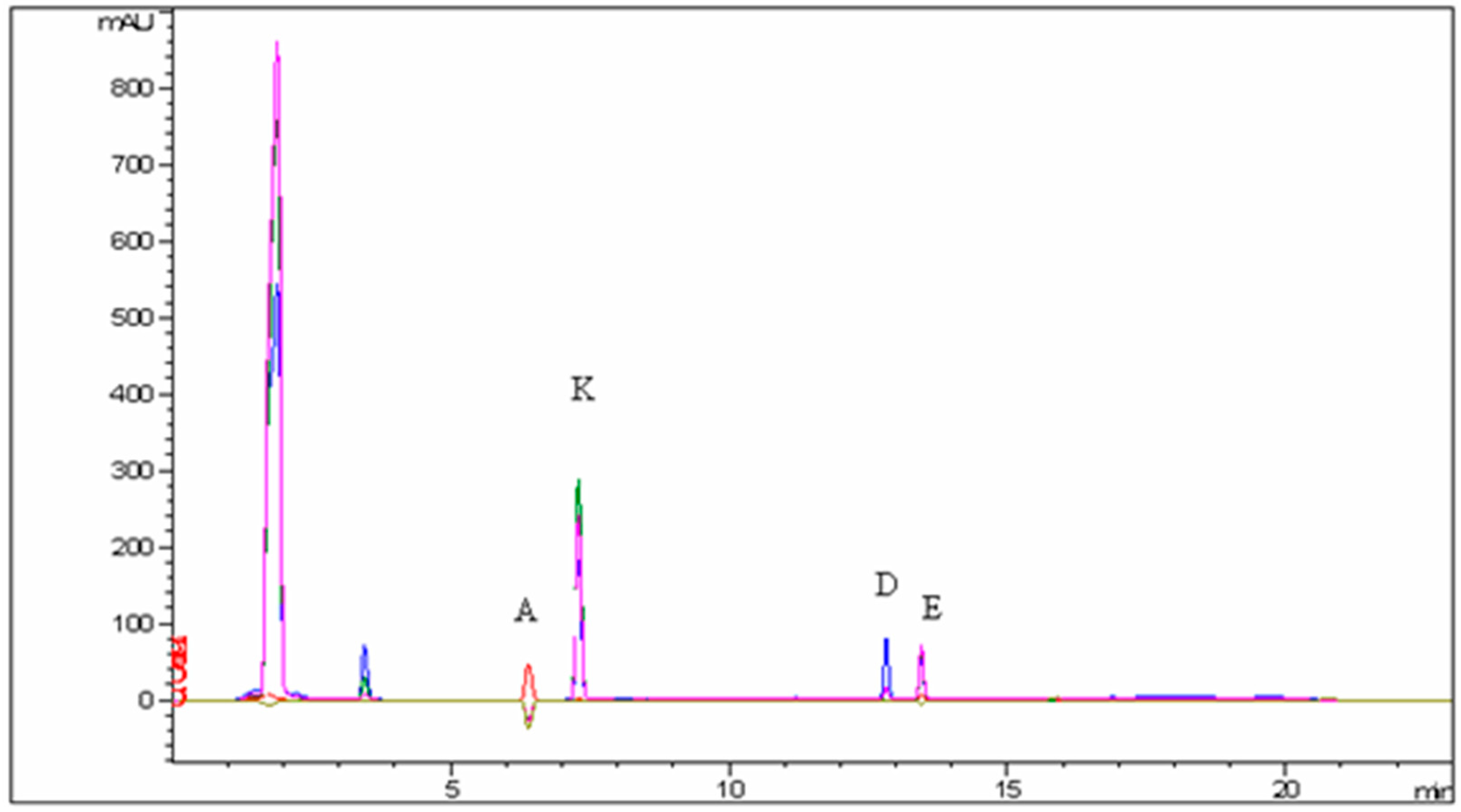
The calibration curve was constructed using the external standard method. The method underwent validation based on key parameters including specificity, linearity, precision, accuracy, and sensitivity. Method validation was performed according to the ICH guidelines. Examination of the placebo solution revealed the absence of interfering peaks at the retention time of the vitamins in the standard solution, affirming the method’s specificity. Linearity assessment involved employing linear regression to process the calibration plot, followed by determination of the linear equation and correlation coefficient. The R2 values demonstrate the robust correlation between the peak areas and analyzed concentration ranges. Calibration curves and linear equations were also utilized for LOD (limit of detection) and LOQ (limit of quantification) calculations. Method accuracy was assessed by computing the mean percentage recovery of individual standard solutions across three different concentration levels (50%, 100%, and 150%), each subjected to three injections. Repeatability and intermediate precision were evaluated through the determination of relative standard deviations. The results indicate that the developed methods exhibited precision within acceptable thresholds.

3. Results

The main objective of this study was to optimally prepare samples for the simultaneous quantification of fat-soluble vitamins in milk and infant food. The optimization of the extraction procedure started with the extraction of solvents (HCl, H2O, phosphate buffer, and EtOH), which were found to be ethanol and water (PR.5). The PR.2 procedure provided unsuitable results considering that the extraction solution used was only ultra-pure water.
All the results obtained in this research are presented in seven tables, including statistical processing.
Table 2 illustrates the efficiency of the most efficient extraction procedure for each vitamin using the HPLC technique, while Table 3 presents the results of sample preparation efficiency concerning the type vitamin. Table 4 presents the results of a statistical analysis using the Kruskal–Wallis test.
Depending on the preparation method, the obtained values of the vitamin mass fractions in the analyzed samples are presented in Table 5, Table 6, Table 7 and Table 8. The mass fractions of added vitamins in all samples comply with the information in the nutritional table, which is an integral part of the product declaration. Vitamins that were not added (i.e., vitamin K) to the products are marked with ND (not declared).
Method accuracy was evaluated by determining the recovery of spiked analytes to the matrix of the sample. Considering the obtained results, the efficiency of sample preparation for the determination of all four vitamins was ranked. For the analysis of vitamins A, D, E, and K in the examined baby food, preparation number 5 (PR.5) proved to be the most effective, followed by preparation number 3 (PR.3) for the analysis of vitamins A, E, and K. As for the analysis of vitamin D, preparation number 7 (PR.7) emerged as the second most effective. The preparation ranked third in efficiency for both vitamin A and vitamin E is preparation number 6 (PR.6), while for vitamin D, preparation number 4 (PR.4) is third in effectiveness, and for vitamin K, preparation number 2 (PR.2) ranks third. Preparation number 1 (PR.1), in terms of efficiency, is fourth for both vitamin A and K, while for vitamin D, preparation number 3 (PR.3) ranks fourth, and for vitamin E, preparation number 7 (PR.3) is fourth.
The best extraction procedures in terms of method recoveries were achieved with the addition of 5.0 mL ethanol and 5 mL ultra-pure water into a centrifuge tube and the addition of 0.025% (m/v) solution of BHT before further sonication (30 min). After centrifugation for 10 min at 25 °C and 5000 rpm, the samples were purified on C18 SPE columns as the best purification selection because of its properties for the extraction of hydrophobic or polar organic analytes from aqueous matrices.
The data were analyzed using the non-parametric Kruskal–Wallis test for each matrix and each vitamin. A result is statistically significant at p < 0.01 with the use of a pairwise comparison with Bonferroni correction. The obtained statistical results indicate that the values obtained for all analyzed vitamins concerning the sample preparation method for milk samples are statistically significant. Similarly, the statistical significance of the results for all analyzed vitamins was determined in the preparation of both dry and liquid porridge samples. In the preparation of chocolate porridge samples, statistically significant values were found related only to the analysis of vitamin K, while for other vitamins, no statistically significant difference was found, considering the sample preparation method.
Table 9 provides a summary of the extraction procedures, depending on their specific conditions.

4. Discussion

A well-balanced and high-quality diet is crucial for the seamless growth and development of every individual, particularly during the growth and development of children. Fat-soluble vitamins are responsible for enzymatic and metabolic functions in the body. Imbalances in the levels of specific vitamins can lead to health issues, especially in children [28]. Some studies have indicated that children’s food rarely contains the declared quantity of vitamin D, and there are instances of hypervitaminosis D in children who consume children’s food, with vitamin D levels exceeding the recommended daily dose for their age group [29]. The highest permitted levels of vitamins in products ready for use as placed on the market, or products prepared according to the manufacturer’s instructions, are prescribed in some national regulations related to processed cereal-based food and baby food for infants and young children [30], with permissible deviations from declared values, including those recommended by the European Union regulation, set at −35% (lower tolerance limit) and +50% (upper tolerance limit) [31].
This research aimed to determine the optimal sample preparation method for milk and baby food for the routine determination of fat-soluble vitamins. This study included seven different extraction procedures for vitamins A, D, E, and K from milk samples and three baby food samples. From the literature, it is evident that various extraction methods are used for extracting vitamins from the analyzed sample group. Solvent extraction proved to be very efficient, although some authors note the need to improve the accuracy and precision of the method in the validation process [32].
Italian researchers explored using supercritical carbon dioxide (SC-CO2) to extract fat-soluble vitamins (A, D, E, and β-carotene) as a substitute for conventional liquid extraction methods, which typically require large volumes of organic solvents. Supercritical fluid extraction (SFE) is a swift method for extracting these vitamins, enabling accurate determination with only small amounts of organic solvents. That study performed extractions on ultra-high temperature sterilized milk, milk powder, pork, liver pâté, infant formula, and canned baby food to compare this new technique with traditional methods. The innovative approach utilizes SC-CO2 with methanol as a modifier for extracting fat-soluble vitamins and their esters. A vitamin analysis was carried out using high-performance liquid chromatography (HPLC) with photometric detection. The findings revealed no significant differences between the methods, suggesting that the SFE method is an effective alternative to traditional solvent-based techniques, particularly for vitamin A and γ-tocopherol [32].
Solvent extraction was used for the determination of vitamin D3 in milk samples [33]. The results were satisfactory, with a recovery of 94%, which could be compared with our vitamin D results according to PR.7 as the second-best purification method for liquid milk porridge.
High recoveries for the determination of tocopherols in cereals when purified on C18 SPE sorbent have been reported, which would correspond to our results. As can be seen from our results, the saponification method did not prove to be relevant for the determination of vitamin K due to its unstable nature in alkaline conditions, as stated by several authors [34,35].
New methodologies based on supercritical fluid extraction (SFE) have been developed for determining fat-soluble vitamins in processed foods. Initial results indicate that SFE is highly suitable for extracting fat-soluble vitamins from food products, although further validation work is needed to establish accuracy and precision. Vitamins A, and E, and beta-carotene were investigated, and the processed food samples included ultra-high temperature (UHT) milk, milk powder, ground meat, liver paste, infant food, canned baby food, and margarine. The extraction equipment utilized analyte collection on solid-phase traps or in solvents. Following extraction, samples were saponified, and vitamins were determined by reverse-phase liquid chromatography with ultraviolet or fluorescent detection. The sample throughput was at least 12 samples per day, i.e., at least twice the number achievable with conventional extraction methodologies. The detection limits for vitamins in various processed foods were significantly below 0.1 micrograms per gram. Recoveries (compared to vitamin levels obtained by conventional solvent extraction) were close to 100% for experienced personnel using modern automated equipment. To achieve this level, vitamins needed protection by antioxidants during various analysis steps; the addition of methanol or ethanol to the extraction cell to facilitate analyte extraction from the food matrix; and when using solid-phase trapping, the utilization of a fractionated extraction-elution procedure to prevent breakthrough losses. The developed methods were subjected to validation exercises among five laboratories involved in method development and intercomparison among ten laboratories, including those with less experience in vitamin determination. The within-laboratory relative standard deviation (RSD) was generally < or = 11%. The average inter-laboratory RSD was around 23% in validation, increasing to about 40% in intercomparison. Robustness testing conducted at various stages of the project showed that different types and models of equipment did not yield significant differences in recoveries. Hence, the increasing RSD can largely be attributed to differences in participants’ experience in vitamin analysis [36]. Enzymatic hydrolysis (PR.3) has been proven to be a relevant method for the determination of vitamins E and A in powdered milk and infant formula, with recoveries ranging from 96 to 112%, which can be compared with our results as another suitable preparation method for milk and liquid milk porridge [37].
Solid-phase extraction, enzymatic hydrolysis, saponification, and alkaline hydrolysis have demonstrated effectiveness based on the product type [38]. To develop and confirm methods for the simultaneous detection of fat-soluble vitamins A, D3, and E in rice-based infant food, researchers employed two liquid chromatography systems: one combined with atmospheric pressure chemical ionization and tandem mass spectrometry (UHPLC-APCI-MS/MS), and the other with high-performance liquid chromatography and a diode array detector (HPLC-DAD). Separation was performed on C18 columns using a methanol/acetonitrile mixture as the mobile phase. The extraction of fat-soluble vitamins included enzymatic hydrolysis with α-amylase, saponification, extraction with petroleum ether or n-hexane, and purification using a silica gel column specifically for vitamin D3. Vitamins D3 and E were quantified with internal standards (IS) D3-d3 and E-d6, while vitamin A was quantified without an IS. The methods were refined and validated for linearity, precision, accuracy, detection limits, and quantification limits. Recovery rates ranged from 85.0 to 107% for retinol, from 92.0 to 105% for α-tocopherol, and from 95.2 to 106% for cholecalciferol, with %RSD (relative standard deviation) values between 6.4% and 15%. Evaluation included an uncertainty assessment, application to commercial samples, and proficiency testing [38].
Enzymatic hydrolysis catalyzed by lipase was effective in breaking down esters of fatty acids and esters of vitamins A and E. In previous studies, this preparation method partially converted retinyl palmitate and α-tocopheryl acetate into alcoholic forms, while vitamins D and K remained unchanged. Saponification is commonly used to release bound or esterified forms of vitamins A, E, and D, and carotenoids. It is important to adjust saponification conditions to achieve optimal extraction and minimal degradation losses [39]. The saponification process is often used in the preparation of samples with complex matrices, such as baby food. Alkaline hydrolysis breaks ester bonds of interfering substances in fatty foods, such as triglycerides, phospholipids, and sterols, and releases vitamins from the lipoprotein complex. Vitamins A and E in food are mostly found in the form of esters, such as retinyl acetate, retinyl palmitate, and α-tocopheryl acetate, and through saponification, they convert into their alcoholic forms: retinol and α-tocopherol. Vitamin D remains unchanged, while vitamin K is not stable in an alkaline medium, and the saponification process is not suitable for its analysis [40].
When comparing the analytical procedures employed by other researchers with those described in this study, it is evident that other investigations have also utilized high-performance liquid chromatography (HPLC) as the fundamental analytical technique for quantifying vitamin content in food [32,36,37,38]. The differences primarily arise in the sample preparation methods, including the use of specific extraction columns and extraction solvents. The application of C18 solid-phase extraction (SPE) columns along with ethanol, ultra-pure water, and butylated hydroxytoluene (BHT) solution in this study yielded favorable results for all analyzed vitamins and matrices (PR.5). The recoveries ranged from 96.1% for vitamin D and 96.2% for vitamin K to 97.4% for vitamin A and 98.3% for vitamin E, with limits of detection (LODs) for all vitamins at 0.010 µg/mL and limits of quantification (LOQs) at 0.100 µg/mL.
Research indicating recoveries, such as 64% for vitamin A in milk and 103% in pâté [36], highlights the necessity for further method development, as noted by the authors, since the employed method is suitable only for certain vitamins in specific food categories. When comparing the results of this study with those obtained in a study focused on the analysis of fat-soluble vitamins in rice cereal-based infant food [38], the extraction efficiency for fat-soluble vitamins largely aligns with the efficiencies observed in the current research. Extraction efficiencies for the analyzed vitamins ranged from 85% to 107%, depending on the vitamin. The extraction process employed a C18 column, using a methanol/acetonitrile mixture as the extraction solvent, and utilized both the HPLC and UHPLC APC-MS/MS techniques for the individual determination of vitamin D and the simultaneous determination of vitamins A, D, and E.
It is evident that, despite using different extraction solvents for fat-soluble vitamins, the results obtained in this study regarding the extraction efficiency of analyzed vitamins in certain food categories can be considered acceptable. Additionally, although some studies [32,36,39] prefer supercritical fluid extraction methods aiming to increase the extraction efficiency of vitamins from food while reducing the use of organic solvents, the extraction method used in this study can also be considered environmentally acceptable given the use of ultra-pure water and BHT as extraction solvents.
Although research highlights the advantages and disadvantages of the methods used, considering the extraction solvents, food categories, and vitamins extracted and quantified using liquid chromatography-based analytical techniques [32,36,37,38,39,40], similar conclusions are indicated by this study. The authors suggest further research involving a broader range of food categories to refine the method shown to be most efficient in this study, utilizing LC-MS/MS techniques alongside HPLC for vitamin quantification.

5. Conclusions

In this study, it was experimentally demonstrated that sample preparation based on reverse-phase extraction, specifically solid-phase extraction (SPE), for preparing baby food samples for the analysis of fat-soluble vitamins proved to be the most efficient. Interfering substances were rapidly and easily separated from the analyte using suitable eluting solvents. All other sample preparation methods yielded inferior results and were less efficient. Based on the obtained results, it can be concluded that for the simultaneous extraction of vitamins A, D, E, and K from milk and baby food, it is advisable to use the ethanol extraction method and SPE C18 column purification followed by dissolution in acetonitrile and methanol.
The limitations of this study are related to the type of samples analyzed, namely milk and baby food. Future plans would include further development of the extraction procedures described in PR.5 and introducing new matrices that would include other food categories, both those artificially enriched with vitamins and those in which vitamins are found naturally.
It is necessary to continue applying the most efficient extraction method for fat-soluble vitamins across all food categories, especially infant and toddler food and foods with higher fat contents. Vitamin quantification should also be conducted using HPLC-MS/MS techniques as a confirmatory method for HPLC results and as a potential technique for determining low levels of vitamins in food.

Author Contributions

Conceptualization, J.B., D.L. and A.R.; methodology, J.B., M.B. (Martina Bevardi), I.H., M.B. (Maja Budeč) and I.J.C.; software, G.J.; validation, G.J., D.L. and M.B. (Martina Bevardi); formal analysis, J.B., M.B. (Martina Bevardi), I.H., M.B. (Maja Budeč) and I.J.C.; investigation R.K., D.L. and A.R.; resources, D.B. and D.L.; writing—original draft preparation, J.B., M.B. (Martina Bevardi), I.H., M.B. (Maja Budeč) and I.J.C.; writing—review and editing, D.L. and A.R.; visualization, M.B. (Maja Budeč), I.J.C., R.K. and G.J.; supervision, J.B., M.B. (Martina Bevardi) and G.J.; project administration R.K. and M.B. (Maja Budeč), funding acquisition, J.B. and A.R. All authors have read and agreed to the published version of the manuscript.

Funding

This research received no external funding.

Data Availability Statement

From the first Author at the Teaching Institute of Public Health A.Štampar, Zagreb.

Acknowledgments

This work was carried out within the project “Food Safety and Quality Center” (KK.01.1.1.02.0004).

Conflicts of Interest

The authors declare no conflicts of interest.

References

  1. Padh, H. Vitamins for Optimal Health. In Functional Foods; Goldberg, I., Ed.; Springer: Boston, MA, USA, 1994; p. 13. [Google Scholar] [CrossRef]
  2. Bender, D.A. Nutritional Biochemistry of the Vitamins, 2nd ed.; Cambridge University Press: Cambridge, UK, 2003. [Google Scholar]
  3. Singh, P.; Kesharwani, R.K.; Keservani, R.K. Antioxidants and Vitamins: Roles in Cellular Function and Metabolism. In Sustained Energy for Enhanced Human Functions and Activity; Academic Press: Cambridge, MA, USA, 2017; pp. 385–407. [Google Scholar] [CrossRef]
  4. Rafeeq, H.; Ahmad, S.; Tareen, M.B.K.; Shahzad, K.A.; Bashir, A.; Jabeen, R.; Tariq, S.; Shehzadi, I. Biochemistry of Fat Soluble Vitamins, Sources, Biochemical Functions and Toxicity. Haya Saudi J. Life Sci. 2020, 5, 188–196. [Google Scholar] [CrossRef]
  5. Głąbska, D.; Kołota, A.; Lachowicz, K.; Skolmowska, D.; Stachoń, M.; Guzek, D. The Influence of Vitamin D Intake and Status on Mental Health in Children: A Systematic Review. Nutrients 2021, 13, 952. [Google Scholar] [CrossRef] [PubMed]
  6. Allen, L.H. Causes of Nutrition-related Public Health Problems of Preschool Children: Available Diet. J. Pediatr. Gastroenterol. Nutr. 2006, 43 (Suppl. S3), S8–S12. [Google Scholar] [CrossRef] [PubMed]
  7. Awasthi, S.; Awasthi, A. Role of vitamin A in child health and nutrition. Clin. Epidemiol. Glob. Health 2020, 8, 1039–1042. [Google Scholar] [CrossRef]
  8. Torquato, P.; Marinelli, R.; Bartolini, D.; Galli, F. Vitamin E: Nutritional aspects. In Molecular Nutrition–Vitamins; Academic Press: Cambridge, MA, USA, 2020; pp. 447–485. [Google Scholar] [CrossRef]
  9. Kozioł-Kozakowska, A.; Maresz, K. The Impact of Vitamin K2 (Menaquinones) in Children’s Health and Diseases: A Review of the Literature. Children 2022, 9, 78. [Google Scholar] [CrossRef] [PubMed]
  10. Alturfan, A.A. A Guide to Vitamins and Their Effects on Diseases; Chapter 4—Water-soluble vitamins; Cambridge Scholars Publishing: Newcastle upon Tyne, UK, 2023. [Google Scholar]
  11. Ofoedu, C.E.; Iwouno, J.O.; Ofoedu, E.O.; Ogueke, C.C.; Igwe, V.S.; Agunwah, I.M.; Ofoedum, A.F.; Chacha, J.S.; Muobike, O.P.; Agunbiade, A.O.; et al. Revisiting food-sourced vitamins for consumer diet and health needs: A perspective review. PeerJ 2021, 9, e11940. [Google Scholar] [CrossRef] [PubMed]
  12. Adamec, J.; Jannasch, A.; Huang, J.; Hohman, E.; Fleet, J.C.; Peacock, M.; Ferruzzi, M.G.; Martin, B.; Weaver, C.M. Development and optimization of an LC-MS/MS-based method for simultaneous quantification of vitamin D2, vitamin D3, 25-hydroxyvitamin D2 and 25-hydroxyvitamin D3. J. Sep. Sci. 2011, 34, 2011. [Google Scholar] [CrossRef] [PubMed]
  13. Ertugrul, S.; Yucel, C.; Sertoglu, E.; Ozkan, Y.; Ozgurtas, T. Development and optimization of simultaneous determination of fat-soluble vitamins by liquid chromatography-tandem mass spectrometry. Chem. Phys. Lipids 2020, 230, 104932. [Google Scholar] [CrossRef] [PubMed]
  14. Ryynänen, M.; Lampi, A.-M.; Salo-Väänänen, P.; Ollilainen, V.; Piironen, V. A small-scale sample preparation method with HPLC analysis for determination of tocopherols and tocotrienols in cereals. J. Food Compos. Anal. 2004, 17, 749–765. [Google Scholar] [CrossRef]
  15. Katsa, M.; Proestos, C.; Komaitis, E. Determination of Fat Soluble Vitamins A and E in Infant Formulas by HPLC-DAD. Curr. Res. Nutr. Food Sci. J. 2016, 4, 92–96. [Google Scholar] [CrossRef]
  16. Álvarez, R.; Vaz, B.; Gronemeyer, H.; de Lera, Á.R. Functions, Therapeutic Applications, and Synthesis of Retinoids and Carotenoids. Chem. Rev. 2014, 114, 1–125. [Google Scholar] [CrossRef]
  17. Das, B.C.; Thapa, P.; Karki, R.; Das, S.; Mahapatra, S.; Liu, T.C.; Torregroza, I.; Wallace, D.P.; Kambhampati, S.; Van Veldhuizen, P.; et al. Retinoic acid signaling pathways in development and diseases. Bioorg. Med. Chem. 2014, 22, 673–683. [Google Scholar] [CrossRef] [PubMed]
  18. Feldman, D.; Malloy, P.J.; Gross, C. Chapter 9—Vitamin D. In Osteoporosis, 2nd ed.; Academic Press: Cambridge, MA, USA, 2001; Volume 1, pp. 257–303. [Google Scholar]
  19. Dominguez, L.J.; Farruggia, M.; Veronese, N.; Barbagallo, M. Vitamin D Sources, Metabolism, and Deficiency: Available Compounds and Guidelines for Its Treatment. Metabolites 2021, 11, 255. [Google Scholar] [CrossRef] [PubMed]
  20. Masuno, H.; Yamamoto, K.; Wang, X.; Choi, M.; Ooizumi, H.; Shinki, T.; Yamada, S. Rational Design, Synthesis, and Biological Activity of Novel Conformationally Restricted Vitamin D Analogues. J. Med. Chem. 2002, 45, 1825–1834. [Google Scholar] [CrossRef] [PubMed]
  21. Nadkarni, S.; Chodynski, M.; Corcoran, A.; Marcinkowska, E.; Brown, G.; Kutner, A. Double Point Modified Analogs of Vitamin D as Potent Activators of Vitamin D Receptor. Curr. Pharm. Des. 2015, 21, 1741–1763. [Google Scholar] [CrossRef]
  22. Prieto, P.; Pineda, M.; Aguilar, M. Spectrophotometric Quantitation of Antioxidant Capacity through the Formation of a Phosphomolybdenum Complex: Specific Application to the Determination of Vitamin E. Anal. Biochem. 1999, 269, 337–341. [Google Scholar] [CrossRef] [PubMed]
  23. Moreno, P.; Salvadó, V. Determination of eight water- and fat-soluble vitamins in multi-vitamin pharmaceutical formulations by high-performance liquid chromatography. J. Chromatogr. A 2000, 870, 207–215. [Google Scholar] [CrossRef] [PubMed]
  24. Lee, B.L.; Chua, S.C.; Ong, H.Y.; Ong, C.N. High-performance liquid chromatographic method for routine determination of vitamins A and E and β-carotene in plasma. J. Chromatogr. B Biomed. Sci. Appl. 1992, 581, 41–47. [Google Scholar] [CrossRef]
  25. Rodas Mendoza, B.; Morera Pons, S.; Castellote Bargalló, A.I.; López-Sabater, M.C. Rapid determination by reversed-phase high-performance liquid chromatography of Vitamins A and E in infant formulas. J. Chromatogr. A 2003, 1018, 197–202. [Google Scholar] [CrossRef] [PubMed]
  26. Shearer, M.J.; Newman, P. Metabolism and cell biology of vitamin K. Thromb. Haemost. 2008, 100, 530–547. [Google Scholar] [CrossRef]
  27. Shearer, M.J.; Newman, P. Recent trends in the metabolism and cell biology of vitamin K with special reference to vitamin K cycling and MK-4 biosynthesis. J. Lipid Res. 2014, 55, 345–362. [Google Scholar] [CrossRef] [PubMed]
  28. Ravisankar, P.; Reddy, A.A.; Nagalakshmi, B.; Koushik, O.S.; Kumar, B.V.; Anvith, P.S. The Comprehensive Review on Fat Soluble Vitamins. IOSR J. Pharm. 2015, 5, 12–28. [Google Scholar]
  29. Nako, Y.; Tomomasa, T.; Morikawa, A. Risk of hypervitaminosis D from prolonged feeding of high vitamin D premature infant formula. Pediatr. Int. 2004, 46, 439–443. [Google Scholar] [CrossRef] [PubMed]
  30. Croatian National Ordinance on Processed Cereal-Based Food and Baby Food for Infants and Young Children-OG 126/2013 (Pravilnik o Prerađenoj Hrani na Bazi žitarica i dječjoj Hrani za Dojenčad i Malu Djecu-NN 126/2013). Available online: https://narodne-novine.nn.hr/clanci/sluzbeni/2013_10_126_2741.html (accessed on 4 July 2024).
  31. Guidance Document for Competent Authorities for the Control of Compliance with EU Legislation. Health and Consumers Directorate-General. 2012. Available online: https://food.ec.europa.eu/system/files/2016-10/labelling_nutrition-vitamins_minerals-guidance_tolerances_1212_en.pdf (accessed on 4 July 2024).
  32. Perretti, G.; Marconi, O.; Montanari, L.; Fantozzi, P. Fat-soluble vitamin extraction by analytical supercritical carbon dioxide. J. Amer. Oil Chem. Soc. 2003, 80, 629–633. [Google Scholar] [CrossRef]
  33. Sazali, N.H.; Alshishani, A.; Saad, B.; Chew, K.Y.; Chong, M.M.; Miskam, M. Salting-out assisted liquid–liquid extraction coupled with high-performance liquid chromatography for the determination of vitamin D3 in milk samples. R Soc. Open Sci. 2019, 6, 190952. [Google Scholar] [CrossRef] [PubMed]
  34. Yueqing, X.; Zhang, L.; Yang, R.; Yu, X.; Yu, L.; Ma, F.; Li, H.; Wang, X.; Li, P. Extraction and Determination of Vitamin K1in Foodsby Ultrasound-Assisted Extraction, SPE, and LC-MS/MS. Molecules 2020, 25, 839–851. [Google Scholar]
  35. Fanali, C.; D’Orazio, G.; Fanali, G.A. Advanced analytical techniques for fat-soluble vitamin analysis. Trends Anal. Chem. 2017, 87, 82–97. [Google Scholar] [CrossRef]
  36. Mathiasson, L.; Turner, C.; Berg, H.; Dahlberg, L.; Theobald, A.; Anklam, E.; Ginn, R.; Sharman, M.; Ulberth, F.; Gabernig, R. Development of methods for the determination of vitamins A, E and β-carotene in processed foods based on supercritical fluid extraction: A collaborative study. Food Addit. Contam. 2002, 19, 632–646. [Google Scholar] [CrossRef]
  37. Turner, C.; Persson, M.; Mathiasson, L.; Adlercreutz, P. Lipase-catalyzed reactions in organic and supercritical solvents:application to fat-soluble vitamin determination in milk powder and infant formula. Enzym. Microb. Technol. 2001, 29, 111–121. [Google Scholar] [CrossRef]
  38. Katsa, M.; Papalouka, N.; Mavrogianni, T.; Papagiannopoulou, I.; Kostakis, M.; Proestos, C.; Thomaidis, N.S. Comparative Study for the Determination of Fat-Soluble Vitamins in Rice Cereal Baby Foods Using HPLC-DAD and UHPLC-APCI-MS/MS. Foods 2021, 10, 648. [Google Scholar] [CrossRef]
  39. Turner, C.; King, J.W.; Mathiasson, L. Supercritical fluid extraction and chromatography for fat-soluble vitamin analysis. J. Chrmatogr. A 2001, 936, 215–237. [Google Scholar] [CrossRef] [PubMed]
  40. Blake, C.J. Status of Methodology for the Determination of Fat-Soluble Vitamins in Foods, Dietary Supplements, and Vitamin Premixes. J. AOAC Int. 2007, 90, 897–910. [Google Scholar] [PubMed]
Figure 1. Chromatogram of the RP-HPLC/DAD analysis of the standard mixture of four evaluated fat-soluble vitamins at four wavelengths; A—vitamin A, K—vitamin K, D—vitamin D, and E—vitamin E.
Processes 12 01530 g001
Figure 2. Chromatogram of the RP-HPLC/DAD analysis of the food sample and four evaluated fat-soluble vitamins at four wavelengths; A—vitamin A, K—vitamin K, D—vitamin D, and E—vitamin E.
Processes 12 01530 g002
Table 2. Range of validation parameters for the optimal procedure (PR.5).
VitaminRange (mg/mL)Recovery (%)Repeatability (%)Intermediate Precision (%)R2LOD (µg/mL)LOQ (µg/mL)
A0.072–28.90097.4 1.521 2.896 0.9990.0100.100
D0.048–19.20096.1 1.325 3.250 0.9990.0100.100
E0.013–27.00098.3 1.021 2.128 1.0000.0100.100
K0.013–25.21396.2 1.102 3.012 0.9990.0100.100
Table 3. Sample preparation efficiency related to vitamin type.
VitaminOrder of Sample Preparation Efficiency
APR.5—PR.3—PR.6—PR.1—PR.7—PR.4—PR.2
DPR.5—PR.7—PR.4—PR.3—PR.6—PR.1—PR.2
EPR.5—PR.3—PR.6—PR.7—PR.4—PR.1—PR.2
KPR.5—PR.3—PR.2—PR.1—PR.4—PR.6—PR.7
PR = preparation.
Table 4. Results of the statistical analysis using the Kruskal–Wallis test.
Test StatisticMean Rank
NKruskal–Wallis HdfAsymp. Sig.P1P2P3P4P5P6
MilkVitamin A9083.5365<0.0138.938.0064.4023.0082.6056.07
Vitamin D9085.0315<0.0152.208.0068.0337.1782.9724.63
Vitamin E9084.1755<0.0138.008.0074.1723.0076.8353.00
Vitamin K9084.4635<0.0138.0053.0072.5323.0078.478.00
CokolinoVitamin A908.79550.11857.0330.2045.6350.4044.2045.53
Vitamin D9010.40250.06533.7746.1751.1335.9346.5359.47
Vitamin E9084.1115<0.0183.0020.6051.0010.4068.0040.00
Vitamin K903.28350.65635.8751.0346.3046.3043.7349.77
Dry milk porridge Vitamin A9085.0105<0.0138.0023.0053.008.0070.8780.13
Vitamin D9086.3415<0.0138.338.0083.0023.0068.0052.67
Vitamin E9086.5465<0.0183.008.0053.0038.0068.0023.00
Vitamin K9043.2305<0.0162.439.0746.1761.4743.6750.20
Liquid milk porridgeVitamin A9084.1635<0.0183.008.0044.4323.0046.5768.00
Vitamin D9084.8965<0.0131.508.0053.0083.0068.0029.50
Vitamin E9016.5895<0.0138.8761.6047.7352.2046.0026.60
Vitamin K9032.5315<0.0156.6772.7337.2345.1734.3326.87
Table 5. Mass fractions of vitamin A in analyzed samples based on the procedure method compared to the declared values. Each column MV ± SD/DV (%) represents the mean value ± SD/DV (% relative to the declared value).
Sample TypeVitamin A (µg/100 g)
Procedure 1Procedure 2Procedure 3Procedure 4Procedure 5Procedure 6Procedure 7
(MV ± SD
/DV) (%)
(MV ± SD
/DV) (%)
(MV ± SD
/DV) (%)
(MV ± SD
/DV) (%)
(MV ± SD
/DV) (%)
(MV ± SD
/DV) (%)
(MV ± SD
/DV) (%)
MILK352.33 ± 9.07
/460 (76.5%)
13.75 ± 1.34
/460 (2.9%)
454.33 ± 5.13/460 (98.7%)199.50 ± 2.12
/460 (43.3%)
523.50 ± 30.41
/460 (114%)
2.70 ± 0.10
/13 (21%)
99.33 ± 2.33
/460 (22%)
COKOLINOND
/no DV
ND
/no DV
ND
/no DV
ND
/no DV
ND
/no DV
ND
/no DV
ND
/no DV
DRY MILK PORRIDGE 218.33 ± 2.08
/375 (58.2%)
32.40 ± 0.44
/375 (8.6%)
279.67 ± 3.51
/375 (74.5%)
13.15 ± 0.78
/375 (3.5%)
40.67 ± 16.80
/375 (108%)
2.00 ± 0.14
/13 (15.38%)
111.67 ± 4.51
/375 (29.7%)
LIQUID MILK PORRIDGE 92.27 ± 0.55
/65 (141.9%)
ND
/65
59.00 ± 1.00
/65 (90%)
3.87 ± 0.15
/65 (5.9%)
59.67 ± 2.52
/65 (91.8%)
76.40 ± 0.98
/65 (117.5%)
24.33 ± 1.53
/65 (37.4%)
SD—standard deviation; DV—declared value; ND—not declared; % = recalculated ratio between the declared value of the vitamin on the product label and the value of the vitamin obtained through analytical procedures, expressed as a percentage.
Table 6. Mass fractions of vitamin D in analyzed samples based on the procedure method compared to the declared values. Each column MV ± SD/DV (%) represents the mean value ± SD/DV (% relative to the declared value).
Sample TypeVitamin D (µg/100 g)
Procedure 1Procedure 2Procedure 3Procedure 4Procedure 5Procedure 6Procedure 7
(MV ± SD
/DV) (%)
(MV ± SD
/DV) (%)
(MV ± SD
/DV) (%)
(MV ± SD
/DV) (%)
(MV ± SD
/DV) (%)
(MV ± SD
/DV) (%)
(MV ± SD
/DV) (%)
MILK4.00 ± 0.14
/13 (30.8%)
0.30 ± 0.00
/13 (2.30%)
9.27 ± 0.35
/13 (71.3%)
3.30 ± 0.42
/13 (25.3%)
11.50 ± 0.71
/13 (88.4%)
2.70 ± 0.10
/13 (21%)
4.63 ± 0.40
/13 (35.6%)
COKOLINOND
/no DV
ND
/no DV
ND
/no DV
ND
/no DV
ND
/no DV
ND
/no DV
ND
/no DV
DRY MILK PORRIDGE 1.55 ± 0.21
/7.1 (21.8%)
ND
/7.1 (0%)
60.15 ± 0.07
/7.1 (847%)
0.85 ± 0.07
/7.1 (11.9%)
7.65 ± 0.35
/7.1 (108%)
2.00 ± 0.14
/13 (15.38%)
6.57 ± 0.31
/1.55 (212%)
LIQUID MILK PORRIDGE 0.40 ± 0.00
/1.1 (36.6%)
ND
/1.1 (0%)
0.70 ± 0.00
/1.1 (63.6%)
1.20 ± 0.10
1.1 (109%)
0.83 ± 0.06
/1.1 (75.4%)
0.40 ± 0.00
1.1 (36.3%)
0.55 ± 0.07
/1.1 (50%)
SD—standard deviation; DV—declared value; ND—not declared; % = recalculated ratio between the declared value of the vitamin on the product label and the value of the vitamin obtained through analytical procedures, expressed as a percentage.
Table 7. Mass fractions of vitamin E in analyzed samples based on the procedure method compared to the declared values. Each column MV ± SD/DV (%) represents the mean value ± SD/DV (% relative to the declared value).
Sample TypeVitamin E (µg/100 g)
Procedure 1Procedure 2Procedure 3Procedure 4Procedure 5Procedure 6Procedure 7
(MV ± SD
/DV) (%)
(MV ± SD
/DV) (%)
(MV ± SD
/DV) (%)
(MV ± SD
/DV) (%)
(MV ± SD
/DV) (%)
(MV ± SD
/DV) (%)
(MV ± SD
/DV) (%)
MILK1.95 ± 0.21
/12 (16.2%)
0.08 ± 0.04
/12 (0.66%)
11.60 ± 0.62
/12 (96.6%)
1.05 ± 0.07
/12 (8.75%)
11.75 ± 0.49
/12 (97.9%)
8.73 ± 0.45
/12 (72.7%)
1.10 ± 0.00
/12 (0.09%)
COKOLINO6.50 ± 0.57
/3.3 (197%)
0.80 ± 0.28
/3.3 (24.2%)
3.27 ± 0.21
/33 (102%)
0.54 ± 0.04
3.3 (16.3%)
4.45 ± 0.49
/3.3 (135%)
3.03 ± 0.21
/3.3 (91.8%)
2.00 ± 0.10
/33 (6.1%)
DRY MILK PORRIDGE 6.53 ± 0.32
/4.8 (136%)
0.80 ± 0.14
/4.8 (167%)
3.87 ± 0.06
/4.8 (80.6%)
3.55 ± 0.07
/4.8 (73.9%)
4.15 ± 0.07
/4.8 (86.4%)
1.50 ± 0.14
/13 (11.5%)
5.30 ± 0.00
/4.8 (110%)
LIQUID MILK PORRIDGE ND
/no DV
ND
/no DV
ND
/no DV
ND
/no DV
ND
/no DV
ND
/no DV
ND
/no DV
SD—standard deviation; DV—declared value; ND—not declared; % = recalculated ratio between the declared value of the vitamin on the product label and the value of the vitamin obtained through analytical procedures, expressed as a percentage.
Table 8. Mass fractions of vitamin K in analyzed samples based on the procedure method compared to the declared values. Each column MV ± SD/DV (%) represents the mean value ± SD/DV (% relative to the declared value).
Sample TypeVitamin K (µg/100 g)
Procedure 1Procedure 2Procedure 3Procedure 4Procedure 5Procedure 6Procedure 7
(MV ± SD
/DV) (%)
(MV ± SD
/DV) (%)
(MV ± SD
/DV) (%)
(MV ± SD
/DV) (%)
(MV ± SD
/DV) (%)
(MV ± SD
/DV) (%)
(MV ± SD
/DV) (%)
MILK17.00 ± 0.62
/38 (44.7%)
21.00 ± 1.41
/38 (55.2%)
36.33 ± 0.58
/38 (95%)
5.80 ± 0.14
/38 (15.2%)
37.50 ± 2.12
/38 (98.6%)
2.05 ± 0.07
/38 (5.39%)
ND
/38 (0%)
COKOLINOND
/no DV
ND
/no DV
ND
/no DV
ND
/no DV
ND
/no DV
ND
/no DV
ND
/no DV
DRY MILK PORRIDGEND
/no DV
ND
/no DV
ND
/no DV
ND
/no DV
ND
/no DV
ND
/no DV
ND
/no DV
LIQUID MILK PORRIDGE ND
/no DV
ND
/no DV
ND
/no DV
ND
/no DV
ND
/no DV
ND
/no DV
ND
/no DV
SD—standard deviation; DV—declared value; ND—not declared; % = recalculated ratio between the declared value of the vitamin on the product label and the value of the vitamin obtained through analytical procedures, expressed as a percentage.
Table 9. Summary of extraction procedures.
Extraction Procedure SolventsAverage Recoveries (%)Time for Completion (h)Temperature
(°C)
Reliability of Results (Y/N)Greenness Aspect (L/I/H)MeritsDemerits
Saponificationcitric acid
ethanol
KOH
NaCl
hexane/ethyl acetate (85:15 v/v)
acetonitrile/methanol (75:25 v/v)
20.3–88.11.570NLlow digestion timeinstability of vitamin K in an alkaline medium
Enzymatic hydrolysislipase
phosphate buffer
ethanol/methanol (95:5 v/v)
K2CO3
hexsane
acetonitrile/methanol (75:25 v/v)
72.1–91.4338YIability to break down complex moleculestime-consuming
Solvent extraction4 N HCl
methanol
ethanol
hexane
acetonitrile/methanol (75:25 v/v)
70.3–90.11roomYIeasy to utilizelarge amounts of solvents
Solid-phase extractionethanol
methanol
isopropanol/acetonitrile (50:50 v/v)
20% ethyl acetate
acetonitrile/methanol (75:25 v/v)
80.2–98.31.5roomYIhigh recoveriesexpensive
Y—Yes; N—No; L—Low, H—High; I—intermediate.
Disclaimer/Publisher’s Note: The statements, opinions and data contained in all publications are solely those of the individual author(s) and contributor(s) and not of MDPI and/or the editor(s). MDPI and/or the editor(s) disclaim responsibility for any injury to people or property resulting from any ideas, methods, instructions or products referred to in the content.

