Article

Neotropical Biodiversity as Microbial Frontline for Obtaining Bioactive Compounds with Potential Insecticidal Action

1
Laboratory of Agroindustrial Processes Engineering (LAPE), Federal University of Santa Maria (UFSM), Cachoeira do Sul 96503-205, RS, Brazil
2
Department of Soils, Federal University of Santa Maria, Santa Maria 97105-900, RS, Brazil
3
Department of Agricultural Engineering, Federal University of Santa Maria, Santa Maria 97105-900, RS, Brazil
4
Integrated Pest Management Laboratory (LabMIP), Federal University of Santa Maria, Santa Maria 97105-900, RS, Brazil
5
Department of Chemical Engineering, Federal University of Santa Maria, Santa Maria 97105-900, RS, Brazil
*
Author to whom correspondence should be addressed.
Processes 2024, 12(8), 1722; https://doi.org/10.3390/pr12081722
Submission received: 19 July 2024 / Revised: 13 August 2024 / Accepted: 14 August 2024 / Published: 16 August 2024
(This article belongs to the Section Biological Processes and Systems)

Abstract

:
The occurrence of insect pests in crops directly affects the yield of plants and grains. This scenario led to the mass investigation of chemical products that overcome these adversities and provide control potential. Nonetheless, over the years, this strategy resulted in high production costs, generation of waste harmful to the environment, and resistance of target insects. The adoption of alternative practices, such as the formulation and production of products of microbial origin, emerges as an encouraging tool compared to control alternatives, indicating a sustainability bias, and allowing a reduction in the risks of human and animal contamination. The purpose of this study was to perform bioprospecting for microbial agents with potential insecticidal effects. The isolated microorganisms were submitted to submerged fermentation, at 28 °C and 120 rpm, for seven days. The fermented broth was filtered using a vacuum pump and centrifuged at 3200× g and 10 °C for 10 min. Initially, 163 microbial agents were collected. Subsequently, a pre-selection of the 50 most promising bioagents was conducted, based on the mortality rates (%) of the applied isolates to target pests. Furthermore, a global mathematical modeling design was created, indicating the best potential microorganisms. Moreover, to stipulate the difference between treatments, dilutions of the fermented broths of each microorganism were conducted (n × 10−5n × 10−8). Mortality was maximum (100%) for Helicoverpa zea and Euschistus heros. Other encouraging results were indicated in the control of Anticarsia gemmatalis and Chrysodeixis includens (up to 87.5%) and Elasmopalpus lignosellus (up to approximately 83.5%). Fungal isolates were identified as Talaromyces piceae. Among the bacteria, based on sequencing of the 16S ribosomal gene, the isolates were identified as Lysinibacillus fusiformis, Paenibacillus ottowii, and Clostridium sphenoides. The results obtained are relevant to the scientific community and, especially, are interesting for companies that are operating in this field in the agricultural sector.

1. Introduction

The use of chemical pesticides triggered a boom in agricultural innovations. The constant and intensive application of these products is the result of a series of factors that are predominant in the contemporary agricultural scenario. The resilience of rural producers to innovation in management programs, higher accessibility to widely marketed chemical pesticides, and the large-scale use verified for centuries are aspects that indicate the strength of the conventional pesticide market today. Consequently, more than 3 billion tons of pesticides are applied annually, generating more than USD 40 billion [1]. Nonetheless, serious environmental contamination and worrying damage to human health have emerged on a larger scale and the indiscriminate use of these products has caused serious setbacks [2]. Furthermore, the emergence of secondary pests, ecological imbalances, development of resistance to synthetic pesticides, and the incidence of chemical residues in food and natural resources have highlighted the search for new alternatives that overcome these adversities [3,4]. Currently, significant advances in biotechnology have driven the emergence and application of biopesticides, with a diverse range of products with high efficacy and specificity [2].
A series of factors can enhance or minimize the severity of the occurrence of invasive species, such as geographic location, edaphoclimatic conditions, crops, and pests [5,6]. A high number of pests can cause significant yield losses and, consequently, serious economic losses [7]. The occurrence and dissemination of insect pests can lead to serious restrictions and trade barriers for agricultural products [8]. In the Neotropical zone, especially in South America, the lepidopteran species Anticarsia gemmatalis, Chrysodeixis includens, Helicoverpa armigera, Spodoptera cosmioides, S. eridania, S. frugiperda, and Elasmopalpus lignosellus are considered the most important pests of soybean and maize, with significant prevalence and severity, and they commonly occur in a matrix of two or more species and mixtures of larval instars [9,10]. Some studies indicate that S. frugiperda, for example, could cause maize yield losses of up to 20.6 million tons annually (up to 53% of production), if not controlled [11]. Also, estimates have projected that S. frugiperda causes annual income losses of USD 9.4 billion on the African continent alone [12]. Furthermore, H. zea has the potential to cause losses of up to USD 30 million in maize and cotton agroecosystems [13]. Estimates indicate that, in 2022, H. zea caused a significant economic loss of approximately USD 74 million to cotton in the United States [14]. For soybean, H. armigera has resulted in more than USD 800 million in losses and control costs since 2012 in Brazil and has spread uncontrollably throughout countries in South America, Central America, and North America [15]. As a result, the main control method for decades was chemical control, where the appearance of synthetic products significantly dominated the market.
Accordingly, reforms were necessary in the agricultural scenario and the encouragement of global initiatives that investigate a predominantly sustainable production system, based on the implementation of essentially ecological and environmentally friendly practices [16]. Consequently, the approach of using biopesticides as an alternative in rule management has emerged as an important frontier for the management of agricultural systems [17]. Recent studies indicate that there are more than 1,400 biopesticides available, representing more than 1,000 active ingredients sold [3]. Nevertheless, biopesticides represent only 4–5% of the global pesticide market [8]. On the other hand, projections indicate that this scenario could increase to up to 20% in the coming years [18]. Therefore, the search for new fungal metabolites is crucial, offering a path to discover potential natural compounds in pest control and in the intensification of ecological strategies for global agricultural sustainability [19,20].
The investigation of biological resources in search of valuable compounds or characteristics can benefit several fields due to their diverse applications [21,22]. The discovery of new fungal metabolites points to significant paradigm shifts in the formulation of bioproducts and the biotechnology industry, shedding light on innovative applications in diverse fields of study [23]. Recent advances have revealed a series of fungal-based bioactive compounds with potential properties for a broad spectrum of applications [24,25,26,27,28,29,30,31,32,33,34,35]. These compounds include enzymes, secondary metabolites, and peptides, and express diverse functionalities, from antimicrobial activities to biocontrol properties against pathogens and pests of high importance in agriculture [36].
Accordingly, the Neotropical region is the home of one of the richest and most diverse ecosystems on the planet [37,38]. Studies have indicated a global estimate of 2.2 to 3.8 million species of fungi in tropical regions, including the Neotropical region [39]. Estimates of bacterial species diversity have yet to be determined. Nonetheless, projections propose a predicted incidence of approximately 2.4 to 3.2 million bacterial species [40].
Brazil is globally recognized as a megadiverse country, housing approximately 15% to 20% of all species in the world [41]. Estimates indicate that the total number of fungal species in Brazilian territory is 13,090 to 14,510 recognized species and 150,300 to 263,900 predicted species [42]. The Brazilian biomes are dynamic and dominated by microbial agents, since the local ecosystem is a unique and appropriate habitat for the incidence and proliferation of microorganisms, and it enhances their bioactive potential based on biochemical mechanisms associated with favorable conditions of temperature, water availability, interactions with plant organisms, exposure and intensity of solar radiation, accessibility to nutrients, etc. [43]. Nevertheless, fluctuations in fungal diversity based on soil and climate conditions indicate complex interactions between environmental factors and ecological niches that shape microbial organisms [44]. Therefore, current microorganism quantification and distribution estimates are subject to change as new research strategies and methods reveal previously unknown microbial diversity [23,45,46,47].
The impacts of microbial diversity transcend sustainable domains, with potential compounds that can play a fundamental role in pest management and serve as a basis for biopesticide production processes with new mechanisms of action. Appropriately, the main purpose of this study was to perform the screening strategy and isolate microorganisms to obtain bioactive molecules with insecticidal potential. Moreover, the complementary objectives of this study are as follows: (a) collect and isolate microorganisms with potential insecticidal effects; (b) perform microbial fermentations in a submerged medium and extraction of secondary metabolites of high biological effect against the following insect pests studied: Euschistus heros, Anticarsia gemmatalis, Helicoverpa armigera, Chrysodeixis includens, Spodoptera frugiperda, Spodoptera eridania, Helicoverpa zea, Spodoptera cosmioides, and Elasmopalpus lignosellus; (c) select the microorganism(s) that showed the most promising results in controlling insect pests in agricultural crops; (d) identify the selected microorganism(s) that indicated the most promising results in the management of the insect pests studied; (e) define the microbial load necessary to obtain the level of commercial control for the investigated insect; and (f) perform bioassays to verify the effectiveness, adequate dilution, and specificity of different microorganisms collected through the screening strategy.

2. Materials and Methods

2.1. Sampling and Selection of Microorganisms

Initially, insect collection regions were established where there was no pest control. Therefore, collections of dead insects were conducted through a detailed manual sweep of the area (Table 1). Insects of agricultural importance, such as caterpillars and naturally dead bugs, were collected. Firstly, the screening procedure was conducted by directly checking insects hanging on plants, attached to leaves, or below plants and trees, in the vegetative canopy. The collection was performed with previously sterilized tweezers and washed with 70% alcohol between each collection. The dead insects were placed in falcon flasks previously sterilized in an autoclave at 121 ± 1 °C and pressure of 1 atm, for 30 min. Subsequently, the insects were stored at 5 °C for subsequent isolation.
Collections were conducted in five locations, four in the southern subtropical region of Brazil, during the summer, autumn, winter, and spring seasons of 2021, and one in the central tropical zone of Brazil during the dry season, May to September 2021. Samples were collected in sterile 50 mL falcon bottles (114 × 28 mm) and immediately taken to the Biotec Factory, Federal University of Santa Maria, Santa Maria, Brazil. The locations were selected based on their proximity to the research site, as well as the predominance of essentially agricultural areas and with intense cultivation of species of great importance in agriculture, such as soybean and cotton. The quantification of the number of samples obtained at each collection point is indicated in Table 1. The isolates were evaluated for the mortality potential (%) of the insect pest species E. heros, A. gemmatalis, H. armigera, C. includens, S. frugiperda, S. eridania, H. zea, S. cosmioides, and E. lignosellus.
For the applications, the caterpillar breeding process was standardized, and the food diets were implemented according to the following specificities (g L−1): white beans, 50.0; wheat germ, 40.0; soybean protein, 20.0; casein, 20.0; brewer yeast, 25.0; carrageenan, 7.5; ascorbic acid, 2.5; sorbic acid, 1.25; methylparaben, 2.0; and tetracycline, 0.025. Finally, the diet was performed by mixing the components and homogenizing them for approximately four to five minutes. The final mixture (5 mL of diet per insect) was directed to individual 50 mL plastic cups (41.7 × 33.5 mm) and maintained at environmental temperature for cooling and solidification before use. Subsequently, the cups were arranged sterilized in a laminar flow chamber (Marconi®, Piracicaba, Brazil) for approximately 30 min [48].

2.2. Isolation of Microorganisms

The isolation stage was conducted in the following steps: (i) sending insect corpses to the laboratory as separate entities in sterile tubes; (ii) observation of insects using a binocular biological microscope (40–1600× Illumination) (TIM-107, Zeiss-Opton®, Oberkochen, Germany), at a resolution of 40 × to verify the level of damage and spread of fungal spores; (iii) superficial sterilization of the insects using 70% ethanol and a 0.5% NaOCl solution, for 3 min, with three subsequent washes with 100 mL of sterilized water and immediately placing the insects in Petri dishes with the culture medium; (iv) cultivation of dead insects with microbial presence in BDA (Potato, Dextrose and Agar) and/or SDA (Sabouraud, Dextrose and Agar) cultivation medium, at concentrations of 39 g BDA/L distilled water and 65 g SDA/L distilled water in a BOD incubator oven, at 25 °C, for one to two weeks, considering the different periods necessary for fungal germination and proliferation. The medium was previously autoclaved at 121 ± 1 °C and 1 atm pressure for 30 min [49]. In case of non-germination, the dead insects were placed in Petri dishes containing the selective (fermentative) medium. (v) Finally, the effectiveness of successive subcultures in PDA and/or SDA culture medium was determined, until pure culture was obtained [50]. The Petri dishes were placed in a Biochemical Oxygen Demand (BOD) incubator at 25 °C for up to two weeks.

2.3. Submerged Fermentation

Submerged fermentation (SF) was performed using Erlenmeyer flasks with a capacity of 250 mL, filled with 125 mL of Potato Dextrose (BD) culture medium, sterilized in an autoclave at 121 ± 1 °C and 1 atm pressure for 30 min [51]. For the fractionation of fungal and bacterial microorganisms, the Petri dishes were checked and the microorganisms were visually identified based on their morphological characteristics and behavior in the Petri dishes. Fungi SF was performed according to the methodology established by [52], which consists of (g L−1) glucose, 10.0; yeast extract, 7.5; peptone, 10.0; (NH4)2SO4, 2.0; FeSO4·7H2O, 1.0; MnSO4·H2O, 1.0; and MgSO4, 0.5. The pH of the sample was adjusted to 6.0. Bacteria SF was conducted according to the methodological procedure described by Rabinovitch and Oliveira (2015), which consists of (g L−1) meat extract, 1; bacteriological peptone, 5; yeast extract, 2; and NaCl, 5. The initial pH was adjusted to 7.4. Subsequently, the fermented broth was subjected to filtration processes using a vacuum pump (SL-61, Solab, Piracicaba, Brazil), with 12.5 cm filters (Qualy®, Jacareí, Brazil), and centrifugation at 3200× g and 10 °C for 10 min (Eppendorf, model 5804R).

2.4. Application of Fermented Broth and Selection of Microorganisms

The applications of the fermented broths to caterpillar species were performed using a DeVries Generation III automatic spray chamber (DeVries Manufacturing, Hollandale, MS, USA). The system was calibrated for field application simulations of approximately 200 L ha−1, at a working speed of 2.7 km h−1. Applications were executed when the caterpillars were in the L1/L2 larval stage.
For E. heros, applications were performed via topical contact, as established by the Insecticide Resistance Action Committee (IRAC), which corresponds to the direct application of 2 µL of solution on the back (in the region between the pronotum and the scutellum) of the insect (Abagli and Alavo 2019). Afterward, the insects that received the application were placed in Petri dishes (100 × 15 mm). Mortality (%) was assessed by directly counting the number of insects killed between 24 and 240 h (h) after application. Subsequently, the results were corrected according to Abbott’s universal equation, determining the effect of each fermented broth, based on Equation (1) [53]:
Mortality ( % ) = t r e a t m e n t mortality % c o n t r o l mortality   ( % ) 100 c o n t r o l mortality ( % )
With the mortality data obtained for the raw fermented broths, a two-parameter log-logistic model with binomial distribution was employed to select the most promising results for each insect pest investigated. The model was adjusted to mortality data recorded up to the moment when the maximum mortality value was obtained for each sample, while the control treatment did not indicate natural mortality. The slopes of the model (b, slope of the curve) were determined according to the mortality behavior in the time interval established after applications, based on the different mortality responses of each sample for each insect pest investigated. As a standardization mechanism, 10 insects were used for each treatment. The model is described in Equation (2) [54].
y = c + d c 1 + e x p b log x log ( e )
where y: probability of mortality; b: slope (slope of the curve); e: inflection point; c: mortality in control treatment; d: mortality in treatment, 1; and x: concentration, conidia mL−1.
After application, the containers with the insects were kept in the laboratory at a controlled temperature of 25 °C. The diet was changed periodically, or whenever necessary, with diets previously sterilized in a laminar flow chamber (Marconi, Piracicaba, Brazil) for approximately 30 min [55].

2.5. Lethal Concentrations

Serial dilutions of four test concentrations of the strains (n × 10−8, n × 10−7, n × 10−6, and n × 10−5 spores mL−1) were prepared for the application of the most promising fermented broths [48]. After application, the insects were placed individually in containers with a previously sterilized solid diet. Insects were examined daily between 24 and 240 h to assess mortality and sporulation. The dead insects were maintained in the containers until the tests were completed to observe sporulation.

2.6. Analytical Procedure

After obtaining the crude broth and diluting the most promising results, the fractions were subjected to pH, surface tension, and specific density analyses.
The pH of the samples was determined using a pH meter (Systronics, 361, New Delhi, India) at room temperature (25 °C). The analysis was conducted in triplicates. Furthermore, the surface tension was performed on a tensiometer (Krüss, K6, Berlin, Germany), using the Du Nouy ring method. The tensiometer was calibrated with milli-Q water and the analysis was conducted in triplicates. Moreover, the specific density of each sample was measured immediately after the fermentation and dilution process with a high-precision automatic densimeter (Rudolph, DDM 2911 Plus, Lawrenceville, GA, USA). The specific density was determined by injecting 3 mL of each sample into the equipment, with a set temperature of 25 °C. Reading of each sample was conducted in triplicates.

Conidia Quantification

The concentration of conidia was determined based on the serial dilution method with visualization in the Neubauer chamber (hemacytometer). The crude fermented broths were placed in test tubes, previously sterilized in an autoclave at 121 ± 1 °C and 1 atm pressure, for 30 min. The test tubes were then shaken in vortex equipment for approximately 15 s (s). Subsequently, 1 mL aliquots of the crude broths were removed and added to test tubes with 9 mL of previously sterilized distilled water, composing an n × 10−1 solution. The same process was conducted by transferring aliquots from the n × 10−1 solution to the n × 10−2 solution. The procedure was repeated until an n × 10−8 solution was obtained. The representative aliquot for quantification of conidia in the Neubauer chamber was not standardized for samples of different microorganisms, to facilitate counting in the established field. Conidia were counted using an inverted optical microscope at ×40 magnification (Axio Vert Zeizz A1, Oslo, Norway). The total concentration of conidia in each sample was determined using Equation (3):
C o n i d i a   mL 1 = n × N × F d
where n: the average number of propagules quantified in the counting field; N: the correction factor for the Neubauer chamber counting field, 10; and Fd: the number of dilutions.
Furthermore, to determine the density, in colony-forming units (CFU), 2 µL aliquots were taken from the samples of each microorganism in n × 10−4 solution, previously agitated in vortex equipment for 15 s, and placed on a Petri dish with PDA culture medium. Afterward, the sample was distributed evenly over the surface of the medium with the aid of a sterilized Drigalski loop. The Petri dishes were transferred to a BOD-type incubator, at 25 °C, until colonies formed on the surface of the plates began to be verified. Quantification was performed in triplicates under an inverted optical microscope at × 40 magnification.
The concentration of conidia was determined based on the serial dilution method with visualization in the Neubauer chamber (hemacytometer). The crude fermented broths were placed in test tubes, previously sterilized in an autoclave at 121 ± 1 °C and 1 atm pressure, for 30 min.

2.7. Identification of Microorganisms

The identification of microorganisms was performed at the Biological Institute of São Paulo, associated with the Department of Agriculture and Supply of the State of São Paulo. DNA extraction from the isolates was conducted according to the method using the CTAB reagent (cetyltrimethylammonium bromide) described by [56], from the fungal mycelium or bacterial suspension. Polymerase chain reaction (PCR) was performed with the following pairs of primers: for amplification of the ITS region (internal transcribed spacer) of the fungi, ITS1 (5′–TCCGTAGGTGAACCTGCGG–3′) and ITS4 (5′–TCCTCCGCTTATTGATATGC–3′) primers were used [57] (White et al. 1990); for the fungal beta-tubulin gene, TUB2Fd (5′–GTBCACCTYCARACCGGYCARTG–3′) and TUB4Rd (5′–CCRGAYTGRCCRAARACRAAGTTGTC–3′) genes were used [58]; for the fungal 28S ribosomal gene segment, LR0R (5′–ACCCGCTGAACTTAAGC–3′) and LR5 (5′–TCCTGAGGGAAACTTCG–3′) were used [57]; and for the 16S ribosomal gene of bacteria, fD1 (5′–AGAGTTTGATCCTGGCTCAG–3′) and rP1 (5′–ACGGTTACCTTGTTACGACTT–3′) were used [59]. The PCR was performed using the primers at a final concentration of 0.2 µM, 0.2 mM dNTPS, and 1U of the enzyme GoTaq Green (Promega, Madison, WI, USA), in a final volume of 25 µL. The program used to amplify the ITS region and the beta-tubulin and 28S ribosomal genes consisted of the following steps: initial denaturation at 94 °C/2 min, 40 cycles of 94 °C/30 s–54 °C/30 s–72 °C/40 s, final extension at 72 °C/4 min.
For the bacterial 16S ribosomal gene, the program was modified with annealing at 60 °C/30 s and extension at 72 °C/90 s. Verification of the amplified products was performed using electrophoresis in a 0.8% agarose gel stained with ethidium bromide. The amplified products were purified by polyethylene glycol precipitation [60], subjected to the sequencing reaction using the chain termination method using the Big Dye 3.1 reagent (Applied Biosystems, Waltham, MA, USA), and analyzed in a 3500 xL automatic capillary sequencer (Applied Biosystems). The sequences obtained were compared with sequences from type specimens deposited at NCBI (National Center for Biotechnology Information www.ncbi.nlm.nih).

2.8. Statistical Analysis

The data were subjected to normality and homogeneity analysis using SigmaPlot® 14.0 software. Also, principal component analysis (PCA) was performed. For the performance of the analysis, the evaluation of multivariate correlation was performed, in which the different variables investigated were represented in a graph with the two components that best indicate the variability of the data. Based on the established correlation and its p-value to the investigated variable, the analysis of the main components was configured by the RStudio® software, version 4.0.5.

3. Results

3.1. Cultural and Phenotypic Characteristics of the Isolates

A total of 163 fungal and bacterial isolates were obtained in this study. Subsequently, the 50 most promising microorganisms were pre-selected based on the effects (mortality, %) of the initial bioassays. The 50 fungi and bacteria that indicated 100% killing potential on a higher number of pests were pre-selected and directed to the following stages. The 50 microorganisms were phenotypically identified. A total of 36 fungi and 14 bacteria constituted the set of pre-selected isolates. The fungi were phenotypically identified at the genus level. Talaromyces sp., Aspergillus sp., Fusarium sp., Penicillium sp., Pythium sp., Trichoderma sp., Botryoderma sp., Nigrospora sp., and Rhizoctonia sp. were obtained, and Fusarium sp. was the predominant genus (10 isolates) (Table 2). Furthermore, the results in Table 2 also revealed the cultural characteristics of the isolates in terms of classification, concentration (conidia mL−1), density (CFU), and time of appearance of the first colony after isolation (h).
Santa Maria, RS, Brazil, indicated the highest number of strains found. Strains of Talaromyces sp., Fusarium sp., Penicillium sp., Pythium sp., Botryoderma sp., Aspergillus sp., Rhizoctonia sp., Nigrospora sp., and Trichoderma sp. were obtained. Luziânia, GO, Brazil, presented strains of Aspergillus sp., Fusarium sp., and Nigrospora sp. Lavras do Sul, RS, Brazil, presented strains of Trichoderma sp. and Penicillium sp. Finally, the city of Cerro Largo, RS, Brazil, presented strains of Trichoderma sp. and Pythium sp. All strains indicated 100% mortality in at least three of the nine insect pests studied.
A total of 48 isolates (36 fungi and 12 bacteria) were collected in the southern subtropical region of Brazil (Santa Maria, Lavras do Sul, Cerro Largo, and São Vicente do Sul). Two isolates (BR7 and BR3.2) were collected in the central tropical zone of Brazil (Luziânia). Furthermore, Fusarium sp. was the predominant genus in the isolates obtained. A total of 10 isolates were classified as Fusarium sp., in addition to Trichoderma sp. (8 isolates), Talaromyces sp. (5 isolates), Aspergillus sp. (3 isolates), Pythium sp. (3 isolates), and Penicillium sp. (3 isolates). The concentration, determined by counting conidia under a microscope, indicated up to 7.6 × 10⁻⁸ conidia mL−1 (isolate CL7). The time for colonies to appear after plating varied from 48 to 120 h.

3.2. Selection of the Most Promising Isolates

A total of 163 fungal and bacterial isolates were obtained. Subsequently, the 50 most promising microorganisms were pre-selected based on the mortality (%) of the initial bioassays. The 50 fungi and bacteria that indicated 100% killing potential on a higher number of pests were pre-selected. The 50 microorganisms were phenotypically identified (Supplementary Table S1). Furthermore, the results in Table 2 also revealed the cultural characteristics of the isolates in terms of classification, concentration (conidia mL−1), density (CFU), and time of appearance of the first colony after isolation (h).
After characterizing the 50 isolates with the most promising results, an objective function was applied based on the mortality potential and the behavior of this parameter over the 240 h of evaluation. The slope of the curve (b) was determined based on the mortality performance of each treatment during the quantification period. The inflection point (e) corresponded to the counting day (1–10 days) in which the treatment reached 100% mortality. Considering a mortality index (d) of 1, or 100% at the end of 240 h of evaluation, x was the concentration, in conidia mL−1, according to the data presented in Supplementary Table S1. The mathematical parameters (b, e, and y) adopted for the application of the log-logistic model are indicated in Supplementary Table S1. The values established as control treatment (c, 0–1) were H. zea (0.44 or 44%), H. armigera (0.00), S. frugiperda (0.00), A. gemmatalis (0.40), E. lignosellus (0.50), S. cosmioides (0.34), C. includens (0.28), S. eridania (0.36), and E. heros (1.00).
The selection based on the determination of y was established to determine the eight microorganisms with the highest insecticidal potential among the 50 pre-selected isolates. Nonetheless, isolates that indicated high y values and were not investigated as potential microorganisms for agricultural use were eliminated. Hence, fungal agents without agricultural interest were eliminated based on the investigation of scientific studies that confirmed their application as a biocontrol agent. Therefore, the fungal isolates FT4.1.1, A6.1, C7, OL1, and MI5, and the bacterial isolates BR7, BR3.2, and P1 were selected.

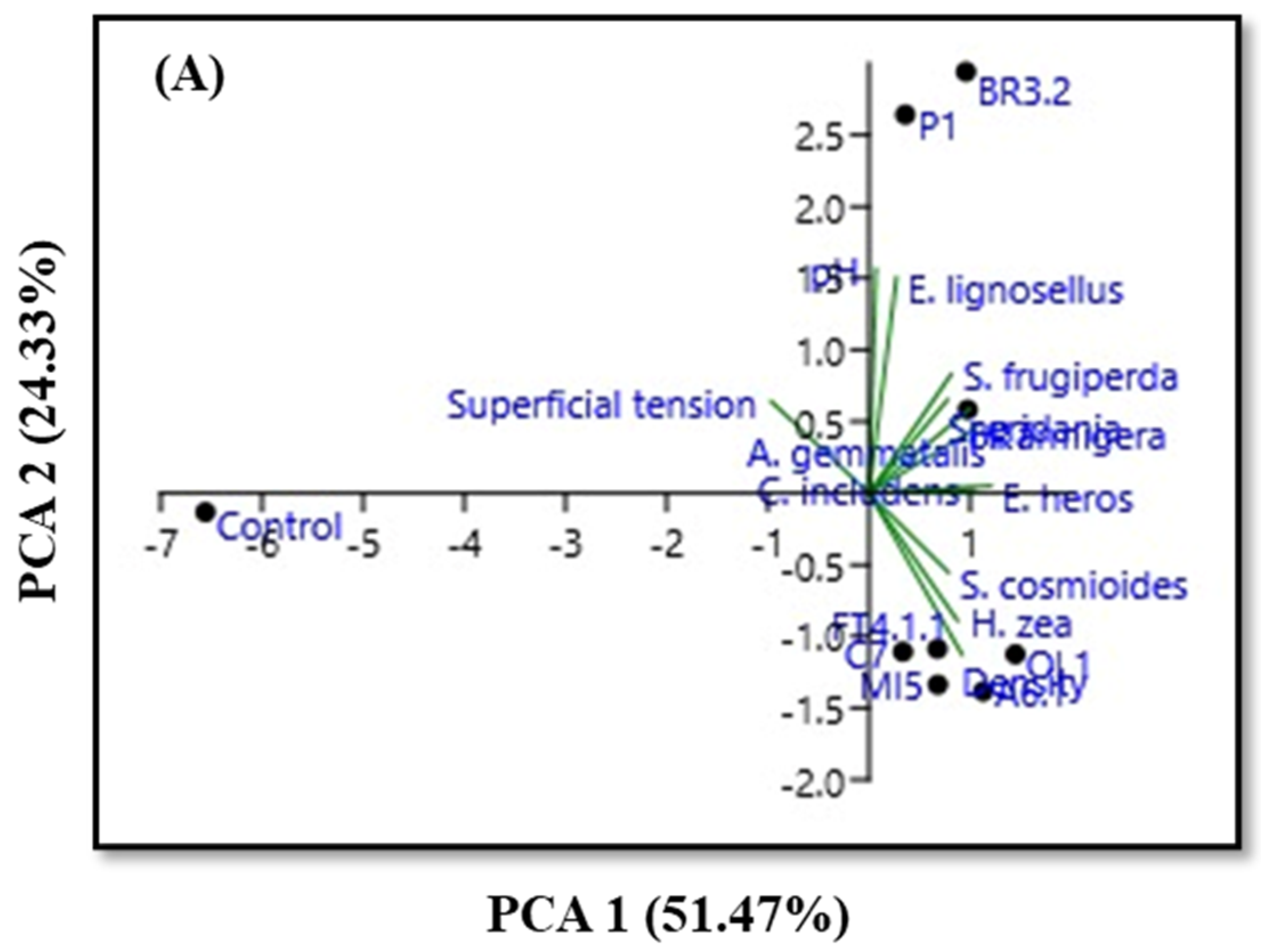
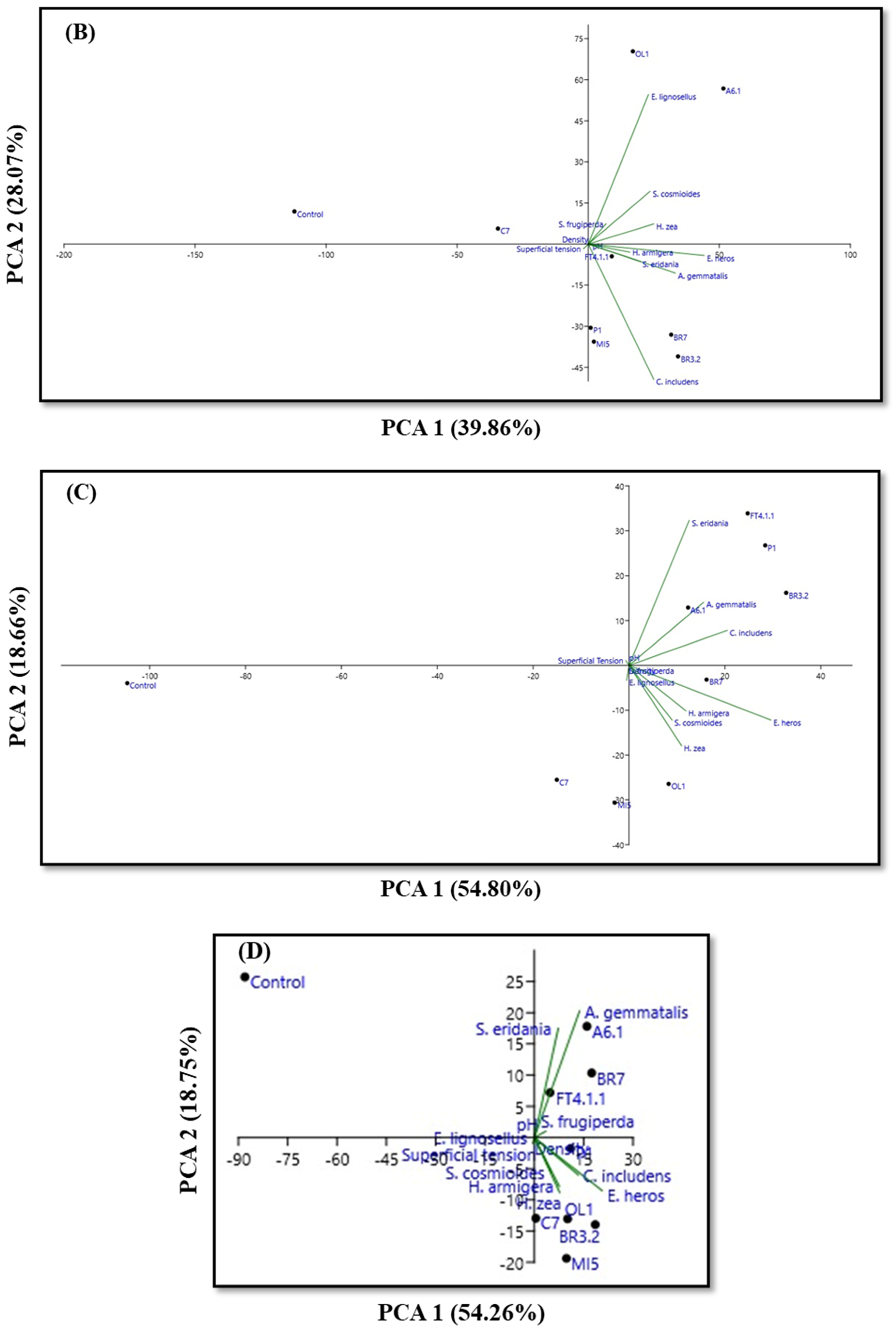
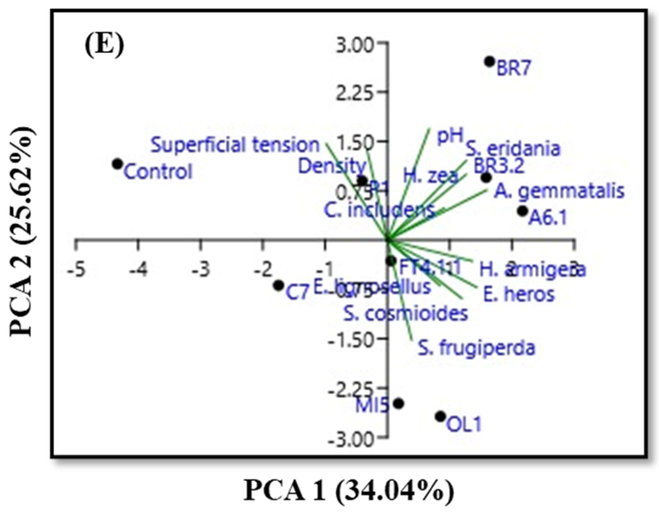
3.3. Physicochemical Characteristics and PCA

For further comparison between the eight samples of fungal and bacterial isolates selected after the pre-selection steps, serial dilutions of four test concentrations of the strains (n × 10−8, n × 10−7, n × 10−6, and n × 10−5 spores mL−1) were applied to the insect pests investigated in this study. Additionally, the crude broths of the eight isolates were applied as a way of checking the potential of the diluted concentrations. Furthermore, the parameters pH, surface tension, and specific density of the samples were evaluated.
The results of mortality and physicochemical characteristics of the samples of fungal and bacterial isolates were summarized in Supplementary Table S2. Mortality (%), in general, was higher for the crude broth treatments, compared to the dilutions. Maximum mortality rates (100%) were indicated for E. heros in almost all treatments, compared to the control treatment (30%). Mortality was also maximum for H. zea and C. includens, in the MI5 fungus broth treatment, compared to the control treatment (60% and 20%, respectively).
The pH of the samples was slightly higher in the raw broth samples, ranging from 8.6 ± 0.00 (BR7) to 8.1 ± 0.02 (P1) (moderately alkaline). In dilutions, it varied from 8.20 ± 0.01 (BR3.2) to 6.98 ± 0.00 (BR7) (from moderately alkaline to slightly acidic). Furthermore, surface tension indicated lower values for raw juice treatments. In these treatments, the value varied between 52.50 mN m−1 (OL1) and 70.72 mN m−1 (P1). In treatments with dilution, the surface tension range observed was between 64.96 ± 4.71 mN m−1 (A6.1) and 75.65 ± 1.40 mN m−1 (MI5). The surface tension of the control treatment was 71.33 ± 0.39 mN m−1. On the other hand, specific density indicated higher values in treatments without dilution. The specific density varied from 1.019408 g cm−3 (OL1) to 1.006 g cm−3 (P1 and BR3.2) in the crude broth treatments. The specific density of the control treatment was 0.997 g cm−3.
Furthermore, a principal component analysis (PCA) was performed based on data from the four dilutions and the crude broth for the eight selected isolates. The analysis indicated a correlation between the variables (mortality, pH, surface tension, and specific density) and the treatments (fungal and bacterial isolates) for crude broth (Figure 1A), n × 10−5 (Figure 1B), n × 10−6 (Figure 1C), n × 10−7 (Figure 1D), and n × 10−8 spores mL−1 (Figure 1E). The PCA separated the samples of fungal and bacterial isolates into three distinct groups for the broth samples (group 1: FT4.1.1, A6.A, C7, OL1, and MI5; group 2: BR7, BR3.2, and P1; and group 3: control) and dilutions of n × 10−5 (A6.1 and OL1; FT4.1.1, C7, MI5, BR7, BR3.2, and P1; and control), n × 10−6 (FT4.1.1, A6.1, BR3.2, and P1; OL1 and BR7; and C7, MI5, and control) and n × 10−7 spores mL−1 (FT4.1.1, A6.1, and BR7; C7, OL1, MI5, BR3.2, and P1; and control). PCA separated the samples at dilution n × 10−8 spores mL−1 into four distinct groups (A6.1, BR3.2, and BR7; FT4.1.1, OL1, and MI5; C7; and P1 and control).
Figure 1A indicates the distribution of mortality of the nine insect pests and the physicochemical variables for the crude broth treatments. PC1 explained 51.47% of the measured variation. All treatments indicated a positive correlation with the variables investigated for the crude broth. The second component, responsible for 24.33% of the total variation, was associated only with the control treatment, which showed a negative correlation with the variables studied. The same scenario was observed for treatments at dilution n × 10−7 (Figure 1D), in which the first and second components explained 54.26% and 18.75% of the variation.
For the n × 10−5 dilution (Figure 1B), PC1 explained 39.86% of the measured variation. All treatments indicated a positive correlation with the variables investigated, except the C7 and control treatments. The second component was responsible for 28.07% of the total variation. For the n × 10−6 dilution (Figure 1C), PC1 explained 54.80% of the measured variation. All treatments indicated a positive correlation with the variables investigated, except the C7, MI5, and control treatments. The second component was responsible for 18.66% of the total variation. Finally, for the n × 10−8 dilution (Figure 1E), PC1 explained 34.04% of the measured variation. All treatments indicated a positive correlation with the variables investigated, except the P1, C7, and control treatments. The second component was responsible for 25.62% of the total variation.

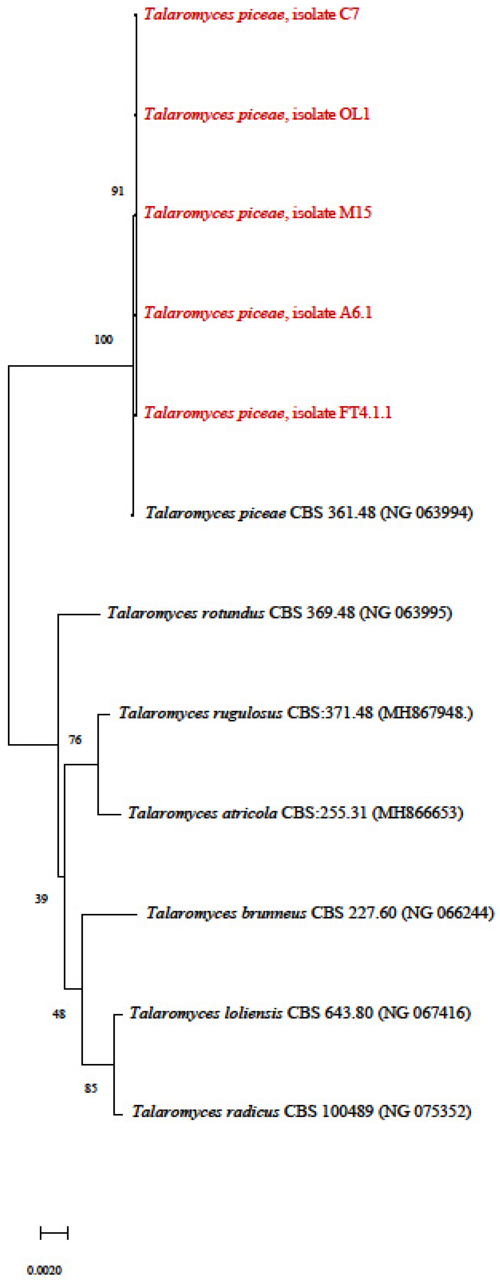
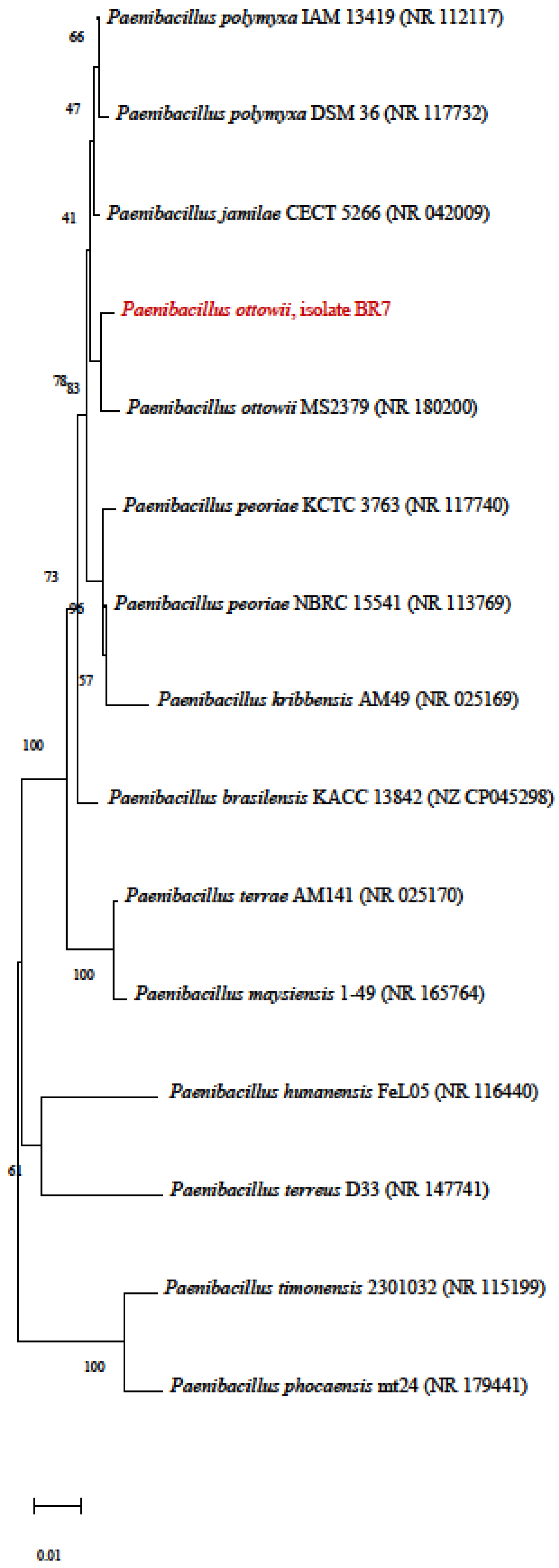
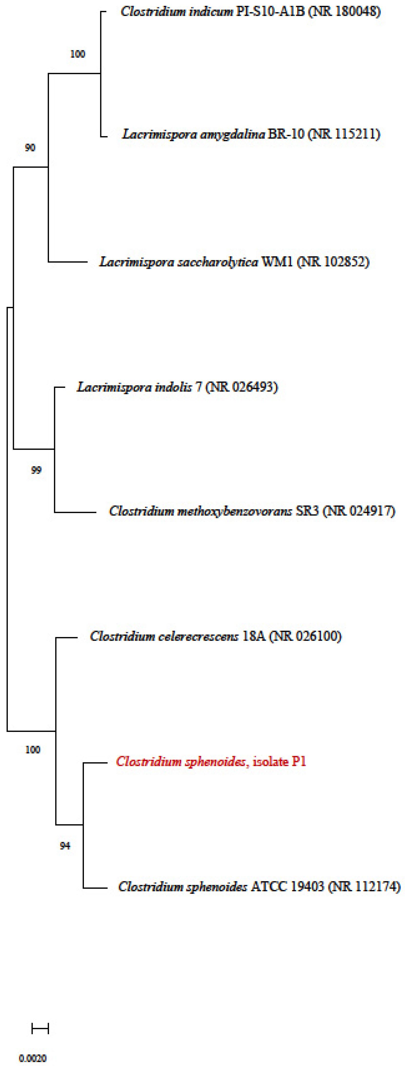
3.4. Phylogenetic Tree

The phylogenetic structures of the fungal and microbial samples revealed that the isolates were grouped according to highly similar clustering criteria. The sequences of the isolates in this study are indicated in red. The sequences obtained were compared with sequences from species deposited at NCBI (National Center for Biotechnology Information, https://www.ncbi.nlm.nih.gov/), using the Blastn tool, and are indicated in black. All fungal isolates were identified as Talaromyces piceae. The isolates were grouped and showed cluster identity of at least 91% with the species sequences deposited at NCBI (Figure 2). Fungal isolate A6.1 was identified based on the ITS sequence. The fungi C7, CL1, and M15 were identified based on the sequence of the 28S ribosomal gene, due to the low quality of the results obtained for the ITS region of these isolates. Likewise, isolate FT4.1.1 was identified based on the beta-tubulin gene sequence.
The bacterial isolate BR7 was identified as Paenibacillus ottowii, based on the sequencing of the 16S ribosomal gene. The isolate was grouped and showed cluster identity of at least 83% with the sequences of species deposited in NCBI (Figure 3).
The bacterial isolate BR3.2 was identified as Lysinibacillus fusiformis, based on the sequencing of the 16S ribosomal gene. The isolate was grouped and showed cluster identity of at least 95% with the sequences of species deposited in NCBI (Figure 4).
Finally, bacterial isolate P1 was identified as Clostridium sphenoides, based on 16S ribosomal gene sequencing. The isolate was clustered and showed at least 94% cluster identity with the sequences of species deposited in NCBI (Figure 5).
Finally, morphological representations of the surface characteristics of the selected isolates are indicated in Figure 6.

4. Discussion

A total of 163 fungi and bacteria were isolated via screening in different locations in the Neotropical region. Based on the methodological design described in this study, eight microorganisms were selected for the final stage. Maximum mortality rates of 100% have been indicated in H. zea and E. heros. Control of A. gemmatalis and C. includens was up to 87.5%. Furthermore, for E. lignosellus and H. armigera, the total mortality was 83.33% and 75%, respectively (Supplementary Table S1). All fungi and bacteria showed phytotoxic effects against the insect pests investigated. This performance highlighted the strong insecticidal control potential of the isolates obtained. Consequently, the region indicated that it is highly suitable for microbial growth and proliferation. Recent reports of microbial species, widely applied in pest control, present in the collection establishment region have indicated the active presence of Beauveria sp. [61], Metarhizium sp. [62], Trichoderma sp. [63], Lecanicillium sp. [64], Paecilomyces sp. [65], Hirsutella sp. [66], Cordyceps sp. [67], Aschersonia sp. [68], Sporothrix sp. [69], and Bacillus sp. [70]. Nonetheless, after the final selection of microorganisms, our results indicated the dominance of T. piceae and the bacteria P. ottowii, L. fusiformis, and, to a lesser extent in the agricultural scenario, C. sphenoides.
T. piceae has demonstrated promising attributes in pest management [71]. Nonetheless, the exploitation of T. piceae as a key factor in pest management programs remains limited compared to other species. On the other hand, species of Talaromyces sp. produce a wide range of bioactive compounds with insecticidal potential. An assessment of the insecticidal potential of T. piceae reported compounds such as duclauxin, 3-O-methylfunicone, chromdrimannin B, pyripyropene A, rugulosin, and vermiculin as anti-insect products that have already been responsible for controlling insect pests [72]. Also, T. piceae can produce high concentrations of CAZymes, enzymes involved in the synthesis, modification and degradation of carbohydrates, including chitin [73]. CAZymes, specifically chitinases, lysozymes, and other enzymes categorized into glycoside hydrolases, are crucial for breaking chitin structures. Chitinases target the β-1,4-glycosidic bonds in chitin molecules, cleaving them into smaller oligomers or monomers, such as N-acetylglucosamine (GlcNAc) units [74]. Therefore, CAZymes have the ability to reach and hydrolyze the chitin polymer, worsening the structural disintegration of insects. The same promising scenario is indicated for P. ottowii and L. fusiformis, although the application of P. ottowii as a potential biological control agent is limited. L. fusiformis produces the toxins Cry and Mtx, which indicate high mortality in mosquito individuals and some species of agricultural interest, such as Spodoptera exigua [75], Galleria mellonella [76], and nematodes and plant pathogens [77]. Furthermore, toxins that kill insects, including mosquitocidal toxins, sphaericolysin, S-layer protein, Cry48/Cry49 toxin, and binary toxin (Bin), are produced by Lysinibacillus sp. [78]. Some studies have indicated the high sporulation rate and control potential of P. ottowii on beetle and lepidopteran species [79].
The effects demonstrated by the crude broth treatments were superior to those observed in the dilution treatments. Nonetheless, some treatments with lower dilutions indicated high mortality rates, such as FT4.1.1 in the control of H. zea (up tp 100%) and all treatments in the control of E. heros (85.71–100%). Moreover, Supplementary Table S1 indicated a uniform performance in terms of surface tension (52.50 ± 1.91 mN m−1–75.65 ± 1.40 mN m−1) and mortality (0.00–100%) results for the crude broth and dilution treatments. Surface tension is closely associated with the cohesive force on the surface of a liquid that acts directly on its surface area [49]. In general, crude broth treatments showed lower surface tension compared to dilution treatments. This performance may be associated with the low water retention capacity on specific surfaces, such as chitin surfaces. The chitin has a strongly hydrophobic nature [80]. Insects have a chitinous surface that repels water due to its hydrophobic properties. When the product comes into contact with the insect surface, the high surface tension of the liquid can prevent it from spreading or easily penetrating the chitin layer [81]. Accordingly, products formulated based on highly diluted fermented broths can have a drastic reduction in effectiveness and control. This indicative justifies the low values for mortality in more diluted treatments. Lower dilutions typically result in lower surface tension due to reduced intermolecular forces and increased mobility of molecules [82]. This reduction in surface tension at lower dilutions can increase the wetting capacity of the solution, aiding its penetration into chitinous surfaces. Additionally, the low surface tension of the applied broth enhances the contact of the product with the target species, promoting higher spreading and penetration [83,84]. A similar performance was verified for the relations between surface tension and specific density.
The balance between specific density and surface tension significantly influenced the effectiveness of fermented broths and dilutions in controlling the insect pests investigated. Droplets with higher specific densities tend to spread less when coming into contact with the chitinous surface, as their increased mass can hinder the fluid ability to disperse and cover a larger area. Lower dilutions generally result in reduced surface tension, increasing the likelihood that droplets will effectively reach the target surface. This explains the direct relationship between the increase in specific density and the reduction in surface tension. Alternatively, higher dilutions can decrease the specific density, potentially affecting droplet spreading and reducing contact between the fluid and the target surface [84].
The general results of this study showed that bioproducts based on T. piceae, P. ottowii, L. fusiformis, and C. sphenoides were effective in controlling the investigated insect pests. These compounds present bioactive properties that can disrupt physiological processes in insects, from growth to reproductive performance. Finally, no study has been conducted to date that indicates a significant mortality of microbial species isolated in a high number of agricultural pests. Accordingly, it is expected that the information obtained in this study can encourage future research that enhances the insecticidal activity of the microorganisms found, providing useful data and adaptable microbial selection models.

5. Conclusions

A total of 163 fungal and bacterial isolates were obtained from the screening strategy in different locations. The application of a mathematical log-logistic model was effective in selecting the most promising microorganisms. Based on the model, eight potential microorganisms were selected (fungi FT4.1.1, A6.1, OL1, C7, and MI5; bacteria BR7, P1, and BR3.2). Mortality was maximum (100%) for H. zea and E. heros and high for A. gemmatalis, C. includens (up to 87.5%), and E. lignosellus (up to approximately 83.5%). For H. armigera, mortality reached 75%. Fungal isolates FT4.1.1, A6.1, C7, OL1, and M15 were identified as T. piceae. Bacterial isolates BR7, BR3.2, and P1 were identified as P. ottowii, L. fusiformis, and C. sphenoides, respectively. The results indicated are extremely relevant to the scientific community as they are part of a line of research at the frontier of knowledge and, especially, may be interesting for companies that are operating in this field in the agricultural scenario.

Supplementary Materials

The following supporting information can be downloaded at https://www.mdpi.com/article/10.3390/pr12081722/s1, Table S1: Mathematical parameters b (curve slope, rad), e (inflection point, days), and y (probability of mortality, 0–1) adopted for the application of the log-logistic model of the 50 most promising isolates after the pre-selection stage; Table S2: Mortality (%), pH, surface tension (mN m−1), and specific density (g cm−3) of the eight selected microorganisms under crude broth conditions and dilutions of n × 10−5, n × 10−6, n × 10−7, and n × 10−8 spores mL−1.

Author Contributions

Conceptualization, M.S.N.S., M.A.M., G.L.Z. and M.V.T.; methodology, M.S.N.S., J.V.C.G., M.A.M., G.L.Z. and M.V.T.; software, M.S.N.S. and I.A.C.; validation, M.S.N.S. and I.A.C.; formal analysis, M.S.N.S., L.P.O., B.D.K., I.A.C., B.d.V. and G.A.U.; investigation, M.S.N.S., M.A.M., G.L.Z. and M.V.T.; resources, M.A.M., G.L.Z. and M.V.T.; data curation, M.S.N.S., I.A.C. and B.d.V.; writing—original draft preparation, M.S.N.S.; writing—review and editing, M.S.N.S. and M.V.T.; visualization, M.V.T.; supervision, M.V.T.; project administration, M.A.M., G.L.Z. and M.V.T.; funding acquisition, M.A.M. and M.V.T. All authors have read and agreed to the published version of the manuscript.

Funding

G. L. Zabot (308067/2021-5) and M. V. Tres (306241/2020-0; 428180/2018-3) extend thanks to the National Council for Scientific and Technological Development (CNPq) for the productivity grants and financial support for development projects. M. V. Tres (21/2551-0002253-1) expresses gratitude to the Research Support Foundation of the State of Rio Grande do Sul (FAPERGS) for providing financial resources for development projects.

Data Availability Statement

The original contributions presented in the study are included in the article and Supplementary Materials, further inquiries can be directed to the corresponding author.

Conflicts of Interest

The authors declare no conflicts of interest.

References

  1. Araújo, M.F.; Castanheira, E.M.S.; Sousa, S.F. The buzz on insecticides: A review of uses, molecular structures, targets, adverse effects, and alternatives. Molecules 2023, 28, 3641. [Google Scholar] [CrossRef] [PubMed]
  2. Malinga, L.N.; Laing, M.D. Efficacy of Biopesticides in the management of the cotton bollworm, Helicoverpa armigera (Noctuidae), under field conditions. Insects 2022, 13, 673. [Google Scholar] [CrossRef]
  3. dos Santos, M.S.N.; Ody, L.P.; Kerber, B.D.; Araujo, B.A.; Oro, C.E.D.; Wancura, J.H.C.; Mazutti, M.A.; Zabot, G.L.; Tres, M.V. New frontiers of soil fungal microbiome and its application for biotechnology in agriculture. World J. Microbiol. Biotechnol. 2023, 39, 287. [Google Scholar] [CrossRef] [PubMed]
  4. Verasoundarapandian, G.; Lim, Z.S.; Batrisyia, S.; Radziff, M.; Taufik, S.H.; Puasa, N.A.; Shaharuddin, N.A.; Merican, F.; Wong, C. Remediation of pesticides by microalgae as feasible approach in agriculture: Bibliometric strategies. Agronomy 2022, 12, 117. [Google Scholar] [CrossRef]
  5. Gullino, M.L.; Albajes, R.; Al-Jboory, I.; Angelotti, F.; Chakraborty, S.; Garrett, K.A.; Hurley, B.P.; Juroszek, P.; Lopian, R.; Makkouk, K.; et al. Climate change and pathways used by pests as challenges to plant health in agriculture and forestry. Sustainability 2022, 14, 12421. [Google Scholar] [CrossRef]
  6. Jaroslow, D.D.; Cunningham, J.P.; Smith, D.I.; Steinbauer, M.J. Seasonal phenology and climate associated feeding activity of introduced Marchalina Hellenica in Southeast Australia. Insects 2023, 14, 305. [Google Scholar] [CrossRef]
  7. Han, D.; Yoo, D.; Kim, T. Analysis of social welfare impact of crop pest and disease damages due to climate change: A case study of dried red peppers. Humanit. Soc. Sci. Commun. 2023, 10, 378. [Google Scholar] [CrossRef]
  8. Šunjka, D.; Mechora, Š. An alternative source of biopesticides and improvement in their formulation—Recent advances. Plants 2022, 11, 3172. [Google Scholar] [CrossRef]
  9. Carpane, P.D.; Llebaria, M.; Nascimento, A.F.; Vivan, L. Feeding injury of major lepidopteran soybean pests in South America. PLoS ONE 2022, 17, e0271084. [Google Scholar] [CrossRef]
  10. Marques, L.H.; Santos, A.C.; Castro, B.A.; Moscardini, V.F.; Rosseto, J.; Silva, O.A.B.N.; Babcock, J.M. Assessing the efficacy of Bacillus thuringiensis (Bt) pyramided proteins Cry1F, Cry1A.105, Cry2Ab2, and Vip3Aa20 expressed in Bt maize against lepidopteran pests in Brazil. J. Econ. Entomol. 2019, 112, 803–811. [Google Scholar] [CrossRef]
  11. De Groote, H.; Kimenju, S.C.; Munyua, B.; Palmas, S.; Kassie, M.; Bruce, A. Spread and impact of fall armyworm (Spodoptera frugiperda J.E. Smith) in maize production areas of Kenya. Agric. Ecosyst. Environ. 2020, 292, 106804. [Google Scholar] [CrossRef]
  12. Eschen, R.; Beale, T.; Bonnin, J.M.; Constantine, K.L.; Duah, S.; Finch, E.A.; Makale, F.; Nunda, W.; Ogunmodede, A.; Pratt, C.F.; et al. Towards estimating the economic cost of invasive alien species to African crop and livestock production. CABI Agric. Biosci. 2021, 2, 18. [Google Scholar] [CrossRef]
  13. Dorman, S.J.; Hopperstad, K.A.; Reich, B.J.; Kennedy, G.; Huseth, A.S. Soybeans as a non-Bt refuge for Helicoverpa zea in maize-cotton agroecosystems. Agric. Ecosyst. Environ. 2021, 322, 107642. [Google Scholar] [CrossRef]
  14. Kerns, D.D.; Yang, F.; Kerns, D.L.; Stewart, S.D.; Jurat-Fuentes, J.L. Reduced toxin binding associated with resistance to Vip3Aa in the corn earworm (Helicoverpa zea). Appl. Environ. Microbiol. 2023, 89, e01644-23. [Google Scholar] [CrossRef] [PubMed]
  15. Arnemann, J.A.; Roxburgh, S.; Walsh, T.; Guedes, J.; Gordon, K.; Smagghe, G.; Tay, W.T. Multiple incursion pathways for Helicoverpa armigera in Brazil show its genetic diversity spreading in a connected world. Sci. Rep. 2019, 9, 19380. [Google Scholar] [CrossRef] [PubMed]
  16. Aioub, A.A.A.; Ghosh, S.; AL-Farga, A.; Khan, A.N.; Bibi, R.; Elwakeel, A.M.; Nawaz, A.; Sherif, N.T.; Elmasry, S.A.; Ammar, E.E. Back to the origins: Biopesticides as promising alternatives to conventional agrochemicals. Eur. J. Plant Pathol. 2024, 169, 697–713. [Google Scholar] [CrossRef]
  17. Manson, S.; Campera, M.; Hedger, K.; Ahmad, N.; Adinda, E.; Nijman, V.; Budiadi, B.; Imron, M.A.; Lukmandaru, G.; Nekaris, K.A.I. The effectiveness of a biopesticide in the reduction of coffee berry borers in coffee plants. Crop Prot. 2022, 161, 106075. [Google Scholar] [CrossRef]
  18. Zhao, J.; Liang, D.; Li, W.; Yan, X.; Qiao, J.; Caiyin, Q. Research progress on the synthetic biology of botanical biopesticides. Bioengineering 2022, 9, 207. [Google Scholar] [CrossRef]
  19. Shahriari, M.; Zibaee, A.; Khodaparast, S.A.; Fazeli-Dinan, M. Screening and virulence of the entomopathogenic fungi associated with chilo suppressalis walker. J. Fungi 2021, 7, 34. [Google Scholar] [CrossRef]
  20. Wendel, J.; Cisneros, J.; Jaronski, S.; Vitek, C.; Ciomperlik, M.; Flores, D. Screening commercial entomopathogenic fungi for the management of Diaphorina citri populations in the Lower Rio Grande Valley, Texas, USA. BioControl 2022, 67, 225–235. [Google Scholar] [CrossRef]
  21. Lahlali, R.; Ezrari, S.; Radouane, N.; Kenfaoui, J.; Esmaeel, Q.; El Hamss, H.; Belabess, Z.; Barka, E.A. Biological Control of Plant Pathogens: A Global Perspective. Microorganisms 2022, 10, 596. [Google Scholar] [CrossRef]
  22. Sabbahi, R.; Hock, V.; Azzaoui, K.; Saoiabi, S.; Hammouti, B. A global perspective of entomopathogens as microbial biocontrol agents of insect pests. J. Agric. Food Res. 2022, 10, 100376. [Google Scholar] [CrossRef]
  23. Ayilara, M.S.; Adeleke, B.S.; Akinola, S.A.; Fayose, C.A.; Adeyemi, U.T.; Gbadegesin, L.A.; Omole, R.K.; Johnson, R.M.; Uthman, Q.O.; Babalola, O.O. Biopesticides as a promising alternative to synthetic pesticides: A case for microbial pesticides, phytopesticides, and nanobiopesticides. Front. Microbiol. 2023, 14, 1040901. [Google Scholar] [CrossRef] [PubMed]
  24. Afroz Toma, M.; Rahman, M.H.; Rahman, M.S.; Arif, M.; Nazir, K.H.M.N.H.; Dufossé, L. Fungal pigments: Carotenoids, riboflavin, and polyketides with diverse applications. J. Fungi 2023, 9, 454. [Google Scholar] [CrossRef] [PubMed]
  25. Amobonye, A.; Lalung, J.; Awasthi, M.K.; Pillai, S. Fungal mycelium as leather alternative: A sustainable biogenic material for the fashion industry. Sustain. Mater. Technol. 2023, 38, e00724. [Google Scholar] [CrossRef]
  26. Takahashi, J.A.; Barbosa, B.V.R.; Martins, B.d.A.; Guirlanda, C.P.; Moura, M.A.F. Use of the versatility of fungal metabolism to meet modern demands for healthy aging, functional foods, and sustainability. J. Fungi 2020, 6, 223. [Google Scholar] [CrossRef] [PubMed]
  27. Vaksmaa, A.; Guerrero-Cruz, S.; Ghosh, P.; Zeghal, E.; Hernando-Morales, V.; Niemann, H. Role of fungi in bioremediation of emerging pollutants. Front. Mar. Sci. 2023, 10, 1070905. [Google Scholar] [CrossRef]
  28. Damavandi, M.S.; Shojaei, H.; Esfahani, B.N. The anticancer and antibacterial potential of bioactive secondary metabolites derived from bacterial endophytes in association with Artemisia absinthium. Sci. Rep. 2023, 13, 18473. [Google Scholar] [CrossRef]
  29. Ghosh, S.; Rusyn, I.; Dmytruk, O.V.; Dmytruk, K.V.; Onyeaka, H.; Gryzenhout, M.; Gafforov, Y. Filamentous fungi for sustainable remediation of pharmaceutical compounds, heavy metal and oil hydrocarbons. Front. Bioeng. Biotechnol. 2023, 11, 1106973. [Google Scholar] [CrossRef] [PubMed]
  30. Mishra, B.; Mishra, A.K.; Kumar, S.; Mandal, S.K.; Lakshmayya, N.S.V.; Kumar, V.; Baek, K.H.; Mohanta, Y.K. Antifungal metabolites as food bio-preservative: Innovation, outlook, and challenges. Metabolites 2022, 12, 12. [Google Scholar] [CrossRef]
  31. Okal, E.J.; Heng, G.; Magige, E.A.; Khan, S.; Wu, S.; Ge, Z.; Zhang, T.; Mortimer, P.E.; Xu, J. Insights into the mechanisms involved in the fungal degradation of plastics. Ecotoxicol. Environ. Saf. 2023, 262, 115202. [Google Scholar] [CrossRef] [PubMed]
  32. Saye, L.M.G.; Navaratna, T.A.; Chong, J.P.J.; O’malley, M.A.; Theodorou, M.K.; Reilly, M. The anaerobic fungi: Challenges and opportunities for industrial lignocellulosic biofuel production. Microorganisms 2021, 9, 694. [Google Scholar] [CrossRef] [PubMed]
  33. Schein, D.; Santos, M.S.N.; Schmaltz, S.; Nicola, L.E.P.; Bianchin, C.F.; Ninaus, R.G.; de Menezes, B.B.; dos Santos, R.C.; Zabot, G.L.; Tres, M.V.; et al. Microbial prospection for bioherbicide production and evaluation of methodologies for maximizing phytotoxic activity. Processes 2022, 10, 2001. [Google Scholar] [CrossRef]
  34. Shankar, A.; Sharma, K.K. Fungal secondary metabolites in food and pharmaceuticals in the era of multi-omics. Appl. Microbiol. Biotechnol. 2022, 106, 3465–3488. [Google Scholar] [CrossRef] [PubMed]
  35. Srikanth, M.; Sandeep, T.S.R.S.; Sucharitha, K.; Godi, S. Biodegradation of plastic polymers by fungi: A brief review. Bioresour. Bioprocess. 2022, 9, 42. [Google Scholar] [CrossRef] [PubMed]
  36. Khan, R.A.A.; Najeeb, S.; Hussain, S.; Xie, B.; Li, Y. Bioactive secondary metabolites from Trichoderma spp. Against phytopathogenic fungi. Microorganisms 2020, 8, 817. [Google Scholar] [CrossRef] [PubMed]
  37. Velastegui-Montoya, A.; Montalván-Burbano, N.; Peña-Villacreses, G.; de Lima, A.; Herrera-Franco, G. Land use and land cover in tropical forest: Global research. Forests 2022, 13, 1709. [Google Scholar] [CrossRef]
  38. Menolli, N.; Sánchez-García, M. Brazilian fungal diversity represented by DNA markers generated over 20 years. Braz. J. Microbiol. 2020, 51, 729–749. [Google Scholar] [CrossRef] [PubMed]
  39. Hawksworth, D.L.; Lücking, R. Fungal diversity revisited: 2.2 to 3.8 million species. Fungal Kingd. 2017, 5, 79–95. [Google Scholar] [CrossRef]
  40. Wiens, J.J. How many species are there on Earth? Progress and problems. PLoS Biol. 2023, 21, 10–13. [Google Scholar] [CrossRef]
  41. Duden, A.S.; Verweij, P.A.; Faaij, A.P.C.; Baisero, D.; Rondinini, C.; Van Der Hilst, F. Biodiversity impacts of increased ethanol production in Brazil. Land 2020, 9, 12. [Google Scholar] [CrossRef]
  42. Valencia, E.Y.; Chambergo, F.S. Mini-review: Brazilian fungi diversity for biomass degradation. Fungal Genet. Biol. 2013, 60, 9–18. [Google Scholar] [CrossRef] [PubMed]
  43. Yao, H.; Sun, X.; He, C.; Maitra, P.; Li, X.; Guo, L. Phyllosphere epiphytic and endophytic fungal community and network structures differ in a tropical mangrove ecosystem. Microbiome 2019, 7, 57. [Google Scholar] [CrossRef] [PubMed]
  44. Crowther, T.W.; Van Den Hoogen, J.; Wan, J.; Mayes, M.A.; Keiser, A.D.; Mo, L.; Averill, C.; Maynard, D.S. The global soil community and its influence on biogeochemistry. Science 2019, 365, eaav0550. [Google Scholar] [CrossRef]
  45. Chalivendra, S. Microbial toxins in insect and nematode pest biocontrol. Int. J. Mol. Sci. 2021, 22, 7657. [Google Scholar] [CrossRef] [PubMed]
  46. Kumar, J.; Ramlal, A.; Mallick, D.; Mishra, V. An overview of some biopesticides and their importance in plant protection for commercial acceptance. Plants 2021, 10, 1185. [Google Scholar] [CrossRef] [PubMed]
  47. Negi, R.; Sharma, B.; Kaur, S.; Kaur, T.; Khan, S.S.; Kumar, S.; Ramniwas, S.; Rustagi, S.; Singh, S.; Rai, A.K.; et al. Microbial antagonists: Diversity, formulation and applications for management of pest–pathogens. Egypt. J. Biol. Pest Control 2023, 33, 105. [Google Scholar] [CrossRef]
  48. Portilla, M.; Jones, W.; Perera, O.; Seiter, N.; Greene, J.; Luttrell, R. Estimation of median lethal concentration of three isolates of Beauveria bassiana for control of Megacopta cribraria (heteroptera: Plataspidae) bioassayed on solid Lygus spp. Diet. Insects 2016, 7, 31. [Google Scholar] [CrossRef] [PubMed]
  49. Brun, T.; Rabuske, J.E.; Confortin, T.C.; Luft, L.; Todero, I.; Fischer, M.; Zabot, G.L.; Mazutti, M.A. Weed control by metabolites produced from Diaporthe schini. Environ. Technol. 2022, 43, 139–148. [Google Scholar] [CrossRef]
  50. Sharma, L.; Bohra, N.; Rajput, V.D.; Quiroz-Figueroa, F.R.; Singh, R.K.; Marques, G. Advances in entomopathogen isolation: A case of bacteria and fungi. Microorganisms 2021, 9, 16. [Google Scholar] [CrossRef]
  51. Todero, I.; Confortin, T.C.; Luft, L.; Brun, T.; Ugalde, G.A.; de Almeida, T.C.; Arnemann, J.A.; Zabot, G.L.; Mazutti, M.A. Formulation of a bioherbicide with metabolites from Phoma sp. Sci. Hortic. 2018, 241, 285–292. [Google Scholar] [CrossRef]
  52. de Souza, A.R.C.; Baldoni, D.B.; Lima, J.; Porto, V.; Marcuz, C.; Machado, C.; Ferraz, R.C.; Kuhn, R.C.; Jacques, R.J.; Guedes, J.V.; et al. Selection, isolation, and identification of fungi for bioherbicide production. Braz. J. Microbiol. 2017, 48, 101–108. [Google Scholar] [CrossRef] [PubMed]
  53. Abbott, W.S. The value of the dry substitutes for liquid lime. J. Econ. Entomol. 1925, 18, 265–267. [Google Scholar] [CrossRef]
  54. Jordan, C.; dos Santos, P.L.; Oliveira, L.R.d.S.; Domingues, M.M.; Gêa, B.C.C.; Ribeiro, M.F.; Mascarin, G.M.; Wilcken, C.F. Entomopathogenic fungi as the microbial frontline against the alien Eucalyptus pest Gonipterus platensis in Brazil. Sci. Rep. 2021, 11, 7233. [Google Scholar] [CrossRef] [PubMed]
  55. Greene, G.L.; Leppla, N.C.; Dickerson, W.A. Velvetbean caterpillar: A rearing procedure and artificial medium. J. Econ. Entomol. 1976, 69, 487–488. [Google Scholar] [CrossRef]
  56. Doyle, J.J.; Doyle, J.L. A rapid DNA isolation procedure for small quantities of fresh leaf tissue. Phytochem. Bull. 1987, 19, 11–15. [Google Scholar]
  57. Vilgalys, R.; Hester, M. Rapid genetic identification and mapping of enzymatically amplified ribosomal DNA from several Cryptococcus species. J. Bacteriol. 1990, 172, 4238–4246. [Google Scholar] [CrossRef]
  58. Aveskamp, M.M.; Verkley, G.J.M.; De Gruyter, J.; Murace, M.A.; Perelló, A.; Woudenberg, J.H.C.; Groenewald, J.Z.; Crous, P.W. DNA phylogeny reveals polyphyly of Phoma section Peyronellaea and multiple taxonomic novelties. Mycologia 2009, 101, 363–382. [Google Scholar] [CrossRef]
  59. Weisburg, W.G.; Barns, S.M.; Pelletier, D.A.; Lane, D.J. 16S ribosomal DNA amplification for phylogenetic study. J. Bacteriol. 1991, 173, 697–703. [Google Scholar] [CrossRef] [PubMed]
  60. Schmitz, A.; Riesner, D. Purification of nucleic acids by selective precipitation with polyethylene glycol 6000. Anal. Biochem. 2006, 354, 311–313. [Google Scholar] [CrossRef]
  61. Matsumura, A.T.S.; da Silva, M.E.; Grassotti, T.T.; Costa, L.d.F.X.; Matsumura, A.S. First report of Beauveria bassiana in the in vivo control of Eriosoma lanigerum in Brazilian apple trees. Rev. Ceres 2023, 70, 97–104. [Google Scholar] [CrossRef]
  62. Franzin, M.L.; Moreira, C.C.; da Silva, L.N.P.; Martins, E.F.; Fadini, M.A.M.; Pallini, A.; Elliot, S.L.; Venzon, M. Metarhizium associated with coffee seedling roots: Positive effects on plant growth and protection against Leucoptera coffeella. Agriculture 2022, 12, 2030. [Google Scholar] [CrossRef]
  63. Nascimento, V.C.; Rodrigues-Santos, K.C.; Carvalho-Alencar, K.L.; Castro, M.B.; Kruger, R.H.; Lopes, F.A.C. Trichoderma: Biological control efficiency and perspectives for the Brazilian Midwest states and Tocantins. Braz. J. Biol. 2022, 82, e260161. [Google Scholar] [CrossRef]
  64. Alves, E.B.; Casarin, N.F.B.; Gonçalves-Gervásio, R.d.C.; Omoto, C. Lime sulfur to control citrus flat mite and its interactions with the entomopathogenic fungus Lecanicillium muscarium. Arq. Inst. Biol. 2022, 89, e00232021. [Google Scholar] [CrossRef]
  65. Brites-Neto, J.; Maimone, N.M.; Piedade, S.M.D.S.; Andrino, F.G.; de Andrade, P.A.M.; de Assis Baroni, F.; Gomes, L.H.; de Lira, S.P. Scorpionicidal activity of secondary metabolites from Paecilomyces sp. CMAA1686 against Tityus serrulatus. J. Invertebr. Pathol. 2021, 179, 107541. [Google Scholar] [CrossRef]
  66. Pérez-González, O.; Gomez-Flores, R.; Tamez-Guerra, P. Insight into biological control potential of Hirsutella citriformis against Asian citrus psyllid as a vector of Citrus Huanglongbing disease in America. J. Fungi 2022, 8, 573. [Google Scholar] [CrossRef]
  67. Domingues, M.M.; Santos, P.L.; Gêa, B.C.C.; Carvalho, V.R.; Oliveira, F.N.; Soliman, E.P.; Silva, W.M.; Zanuncio, J.C.; Santos Junior, V.C.; Wilcken, C.F. Isolation and molecular characterization of Cordyceps sp. from Bemisia tabaci (Hemiptera: Aleyrodidae) and pathogenic to Glycaspis brimblecombei (Hemiptera: Aphalaridae). Braz. J. Biol. 2024, 84, e253028. [Google Scholar] [CrossRef]
  68. Homrahud, D.; Uraichuen, S.; Attathom, T. Cultivation of Aschersonia placenta Berkeley and Broom and its efficacy for controlling Parlatoria ziziphi (Lucas) (Hemiptera: Diaspididae). Agric. Nat. Resour. 2016, 50, 179–185. [Google Scholar] [CrossRef]
  69. Etchecopaz, A.; Toscanini, M.A.; Gisbert, A.; Mas, J.; Scarpa, M.; Iovannitti, C.A.; Bendezú, K.; Nusblat, A.D.; Iachini, R.; Cuestas, M.L. Sporothrix brasiliensis: A review of an emerging south american fungal pathogen, its related disease, presentation and spread in Argentina. J. Fungi 2021, 7, 170. [Google Scholar] [CrossRef]
  70. Mashtoly, T.A.; El-Beltagi, H.S.; Almujam, A.N.; Othman, M.N. The potential of a novel concept of an integrated bio and chemical formulate based on an entomopathogenic bacteria, Bacillus thuringiensis, and a chemical insecticide to control tomato leafminer, Tuta absoluta ‘(Meyrick)’ (Lepidoptera: Gelechiidae). Sustainability 2022, 14, 10582. [Google Scholar] [CrossRef]
  71. Chen, J.; Xu, Z.; Liu, Y.; Yang, F.; Guan, L.; Yang, J.; Li, J.; Niu, G.; Li, J.; Jin, L. Talaromyces sp. ethyl acetate crude extract as potential mosquitocide to control Culex pipiens quinquefasciatus. Molecules 2023, 28, 6642. [Google Scholar] [CrossRef] [PubMed]
  72. Nicoletti, R. Talaromyces–Insect Relationships. Microorganisms 2022, 10, 45. [Google Scholar] [CrossRef]
  73. Tian, Y.; Zhao, Y.; Fu, X.; Yu, C.; Gao, K.; Liu, H. Isolation and identification of Talaromyces sp. strain Q2 and its biocontrol mechanisms involved in the control of Fusarium wilt. Front. Microbiol. 2021, 12, 724842. [Google Scholar] [CrossRef]
  74. Raheja, Y.; Kaur, B.; Falco, M.; Tsang, A.; Chadha, B.S. Secretome analysis of Talaromyces emersonii reveals distinct CAZymes profile and enhanced cellulase production through response surface methodology. Ind. Crops Prod. 2020, 152, 112554. [Google Scholar] [CrossRef]
  75. Eski, A.; Demir, İ.; Güllü, M.; Demirbağ, Z. Biodiversity and pathogenicity of bacteria associated with the gut microbiota of beet armyworm, Spodoptera exigua Hübner (Lepidoptera: Noctuidae). Microb. Pathog. 2018, 121, 350–358. [Google Scholar] [CrossRef]
  76. Loulou, A.; Mastore, M.; Caramella, S.; Bhat, A.H.; Brivio, M.F.; Machado, R.A.R.; Kallel, S. Entomopathogenic potential of bacteria associated with soil-borne nematodes and insect immune responses to their infection. PLoS ONE 2023, 18, e0280675. [Google Scholar] [CrossRef]
  77. Passera, A.; Rossato, M.; Oliver, J.S.; Battelli, G.; Shahzad, G.I.R.; Cosentino, E.; Sage, J.M.; Toffolatti, S.L.; Lopatriello, G.; Davis, J.R.; et al. Characterization of Lysinibacillus fusiformis strain S4C11: In vitro, in planta, and in silico analyses reveal a plant-beneficial microbe. Microbiol. Res. 2021, 244, 126665. [Google Scholar] [CrossRef] [PubMed]
  78. Mohammad, Q.; Jamal, S. Lysinibacilli: A biological factories intended for diseases genus Lysinibacillus. J. Fungi 2022, 8, 1288. [Google Scholar]
  79. Grady, E.N.; MacDonald, J.; Liu, L.; Richman, A.; Yuan, Z.C. Current knowledge and perspectives of Paenibacillus: A review. Microb. Cell Fact. 2016, 15, 1–18. [Google Scholar] [CrossRef]
  80. Henriques, B.S.; Garcia, E.S.; Azambuja, P.; Genta, F.A. Determination of chitin content in insects: An alternate method based on calcofluor staining. Front. Physiol. 2020, 11, 117. [Google Scholar] [CrossRef]
  81. Bello, E.; Chen, Y.; Alleyne, M. Staying dry and clean: An insect’s guide to hydrophobicity. Insects 2023, 14, 42. [Google Scholar] [CrossRef]
  82. Rizza, M.A.; Wijayanti, W.; Hamidi, N.; Wardana, I.N.G. Role of intermolecular forces on the contact angle of vegetable oil droplets during the cooling process. Sci. World J. 2018, 2018, 5283753. [Google Scholar] [CrossRef] [PubMed]
  83. Chaves Neto, J.R.; Nascimento dos Santos, M.S.; Mazutti, M.A.; Zabot, G.L.; Tres, M.V. Phoma dimorpha phytotoxic activity potentialization for bioherbicide production. Biocatal. Agric. Biotechnol. 2021, 33, 101986. [Google Scholar] [CrossRef]
  84. Yong, J.; Yang, Q.; Hou, X.; Chen, F. Emerging separation applications of surface superwettability. Nanomaterials 2022, 12, 688. [Google Scholar] [CrossRef]
Figure 1. Principal component analysis (PCA) on mortality (%) and physicochemical properties (surface tension, mN m−1; density, g cm−3; and pH) of the eight most promising microorganisms obtained after the selection stage: (A) crude broth, (B) n × 10−5 dilution, (C) n × 10−6 dilution, (D) n × 10−7 dilution, and (E) n × 10−8 dilution.
Processes 12 01722 g001aProcesses 12 01722 g001bProcesses 12 01722 g001c
Figure 2. Phylogenetic tree of sequences by maximum likelihood. Isolate A6.1 was identified based on the ITS sequence. Isolates C7, CL1, and M15 were identified based on the sequence of the 28S ribosomal gene. Isolate FT4.1.1 was identified based on the beta-tubulin gene sequence. The sequences of the isolates in this study are indicated in red. Bootstrap values (%) correspond to maximum likelihood (ML) analysis (1000 bootstraps).
Processes 12 01722 g002
Figure 3. Phylogenetic tree of sequences by maximum likelihood. Isolate BR7 was identified based on sequencing of the 16S ribosomal gene. The sequences of the isolates in this study are indicated in red. Bootstrap values (%) correspond to maximum likelihood (ML) analysis (1000 bootstraps).
Processes 12 01722 g003
Figure 4. Phylogenetic tree of sequences by maximum likelihood. Isolate BR3.2 was identified based on sequencing of the 16S ribosomal gene. The sequences of the isolates in this study are indicated in red. Bootstrap values (%) correspond to maximum likelihood (ML) analysis (1000 bootstraps).
Processes 12 01722 g004
Figure 5. Phylogenetic tree of sequences by maximum likelihood. Isolate P1 was identified based on sequencing of the 16S ribosomal gene. The sequences of the isolates in this study are indicated in red. Bootstrap values (%) correspond to maximum likelihood (ML) analysis (1000 bootstraps).
Processes 12 01722 g005
Figure 6. Cultural images of the eight most promising fungal and bacterial isolates, obtained via screening performed in different locations in the southern subtropical region of Brazil (Santa Maria, Lavras do Sul, Cerro Largo, and São Vicente do Sul), during the summer, autumn, winter, and spring 2021, and central tropical region of Brazil (Luziania), during the dry season, May to September 2021.
Processes 12 01722 g006
Table 1. Geographic characterization of collection points and quantification of microbial agents isolated in this study.
Collection LocationStateGeographic CoordinatesNumber of Microorganisms Collected
LatitudeLongitude
Santa MariaRS−29.4129−53.4803125
Lavras do SulRS−30.4857−53.533515
Cerro LargoRS−28.1506−54.76869
São Vicente do SulRS−29.4132−54.40473
LuziâniaGO−16.2345−47.916711
Table 2. Cultural characteristics of the 50 most promising isolates after the pre-selection stage, in terms of classification, concentration (conidia mL−1), density (CFU), and time of appearance of the first colony after isolation (h).
MicroorganismIdentificationClassificationCollection LocationConcentration (Conidia mL−1)Density (CFU)Time (h)Genre-Level Classification
1FT4.1.1FungiSanta Maria, RS, Brazil1.27 × 108585.0 ± 71.048Talaromyces sp.
2BR3FungiLuziânia, GO, Brazil5.10 × 1061098.3 ± 336.596Aspergillus sp.
3BR2.1FungiLuziânia, GO, Brazil3.00 × 108922.6 ± 79.596Aspergillus sp.
4MI2FungiSanta Maria, RS, Brazil2.30 × 106127.0 ± 12.196Fusarium sp.
5MI5FungiSanta Maria, RS, Brazil3.30 × 108466.0 ± 0.072Talaromyces sp.
6SHFungiSanta Maria, RS, Brazil7.13 × 107522.6 ± 147.096Penicillium sp.
7MI3F2FungiSanta Maria, RS, Brazil3.20 × 108612.3 ± 24.672Fusarium sp.
8TR3.1FungiSanta Maria, RS, Brazil1.00 × 10719.6 ± 7.396Fusarium sp.
9C7FungiSanta Maria, RS, Brazil1.05 × 107206.0 ± 27.0120Talaromyces sp.
10BR5.4FungiLuziânia, GO, Brazil3.30 × 107214.6 ± 28.396Fusarium sp.
11C9FungiSanta Maria, RS, Brazil7.00 × 108231.6 ± 88.696Fusarium sp.
12MI7FungiSanta Maria, RS, Brazil1.96 × 1081194.0 ± 212.696Pythium sp.
13OL1FungiSanta Maria, RS, Brazil1.40 × 10781.6 ± 6.672Talaromyces sp.
14CL4FungiCerro Largo, RS, Brazil2.73 × 1082.3 ± 1.348Trichoderma sp.
15A6.1FungiSanta Maria, RS, Brazil2.10 × 1073.0 ± 1.096Talaromyces sp.
16MI3FungiSanta Maria, RS, Brazil6.00 × 10842.0 ± 5.696Fusarium sp.
17LV4FungiLavras do Sul, RS, Brazil3.70 × 102395.6 ± 88.596Trichoderma sp.
18MIAFungiSanta Maria, RS, Brazil7.10 × 107267.0 ± 76.296Botryoderma sp.
19SN5FungiSanta Maria, RS, Brazil2.40 × 10831.6 ± 4.048Fusarium sp.
20LV1FungiLavras do Sul, RS, Brazil4.00 × 108138.5 ± 44.596Trichoderma sp.
21CL2FungiCerro Largo, RS, Brazil6.10 × 108200.0 ± 15.596Pythium sp.
22BR4.1FungiLuziânia, GO, Brazil1.75 × 10845.6 ± 23.096Nigrospora sp.
23MI6FungiSanta Maria, RS, Brazil3.40 × 108155.30 ± 71.6096Aspergillus sp.
24CL3FungiCerro Largo, RS, Brazil1.00 × 10813.00 ± 1.7096Trichoderma sp.
25OL4FungiSanta Maria, RS, Brazil2.10 × 108271.00 ± 143.1096Rhizoctonia sp.
26MA8FungiSanta Maria, RS, Brazil2.00 × 108674.30 ± 169.1096Pythium sp.
27FT4.1FFungiSanta Maria, RS, Brazil1.71 × 108118.00 ± 20.6048Nigrospora sp.
28LV12FungiLavras do Sul, RS, Brazil1.90 × 1072.00 ± 0.0096Trichoderma sp.
29LV7FungiLavras do Sul, RS, Brazil2.30 × 1081420.50 ± 47.3096Penicillium sp.
30MI7F2FungiSanta Maria, RS, Brazil1.05 × 108154.60 ± 47.5096Penicillium sp.
31CL12FungiCerro Largo, RS, Brazil3.50 × 1082.00 ± 0.00120Trichoderma sp.
32SN4FungiSanta Maria, RS, Brazil5.77 × 108584.00 ± 191.1096Fusarium sp.
33MI5F1FungiSanta Maria, RS, Brazil3.30 × 108466.00 ± 0.0072Trichoderma sp.
34CL7FungiCerro Largo, RS, Brazil7.60 × 108571.00 ± 87.6096Trichoderma sp.
35SN9FungiSanta Maria, RS, Brazil4.33 × 10731.30 ± 5.5096Fusarium sp.
36MI2.2FungiSanta Maria, RS, Brazil3.40 × 10737.20 ± 5.5096Fusarium sp.
37P1BacteriaSanta Maria, RS, Brazil-620.00 ± 0.0096-
38CBMBacteriaSanta Maria, RS, Brazil-3.60 ± 3.00120-
39X1BacteriaSanta Maria, RS, Brazil-290.60 ± 22.5072-
40MIDBacteriaSanta Maria, RS, Brazil-1109.60 ± 197.4096-
41UMB1BacteriaSão Vicente do Sul, RS, Brazil-866.30 ± 106.8096-
42C1BacteriaSanta Maria, RS, Brazil-550.00 ± 86.2048-
43LV19BacteriaLavras do Sul, RS, Brazil-61.00 ± 4.3096-
44UMB2BacteriaSão Vicente do Sul, RS, Brazil-474.00 ± 80.9048-
45OL5BacteriaSanta Maria, RS, Brazil-138.50 ± 44.5096-
46FTGBacteriaSanta Maria, RS, Brazil-31.30 ± 5.5096-
47BR7BacteriaLuziânia, GO, Brazil-51.00 ± 31.9096-
48UMB6BacteriaSão Vicente do Sul, RS, Brazil-138.50 ± 44.5048-
49BR3.2BacteriaLuziânia, GO, Brazil-77.60 ± 25.5048-
50A8BacteriaSanta Maria, RS, Brazil-10.60 ± 0.5096-
Disclaimer/Publisher’s Note: The statements, opinions and data contained in all publications are solely those of the individual author(s) and contributor(s) and not of MDPI and/or the editor(s). MDPI and/or the editor(s) disclaim responsibility for any injury to people or property resulting from any ideas, methods, instructions or products referred to in the content.

