Article

Parameter Optimization and Capacitance-Based Monitoring of In Situ Cell Detachment in Microcarrier Cultures

1
ACIB—Austrian Centre of Industrial Biotechnology, Krenngasse 37, 8010 Graz, Austria
2
Department of Applied Life Science, Bioengineering, FH Campus Wien, 1100 Vienna, Austria
3
Department of Biotechnology, University of Natural Resources and Life Sciences, 1190 Vienna, Austria
4
Novasign GmbH, 1020 Vienna, Austria
*
Author to whom correspondence should be addressed.
Processes 2024, 12(9), 1887; https://doi.org/10.3390/pr12091887
Submission received: 4 July 2024 / Revised: 29 July 2024 / Accepted: 29 August 2024 / Published: 3 September 2024
(This article belongs to the Section Biological Processes and Systems)

Abstract

:
This study delves into the scale-down optimization of the in situ cell detachment process for MA 104 cells cultivated on Cytodex 1 microcarriers (MCs). Through a systematic exploration, critical operational parameters—the agitation speed, incubation time, Trypsin–EDTA volume and corresponding activity, and washing steps—were identified as key factors influencing the efficiency and scalability of in situ cell detachment in microcarrier-based cell culture. Maintaining an appropriate agitation speed (1.25 × Njs, minimum agitation speed at which no microcarriers remain stationary for the signification period of 5 s), optimizing the Trypsinization incubation time (up to 60 min), and implementing multiple washing steps (two times) post-medium removal were found to be crucial for efficient cell detachment and subsequent growth. Our study demonstrates the feasibility of reducing the final Trypsin volume to 50 mL per gram of microcarrier while maintaining a Trypsin activity above 380 USP/mL. These conditions ensure complete cell dissociation and improve the cost effectiveness in large-scale productions. Additionally, we introduced real-time monitoring using a capacitance sensor during in situ cell detachment. This method has proven to be an effective process analytical technology (PAT) tool for tracking the cell detachment progress and efficiency. It allows for the prediction of cell detachment based on signals recorded between 3 and 7 min of Trypsinization, enabling rapid process decisions without the need for offline sampling, thereby enhancing the overall process control. This systematic approach not only optimizes in situ cell detachment processes but also has significant implications for the scalability and efficiency of microcarrier-based cell culture systems.

1. Introduction

Microcarrier (MC) culture, a method introduced by VanWezel in 1967 [1], significantly transformed the mass production of viral vaccines and biological cell products using cells that require attachment to surfaces [2]. Although new vaccine production technologies like mRNA have emerged, the MC culture system still remains crucial for pandemic defense [3]. The primary challenges in producing mRNA vaccines include shortages of essential raw materials and the complexities involved in scaling up production and distribution [4]. Large-scale vaccine production with a low capital cost is required to meet the potential demand for large vaccine quantities against pathogens like SARS-CoV-2, especially in low-income countries [4,5,6].
MC-based cell culture reduces the time and cost of the manufacturing process compared to traditional static cell culture for host cell lines like Vero, HEK, MDCK, and MA 104 [2,7,8,9]. Additionally, this method minimizes the space requirements, labor, and storage needs for routine production, with large-scale applications feasible in conventional and well-characterized stirred-tank bioreactors [7]. In alignment with the need for quick and efficient manufacturing, single-use bioreactors emerge as an appealing choice due to their ability to replicate the scalability and operational parameters of stainless steel bioreactors but with added convenience [3,10]
In MC-based cell culture, cell adhesion, proliferation, and harvesting from MCs are critical stages influencing the final cell productivity and quality [11,12]. Cell attachment to MCs and proliferation have been extensively studied by different research groups under different conditions such as media components [13,14], seeding densities (cells/bead ratio) [14], MC concentrations [9,13,14,15], agitation speeds [9], and culture volumes [11]. However, they cover the cell attachment and proliferation optimization on the MC before undergoing in situ cell detachment, which is the cell harvest from MCs using an enzymatic solution. In our previous study [16], we reported a holistic cell growth optimization after in situ cell detachment by applying a hybrid model approach for the cell attachment and proliferation of the MA 104 cell line on Cytodex 1.
For the step of in situ cell detachment and harvesting from MCs, there are few reports on evaluating operational parameters with the purpose of scale-up and seed-train improvement in the field of vaccine manufacturing [17,18]. Sousa et al. (2019) examined Vero cell detachment from Cytodex 1 using a one-time PBS washing step and different enzyme volume ratios but did not explore other factors or the scalability of the operating ranges [17]. Ton et al. (2023) reported the scale-up of vaccine production using Vero cells in single-use bioreactors but did not evaluate parameters during cell detachment, impacting the subsequent cell attachment to MCs [3]. Some other studies focus on harvesting human mesenchymal stem cells (hMSC), for which maintaining a high cell yield is crucial. However, these studies mainly concentrate on optimizing mechanical agitation for cell harvesting while preserving the cell quality [11,12,13].
This study aims to propose a scalable method for in situ cell detachment of MA 104 cells grown on Cytodex 1 MCs. The approach builds on earlier research demonstrating that MA 104 cells can undergo up to five consecutive in situ cell detachments [16], and in the optimized culture condition, a maximum cell density of over 2.0 × 10⁶ cells/mL can be achieved. We explore multiple operational parameters, including the time, volume of Trypsin–EDTA solution (0.25%), which result in different enzyme activities, number of PBS washing steps post-spent medium removal, and agitation speed. This investigation, grounded in theoretical concepts and empirical data, establishes a solid foundation for scaling up single-use bioreactors, aiming for consecutive cell propagation on MCs, which requires in situ cell detachment. The same methodology is also applicable for the harvesting of human mesenchymal stem cells (hMSCs) on MCs.
We also developed a decision model tool for monitoring cell detachment from MCs using impedance spectroscopy with a cell capacitance sensor. This sensor works on the principle of frequency-dependent polarization of dielectric material, allowing for the detection of cells with intact membranes [19].
The use of capacitance sensors to monitor the proliferation of various cell lines, such as CHO, HCT, Vero, and hMSC, on MCs like Cytodex 1, Cytodex 3, and Cytopore, is widely established [20,21,22,23]. Furthermore, the application of biocapacitance sensors in viral vaccine production has demonstrated their potential as effective and non-invasive tool for the real-time monitoring of cell growth on MCs [24]. These sensors can detect when cells have reached confluence and have been proposed for use in identifying apoptosis resulting from viral infections [25]. Apart from capacitance sensors, alternative approaches like in situ microscopy [26,27,28], laser scanning cytometry [29], and optical coherence tomography (OCT) [30] have been devised to directly monitor and trace cell proliferation on MCs within bioreactors. These imaging techniques offer valuable real-time data and high-resolution images but have drawbacks such as a limited field of view or penetration depth, high costs, and technical complexity [31]. The main advantage of capacitance sensors over the other methods is their ability to provide continuous, non-invasive, and real-time monitoring of the viable cell density without the need for complex and costly imaging setups. Additionally, capacitance sensors do not require visual access to the cells, making them easier to maintain and more adaptable to various bioreactor configurations. However, all the methods, including the use of capacitance sensors, have been applied only for monitoring cell growth on MCs. There are no reports of using any non-invasive methods for monitoring cell detachment from MCs.
This study addresses this gap by demonstrating the effective use of capacitance sensors for the real-time monitoring of cell detachment processes. Capacitance sensors enable the real-time monitoring of cell dissociation and can potentially improve process control when scaling up microcarrier-based cultures. This monitoring is particularly crucial at larger scales where sampling is less convenient for identifying whether the dissociation is happening or not. The methodology outlined in this study holds promise as a process analytical technology (PAT) instrument for the comprehensive monitoring of cellular dissociation during in situ cell detachment. Additionally, it may function as a decision-support tool for determining subsequent actions, such as transferring cells to the next bioreactor or adding more enzyme to facilitate the detachment from MCs. This method is applicable not only to the MA 104 cell line but also to other cell lines such as hMSCs. This broad applicability is important where extracellular matrix (ECM) proteins and the cellular morphology/phenotype are significant in these cell types [32].

2. Materials and Methods

2.1. Static Culture in T-Flask

The MA 104 cell line derived from the African Green monkey kidney was employed in this study. Routinely, the cells were cultured in T-flasks (175 or 225 cm2) with a seeding density of 15,000–20,000 cells/cm2. The culture medium was Glasgow medium (ThermoFisher Scientific, Inchinnan, Scotland) supplemented with 10% FBS (SAFC Bioscience Inc., Brighton, VIC, Australia), 2 g/L of peptone (NEOGEN, Lansing, MI, USA), and 1% of Penicillin–Streptomycin solution (5000 U/mL) (Gibco, New York, NY, USA). The cells were passaged every 2 to 3 days with 0.25% Trypsin -EDTA solution (ThermoFisher Scientific, Waltham, MA, USA) once they reached confluency.

2.2. MC Cell Culture in Bioreactor

Bioreactor culture was conducted using the DASGIP system (Eppendorf, DASGIP® Bioblock parallel system, Germany, 700 mL). All details of the bioreactor preparation and parameter settings are explained in our earlier research article [16]. Normally, two bioreactors out of four available bioreactors were used as seeding bioreactors for the other two bioreactors after undergoing in situ cell detachment.
In all bioreactors, Cytodex 1 (Cytiva) and the seeding density were set at 3 g/L and 15–20 cells/bead, respectively. Bioreactors were seeded either from harvested cells in T-flasks or from cells detached from MCs, as described in Section 2.3. After seeding, the agitation speed was set based on the optimized conditions from a previous study [16]. The bioreactors were initially agitated at a minimum speed of 80 rpm, identified as the just-suspended agitation speed (Njs) in the earlier study, which prevents MCs from settling at the bottom. The speed gradually increased to 100 rpm by the final day of culture. Every 2 days, 50% of the culture medium was replaced with fresh medium. While the first medium exchange on the second day of cultivation utilized a medium supplemented with 10% FBS, the second exchange exclusively used the Glasgow medium without serum addition.

2.3. In Situ Cell Detachment in Bioreactor and Subculturing to the Subsequent Bioreactor

Once the MCs in the first bioreactors were fully covered by cells, with a typical cell concentration of over 1.5 × 10⁶ cells/mL after 4 days of culture, in situ cell detachment was initiated. Initially, after stopping agitation and aeration and allowing the MCs to settle, 80% of the medium was removed using a deep tube. The reason for only 80% removal is explained in our previous study [16]. After the medium removal, the cells and MCs were rinsed with PBS buffer without Ca2+ and Mg2+ ions (pH 7.4), using a ratio of 40% of the initial volume (iv). To perform cell detachment, a solution of Trypsin–EDTA (0.25%) was used under agitation. The number of PBS washing steps, volume of Trypsin, agitation value, and exposure time to the enzyme were varied based on the defined experimental conditions (Section 2.4). The progress of cell detachment was monitored by taking samples and observing them under an inverted microscope (Olympus, Tokyo, Japan), with the cell viability measured as described in Section 2.5.1.
Upon completion of the cell detachment process and in alignment with the pre-established experimental design planned in this phase (Section 2.4), a volume of cell suspension containing the necessary quantity of cells as well as spent MCs was transferred in a sterile manner using a pump to the prepared subsequent DASGIP bioreactor (outlined in Section 2.2).

2.4. Selection of Critical Parameters for Experimental Design in In Situ Cell Detachment Studies

We identified potential variables that could influence the desired quality attributes, specifically the efficiency of cell detachment and subsequent cell growth on MCs, and developed a fishbone diagram to illustrate these factors (Supplementary Figure S1). Among the parameters considered, we determined four key variables—the agitation speed, exposure time to the Trypsin–EDTA solution (0.25%), the frequency of PBS washes, and the volume of enzyme used—as critical to investigate during the in situ cell detachment process. This identification was based on a risk assessment conducted using the FMEA method (detailed in Supplementary File S1). The selected critical parameters were optimized through a scaled-down approach in a 1 L DASGIP bioreactor (outlined in Section 2.3) based on the strategy of one factor at a time. The range of parameters is presented in Table 1. After cell detachment, cells were transferred to the subsequent bioreactor to observe the impact of the changed parameter during in situ cell detachment on continued cell growth.
The determination of the lower limit of the enzyme volume is based on the minimum required volume to cover the impeller at different scales. As we demonstrated in our earlier study, MA 104 cells can at least sustain 5 consecutive in situ cell detachments while ensuring that both the single cells and previously utilized MCs aretransferred to the next scale [16]. We proposed that MA 104 cells have the potential for scalable growth, enabling cultivation in single-use bioreactors of 3c, 10c, 50c (Eppendorf, Hamburg, Germany), 250 L, and 1000 L (ThermoFisher, MA, USA) volumes. Therefore, the minimum required volume of Trypsin–EDTA solution to cover the impeller is different at each scale (presented in Table 2).

2.5. Analytical Techniques

2.5.1. Cell Count Determination

To determine the total cell density during the culture, samples were taken daily and measured with a 0.1% crystal violet solution (CESCO Bioengineering, Taiwan). In this method, stained cell nuclei were counted with a Neubauer Chamber. The total cell concentration and viability of single cells from in situ cell detachment or harvested from T-flasks were measured using a 0.4% Trypan Blue dye solution with Countess (ThermoFisher Scientific, Waltham, MA, USA).
Cell recovery during in situ detachment and harvesting is determined by comparing the number of viable single cells counted after complete dissociation from microcarriers with the total cell concentration measured prior to detachment using the crystal violet assay.
The cell detachment efficiency throughout the process of cell dissociation is calculated based on the number of real-time single cells over the final number of single cells achieved upon complete cell dissociation. Complete cell dissociation is confirmed via microscopic observation.

2.5.2. Trypsin Activity Assay

The method for the Trypsin activity assay was performed according to the protocol provided by Merck Co. (Rahway, NJ, USA) [33] and the United Sated Pharmacopoeia (USP) monograph for Trypsin [34,35], which uses Nα-Benzoyl-L-arginine ethyl ester (BAEE, Sigma, Darmstadt, Germany) as the substrate. The procedure involves a continuous spectrophotometric rate determination (A253, light path = 1 cm) at a 3.2 mL volume in a quartz cuvette (VWR, Vienna, Austria) using a spectrophotometer (TECAN, Männedorf, Switzerland), based on the following reaction (1):
BAEE + H2O + Trypsin → Nα-Benzoyl-L-arginine + Ethanol
From this assay, the Trypsin activity assay will be based on BAEE units/mL enzyme. The three units of BAEE digested by Trypsin is defined as 1 USP, and the activity per mg of enzyme is as follows:
(   U S P m g ) e n z y m e = (   B A E E m L ) e n z y m e 3 × (   m g m L ) e n z y m e

2.5.3. Apoptosis and Necrosis Analysis

The assessment of apoptotic and necrotic cells was performed using the RealTime-Glo™ Annexin V Apoptosis and Necrosis Assay kit (Promega, Walldorf, Germany). A cell concentration of 1,000,000 cells/mL underwent incubation for varying durations (20, 60, 90, and 120 min) in a Trypsin solution (0.25%). The enzyme solution was inactivated subsequently using a complete Glasgow medium (explained in Section 2.1), followed by centrifugation and resuspension in a complete Glasgow medium to achieve a final density of 200,000 cells/mL. A total of 50 μL of the suspension was transferred to each well of 96-well plates containing 50 μL of complete Glasgow medium and incubated for 3 to 4 h (equating to a seeding density of 10,000 cells/well). Once cell attachment occurred, 100 μL of freshly prepared detection mixture solution, following the manufacturer’s instructions, was added to each well. Luminescence and green fluorescence signals (485 nmEx/525–530 nmEm) were measured using a microplate reader (TECAN, Switzerland). In this assay, early apoptotic cells were identified through the binding of two Annexin V fusion proteins (Annexin V-LgBiT and Annexin V-SmBiT) to exposed Phosphatidylserine (PS) sites. This binding led to the complementary interaction of NanoBit® Luciferase subunits on the Annexin V fusion proteins, inducing the release of a luminescence signal (RLU) [36]. Necrotic cells were differentiated by including a cell-impermeant fluorescent DNA dye, which emits a fluorescence signal upon encountering membrane-compromised cells.

2.5.4. Online Viable Cell Monitoring Using Capacitance Sensor

During the cell growth on the MC and in situ cell detachment process in the bioreactor, the real-time permittivity signal was monitored using the Incyte Arc sensor (Hamilton Co., Bonaduz, Switzerland) as a capacitance sensor. The signal value correlates to viable cell density, as cells act as capacitors. The principle of this measurement approach has been previously detailed [22]. In this study, two older-generation sensors (Incyte Unit DN12—320, Incyte Unit DN12—420) and one new generation Incyte Arc sensor (Incyte Arc Expert—320) were employed. The new-generation sensor differs by incorporating an embedded preamplifier. The capacitance sensors operated at 1 MHz, which is recommended for mammalian cells in all experiments.

2.6. Experimental Design for Model Development of Cell Detachment from MCs

After evaluating the critical parameters and their levels (Section 2, Section 3 and Section 4), the design of an experiment for in situ cell detachment with varying final Trypsin concentrations was conducted (Table 3). The final volume of the Trypsin concentration was determined by removing 80% of the culture volume (700 mL), followed by two PBS washes. The agitation speed for all in situ cell detachment processes was maintained at 100 rpm.
In all conditions, the permittivity sensor was employed during in situ cell detachment, capturing the permittivity signal every 6 s. The permittivity signals were monitored and collected using Arc View software, basic version 3.9.2 (Hamilton, Switzerland) for new generation sensor (Incyte Arc Expert—320) and Cell Density ComBox Software, Version 3.7.2 for old generation sensors (Incyte Unit DN12—320, Incyte Unit DN12—420) throughout the in situ cell detachment process. The endpoint for total cell detachment in each condition was confirmed via microscopic observation of sampled cells and by measuring the detached cell density using the Trypan Blue method (explained in Section 2.5.1).

2.7. Model Development of In Situ Cell Detachment from MC

For process modeling, the online datafiles for each in situ cell detachment (explained in Section 2.5.3) were exported from Arc view software (Hamilton, Switzerland). The collected datafile was converted into data frames representing permittivity measurements over time. An exponential decay model (3) was built in Python and fitted to data that were filtered between 4 and 7 min to exclude the initial minutes, where the signal decreased due to dilution with Trypsin solution. If insufficient data points met these criteria, the range was adjusted to 4 to 7 min. In addition to exported data, other input parameters, including the culture volume, MC concentration, and enzyme volume ratio per g of MC, should be inserted for the prediction.
P e r m i t i v i t y   s i g n a l   p F c m = a   e x p ( b × ( t i m e ) ) + c  
The model predicted the permittivity signal at 20 min, determining the enzyme activity and detachment status based on the prediction. The model conditions for predicting the cell detachment status were defined as follows:
If the predicted signal value at 20 min is predicted to be less than 10 pF/cm, complete cell detachment is expected at 20 min, allowing for cells and MCs to be transferred to the next scale for further recolonization.
If the predicted signal is between 10 and 20 pF/cm, complete cell detachment will occur but more slowly, indicating that low enzyme activity is still sufficient for complete cell dissociation, but complete cell detachment is expected at 60 min.
If the predicted signal exceeds 20 pF/cm, the enzyme is not active enough for cell detachment, necessitating additional enzyme to progress the detachment process.
Actual signal values at 20 min were recorded or interpolated for comparison, and the model accuracy was evaluated using the Root Mean Square Error (RMSE). The datafile for modeling and the code script are available in Supplementary Materials File S2.rar and File S3.doc, respectively.

2.8. Statistical Analysis

The data in this study were analyzed using GraphPad Prism, Version 10 (La Jolla, CA, USA), and presented as the mean ± SD (standard deviation). A significance level of p value < 0.05 in the methods of t-test and one-way ANOVA was used to determine statistical significance.

3. Results and Discussion

3.1. Effect of Agitation Speed on Cell Detachment from MCs and Subsequent Cell Growth

Employing mechanical agitation in combination with enzymatic solutions is necessary for efficient cell detachment in a short time [17,37,38]. The study involved a one-factor-at-a-time comparison of cell detachment efficiency by using Trypsin–EDTA solution (0.25%) with a volume ratio of 90 mL/gMC at two agitation speeds of 100 rpm (1.25 × Njs) and 200 rpm (2.5 × Njs) according to the method explained in Section 2.3.
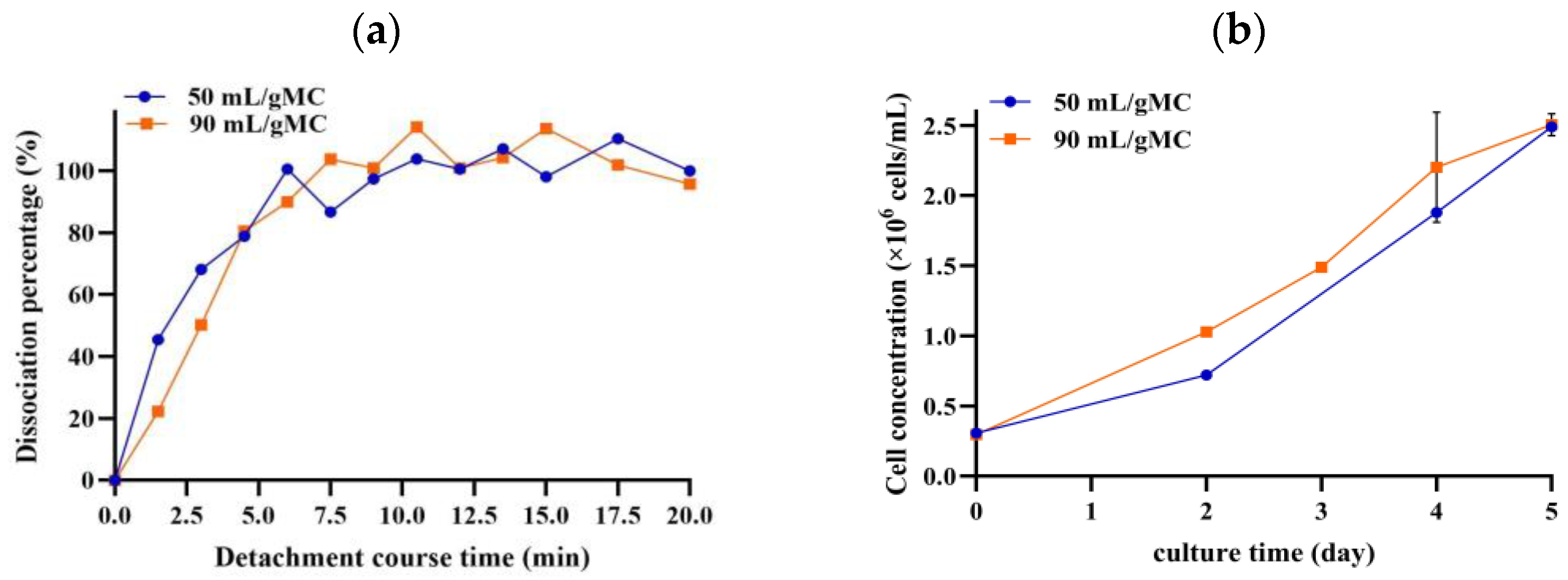
Figure 1 illustrates the outcomes. For both agitation speeds, complete cell detachment was achieved in under 20 min with a cell recovery exceeding 70% (Figure 1a). The cell detachment progress was observed by sampling and observing under the optical inverted microscope. For both conditions, the cells viability was above 98%. This indicates that there was no notable difference in terms of achieving cell detachment within a shorter period at the higher agitation speed.
Following cell detachment, as described in Section 2.3, the cells were transferred jointly with the spent MCs to a subsequent bioreactor to monitor the cell growth trend (Figure 1b). The data depicted in Figure 1b indicate that there were no discernible differences in cell growth after the two different treatments at the time of cell detachment. This suggests that the higher agitation speed did not negatively impact the cell properties in ways that could hinder subsequent cell culture.
To theoretically evaluate the impact of the agitation speed on cells, the Kolmogorov size, a measure of the forces generated by the agitation speed on cells [39], was calculated. This calculation (presented in Table 4), performed for the geometrical properties of a 1 L DASGIP bioreactor for two different volumes at the time of cell detachment, employed the formula presented in Table 4 and has been explained in detailed in [17,37].
As shown in Table 4, even at the higher agitation speed, the smallest eddy size (λmin) which is created near the impeller, is larger than the cell diameter (~18 μm), suggesting that cell damage is unlikely [40], which supports our findings, as illustrated in Figure 1a. However, we observed that in the reactor operating at a higher agitation speed (200 rpm), the temperature increased to 39.5 °C due to the elevated energy dissipation rate. This is confirmed in Table 4, where the mean specific dissipation energy increases by a factor of approximately 10. The amount of heat generated during high-speed agitation depends, among other factors, on the reactor volume. When using a smaller volume of enzymatic solution, the heat generated may have a more pronounced effect on the cells (see Table 4).
Previous studies [17,37,38], primarily conducted on the hMSc cell line, demonstrated that increasing the agitation speed up to five times the minimum agitation speed (Njs) is still suitable for maintaining cell viability. However, they have not reported heat generation during cell detachment. Considering the challenges associated with temperature control in single-use bioreactors, especially in the context of scaling up, it is advisable to opt for an agitation speed of 1.25 times Njs to ensure consistent and reliable results.
Table 4. Operational conditions at the time of cell detachment for two different agitation speeds at two different volumes.
Trypsin ratio (mL/gMC)5090
Volume (V) (mL) *245329
Agitation speed (N) (rpm)100200100200
Tip   speed   ( m / s ) ;   π × D i × N 0.2610.5230.2610.523
Reynolds   number   ( Re )   ρ × N × D i 2 μ 4429885944298859
Mean   specific   energy   dissipation   rate   ε ¯   ( m 2 / s 3 )   ε ¯ = P ρ × V   0.00880.07080.00660.052
λ   ( μ m ) ;   ϑ 3 ε ¯ 0.25 98.558.5106.063.0
Smallest possible eddy size λ min (μm) ** ϑ 3 ε m a x 0.25 68.9–78.340.9–46.568.9–78.340.9–46.5
* Volume at the time of Trypsinization, which is calculated based on MC concentration of 3.0 g/L upon removal of 80% of spent medium and two washes with PBS. Bioreactor type: DASGIP DASBOX Eppendorf Reactor. Tank diameter (DT) = 10.5 cm, which was used to calculate the height (T). Impeller type: 3-blade segment, 30° angled. Power consumption (kg.m2/S3): P = N p × ρ × N 3 × D i 5 . Power number, Np = 1.5 [41]. Impeller diameter (Di) = 5 cm; impeller blade depth (Wi) = 3 cm. Viscosity (ν) = 0.95 cP; density ( ρ ) = 1.01 g/cm3; kinematic viscosity (m2/S): ϑ = ν/ ρ . ** Smallest possible eddy size will be calculated based on the following formula for the maximum specific energy dissipation rate [17]:
ε m a x = P 0 N 3 D i 2 T 2 H V
ε m a x = P 0 N 3 D i 3 π 4 W i

3.2. Effect of Incubation Time on Cell Detachment from MCs and Subsequent Cell Growth

Based on the experiments performed in the previous section, cells were released completely from MCs within 10 to 15 min, facilitated by mechanical agitation of at least 1.25 × Njs. This outcome aligns with the findings of other studies [13,37,38,42,43,44]. To develop an approach suitable for larger operations, we investigated extended incubation periods of up to 150 min. This was driven by the need for a higher enzyme volume for cell detachment at larger scales (as indicated in Table 1), resulting in a longer time of liquid handling.
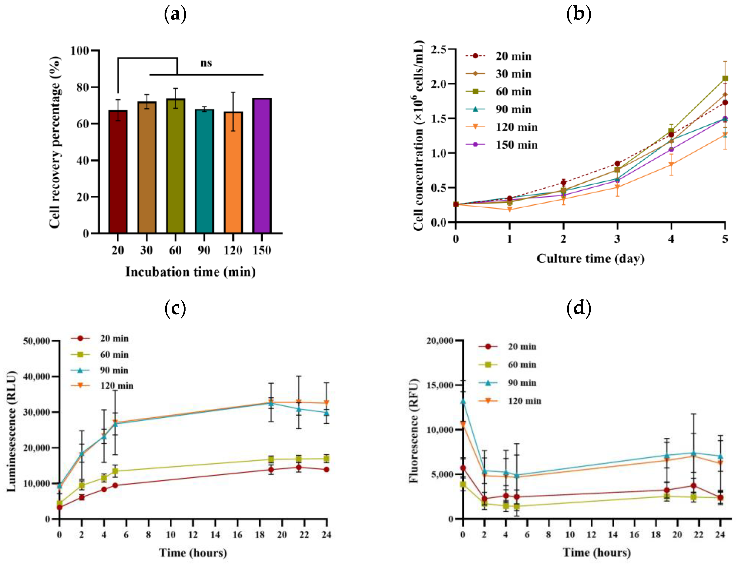
Cell detachment was performed with different incubation times of 20, 30, 60, 90, 120, and 150 min with Trypsin–EDTA (0.25%) solution at a concentration of 90 mL/gMC. As presented in Figure 2a, the cell recovery remained interestingly consistent across various incubation periods. The cell growth trend after cell detachment and subsequent cell culture on the MCs was monitored. According to Figure 2b, longer Trypsin incubation durations resulted in a notable delay in cell growth.
These findings were further supported by the analysis of apoptotic and necrotic single cells obtained from flask culture at different incubation times. A conspicuous increase in the relative luminescence signal, as presented in Figure 2c, was observed with longer incubation times using the Trypsin–EDTA (0.25%) solution, mirroring the trend of diminished growth after cell detachment on MCs. Additionally, a higher fluorescence signal for longer incubation times, even a few hours after seeding, indicated that a larger number of cells were affected during cell detachment by longer incubation times, subsequently appearing as dead cells after seeding (Figure 2d).
The adverse impact of longer incubation times with the Trypsin–EDTA (0.25%) solution may be attributed to the downregulation of proteins that regulate cell metabolism and growth, as elucidated in the work by Huang and colleagues [45].
Considering these findings, it is crucial to note that employing an incubation time exceeding 60 min with the Trypsin–EDTA (0.25%) solution has detrimental effects on subsequent cell growth. This holds even on a larger scale, emphasizing the importance of completing all steps, including the transfer of the enzyme solution, cell detachment, and transfer to the next culture unit, within 60 min.

3.3. Effect of Trypsin Volume on Cell Detachment from MCs and Subsequent Cell Growth

In prior experiments, utilizing Trypsin–EDTA solution (0.25%, with a measured activity of 890 ± 63 USP per mg of enzyme according to Section 2.5.2) at a ratio of 90 mL/gMC resulted in a 100% cell detachment efficiency while maintaining the cell viability above 98%. Given that 20% of the initial volume remained after medium removal and subsequent washing with PBS, the final Trypsin concentration in the bioreactor after adding Trypsin solution was 1.64 mg/mL (or 1465.2 USP/mL).
The ratio of 90 mL/gMC is equivalent to 20 μL/cm2 of MC surface, as 1 g of Cytodex 1 provides 4400 cm2. Some reports even indicated usage of up to 1000 μL/cm2 [46],while the Cytodex I manual recommends 30–50 mL/gMC which is equivalent to 7.5–12.5 μL/cm2 of Trypsin–EDTA solution (0.25%) for effective cell detachment [47]. This suggests the possibility of reducing the Trypsin ratio while maintaining a high detachment efficiency. In this study, Trypsin–EDTA solution (0.25%) at a ratio of 50 mL/gMC or 11 μL/cm2 of MC surface was explored. At this ratio, the final available active Trypsin was 1.29 mg/mL, or 1150.9 USP/mL.
We did not explore lower ratios of Trypsin–EDTA solution (0.25%) per g of MCs, as the focus of this study was developing a method for cell detachment across all scales up to 250 L in a single-use bioreactor. Referring to the data in Table 1, it is apparent that an enzyme volume below 50 mL/gMC will not adequately cover the impeller. This can lead to undesirable effects on mixing due to the impeller’s designed function being compromised. An inadequate volume during cell detachment may trigger vortex formation, entrapping bubbles in the fluid, and creating high shear stress through bubble bursting [48,49]. Therefore, maintaining an appropriate volume relative to the impeller height is crucial to ensure effective and controlled mixing without causing excessive turbulence or shear stress.
As shown in Figure 3a, reducing the Trypsin–EDTA solution ratio (0.25%) to 50 mL/gMC yielded no significant impact on the cell detachment efficiency, and the cells completely detached after 10 min of incubation at an agitation speed of 1.25 × Njs (100 rpm). This figure was developed by taking offline samples and measuring the single cell density using the Trypan Blue assay explained in Section 2.5.1. The data presented in Figure 3a reflect single experimental runs, but after cell detachment, subsequent cell culture was performed in duplicate (Figure 3b). Figure 3b indicates that the subsequent cell cultivation exhibited a similar growth trend to the previously observed results after 90 mL/gMC for detachment, with a comparable specific growth rate during the logarithmic growth phase (0.0178 h−1 and 0.0175 h−1, respectively). This study demonstrates that reducing the Trypsin volume, while ensuring sufficient coverage of the impeller across all scales and maintaining a high enzymatic activity, can result in a more cost-effective process.

3.4. Effect of Washing Steps on Cell Detachment Efficiency and Subsequent Cell Growth

Rinsing MCs post-medium removal is vital for effective cell detachment. The existing literature outlines various PBS solution ratios per gram of MC and recommends multiple washing steps (as summarized in Table 5). However, in these studies, the spent culture medium was removed completely, while in real conditions in single-use bioreactors, the removal of the whole medium is not feasible, as there is no deep tube that reaches exactly above the height of the settled MCs [16].
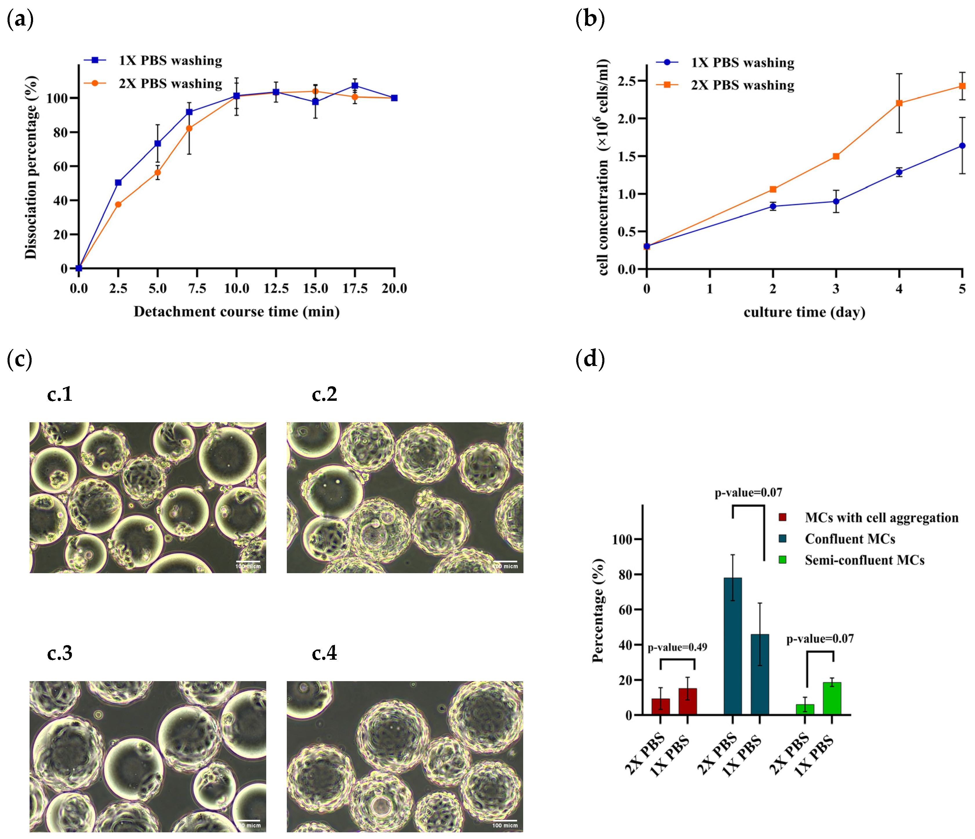
In this study, performing one step of PBS washing after medium removal, with a washing ratio of 40% of the initial volume, enabled effective cell detachment at a Trypsin–EDTA solution ratio of 90 mL/gMC (Figure 4a). However, reducing the washing steps to one, despite its benefits on a larger scale including time reduction, notably hindered subsequent cell growth, as depicted in Figure 4b. Furthermore, as illustrated in Figure 4c, the number of MCs fully covered with cells (confluent MCs) decreased within the same culture time, necessitating an extra day of culture to achieve the target cell concentration.
Additionally, a tendency for cell aggregation on MCs was observed over the culture period when one-time PBS washing was used. Figure 4c compares the cell aggregation forms for two cultures that differed only in their PBS wash times. Figure 4d compares the proportion of MCs carrying aggregated cells between the single PBS wash (1X PBS) condition and the two-times PBS wash (2X PBS) condition. Although the difference was not significant, it holds prominence. It is assumed that with only one PBS wash, cellular debris remains in the culture medium. This debris, following cell detachment, could adhere to both fresh and previously used MCs in subsequent stages, hindering optimal cell growth on MCs. Moreover, the debris could act as a nucleus for cell clumping atop MCs. In Figure 4b, a similar growth pattern was observed up to day two, followed by a decline. This pattern indicates that aggregated cells proliferated until day two, after which they mostly detached, resulting in a decrease in the total cell concentration.

3.5. Monitoring Cell Growth on MCs Using Capacitance Sensor

In this study, cell growth was monitored by using the Incyte Arc sensor as a capacitance sensor. It was observed that the permittivity signal has a linear correlation with the cell density on MCs up to 1.5 × 10⁶ cells/mL (Supplementary Materials, Figure S2a,b). This finding was in line with other studies that also proved that the value of the permittivity (pF/cm) has a linear correlation with the cell density up to a certain cell concentration while cells are growing on MCs [21,52]. Employing capacitance sensors for cell growth monitoring offers the benefit of minimizing the need for offline sampling and conventional cell density measurements, such as the crystal violet assay or viability assessments using staining methods like the Trypan Blue assay. Moreover, by estimating the cell density from the capacitance sensor data, the timing of infection can be determined without offline sampling, making the process more controllable and automated overall.
By the time MC confluence is reached and, hence, there is a lower available surface area, the size of the cells decreases, while the cells continue to proliferate. Due to the decreasing biovolume of cells, the permittivity signal at the given frequency is no longer representative of cell density [21,52].
Additionally, as shown in the Supplementary Materials, Figure S2a, the permittivity signal at a cell concentration of 1.5 × 10⁶ cells/mL is approximately 40 pF/cm. Reports from other research groups have also documented higher permittivity signals at this cell concentrations when cells are cultured on MCs [52]. This value is notably higher than the permittivity signals typically observed in suspension cultures of other cell lines at the same cell concentration, such as CHO [53] or SF9 [54], which exhibit a lower permittivity at various frequencies, <10 pF/cm. It is important to consider that the dielectric model was designed for suspension cells, indicating that adherent cells may respond differently to the electrical field [52]. This difference can be attributed to the fact that cells on MCs spread out and flatten, altering their morphology and potentially increasing their effective surface area, which leads to a higher permittivity signal due to greater interaction with the electric field.

3.6. Monitoring In Situ Cell Detachment by Capacitance Sensor

In addition to using the capacitance sensor to monitor cell growth, capacitance measurement was utilized during in situ cell detachment for various final Trypsin concentrations, as designed in Section 2.6.
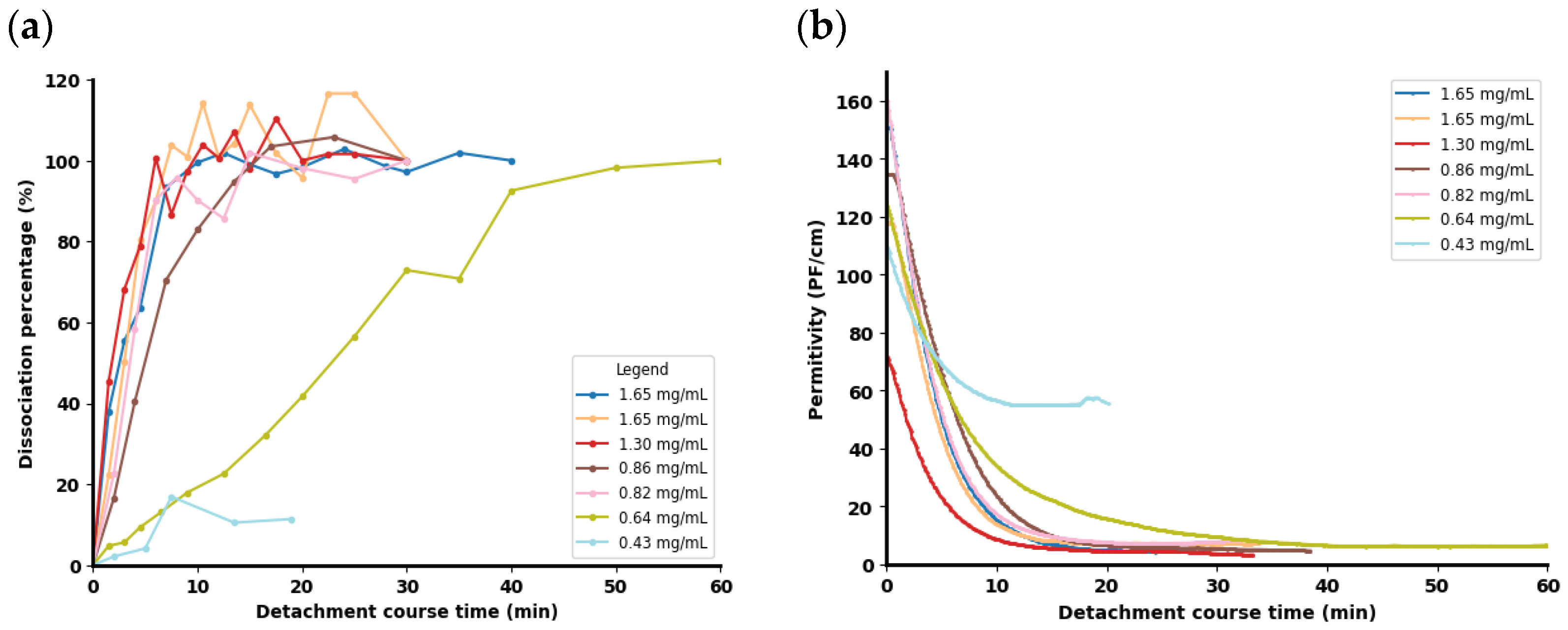
Figure 5a illustrates the detachment efficiency (calculated according to Section 2.5.1) during the Trypsinization process for different final enzyme concentrations. The data presented in this figure reflect single experimental runs. Given the extensive volume of experimental conditions, multiple repetitions were not feasible within this study.
In all experiments, cell viability following complete detachment exceeded 98%, and complete dissociation was achieved at enzyme concentrations above 0.64 mg/mL (575.43 USP/mL). However, at this concentration, it took 40 min to reach complete cell detachment. At an enzyme concentration of 0.43 mg/mL (383.6 USP/mL), cell detachment did not occur, as confirmed by the offline samples observed under the microscope, showing no cell detachment at 20 min; this was compared with higher enzyme concentrations, in which total cell detachment occurred (Supplementary Materials, Figure S3a,b). Although longer incubation times were effective for the 0.68 mg/mL Trypsin enzyme (Supplementary Materials Figure S4), a similar efficiency was not anticipated for the 0.43 mg/mL concentration. According to Figures S2a and S3, it was demonstrated that further extension of the incubation time for the 0.43 mg/mL trypsin did not significantly enhance cell detachment, indicating a concentration-dependent effect rather than a time-dependent improvement.
Figure 5b illustrates that the permittivity signal decreased upon the addition of the enzyme solution. Initially, the permittivity signal ranged from 120 to 160 pF/cm, indicating a high concentration of cells in the remaining volume after the removal of the conditioning medium and PBS washing. This signal corresponds to a cell concentration of 6–8 × 10⁶ cells/mL in the residual solution. As shown in Figure 5b, complete cell dissociation from the MCs results in a decrease in the permittivity signal to less than 10 pF/cm. Higher enzyme concentrations produce a sharper decline in the signal compared to lower concentrations. However, at very low enzyme concentrations, the permittivity signal decreases due to solution dilution but does not reach the same low permittivity value, instead plateauing at a higher signal value.
The drop in the signal value upon cell detachment from MCs can be attributed to morphological changes, with the cells becoming more spherical and uniform in shape, which affects their interaction with the electric field [52]. As discussed in the previous section (Section 3.5), at the same cell concentration, the permittivity signal value is higher when the cells are flattened on the MCs compared to when they are in a form of single cells in a suspended culture.

3.7. Decision-Making Tools for In Situ Cell Detachment

Based on the obtained result from the online measurement of the permittivity signal during in situ cell detachment, the capacitance sensor shows potential as a sensor to monitor how the enzyme is performing and predicting whether complete cell detachment will occur. The predictive model, developed according to Section 3.6, forecasts the permittivity signal value at 20 min of in situ cell detachment based on the signals obtained from minutes 4 to 7 of the detachment process. Figure 6 indicates the predictive performance of the model on the collected signals from the previous section, from which all the collected signals are from the older generation sensors (Incyte Unit DN12—320 and Incyte Unit DN12—420).
Figure 6 indicates the predictive performance of the model developed according to Section 2.7. It predicts the permittivity signal value at 20 min of in situ cell detachment based the obtained signal from minutes 4 to 7 of the cell detachment course. The model performance, based on the defined conditions in Section 3.6, was evaluated using the Root Mean Square Error (RMSE), which was 4.5.
To validate the model’s prediction performance, an experiment was conducted using an enzyme with a high concentration but low activity, stored in the refrigerator for 24 h. The activity of Trypsin–EDTA (0.25%) was assessed at different incubation times at room temperature and in a cold environment, as detailed in Supplementary Figure S5. After 24 h in the cold room (2–8 °C), the enzyme activity decreased to 378.3 USP/mg. In situ cell detachment was performed in a bioreactor with an enzyme volume ratio of 50 mL/gMC for a 3 g/L MC and a 700 mL culture volume.
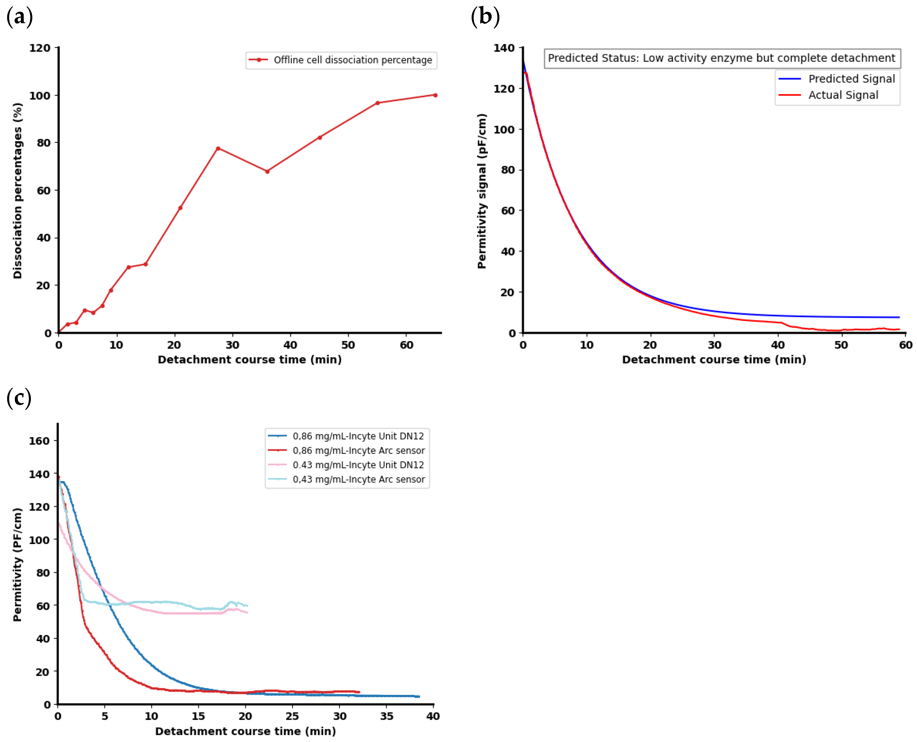
During the in situ cell detachment process, samples were collected at various time points and analyzed microscopically to measure the number of single cells using the Trypan Blue assay, as described in Section 2.5.1. Figure 7a illustrates the progress of cell detachment under these conditions, where complete detachment occurred at 60 min.
The permittivity signal was measured using two older generation sensors (Incyte Unit DN12–320 and Incyte Unit DN12–420). In Figure 7b, the average signal from these two sensors is depicted in red. The predictive signal generated by the developed model, based on the signal data collected from 4 to 7 min, for the whole course of cell detachment is presented in blue. The predicted signal at 20 min was determined to be 17.88 pF/cm. This value falls between 10 pF/cm and 20 pF/cm, which, according to the conditions defined in Section 2.7, indicates a slow but complete cell detachment at 60 min. The predicted signal closely matched the actual signal observed at 20 min, which was 17.27 pF/cm. The prediction error was minimal, with a deviation of only 0.6 pF/cm.
Additionally, to confirm the consistency and reproducibility of the obtained signal value from the older generation capacitance sensor (Incyte Unit DN12), a new generation of the cell capacitance sensor (Incyte Arc sensor) was also used in the experiments with enzyme concentrations of 0.86 g/L and 0.43 g/L, corresponding to 767.2 USP/mL and 383.6 USP/mL. The data were compared with those acquired using the older generation capacitance sensor. As illustrated in Figure 7c, similar outcomes were achieved, with the only observed difference being the slope of decrease, which shows a quicker response from the Incyte Arc sensor. The faster response observed with the Incyte Arc sensor suggests an improvement in the sensor technology, which might be attributed to advancements in the sensor design or enhanced sensitivity.
From the data presented in Table 6, it is evident that both the Incyte Unit DN12 and the Incyte Arc sensors show high consistency and reproducibility in detecting cell detachment events, with minor differences in the actual signal values obtained at 20 min during the cell detachment process. Additionally, for both sensors and enzyme concentrations, the predicted values from the developed model accurately predicted the status, demonstrating that the selected criteria were suitable for both types of sensors.
Based on these findings, the use of capacitance sensors to predict cell detachment in bioreactors during the initial stages of processes shows promise. This approach offers significant advantages by eliminating the need for offline sampling and observing the cell detachment under microscope, particularly in larger reactors where frequent sampling poses contamination risks and is time intensive in Good Manufacturing Practice (GMP) settings. Maintaining a signal value below 10 pF/cm can serve as a criterion for determining the optimal time to transfer processes to the next scale. These criteria help to minimize cell exposure to Trypsin solution and mitigate potential negative impacts during subsequent recolonization on MCs, which was observed in Section 3.2.
Furthermore, the capacitance sensor and predictive model provide valuable assistance in scenarios where the enzyme activity levels are low or unknown to operators. By predicting the signal value and cell detachment status in 20 min, operators can make informed decisions, such as adding more enzyme to accelerate cell detachment. Thereby, this methodology will facilitate streamlined decision making throughout the manufacturing process.

4. Conclusions

In conclusion, this study focused on optimizing the in situ cell detachment process for MA 104 cells cultivated on Cytodex 1 MCs in a scale-down approach by systematically investigating critical operational parameters such as the agitation speed, incubation time, Trypsin concentration, and washing steps.
Our findings underscore the significance of maintaining an optimal agitation speed, approximately 1.25 × Njs (minimum agitation speed), to ensure uniform MC distribution, prevent clumping, and support effective enzymatic cell dissociation. Prolonged incubation times of more than 1 h during Trypsinization were found to adversely affect cell growth rates, highlighting the need for precise control in larger-scale processes.
Moreover, our study demonstrates the feasibility of reducing Trypsin consumption by using lower volumes per gram of MC without compromising the cell detachment efficiency. This finding has practical implications for cost-effective, large-scale production, although maintaining a final Trypsin activity of at least 500 USP/mL is crucial. We also observed that two times washing steps after medium removal are essential for effective cell detachment and subsequent growth. Single washes resulted in decreased cell growth and increased cell aggregation on MCs.
The optimized range of critical parameters was selected not solely for achieving a higher yield of viable cells at the end of the detachment process, which was one of the objectives. Instead, the chosen optimized conditions were primarily based on their impact on subsequent cell growth on MCs, ensuring optimal cell proliferation post-detachment. This approach addresses a gap in the existing literature in this field [17,18]. While this study addresses scalability by selecting scale-independent parameters, practical challenges in large-scale implementation may still arise, which could potentially affect the overall efficiency and cost-effectiveness of the process.
Introducing real-time monitoring of cell detachment using a capacitance sensor showed promise in tracking detachment efficiency and predicting detachment status. This capability facilitates timely decision making for subsequent process steps and enhances overall process control. Furthermore, it reduces the need for offline sampling.
Additionally, the proposed technique for real-time monitoring of cell detachment holds significant potential, especially for detaching hMSc cells from MCs. Given the sensitivity of these cells, predicting the optimal moment for Trypsin inactivation can help mitigate potential damage to vital surface markers that are crucial for their differentiation.
In conclusion, this study provides significant insights and a structured methodology for optimizing in situ cell detachment processes, especially in microcarrier-based (MC-based) cell culture systems. These results are highly relevant for applications in bioprocessing, vaccine production, and the manufacturing of biologics, offering potential improvements in the cost efficiency and product quality. Additionally, this study, building on our previous research on MC-based cell propagation, demonstrates economic benefits in terms of reduced material and consumable usage. The accompanying process flow chart and comparison tables, detailed in Supplementary Figure S6 and Tables S1 and S2, illustrate the cost reductions achieved while ensuring a more consistent and less labor-intensive process.

Supplementary Materials

The following supporting information can be downloaded at: https://www.mdpi.com/article/10.3390/pr12091887/s1, Figure S1. Fishbone diagram of the parameters involved in in situ cell detachment, Figure S2. Kinetics of MA 104 cell cultures performed under condition explained in Section 2.2: (a) offline cell density measurements ( ) and online permittivity (-) for four independent experiments; (b) linear correlation of permittivity signal vs. cell concentration in culture. Figure S3. Visual observation of cell dissociation with an inverted microscope for cell dissociation, with final enzyme concentrations of 0.43 g/L (a) and 0.86 g/L (b) over in situ cell detachment course. Figure S4. Visual observation of cell dissociation with an inverted microscope for cell dissociation with final enzyme concentration of 0.64 g/L. Figure S5. (Trypsin–EDTA (0.25%) activity over different incubation times at room temperature and in a cold room (2–8 °C), File S1. xlsx, File S2. rar, File S3. doc, Figure S6. Schematic representation of the process flow for cell expansion and culture (a) using roller bottles, followed by transfer to the final bioreactor for viral vaccine production. (b) using single-use bioreactor and cultivation on microcarrier, Table S1. Comparison of costs associated with static cell culture and microcarrier-based (MC-based) cell culture systems across different passages. Table S2. Comparison of resource consumption in microcarrier-based (MC-based) and static-based cell propagation.

Author Contributions

Conceptualization, A.E., M.D., M.M., R.B. and H.K.; data curation, A.E. and H.K.; formal analysis, A.E. and M.S.; funding acquisition, M.M. and R.B.; investigation, A.E. and M.S.; methodology, A.E. and H.K.; project administration, H.K.; resources, M.M.; software, A.E.; supervision, M.D., M.M., R.B. and H.K.; validation, A.E., R.B. and H.K.; visualization, A.E.; writing—original draft, A.E.; writing—review and editing, M.S., M.D., R.B. and H.K. All authors have read and agreed to the published version of the manuscript.

Funding

This research was conducted in the framework of ACIB Co. and in the scope of COMET: The COMET center ACIB was funded by BMK, BMDW, SFG, Standortagentur Tirol, Government of Lower Austria und Vienna Business Agency in the framework of COMET—Competence Centers for Excellent Technologies. The COMET Funding Program is managed by the Austrian Research Promotion Agency FFG.

Data Availability Statement

The original contributions presented in the study are included in the article/Supplementary Materials, further inquiries can be directed to the corresponding author.

Acknowledgments

The authors would like to extend their appreciation to Lukas Herzog for his their assistance in the laboratory workplace at FH Campus Wien. Thanks to Dominik Wolfsberger from Eppendorf Co. for giving us the detailed drawing for the single-use bioreactor. Special thanks to Jochen Uhlenküken and William Tima from Hamilton company for their support and providing us the new generation sensor of cell capacitance in this study.

Conflicts of Interest

Author Mark Dürkop was employed by the company Novasign GmbH. The remaining authors declare that the research was conducted in the absence of any commercial or financial relationships that could be construed as a potential conflict of interest. The Novasign GmbH had no role in the design of the study; in the collection, analyses, or interpretation of data; in the writing of the manuscript, or in the decision to publish the results.

References

  1. van Wezel, A.L. Growth of cell-strains and primary cells on micro-carriers in homogeneous culture. Nature 1967, 216, 64–65. [Google Scholar] [CrossRef]
  2. Malda, J.; Frondoza, C.G. Microcarriers in the engineering of cartilage and bone. Trends Biotechnol. 2006, 24, 299–304. [Google Scholar] [CrossRef]
  3. Ton, C.; Stabile, V.; Carey, E.; Maraikar, A.; Whitmer, T.; Marrone, S.; Afanador, N.L.; Zabrodin, I.; Manomohan, G.; Whiteman, M.; et al. Development and scale-up of rVSV-SARS-CoV-2 vaccine process using single use bioreactor. Biotechnol. Rep. 2023, 37, e00782. [Google Scholar] [CrossRef] [PubMed]
  4. Fang, Z.; Lyu, J.; Li, J.; Li, C.; Zhang, Y.; Guo, Y.; Wang, Y.; Zhang, Y.; Chen, K. Application of bioreactor technology for cell culture-based viral vaccine production: Present status and future prospects. Front. Bioeng. Biotechnol. 2022, 10, 921755. [Google Scholar] [CrossRef] [PubMed]
  5. Kumraj, G.; Pathak, S.; Shah, S.; Majumder, P.; Jain, J.; Bhati, D.; Hanif, S.; Mukherjee, S.; Ahmed, S. Capacity Building for Vaccine Manufacturing Across Developing Countries: The Way Forward. Hum. Vaccin. Immunother. 2022, 18, 2020529. [Google Scholar] [CrossRef]
  6. Smeaton, J. Manufacturing COVID-19 Vaccines. Available online: https://post.parliament.uk/manufacturing-covid-19-vaccines (accessed on 14 January 2021).
  7. Badenes, S.M.; Fernandes-Platzgummer, A.; Rodrigues, C.; Diogo, M.M.; Da Silva, C.L.; Cabral, J. Microcarrier Culture Systems for Stem Cell Manufacturing. In Stem Cell Manufacturing; Elsevier: Amsterdam, The Netherlands, 2016; pp. 77–104. ISBN 9780444632654. [Google Scholar]
  8. Chen, X.-Y.; Chen, J.-Y.; Tong, X.-M.; Mei, J.-G.; Chen, Y.-F.; Mou, X.-Z. Recent advances in the use of microcarriers for cell cultures and their ex vivo and in vivo applications. Biotechnol. Lett. 2020, 42, 1–10. [Google Scholar] [CrossRef] [PubMed]
  9. Luo, X.; Niu, Y.; Fu, X.; Lin, Q.; Liang, H.; Liu, L.; Li, N. Large-Scale Microcarrier Culture of Chinese Perch Brain Cell for Viral Vaccine Production in a Stirred Bioreactor. Vaccines 2021, 9, 1003. [Google Scholar] [CrossRef]
  10. George, M.; Farooq, M.; Dang, T.; Cortes, B.; Liu, J.; Maranga, L. Production of cell culture (MDCK) derived live attenuated influenza vaccine (LAIV) in a fully disposable platform process. Biotechnol. Bioeng. 2010, 106, 906–917. [Google Scholar] [CrossRef]
  11. Derakhti, S.; Safiabadi-Tali, S.H.; Amoabediny, G.; Sheikhpour, M. Attachment and detachment strategies in microcarrier-based cell culture technology: A comprehensive review. Mater. Sci. Eng. C 2019, 103, 109782. [Google Scholar] [CrossRef]
  12. Kiesslich, S.; Kamen, A.A. Vero cell upstream bioprocess development for the production of viral vectors and vaccines. Biotechnol. Adv. 2020, 44, 107608. [Google Scholar] [CrossRef]
  13. Souza, M.C.d.O.; Da Freire, M.S.; Castilho, L.d.R. Influence of culture conditions on Vero cell propagation on non-porous microcarriers. Braz. Arch. Biol. Technol. 2005, 48, 71–77. [Google Scholar] [CrossRef]
  14. Mendonça, R.Z.; Prado, J.C.M.; Pereira, C.A. Attachment, spreading and growth of Vero cells on microcarriers for the optimization of large scale cultures. Bioprocess Biosyst. Eng. 1999, 20, 565. [Google Scholar] [CrossRef]
  15. Maillot, C.; Isla, N.; de Loubiere, C.; Toye, D.; Olmos, E. Impact of microcarrier concentration on mesenchymal stem cell growth and death: Experiments and modeling. Biotechnol. Bioeng. 2022, 119, 3537–3548. [Google Scholar] [CrossRef] [PubMed]
  16. Ebrahimian, A.; Schalk, M.; Dürkop, M.; Maurer, M.; Bliem, R.; Kühnel, H. Seed Train Optimization in Microcarrier-Based Cell Culture Post In Situ Cell Detachment through Scale-Down Hybrid Modeling. Bioengineering 2024, 11, 268. [Google Scholar] [CrossRef]
  17. Sousa, M.; Fenge, C.; Rupprecht, J.; Tappe, A.; Greller, G.; Alves, P.; Carrondo, M.; Roldão, A. Process intensification for Peste des Petites Ruminants Virus vaccine production. Vaccine 2019, 37, 7041–7051. [Google Scholar] [CrossRef] [PubMed]
  18. Rourou, S.; Riahi, N.; Majoul, S.; Trabelsi, K.; Kallel, H. Development of an in situ detachment protocol of Vero cells grown on Cytodex1 microcarriers under animal component-free conditions in stirred bioreactor. Appl. Biochem. Biotechnol. 2013, 170, 1724–1737. [Google Scholar] [CrossRef]
  19. Dabros, M.; Dennewald, D.; Currie, D.J.; Lee, M.H.; Todd, R.W.; Marison, I.W.; Stockar, U. von. Cole-Cole, linear and multivariate modeling of capacitance data for on-line monitoring of biomass. Bioprocess Biosyst. Eng. 2009, 32, 161–173. [Google Scholar] [CrossRef] [PubMed]
  20. Degouys, V.; Cerckel, I.; Garcia, A.; Harfield, J.; Dubois, D.; Fabry, L.; Miller, A.O. Dielectric spectroscopy of mammalian cells. 2. Simultaneous in situ evaluation by aperture impedance pulse spectroscopy and low frequency dielectric spectroscopy of the biomass of HTC cells on Cytodex 3. Cytotechnology 1993, 13, 195–202. [Google Scholar] [CrossRef]
  21. El Wajgali, A.; Esteban, G.; Fournier, F.; Pinton, H.; Marc, A. Impact of microcarrier coverage on using permittivity for on-line monitoring high adherent Vero cell densities in perfusion bioreactors. Biochem. Eng. J. 2013, 70, 173–179. [Google Scholar] [CrossRef]
  22. Justice, C.; Leber, J.; Freimark, D.; Pino Grace, P.; Kraume, M.; Czermak, P. Online- and offline- monitoring of stem cell expansion on microcarrier. Cytotechnology 2011, 63, 325–335. [Google Scholar] [CrossRef]
  23. Sion, C.; Ghannoum, D.; Ebel, B.; Gallo, F.; Isla, N.; de Guedon, E.; Chevalot, I.; Olmos, E. A new perfusion mode of culture for WJ-MSCs expansion in a stirred and online monitored bioreactor. Biotechnol. Bioeng. 2021, 118, 4453–4464. [Google Scholar] [CrossRef] [PubMed]
  24. Juanola, S.; Garcia, L.; Mouriño, M.; Cheeseman, S.; Urniza, A.; Cheeseman, S.; Scholz, J.; Boulais, A. Control and Scale-Up of A Microcarrier-Based Viral Vaccine Process Control and Scale-Up of A Microcarrier-Based Viral Vaccine Process Using BioPAT® ViaMass for Inline Viable Cell Density Measurement. Available online: https://www.sartorius.com/download/457214/5/appl-note-biopat-viamass-zoetis-2568350-000-e-data.pdf (accessed on 21 January 2020).
  25. Petiot, E.; Ansorge, S.; Rosa-Calatrava, M.; Kamen, A. Critical phases of viral production processes monitored by capacitance. J. Biotechnol. 2017, 242, 19–29. [Google Scholar] [CrossRef] [PubMed]
  26. Farrell, C.J.; Cicalese, S.M.; Davis, H.B.; Dogdas, B.; Shah, T.; Culp, T.; van Hoang, M. Cell confluency analysis on microcarriers by micro-flow imaging. Cytotechnology 2016, 68, 2469–2478. [Google Scholar] [CrossRef]
  27. Odeleye, A.O.O.; Castillo-Avila, S.; Boon, M.; Martin, H.; Coopman, K. Development of an optical system for the non-invasive tracking of stem cell growth on microcarriers. Biotechnol. Bioeng. 2017, 114, 2032–2042. [Google Scholar] [CrossRef]
  28. Anton, F.; Burzlaff, A.; Kasper, C.; Brückerhoff, T.; Scheper, T. Preliminary Study towards the Use of In-situ Microscopy for the Online Analysis of Microcarrier Cultivations. Eng. Life Sci. 2007, 7, 91–96. [Google Scholar] [CrossRef]
  29. Benavides, O.R.; Gibbs, H.C.; White, B.P.; Kaunas, R.; Gregory, C.A.; Walsh, A.J.; Maitland, K.C. Volumetric imaging of human mesenchymal stem cells (hMSCs) for non-destructive quantification of 3D cell culture growth. PLoS ONE 2023, 18, e0282298. [Google Scholar] [CrossRef] [PubMed]
  30. Yamaguchi, J.; Onodera, T.; Homan, K.; Liang, X.; Matsuoka, M.; Miyazaki, T.; Yoshiaki, H.; Saito, M.; Iwasaki, N. Optical coherence tomography evaluation of the spatiotemporal effects of 3D bone marrow stromal cell culture using a bioreactor. J. Biomed. Mater. Res. B Appl. Biomater. 2022, 110, 1853–1861. [Google Scholar] [CrossRef] [PubMed]
  31. Bournonville, S.; de Geris, L.; Kerckhofs, G. Micro computed tomography with and without contrast enhancement for the characterization of microcarriers in dry and wet state. Sci. Rep. 2021, 11, 2819. [Google Scholar] [CrossRef]
  32. Schnitzler, A.C.; Verma, A.; Kehoe, D.E.; Jing, D.; Murrell, J.R.; Der, K.A.; Aysola, M.; Rapiejko, P.J.; Punreddy, S.; Rook, M.S. Bioprocessing of human mesenchymal stem/stromal cells for therapeutic use: Current technologies and challenges. Biochem. Eng. J. 2016, 108, 3–13. [Google Scholar] [CrossRef]
  33. Merck. Trypsin Assay Procedure. Available online: https://www.sigmaaldrich.com/AT/de/technical-documents/protocol/protein-biology/enzyme-activity-assays/enzymatic-assay-of-trypsin (accessed on 28 August 2024).
  34. United Sated Pharmacopoeia. Official Monographs/Trypsin 5969 (35). Available online: https://www.drugfuture.com/Pharmacopoeia/usp35/PDF/4969-4970%20Crystallized%20Trypsin.pdf (accessed on 1 May 2012).
  35. Liu, K. Trypsin Inhibitor Assay: Expressing, Calculating, and Standardizing Inhibitor Activity in Absolute Amounts of Trypsin Inhibited or Trypsin Inhibitors. J. Am. Oil Chem. Soc. 2021, 98, 355–373. [Google Scholar] [CrossRef]
  36. Kupcho, K.; Shultz, J.; Hurst, R.; Hartnett, J.; Zhou, W.; Machleidt, T.; Grailer, J.; Worzella, T.; Riss, T.; Lazar, D.; et al. A real-time, bioluminescent annexin V assay for the assessment of apoptosis. Apoptosis 2019, 24, 184–197. [Google Scholar] [CrossRef] [PubMed]
  37. Nienow, A.W.; Hewitt, C.J.; Heathman, T.R.; Glyn, V.A.; Fonte, G.N.; Hanga, M.P.; Coopman, K.; Rafiq, Q.A. Agitation conditions for the culture and detachment of hMSCs from microcarriers in multiple bioreactor platforms. Biochem. Eng. J. 2016, 108, 24–29. [Google Scholar] [CrossRef]
  38. Heathman, T.R.; Nienow, A.W.; Rafiq, Q.A.; Coopman, K.; Kara, B.; Hewitt, C.J. Agitation and aeration of stirred-bioreactors for the microcarrier culture of human mesenchymal stem cells and potential implications for large-scale bioprocess development. Biochem. Eng. J. 2018, 136, 9–17. [Google Scholar] [CrossRef]
  39. Petry, F.; Salzig, D. Impact of Bioreactor Geometry on Mesenchymal Stem Cell Production in Stirred-Tank Bioreactors. Chem. Ing. Tech. 2021, 93, 1537–1554. [Google Scholar] [CrossRef]
  40. Nienow, A.W. Reactor engineering in large scale animal cell culture. Cytotechnology 2006, 50, 9–33. [Google Scholar] [CrossRef]
  41. Glaser, R.; Greenlea, Z.; Sha, M. Stimulating Growth. Cultivating Solutions: Power Number for Cell Culture Glass Vessels. Available online: https://www.eppendorf.com/product-media/doc/en/70270/Fermentors-Bioreactors_Brochure_Bioprocess-Family_Stimulating-Growth-Cultivating-Solutions.pdf (accessed on 28 August 2024).
  42. Yang, J.; Guertin, P.; Jia, G.; Lv, Z.; Yang, H.; Ju, D. Large-scale microcarrier culture of HEK293T cells and Vero cells in single-use bioreactors. AMB Express 2019, 9, 70. [Google Scholar] [CrossRef]
  43. Hewitt, C.J.; Lee, K.; Nienow, A.W.; Thomas, R.J.; Smith, M.; Thomas, C.R. Expansion of human mesenchymal stem cells on microcarriers. Biotechnol. Lett. 2011, 33, 2325–2335. [Google Scholar] [CrossRef]
  44. Goh, T.K.-P.; Zhang, Z.-Y.; Chen, A.K.-L.; Reuveny, S.; Choolani, M.; Chan, J.K.Y.; Oh, S.K.-W. Microcarrier culture for efficient expansion and osteogenic differentiation of human fetal mesenchymal stem cells. Biores. Open Access 2013, 2, 84–97. [Google Scholar] [CrossRef]
  45. Huang, H.-L.; Hsing, H.-W.; Lai, T.-C.; Chen, Y.-W.; Lee, T.-R.; Chan, H.-T.; Lyu, P.-C.; Wu, C.-L.; Lu, Y.-C.; Lin, S.-T.; et al. Trypsin-induced proteome alteration during cell subculture in mammalian cells. J. Biomed. Sci. 2010, 17, 36. [Google Scholar] [CrossRef]
  46. Fonte, G. The Effect of Key Process Parameters on Human Mesenchymal Stem Cell Expansion and Harvest. Available online: https://tinyurl.com/9w968chj (accessed on 1 November 2014).
  47. Cytiva. Cytodex 1, Cytodex 3 Cell Culture. Available online: https://cytiva-delivery.sitecorecontenthub.cloud/api/public/content/digi-11574-pdf (accessed on 1 June 2020).
  48. Motamedvaziri, S.; Armenante, P.M. Flow regimes and surface air entrainment in partially filled stirred vessels for different fill ratios. Chem. Eng. Sci. 2012, 81, 231–250. [Google Scholar] [CrossRef]
  49. Martín, M.; Montes, F.J.; Galán, M.A. Influence of Impeller Type on the Bubble Breakup Process in Stirred Tanks. Ind. Eng. Chem. Res. 2008, 47, 6251–6263. [Google Scholar] [CrossRef]
  50. Souza, M.C.O.; Freire, M.S.; Schulze, E.A.; Gaspar, L.P.; Castilho, L.R. Production of yellow fever virus in microcarrier-based Vero cell cultures. Vaccine 2009, 27, 6420–6423. [Google Scholar] [CrossRef]
  51. Nienow, A.W.; Rafiq, Q.A.; Coopman, K.; Hewitt, C.J. A potentially scalable method for the harvesting of hMSCs from microcarriers. Biochem. Eng. J. 2014, 85, 79–88. [Google Scholar] [CrossRef]
  52. Petiot, E.; El-Wajgali, A.; Esteban, G.; Gény, C.; Pinton, H.; Marc, A. Real-time monitoring of adherent Vero cell density and apoptosis in bioreactor processes. Cytotechnology 2012, 64, 429–441. [Google Scholar] [CrossRef] [PubMed]
  53. Metze, S.; Ruhl, S.; Greller, G.; Grimm, C.; Scholz, J. Monitoring online biomass with a capacitance sensor during scale-up of industrially relevant CHO cell culture fed-batch processes in single-use bioreactors. Bioprocess Biosyst. Eng. 2020, 43, 193–205. [Google Scholar] [CrossRef] [PubMed]
  54. Negrete, A.; Esteban, G.; Kotin, R.M. Process optimization of large-scale production of recombinant adeno-associated vectors using dielectric spectroscopy. Appl. Microbiol. Biotechnol. 2007, 76, 761–772. [Google Scholar] [CrossRef]
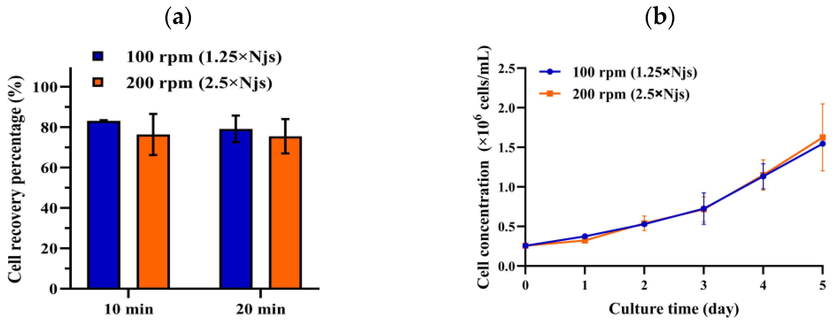
Figure 1. (a) Cell recovery percentage during Trypsinization at two periods, 10 and 20 min, for two different agitation speeds, 100 rpm (1.25 × Njs) and 200 rpm (2.5 × Njs), in 1 L DASGIP bioreactor; (b) cell growth on MCs after undergoing in situ cell detachment. For each condition, two separate experiments were performed.
Processes 12 01887 g001
Figure 2. Effect of incubation time: (a) cell detachment efficiency over different incubation times with Trypsin solution (0.25%) (ns: not significant), (b) cell growth monitoring on MCs post-detachment using Trypsin solution (0.25%) at different incubation times, and (c,d) single cells subjected with different incubation time with Trypsin solution (0.25%) and subsequently seeded on plates, with the monitoring of apoptotic (RLU) and necrotic (RFU) cells over 24 h.
Processes 12 01887 g002
Figure 3. Impact of different volume ratios of Trypsin–EDTA solution (0.25%) at the time of cell detachment: (a) cell detachment efficiency over cell treatment with Trypsin solution (0.25%) with two ratios; (b) subsequent cell growth after cell detachment with two different ratios of Trypsin solution (0.25%).
Processes 12 01887 g003
Figure 4. Impact of washing step times on cell detachment and subsequent cell growth: (a) efficiency of cell detachment during treatment with Trypsin–EDTA (0.25%) solution combined with one or two steps of PBS washing at a 40% iv; (b) subsequent cell growth post detachment; (c) monitoring the cell aggregation on MCs during the culture after undergoing one-time and two-time PBS washing during in situ cell detachment: (c.1,c.2) notable presence of cell aggregation on MCs at 2nd day and 4th day of culture for the condition of one-time PBS washing, and (c.3,c.4) no notable cell aggregation on MCs at 2nd day and 4th day of culture for condition of two time-PBS washing; (d) comparison of observed aggregated cells on MCs, and the level of full (confluent) or partial (semi-confluent) coverage of MCs at the end of culture with one or two PBS washing steps (1X PBS or 2X PBS) before enzymatic cell detachment.
Processes 12 01887 g004
Figure 5. Cell dissociation monitoring: (a) development of cell dissociation over the course of in situ cell detachment monitored by taking samples and offline measurement of cell density; (b) permittivity signal over cell dissociation with different final Trypsin concentrations (measured with two independent capacitance sensors per each enzyme concentration).
Processes 12 01887 g005
Figure 6. The predictive performance of the model using permittivity signals obtained from capacitance sensors across seven experimental runs with different Trypsin concentrations: 1.65 g/L (runs 1 and 2), 1.29 g/L (run 3), 0.86 g/L (run 4), 0.82 g/L (run 5), 0.64 g/L (run 6), and 0.43 g/L (run 7).
Processes 12 01887 g006
Figure 7. (a) The effectiveness of cell release during trypsin treatment; (b) measured permittivity signal (red) and predicted permittivity signal (blue) during in situ cell detachment using Trypsin solutions with high enzyme concentration but lower activity (1.29 g/L, 488 USP/mL); (c) permittivity signals from two generations of capacitance sensors for two distinct enzyme concentrations: 0.86 g/L and 0.43 g/L.
Processes 12 01887 g007
Table 1. Range of selected critical parameters during in situ cell detachment.
ParameterLower LimitUpper LimitOther Parameters’ Setpoints
Agitation speed (rpm)100 *200 *90 mL/gMC, 30 min, 2X PBS wash
Incubation time with enzyme (min)2015090 mL/gMC; 100 rpm, 2X PBS wash
Trypsin–EDTA (0.25%) volume, mL/gMC509020 min, 100 rpm, 2X PBS wash
PBS washing steps (40% of initial volume)1220 min, 100 rpm, 90 mL/gMC
* 100 and 200 rpm correspond to 1.25 and 2.5 times the Njs (minimum agitation speed) in 1 L bioreactor, respectively.
Table 2. Assumed process conditions on a larger scale at the time of in situ cell detachment.
3c10c50c250 L1000 L
Bioreactor working volume (L)1.25–3.753.3–1018–40 L125–250500–1000
Actual culture volume (L)2.51040190900
MC concentration (g/L) *3.03.83.93.83.8
Vessel diameter (cm)14.720.433.759.7NA **
Blade width + off-bottom clearance (cm)7.09.016.530.0NA
Minimum required volume to cover the impeller1.182.94314.7183.93NA
20% remaining (L) ***0.72850NA
Minimum volume of enzyme solution (L)0.440.946.7133.93NA
Minimum volume of enzyme solution/gMC46.224.843.147.0NA
* MC concentration before cell transfer from the previous reactor is 3 g/L, which increases after in situ cell detachment, as the previously used MCs are transferred with the detached cells [16]; ** NA: not applicable, as 1000 L is considered the final scale for antigen manufacturing. *** The remaining volume after conditioned medium removal [16].
Table 3. Experimental design for in situ cell detachment with varying Trypsin–EDTA concentrations.
Run No.MC Conc. (g/L)Culture Volume (mL)Trypsin–EDTA Conc. (mg/mL) *Trypsin–EDTA Volume
(mL/gMC)
Final Trypsin Conc.
(mg/mL)
Final Trypsin Activity (USP/mL) **
1 ***37002.5901.651465.2
2 ****37002.5501.301150.9
337001.25900.86767.2
437001.25900.82732.6
537001.25500.64575.4
637000.830500.43383.6
* Other concentration of Trypsin except 2.5 mg/mL was prepared by diluting Trypsin- EDTA (0.25%) with PBS, ** The final Trypsin activity, measured using the Trypsin–EDTA (0.25%) activity method detailed in Section 2.5.2, was determined to be 890 ± 63 USP/mg. *** This experiment was conducted in duplicate. **** In all experiments except Run No. 2, two older-generation sensors (Incyte Unit DN12—320 and Incyte Unit DN12—420) were used.
Table 5. PBS washing ratios and steps for effective cell detachment from MCs in different studies.
StrainMedium ConditionWashing Ratio (%iv *)Washing StepsReference
VeroSCM **302–3[42]
hMSCSCM41.53[44]
hMSCSFM/SCMNA2[37]
VeroSFM ***-0[17,50]
hMSCSFM202[38]
hMSCnm ****1001[51]
* iv: initial volume, ** SCM: serum-containing medium, *** SFM: serum-free medium, **** nm: Not mentioned.
Table 6. Comparison of predicted and actual signal values and statuses.
Sensor Final Enzyme Conc.
(g/L)
Predicted Signal at 20 minPredicted StatusMeasured Signal at 20 minActual Status
Incyte Unit DN120.4350.3No active enzyme, no detachment55.6No cell detachment
Incyte Arc 57.1No active enzyme, no detachment59.8No cell detachment
Incyte Unit DN120.860Fast and complete detachment6.54Complete cell detachment but slow
Incyte Arc 0Fast and complete detachment6.91Complete cell detachment but slow
Disclaimer/Publisher’s Note: The statements, opinions and data contained in all publications are solely those of the individual author(s) and contributor(s) and not of MDPI and/or the editor(s). MDPI and/or the editor(s) disclaim responsibility for any injury to people or property resulting from any ideas, methods, instructions or products referred to in the content.