Article

Drug Repurposing in Alternative Medicine: Sochehwan, a Polyherbal Traditional Korean Digestant, Protects against Alcoholic Steatohepatitis by Regulating Cytochrome P450 2E1 Expression

1
Department of Diagnostics, College of Korean Medicine, Dongguk University, Dongguk-ro 32, Goyang 10326, Korea
2
Department of Pathology, College of Korean Medicine, Dongguk University, Dongguk-ro 32, Goyang 10326, Korea
3
Institute of Korean Medicine, Dongguk University, Dongguk-ro 32, Goyang 10326, Korea
*
Authors to whom correspondence should be addressed.
Processes 2021, 9(10), 1760; https://doi.org/10.3390/pr9101760
Submission received: 30 August 2021 / Revised: 10 September 2021 / Accepted: 28 September 2021 / Published: 30 September 2021
(This article belongs to the Section Pharmaceutical Processes)

Abstract

:
Sochehwan (SCH) is an herbal prescription from traditional oriental medicine and is currently used to treat digestive ailments. In a previous study, SCH was found to have the potential to attenuate metabolic syndrome (MetS) by activating AMPK and downstream signaling. From the view of drug repurposing, the efficacy of SCH on alcoholic liver injury is implied in classic medical texts but is yet to be proven. C57BL/6J mice were pre-treated with SCH orally for 5 days and challenged by providing a pair-fed Lieber DeCarli diet containing alcohol for 20 days. Hepatic enzyme and triglyceride levels and endoplasmic reticulum (ER) stress-related markers were analyzed. Moreover, mitogen-activated protein kinases (MAPKs) and cytochrome P450 2E1 (CYP2E1) levels were determined. CYP2E1-transfected HepG2 cells were used to test the cytoprotective efficacy of SCH against the adverse effects of alcohol in vitro. In mice, SCH administration notably reduced hepatic enzyme activity and neural lipid levels. Furthermore, ER-stress markers and MAPK phosphorylation were reduced due to ROS suppression, which was attributed to decreased CYP2E1 expression in liver tissue. In addition, SCH successfully protected CYP2E1-transfected HepG2 cells against ethanol. Our findings suggest SCH attenuated alcohol-induced liver injury by inhibiting CYP2E1 expression and indicate drug repurposing should be considered as a valuable option for drug development in traditional herbal medicines.

1. Introduction

Excessive alcohol consumption is considered a major etiology of liver-related diseases [1]. Alcoholic liver disease (ALD) (also referred to as alcohol-related liver disease, ARLD) is widely acknowledged by hepatologists to be a pandemic and presents serious economic burdens and health issues worldwide [2,3]. ALD includes a wide-ranging spectrum of alcohol-induced liver damage from simple steatosis, to alcoholic hepatitis, and hepatic cirrhosis and fibrosis [4].
As a member of the classical cytochrome P450 family (CYP 450), CYP2E1 is regarded as a major source of alcohol- or drug-induced reactive oxygen species (ROS) in the liver [5]. CYP2E1 is directly involved in alcohol metabolism and converts ethanol into acetaldehyde [6]. Ethanol breakdown by cytochrome P450 is referred to as the microsomal ethanol oxidizing system (MEOS), and this system generates ROS as a by-product, which causes a transient intracellular imbalance in redox status [7]. Furthermore, CYP2E1 causes significantly higher NADPH activity than other members of the cytochrome P450 family and can have deleterious effects on mitochondrial function [8].
Numerous authors have suggested that the inhibition of CYP2E1 should be considered a core pharmaceutical target to address the harmful effects of xenobiotic exposure [9,10]. The precise pathophysiological roles of specific CYP450 isoforms in alcoholic steatohepatitis remain unclear; however, alterations in the expression of CYP2E1 have been implicated in liver diseases [11]. In addition, CYP2E1-related endoplasmic reticulum (ER) stress and the cellular inflammatory response with signaling molecules are emerging as potent aggravating factors of steatohepatitis [12].
Drug repurposing, also referred to as drug repositioning, is now recognized as common practice in developing new agents in pharmaceutical industries [13,14], and is also applicable in liver-related diseases [15]. Based on the well-studied pharmacokinetic profiles and safety data from clinical trials, it provides an innovative means of discovering new indications for existing drugs, which can save cost, time, and resources [16,17].Until fairly recently, drug repurposing of herbal medicine was barely discussed. However, interest in herbal prescriptions as sources of novel natural-product drugs has increased dramatically [18].
SCH is a centuries-old traditional herbal digestant prescription consisting of Pharbitis Semen, Trogopterori Faeces, and Cyperi Rihzoma, and is currently commercialized as an over-the-counter drug for its efficacy and safety [19]. Although the prescription has been well-recognized for its efficacy on digestive disorders, we further expected it has an effect on a wide range of diseases associated with diet.
In a previous study, we showed that SCH has multifaceted effects on metabolic syndromes by activating the AMP-activated protein kinase (AMPK) and its downstream signaling in the hepatic steatosis model in vitro (concentration at 50 μg/mL) and in vivo (administrated 200 mg/kg/day) [19]. Pathological marker analysis in an NAFLD mouse model revealed reduced hepatic enzyme activities and serum pro-inflammatory markers, which implied that SCH might be active against steatohepatitis. Moreover, the description of SCH in classic medical texts implies its efficacy against alcohol-related digestive disorders [20]. However, the efficacy and mode of action of SCH on alcoholic steatohepatitis have yet to be investigated.
We investigated the efficacy of SCH on alcoholic steatohepatitis using an in vitro CYP2E1-transfected HepG2 cell model and a pair-fed alcohol-induced liver injury mouse model (in vivo). Various mechanisms and significant markers of SCH on alcohol metabolism via CYP family members, antioxidant enzymes, and endoplasmic reticulum stress (ER stress) were explored. By scrutinizing the repurposing process and data we produced, we review the application of drug repurposing in traditional herbal medicine.

2. Materials and Methods

2.1. Antibodies

Antibodies against c-Jun N-terminal kinases (JNK, no. 9258S), p-JNK (no. 9251S), p38 mitogen-activated protein kinases (p38, no. 9212S), p-p38 (no. 9211S), interleukin-6 (IL-6, no. 12153S), and tumor necrosis factor alpha (TNF-α, no. 3707S) were obtained from Cell Signaling Technology (Beverly, MA, USA). Antibodies specific for cytochrome P450 2E1 (CYP2E1), inositol-requiring enzyme 1 alpha (IRE-1α, no. sc-390960), C/EBP-homologous protein (CHOP, no. sc-7351), activating transcription factor 6 alpha (ATF6α, no. sc-166659), extracellular signal-regulated kinase (ERK, no. sc-94), and p-ERK (no. sc-7383) were purchased from Santa Cruz Biotechnology (Santa Cruz, CA, USA). The β-Actin antibody was from Santa Cruz Biotechnology, and horseradish peroxidase (HRP)-conjugated secondary antibodies were supplied by Santa Cruz Biotechnology.

2.2. Preparation of SCH Sample

All herbs were purchased from Humanherb (Gyeongsangbuk-do, Korea). Pharbitis Semen, Trogopterorum Faeces, and Cyperi Rhizoma (2:1:1, w/w) were extracted in distilled water for 3 h at 95 °C by reflux extraction, as previously described [19]. The hot water extract obtained was filtered twice through 8 μm-pore-size Whatman filter paper, rotary evaporated, and freeze-dried to obtain lyophilized SCH (yield 15.53%, referred to hereafter as SCH). SCH was stored at −20 °C until required.

2.3. Animals

Eight-week-old male C57BL/6J mice (n = 24) were purchased from Orientbio (Gyeonggi-do, South Korea) and fed a Lieber-DeCarli (LDC) liquid diet (Supplementary Table S1). Mice were housed in a temperature- and humidity-controlled environment under a 12 h dark/light cycle and given water ad libitum. After 4 days of acclimatization, mice were randomized into four groups of six mice: Group 1’s (pair-fed, PF) diet did not contain alcohol, but mice were pair-fed the same calories of the LDC diet that mice in the alcohol-induced group consumed during the experimental period. Mice in group 2 (alcohol-fed, AF), group 3 (alcohol-fed with SCH, AF+SCH), and group 4 (alcohol-fed with silymarin, AF+PC) received the LDC diet containing ethanol (5%) for 3 weeks with an ethanol binge (on the 15th and 21st days of the experiment, 10% and 20%, w/w, respectively) (Supplementary Figure S1). Groups 3 and 4 were administered SCH (100 mg/kg body weight) or silymarin (50 mg/kg body weight), respectively, once a day for the first and last 5 days of the experimental period by gavage. Bodyweights were checked daily from the beginning of the feeding period. After sacrifice on experimental day 21, whole blood was collected from abdominal aortas, and livers were harvested under zoletil anesthesia, as previously described [21]. Animal experiments were conducted in accordance with the guidelines issued by Dongguk University Ethics Committee (Approval No. IACUC-2017-026-1), which also approved the study protocols before study commencement.

2.4. Serum Biochemistry

Aspartate aminotransferase (AST), alanine aminotransferase (ALT), and triglyceride (TG) were measured in mouse sera. Serum hepatic enzymes were assayed using a commercial ALT, AST, and TG colorimetric test kit (Asan Pharmaceutical, Seoul, Korea) according to the manufacturer’s protocols with slight modification [9]. Optical densities (OD) were measured using a microplate reader (VersaMax, Molecular Devices, CA, USA).

2.5. mRNA Isolation and Real-Time Polymerase Chain Reaction (qPCR)

Total mRNA was extracted from mouse liver tissues using the Trizol reagent (Thermo Scientific, Waltham, MA, USA) according to the manufacturer’s instructions. mRNA quantities and integrities were checked to ensure sample qualities. The relative expression of CYP2E1 mRNA was determined by quantitative real-time PCR (qPCR). Briefly, reverse transcription was conducted using AccuPower RT PreMix (Bioneer, Daejeon, Korea) and oligo dT (18) primers (Invitrogen, Carlsbad, CA, USA). The amplification of cDNA was performed using target-specific primers and a LightCycler 480 PCR system (Roche Applied Science, Basel, Switzerland) and the following program: Initial denaturation 10 min at 95 °C followed by 45 cycles of denaturation at 95 °C for 10 s, annealing at 52 °C for 20 s, and extension at for 72 °C for 20 s. The following primers were used: CYP2E1 sense 5′-CCAGTCCAGGACCTCATTTT-3′ and antisense 5′-CACCTTTGCCCAAGTCATCT-3′, β-actin sense 5′-GCAAGTGCTTCTAGGCGGAC-3′, and antisense 5′-AAGAAAGGGTGTAAAACGCAGC-3′ were used as the internal control. Results were normalized using Ct values of β-actin gene expression.

2.6. Cell Culture and CYP2E1 Transfection

HepG2 human hepatocellular carcinoma cells were obtained from the Korea Cell Line Bank (KCLB, Seoul, Korea) and cultured in Dulbecco’s Modified Eagle Medium (DMEM) (Hyclone, Logan, UT, USA) supplemented with 10% fetal bovine serum (FBS) (Invitrogen, Carlsbad, CA, USA), 100 U/ml penicillin, and streptomycin. Cells were plated and incubated in a humidified 5% CO2 environment at 37 °C. For CYP2E1 gene transfection, HepG2 cells were seeded at 1 × 10⁶ cells/well on 6-well plates, and 24 h later, the culture medium was replaced with FBS-free opti-MEM, and cells were transfected with the human CYP2E1 plasmid containing the T7 promoter and the myc-DDK tag (OriGene Technologies Inc., Rockville, MD, USA) using the Lipofectamine 3000 (Invitrogen) transfection reagent according to the manufacturer’s instructions. In brief, 96 µL of OPTI-MEM (Invitrogen) including 4 µL of the transfection reagent and 2 µg of plasmid DNA were added to each culture well of 6-well plates and incubated at 37 °C for 6 h. After transfection, the culture medium was replaced, and SCH and different concentrations (0–500 mM) of pure ethanol were treated for 72 h (media were changed every 24 h). CYP2E1-transfected HepG2 cells did not show any morphological changes and no significant cytotoxicity was observed.

2.7. Western Blot Analysis

Total proteins were isolated using the PRO-PREP tissue protein extraction buffer (iNtRON Biotechnology, Burlington, MA, USA) and quantified using the BCA protein assay kit (Thermo Scientific). Briefly, samples were washed with ice-cold DPBS and lysed using a protein extraction buffer containing a protease inhibitor and phosphatase inhibitor cocktail (GenDEPOT, Barker, TX, USA). Equal amounts of proteins (25 µg/lanes) mixed with the Lane Marker Reducing sample buffer (Thermo Scientific) were loaded onto 10% SDS-PAGE, separated, and transferred to polyvinylidene difluoride (PVDF) membranes (GE Healthcare, Buckinghamshire, BK). Membranes were blocked with 5% skim milk in Tris-buffered saline contacting 0.1% Tween (TBST) for 1 h, incubated overnight at 4 °C with primary antibodies (1:1000) diluted in cold TBST containing 3% skim milk, washed with TBST three times, and incubated with horseradish peroxidase (HRP)-conjugated secondary antibodies (1:2000 dilutions in TBST containing 1% skim milk) at room temperature for 1 h. Blots were detected using a Fusion Solo imaging system (Vilber Lourmat, Marne-la-Vallée, France), and results were visualized using a SuperSignal West Pico PLUS chemiluminescent substrate (Thermo Scientific) and normalized using Bio-1D software (Vilber Lourmat) versus β-actin.

2.8. Statistical Analyses

The results were analyzed using Graph Pad Prism version 5.0 (Graph Pad, La Jolla, CA, USA). Standard curves were plotted using Excel (Microsoft, Redmond, WA, USA). Analysis of variance and One-Way ANOVA with Dunnett’s multiple comparison tests were used to determine the significances of differences. Results are presented as means ± SDs, and statistical significance was accepted for p values < 0.05.

3. Results

3.1. Body Weights and Serum Analysis

At sacrifice day, no difference was observed between mean body weights in the three alcohol-administered groups (AF, AF+SCH, AF+PC) and PF group (Figure 1a). However, the mean liver weight/body weight ratio of AF mice was significantly greater than in the PF group, and this ratio was markedly reduced in the AF+SCH and AF+PC groups (Figure 1b). As shown in Figure 1c,d, SCH significantly decreased plasma glutamic oxaloacetic transaminase (GOT) and glutamic pyruvic transaminase (GPT) by 4.13 and 3.75 IU/L, respectively (Figure 1c,d). Serum TG levels were also significantly lower in the alcohol+SCH group than in the AF+SCH group (Figure 1e). Serum GOT, GPT, and TG were also lower in the AF+PC group than in the alcohol group.

3.2. Reduction of Endoplasmic Reticulum (ER) Stress

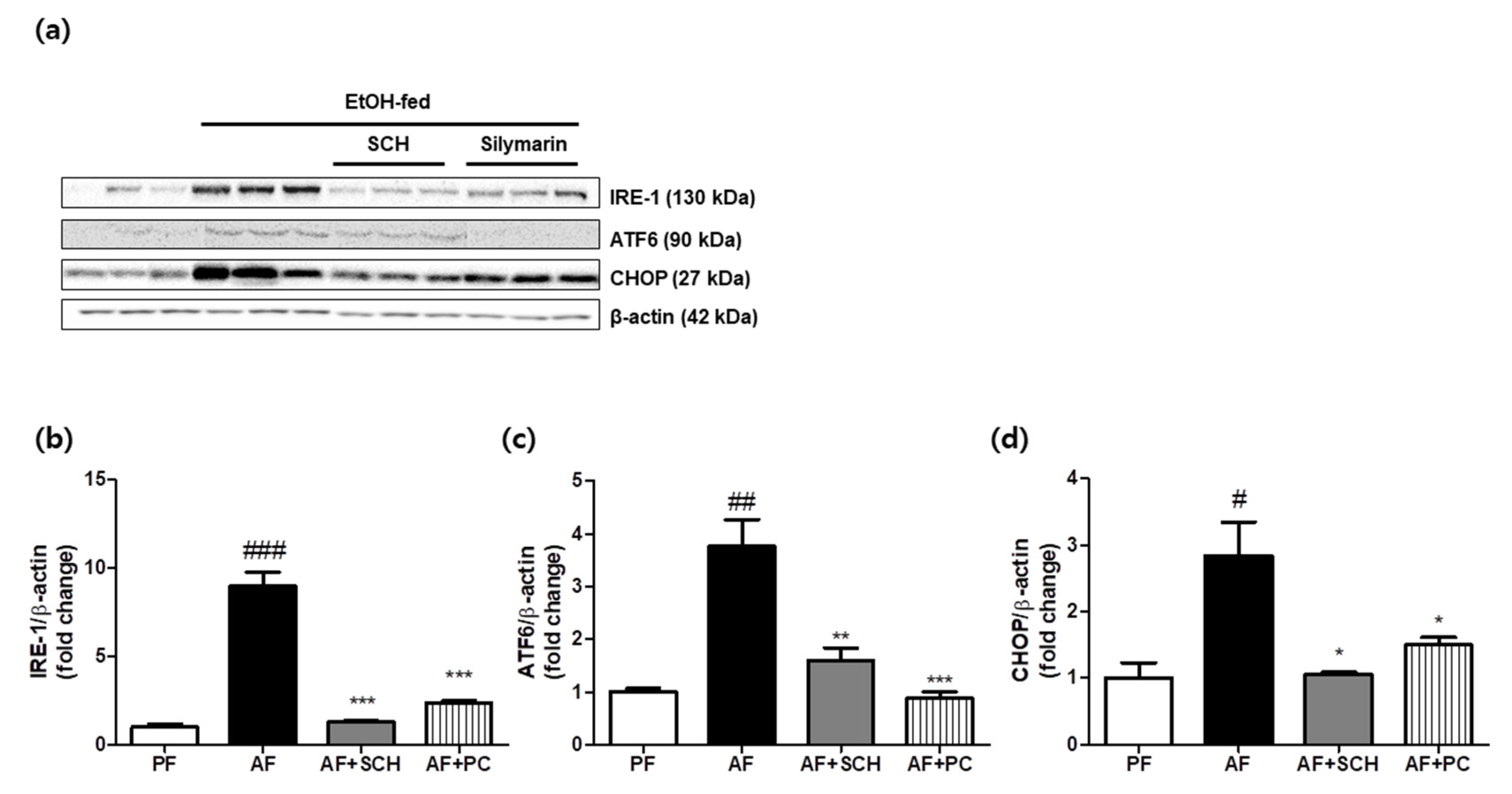
IRE1, an important ER stress sensor, was significantly induced in the liver in the alcohol group (p < 0.005) and was reduced by SCH administration more so than by silymarin (Figure 2a,b). To investigate the anti-apoptotic effect of SCH, we performed immunoblot analysis for the cell signals ATF6 and CHOP. Densitometry showed the ethanol diet increased these protein levels by 3.77- and 2.84-fold compared with non-alcohol-fed controls (Figure 2c,d). Silymarin treatment significantly reduced these increases (p < 0.005 and p < 0.01, respectively). Similarly, SCH administration also remarkably decreased these alcohol-induced increases in ATF6 and CHOP protein levels (p < 0.01 and p < 0.05, respectively).

3.3. Regulation of Antioxidative Enzyme Expression

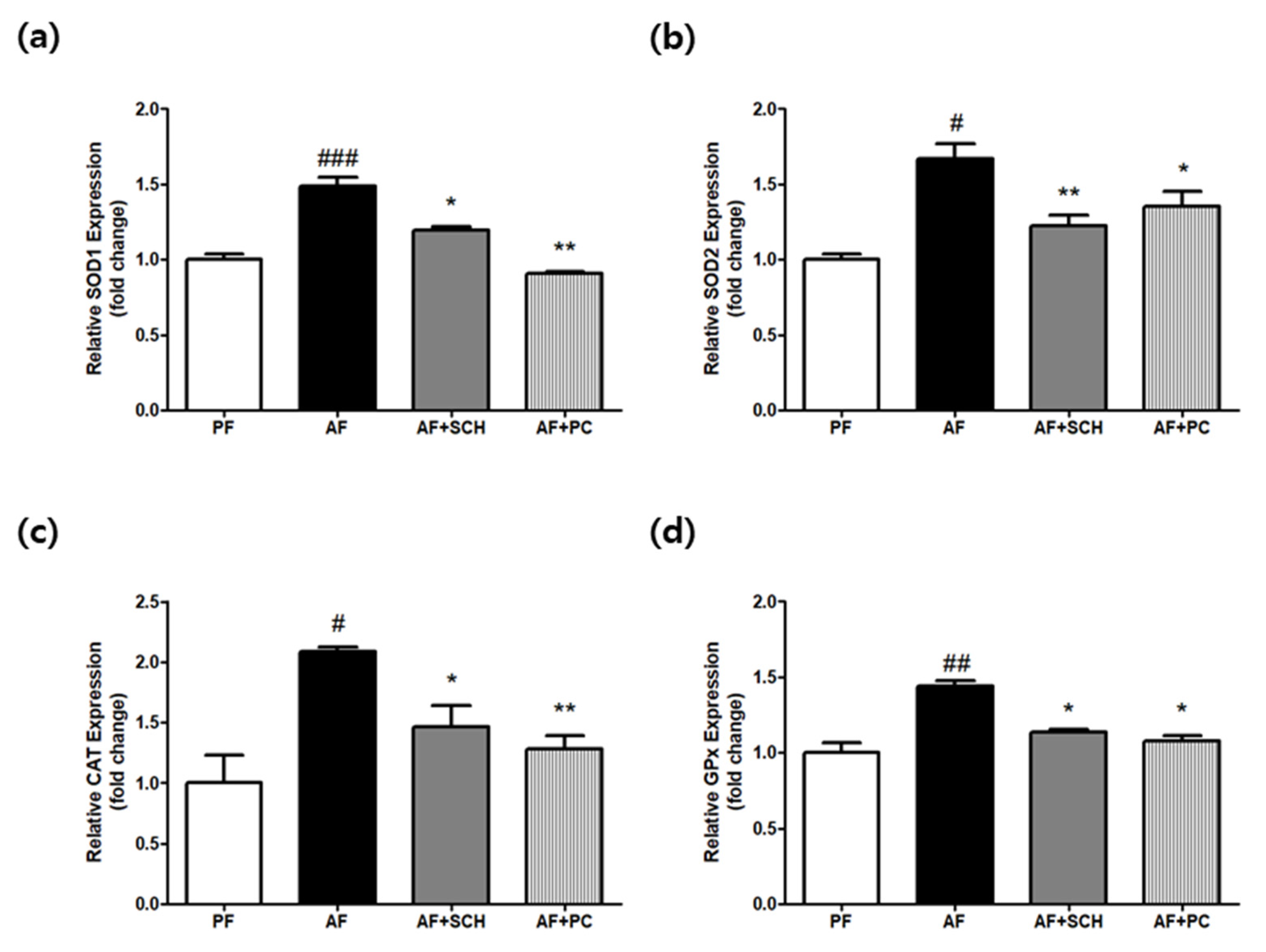
Antioxidative enzyme gene expressions of catalase (CAT), superoxide dismutase (SOD), and glutathione peroxidase (GPx) were evaluated in mouse livers. The expressions of all three genes were significantly upregulated in the three alcohol-administered groups (Figure 3a–d). However, their expressions were significantly lower in the AF+SCH and AF+PC groups than in the alcohol group, indicating that ROS levels were relatively resolved in these groups.

3.4. Regulation of MAPK Signals and Cytokine Production

The MAPK signaling pathway was investigated in liver tissues to investigate the effect of SCH on alcohol-induced steatohepatitis. MAPKs, including ERK, JNK, and p38, were significantly (p < 0.005) phosphorylated in ethanol-fed mice, which indicated stress-induced MAPK activation was associated with hepatic damage. As shown in Figure 4a, silymarin treatment successfully blocked MAPK phosphorylation to the normal control level. Moreover, alcohol-induced ERK, JNK, and p38 phosphorylation were significantly suppressed by SCH or silymarin (Figure 4b–d).
Subsequently, we found SCH prevented ethanol-induced pro-inflammatory cytokine production. Ethanol-fed mice showed 4.10- and 2.07-fold increases in IL-6 and TNF-α expression as compared with normal controls, and SCH significantly (p < 0.005) reduced this IL-6 increased to 0.21-fold, whereas silymarin reduced it to 0.46-fold (Figure 5b). Furthermore, densitometry showed SCH dramatically reduced alcohol-induced TNF-α production, whereas silymarin had little effect (Figure 5c).

3.5. Reduction of CYP2E1 Expression in Mouse Liver Tissue

To investigate the mechanism responsible for the effect of SCH, we performed immunoblot analysis and qPCR for the CYP2E1 protein and mRNA. CYP2E1 protein levels were 45.36-fold higher in the alcohol-fed group than in normal controls (p < 0.005). SCH administration significantly (p < 0.05) reduced these CYP2E1 protein and mRNA level increases (Figure 6a–c), and silymarin also significantly reduced these increases.

3.6. Reduction of Ethanol-Induced Injury in CYP2E1-Transfected HepG2 Cells

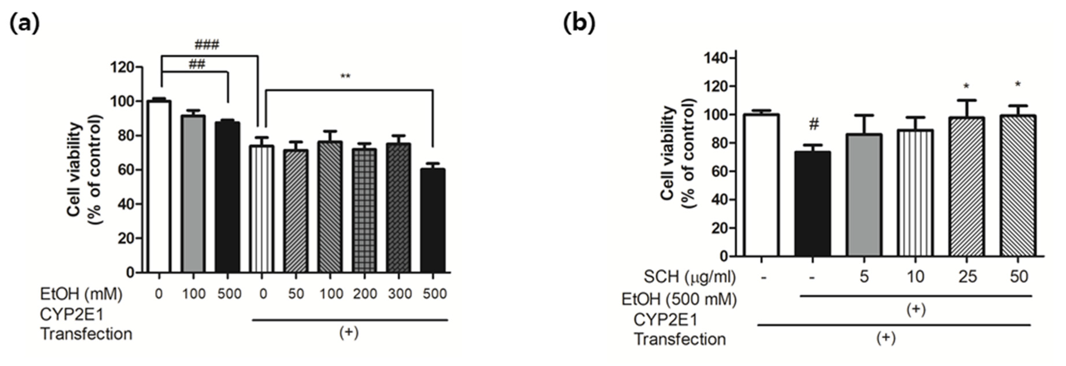
To evaluate the protective effect of SCH against ethanol, we transfected the CYP2E1 plasmid into hepatocellular carcinoma HepG2 cells and exposed them to ethanol concentrations in culture media. CYP2E1-transfected HepG2 cells proliferated less than non-transfected intact cells, which was attributed to the toxicity of liposome as a transfection vehicle. In addition, the comparison between CYP2E1-transfected HepG2 cells with different ethanol concentrations demonstrated that ethanol at a concentration of 500 mM significantly decreased cell viability. However, when these CYP2E1-transfected HepG2 cells were co-treated with ethanol (500 mM) and SCH, the cell toxicity of the ethanol binge was removed, at concentrations of 25 and 50 µg/mL (p < 0.05) (Figure 7b).

4. Discussion

In the present study, we investigated the effect of SCH on alcoholic steatohepatitis with various molecular markers or signaling pathways as its mechanisms. The NIAAA model used in this study is a representative alcoholic liver injury model involving chronic ethanol feeding and binge ethanol administration and induces the major hallmarks of human ALD [22].
GOT and GPT activity levels (widely used parameters of hepatic damage [23]) were determined in mice sera after sacrifice, and not surprisingly, the results obtained indicated alcohol feeding markedly increased hepatic enzyme activities (p < 0.001), which may reflect the development of alcoholic hepatitis in murine models [24]. Notable changes in the liver weight/body weight ratio and increases in serum triglyceride also implied steatotic changes in ethanol-fed mice [25,26]. As SCH administration was found to successfully block these changes, we further tried to deduce a reason for the effect regarding the pathological mechanism of alcoholic steatohepatitis.
Steatosis related to chronic alcohol consumption is caused by dysregulated lipid metabolism [27]. In brief, ethanol exposure reduces AMPK, sirtuin1 (SIRT1), and peroxisome proliferator activated receptor alpha (PPARα) activities and upregulates the expressions and activities of sterol regulatory element binding protein-1c (SREBP-1c), acetyl-CoA carboxylase 1 (ACC1), and peroxisome proliferator activated receptor gamma (PPARγ), which promote lipogenesis [27]. A previous study conducted on SCH showed SCH can activate AMPK and modulate its downstream signals, including ACC and PPARγ, in the liver [19], which may partially explain the suppression of alcohol-induced hepatosteatosis by SCH.
ER stress is induced during ethanol metabolism and is related to the accumulation of unfolded/misfolded proteins [28]. Under prolonged or severe ER stress, the unfolded protein response (UPR) can become cytotoxic, and even induce apoptosis, rather than cytoprotective [29]. Inositol-requiring enzyme 1α (IRE1α), the C/EBP-homologous protein (CHOP), and activating transcription factor 6 (ATF6) are predominant and unique signaling transduction pathways of UPR [30] and can detect abnormal conditions in the ER lumen and transmit signals to activate nuclear transcription factors [31]. We observed SCH administration reduce alcohol-induced IRE1α, CHOP, and ATF6 upregulations.
Alcohol-induced ER stress had a pro-inflammatory effect by activating the NF-κB and JNK signaling pathways [28]. IRE1α activates apoptotic signaling kinase-1 (ASK-1), and thus, activates downstream factors such as JNK (Jun-N-terminal kinase) and p38 MAPK (p38 mitogen-activated protein kinase), which enhance apoptosis in the liver. Moreover, activation of the MAPK pathway induces transcription factor activation [32], which in turn promotes macrophage infiltration and inflammatory cytokine and chemokine production, which lead to liver damage [33]. However, SCH inhibited the alcohol-induced phosphorylation of JNK, ERK, and P38 in the MAPK pathway in alcohol-fed mice.
Alcohol-induced liver disease is fundamentally associated with a state of excessive oxidative stress [34]. The metabolism of alcohol by alcohol dehydrogenase (ADH) and cytochrome P450 2E1 (CYP2E1) leads to the formation of acetaldehyde [6]. CYP2E1 metabolizes and activates ethanol to produce more reactive, toxic products, and thus, increases ROS production, especially the production of H2O2 and the superoxide anion (O2−), which causes cell damage [35]. ROS, a by-product formed during this process, can also activate MAPK family members, and therefore, contribute to the development of ALD [36]. However, SCH inhibited oxidative stress and ROS production in liver tissue by reducing CYP2E1 expression, and thereby, suppressed ER stress and reduced lipid accumulation and the inflammatory response, which suggests SCH might alleviate or prevent alcoholic liver injury or steatohepatitis.
Studies conducted by our team consistently suggest that the application of drug repurposing to traditional herbal medicine provides a potent means of discovering new natural-product drugs because these medicines represent a huge resource of medically interesting natural products with multi-target characteristics [37,38]. There are even more possibilities of new drug developments in herbal concoctions, with numerous candidates and accumulated experience of medical usages. Furthermore, the application of up-to-date methodologies of drug repurposing such as network pharmacology and molecular docking simulation provides a means of rapidly obtaining more objective, practical, and valuable results.

5. Conclusions

In summary, SCH did not affect the body weights in alcohol-fed mice but significantly reduced liver weights to the normal control level. SCH administration reduced alcohol-induced upregulations of ER-stress markers (IRE1α, ATF6, and CHOP), which indicated it reduced alcohol-induced ER stress. Furthermore, SCH inhibited alcohol-induced MAPK signaling and inflammatory responses. We believe that these effects of SCH were caused by its suppressive effect on CYP2E1 expression. Our findings suggest SCH might have therapeutic effects on metabolic syndromes and alcoholic liver injury.

Supplementary Materials

The following are available online at https://www.mdpi.com/article/10.3390/pr9101760/s1, Figure S1: The schematic diagram of animal experimental design of alcohol-induced steatohepatitis in C57BL/6J mice. Table S1: Composition of the standard Lieber-DeCarli liquid diet used in this study.

Author Contributions

G.-R.Y. performed the animal surgery and wrote the manuscript. S.-J.L., D.-W.L. and H.K. reviewed the study design and results. J.-E.K. designed the experiments and analyzed the data. W.-H.P. supervised the project and contributed to the final draft of the paper. All authors have read and agreed to the published version of the manuscript.

Funding

This work was supported by the Basic Science Research Program through the National Research Foundation of Korea by the Ministry of Science and ICT (NRF-2019R1F1A1051652).

Institutional Review Board Statement

Animal experiments were conducted in accordance with the guidelines issued by Dongguk University Ethics Committee (Approval No. IACUC-2017-026-1), which also approved the study protocols before study commencement.

Informed Consent Statement

Not applicable.

Data Availability Statement

The authors declare all data are available within the article or its Supplementary Materials.

Conflicts of Interest

The authors have no conflict of interest to declare.

References

  1. Bruha, R.; Dvořák, K.; Petrtyl, J. Alcoholic liver disease. World J. Hepatol. 2012, 4, 81–90. [Google Scholar] [CrossRef] [PubMed]
  2. Seitz, H.; Neuman, M. The History of Alcoholic Liver Disease: From an Unrecognized Disease to One of the Most Frequent Diseases in Hepatology. J. Clin. Med. 2021, 10, 858. [Google Scholar] [CrossRef]
  3. Younossi, Z.; Henry, L. Contribution of alcoholic and nonalcoholic fatty liver disease to the burden of liver-related morbidity and mortality. Gastroenterology 2016, 150, 1778–1785. [Google Scholar] [CrossRef]
  4. Gao, B.; Bataller, R. Alcoholic Liver Disease: Pathogenesis and New Therapeutic Targets. Gastroenterology 2011, 141, 1572–1585. [Google Scholar] [CrossRef] [Green Version]
  5. Harjumäki, R.; Pridgeon, C.; Ingelman-Sundberg, M. CYP2E1 in Alcoholic and Non-Alcoholic Liver Injury. Roles of ROS, Reactive Intermediates and Lipid Overload. Int. J. Mol. Sci. 2021, 22, 8221. [Google Scholar] [CrossRef]
  6. Heit, C.; Dong, H.; Chen, Y.; Thompson, D.C.; Deitrich, R.A.; Vasiliou, V.K. The Role of CYP2E1 in Alcohol Metabolism and Sensitivity in the Central Nervous System. Subcell. Biochem. 2013, 67, 235–247. [Google Scholar] [CrossRef] [Green Version]
  7. Koop, D.R. Alcohol metabolism’s damaging effects on the cell: A focus on reactive oxygen generation by the enzyme cy-tochrome P450 2E1. Alcohol Res. Health 2006, 29, 274–280. [Google Scholar] [PubMed]
  8. Abdelmegeed, A.M.; Ha, S.-K.; Choi, Y.; Akbar, M.; Song, B.-J. Role of CYP2E1 in mitochondrial dysfunction and hepat-ic injury by alcohol and non-alcoholic substances. Curr. Mol. Pharmacol. 2017, 10, 207–225. [Google Scholar] [CrossRef] [Green Version]
  9. Wang, Y.; Millonig, G.; Nair, J.; Patsenker, E.; Stickel, F.; Mueller, S.; Bartsch, H.; Seitz, H.K. Ethanol-induced cytochrome P4502E1 causes carcinogenic etheno-DNA lesions in alcoholic liver disease. Hepatology 2009, 50, 453–461. [Google Scholar] [CrossRef]
  10. Aubert, J.; Begriche, K.; Knockaert, L.; Robin, M.; Fromenty, B. Increased expression of cytochrome P450 2E1 in nonalcoholic fatty liver disease: Mechanisms and pathophysiological role. Clin. Res. Hepatol. Gastroenterol. 2011, 35, 630–637. [Google Scholar] [CrossRef] [PubMed]
  11. Cho, Y.E.; Mezey, E.; Hardwick, J.P.; Salem, N., Jr.; Clemens, D.L.; Song, B.J. Increased ethanol-inducible cytochrome P450-2E1 and cytochrome P450 isoforms in exosomes of alcohol-exposed rodents and patients with alcoholism through oxidative and endoplasmic reticulum stress. Hepatol. Commun. 2017, 1, 675–690. [Google Scholar] [CrossRef] [PubMed]
  12. Farha, M.A.; Brown, E.D. Drug repurposing for antimicrobial discovery. Nat. Microbiol. 2019, 4, 565–577. [Google Scholar] [CrossRef]
  13. Pushpakom, S.; Iorio, F.; Eyers, P.A.; Escott, K.J.; Hopper, S.; Wells, A.; Doig, A.; Guilliams, T.; Latimer, J.; McNamee, C.; et al. Drug repurposing: Progress, challenges and recommendations. Nat. Rev. Drug Discov. 2018, 18, 41–58. [Google Scholar] [CrossRef] [PubMed]
  14. Sookoian, S.; Pirola, C.J. Repurposing drugs to target nonalcoholic steatohepatitis. World J. Gastroenterol. 2019, 25, 1783–1796. [Google Scholar] [CrossRef]
  15. Tobinick, E.L. The value of drug repositioning in the current pharmaceutical market. Drug News Perspect. 2009, 22, 119–125. [Google Scholar] [CrossRef]
  16. Parvathaneni, V.; Kulkarni, N.S.; Muth, A.; Gupta, V. Drug repurposing: A promising tool to accelerate the drug discovery process. Drug Discov. Today 2019, 24, 2076–2085. [Google Scholar] [CrossRef]
  17. Sucher, N.J. The application of Chinese medicine to novel drug discovery. Expert Opin. Drug Discov. 2012, 8, 21–34. [Google Scholar] [CrossRef]
  18. Lim, D.; Kim, H.; Kim, Y.-M.; Chin, Y.-W.; Park, W.-H.; Kim, J.-E. Drug repurposing in alternative medicine: Herbal digestive Sochehwan exerts multifaceted effects against metabolic syndrome. Sci. Rep. 2019, 9, 1–12. [Google Scholar] [CrossRef]
  19. Heo, J. Dongeuibogam; Bupin Publisher: Seoul, Korea, 1999. [Google Scholar]
  20. Lim, D.-W.; Kim, H.; Lee, S.-J.; Yu, G.-R.; Kim, J.-E.; Park, W.-H. Jwa Kum Whan Attenuates Nonalcoholic Fatty Liver Disease by Modulating Glucose Metabolism and the Insulin Signaling Pathway. Evid.-Based Complement. Altern. Med. 2019, 2019, 1–10. [Google Scholar] [CrossRef] [PubMed]
  21. Lim, D.-W.; Kim, H.; Park, J.-Y.; Kim, J.-E.; Moon, J.-Y.; Park, S.-D.; Park, W.-H. Amomum cardamomum L. ethyl acetate fraction protects against carbon tetrachloride-induced liver injury via an antioxidant mechanism in rats. BMC Complement. Altern. Med. 2016, 16, 155. [Google Scholar] [CrossRef] [Green Version]
  22. Bertola, A.; Mathews, S.; Ki, S.H.; Wang, H.; Gao, B. Mouse model of chronic and binge ethanol feeding (the NIAAA model). Nat. Protoc. 2013, 8, 627–637. [Google Scholar] [CrossRef] [Green Version]
  23. Karimi-Khouzani, O.; Heidarian, E.; Amini, S.A. Anti-inflammatory and ameliorative effects of gallic acid on fluoxetine-induced oxidative stress and liver damage in rats. Pharmacol. Rep. 2017, 69, 830–835. [Google Scholar] [CrossRef] [PubMed]
  24. Vogle, A.; Qian, T.; Zhu, S.; Burnett, E.; Fey, H.; Zhu, Z.; Keshavarzian, A.; Shaikh, M.; Hoshida, Y.; Kim, M.; et al. Restricted immunological and cellular pathways are shared by murine models of chronic alcohol consumption. Sci. Rep. 2020, 10, 1–14. [Google Scholar] [CrossRef] [Green Version]
  25. Yao, X.; Hou, S.; Zhang, D.; Xia, H.; Wang, Y.-C.; Jiang, J.; Yin, H.; Ying, H. Regulation of fatty acid composition and lipid storage by thyroid hormone in mouse liver. Cell Biosci. 2014, 4, 38. [Google Scholar] [CrossRef] [Green Version]
  26. Simon, G.; Heckmann, V.; Tóth, D.; Pauka, D.; Petrus, K.; Molnár, T.F. The effect of hepatic steatosis and fibrosis on liver weight and dimensions. Leg. Med. 2020, 47, 101781. [Google Scholar] [CrossRef]
  27. You, M.; Arteel, G.E. Effect of ethanol on lipid metabolism. J. Hepatol. 2019, 70, 237–248. [Google Scholar] [CrossRef] [Green Version]
  28. Liu, X.; Green, R.M. Endoplasmic reticulum stress and liver diseases. Liver Res. 2019, 3, 55–64. [Google Scholar] [CrossRef]
  29. Verfaillie, T.; Salazar, M.; Velasco, G.; Agostinis, P. Linking ER Stress to Autophagy: Potential Implications for Cancer Therapy. Int. J. Cell Biol. 2010, 2010, 1–19. [Google Scholar] [CrossRef] [PubMed] [Green Version]
  30. Szegezdi, E.; Logue, S.; Gorman, A.; Samali, A. Mediators of endoplasmic reticulum stress-induced apoptosis. EMBO Rep. 2006, 7, 880–885. [Google Scholar] [CrossRef] [Green Version]
  31. Madkour, L.H. Reactive Oxygen Species (ROS), Nanoparticles, and Endoplasmic Reticulum(er) Stress-Induced Cell Death Mechanisms; Academic Press: Cambridge, MA, USA, 2020. [Google Scholar]
  32. Whitmarsh, A.J. Regulation of gene transcription by mitogen-activated protein kinase signaling pathways. Biochim. Biophys. Acta (BBA)-Bioenerg. 2007, 1773, 1285–1298. [Google Scholar] [CrossRef] [PubMed]
  33. Mandrekar, P.; Szabo, G. Signalling pathways in alcohol-induced liver inflammation. J. Hepatol. 2009, 50, 1258–1266. [Google Scholar] [CrossRef] [PubMed] [Green Version]
  34. Albano, E. Alcohol, oxidative stress and free radical damage. Proc. Nutr. Soc. 2006, 65, 278–290. [Google Scholar] [CrossRef] [PubMed] [Green Version]
  35. Gaitantzi, H.; Meyer, C.; Rakoczy, P.; Thomas, M.; Wahl, K.; Wandrer, F.; Bantel, H.; Alborzinia, H.; Wölfl, S.; Ehnert, S.; et al. Ethanol sensitizes hepatocytes for TGF-β-triggered apoptosis. Cell Death Dis. 2018, 9, 51. [Google Scholar] [CrossRef] [PubMed] [Green Version]
  36. Schattenberg, J.M.; Czaja, M.J. Regulation of the effects of CYP2E1-induced oxidative stress by JNK signaling. Redox Biol. 2014, 3, 7–15. [Google Scholar] [CrossRef] [Green Version]
  37. McChesney, J.D. Natural products in drug discovery—Organizing for success. P. R. Health Sci. J. 2002, 21. [Google Scholar]
  38. Shen, B. A New Golden Age of Natural Products Drug Discovery. Cell 2015, 163, 1297–1300. [Google Scholar] [CrossRef] [Green Version]
Figure 1. SCH protected mouse livers from alcohol-induced hepatic injury. (a) Weekly body weight changes in the study groups normal pair-fed controls (PF), alcohol-fed group (AF), alcohol-fed and SCH administered group (AF+SCH), and alcohol-fed mice and silymarin administered group (AF+PC). (b) Liver weight/body weight ratio. (c) GOT, (d) GPT, (e) TG levels in mouse serum. Results are presented as means ± SDs. ### p < 0.001 versus non-alcohol fed controls, and * p < 0.05, ** p < 0.01, *** p < 0.001 versus the alcohol-fed group.
Processes 09 01760 g001
Figure 2. SCH inhibited endoplasmic reticulum stress in mice with alcohol-induced hepatic injury. (a) Representative immunoblot images of IRE-1, ATF6, and CHOP. (b) Inositol-requiring enzyme 1 (IRE-1) expressions levels, (c) activating transcription factor6 (ATF6) levels, and (d) C/EBP-homologous protein (CHOP) levels as determined by Western blot. Band intensities were measured by densitometry and normalized versus β-actin. Results are presented as the means ± SDs of three different experiments. # p < 0.05, ## p < 0.01, ### p < 0.001 versus non-alcohol fed controls, and * p < 0.05, ** p < 0.01, *** p < 0.001 versus alcohol fed mice.
Processes 09 01760 g002
Figure 3. SCH upregulated antioxidant enzyme expressions in mice with alcohol-induced hepatic injury. (a) The expressions of SOD1, (b) SOD2, (c) CAT, and (d) GPx were determined by qPCR. Relative gene expression levels were normalized versus β-actin. Results are presented as means ± SDs. # p < 0.05, ## p < 0.01, ### p < 0.001 versus non-alcohol fed controls, and * p < 0.05, ** p < 0.01 versus the alcohol fed group.
Processes 09 01760 g003
Figure 4. SCH inhibited MAPK phosphorylations in alcohol-fed mice. (a) Representative immunoblot images of p-ERK, p-JNK, and p-P38 levels. (b) p-ERK, (c) p-JNK, and (d) p-P38 levels as determined by Western blot. Band intensities were determined by densitometry and normalized versus those of corresponding non-phosphorylated forms and β-actin. Results are presented as the means ± SDs of three different experiments. ### p < 0.001 versus non-alcohol controls, and ** p < 0.01, *** p < 0.001 versus the alcohol-fed group.
Processes 09 01760 g004
Figure 5. SCH inhibited pro-inflammatory cytokine production in alcohol-fed mice. (a) Representative immunoblot images of IL-6 and TNFα levels. (b) IL-6 and (c) TNFα levels as determined by Western blot. Band intensities were measured by densitometry and normalized versus β-actin. Results are presented as the means ± SDs of three different experiments. ### p < 0.001 versus non-alcohol fed controls, and * p < 0.05, *** p < 0.001 versus the alcohol group.
Processes 09 01760 g005
Figure 6. SCH inhibited induction of the CYP2E1 gene in alcohol-fed mice. (a) Cytochrome P450 2E1 (CYP2E1) levels as determined by Western blot, and band intensities determined by densitometry and normalized versus β-actin. (b) CYP2E1 gene expression as determined by real-time PCR. (c) CYP2E1 levels were determined by Western blot, and band intensities were measured by densitometry and normalized versus β-actin. Results are presented as the means ± SDs of three different experiments. ## p < 0.01, ### p < 0.001 versus normal controls, and * p < 0.05, ** p < 0.01 versus the alcohol-fed group.
Processes 09 01760 g006
Figure 7. SCH protected CYP2E1-transfected HepG2 cells from ethanol. (a) Cell viabilities of non-transfected HepG2 cells and CYP2E1-transfected HepG2 cells exposed to different concentrations of ethanol (0-500 mM) for 72 h. (b) Cell viability of CYP2E1-transfected HepG2 cells in the presence of ethanol (500 mM) with or without SCH for 72 h. Results are presented as the means ± SDs of three different experiments. # p < 0.05, ## p < 0.01, ### p < 0.001 versus non-treated intact control group (a and b), and * p < 0.05, ** p < 0.01 versus CYP2E1-transfected HepG2 cells (a) and CYP2E1-transfected HepG2 cells treated with 500 mM ethanol (b).
Processes 09 01760 g007
Publisher’s Note: MDPI stays neutral with regard to jurisdictional claims in published maps and institutional affiliations.