
Toxicological Profile of Biological Environment of Two Elastodontic Devices

 , , ,
, , ,  , and
, and
Abstract
:1. Introduction
2. Materials and Methods
2.1. Reagents
2.2. Preparation of the Artificial Saliva with Different pH Values
2.3. Storage Period of Samples in Artificial Saliva
2.4. Scanning Electron Microscopy Analysis
2.5. Cell Culture
2.6. Cell Viability Assessment
2.7. Gene Expression
2.8. Hen’s Egg Test—Chorioallantoic Membrane (HET-CAM) Assay
2.9. Statistical Evaluation
3. Results
3.1. Scanning Electron Microscopy
3.2. Cell Viability
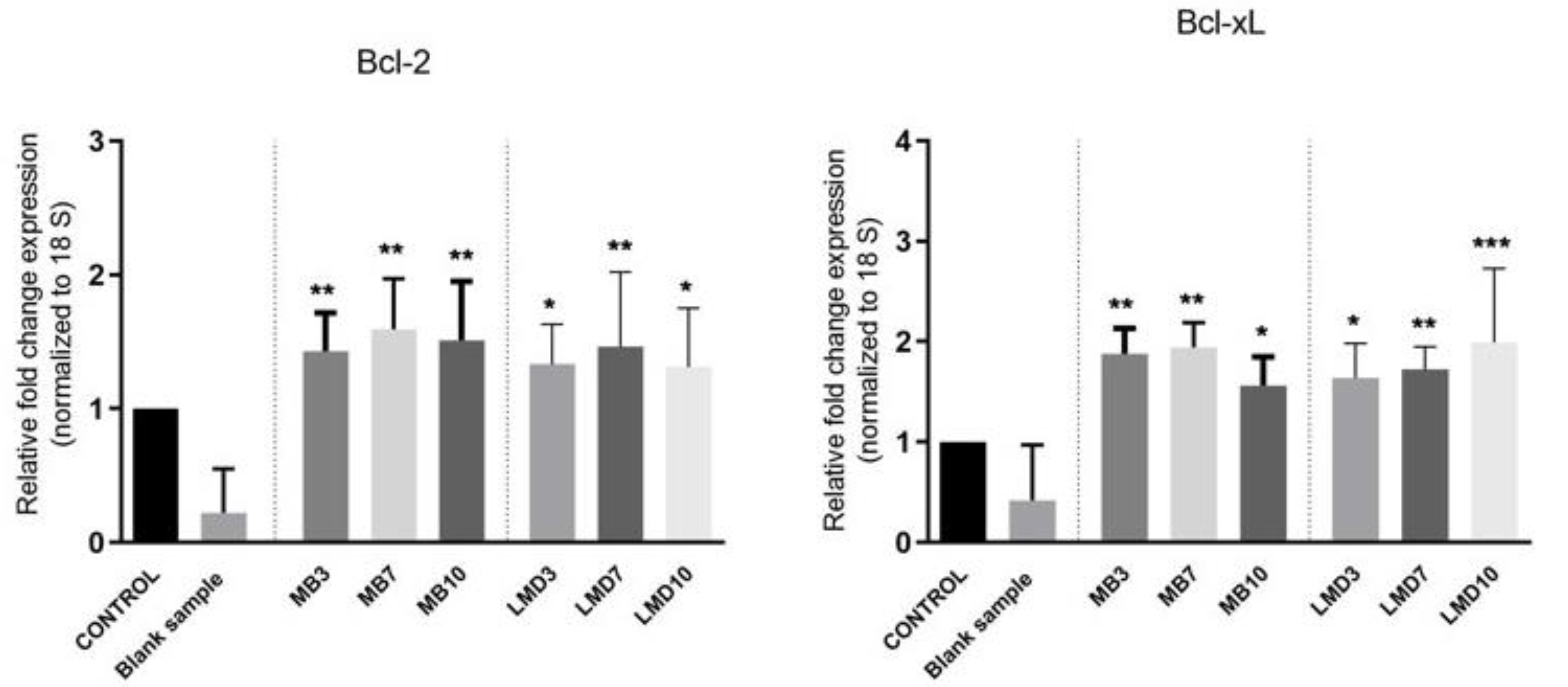
3.3. RT-PCR
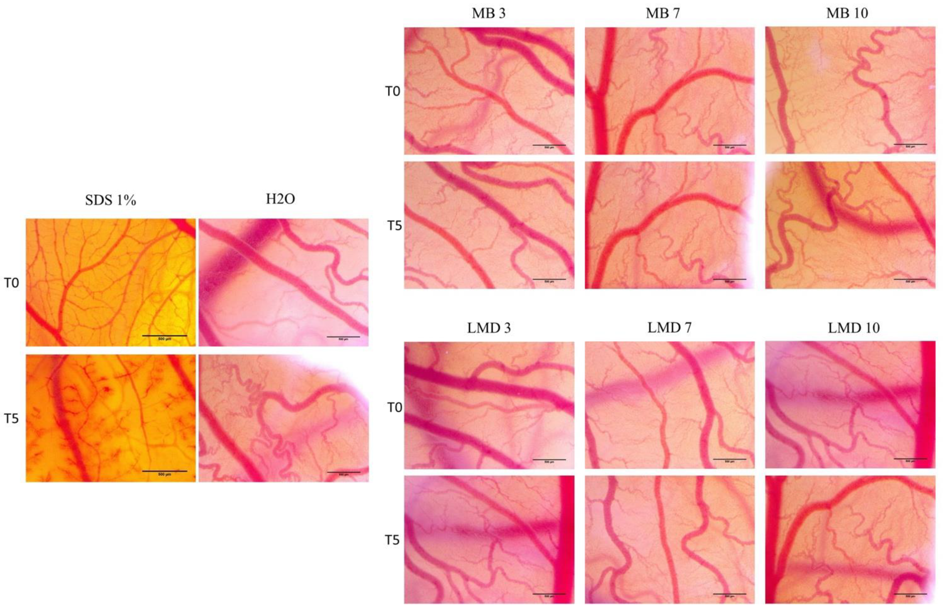
3.4. MB and LMD Has No Irritative Effect in Ovo
4. Discussion
5. Conclusions
Author Contributions
Funding
Institutional Review Board Statement
Informed Consent Statement
Conflicts of Interest
References
- Rapeepattana, S.; Thearmontree, A.; Suntornlohanakul, S. Etiology of malocclusion and dominant orthodontic problems in mixed dentition: A cross-sectional study in a group of Thai children aged 8–9 years. J. Int. Soc. Prev. Community Dent. 2019, 9, 383–399. [Google Scholar] [CrossRef] [PubMed]
- Ahammed, A.R.; Shetty, V.; Panda, A.K.; Gunda, S.; Pradhan, D.; Husain, N.; Gugwad, S. Prevalence of malocclusion among 12 to 15 years age group orphan children using dental aesthetic index. J. Contemp. Dent. Pract. 2013, 14, 111–114. [Google Scholar] [CrossRef]
- Anthony, S.N.; Zimba, K.; Subramanian, B. Impact of Malocclusions on the Oral Health-Related Quality of Life of Early Adolescents in Ndola, Zambia. Int. J. Dent. 2018, 2018, 7920973. [Google Scholar] [CrossRef]
- Dutra, S.R.; Pretti, H.; Martins, M.T.; Bendo, C.B.; Vale, M.P. Impact of malocclusion on the quality of life of children aged 8 to 10 years. Dent. Press J. Orthod. 2018, 23, 46–53. [Google Scholar] [CrossRef]
- Mageet, A.O. Classification of Skeletal and Dental Malocclusion: Revisited. Stomatol. Edu. J. 2016, 3, 205–211. [Google Scholar] [CrossRef]
- Yadav, D.; Rani, M.; Shailaja, A.; Anand, D.; Sood, N.; Gothi, R. Angle’s Molar Classification Revisited. J. Indian Orthod. Soc. 2014, 48, 382–387. [Google Scholar] [CrossRef]
- Buschang, P.H. Class I malocclusions—The development and etiology of mandibular malalignments. Semin. Orthod. 2014, 20, 3–15. [Google Scholar] [CrossRef]
- Eliades, T.; Hegdvedt, A.K. Orthodontic-surgical correction of a Class II, Division 2 malocclusion. Am. J. Orthod. Dentofac. Orthop. 1996, 110, 351–357. [Google Scholar] [CrossRef]
- Li, C.; Cai, Y.; Chen, S.; Chen, F. Classification and characterization of class III malocclusion in Chinese individuals. Head Face Med. 2016, 12, 1–8. [Google Scholar] [CrossRef] [PubMed] [Green Version]
- American Academy on Pediatric Dentistry Clinical Affairs Committee-Developing Dentition Subcommittee. American Academy on Pediatric Dentistry Council on Clinical Affairs. Management of the Developing Dentition and Occlusion in Pediatric Dentistry. Pediatr. Dent. 2017, 39, 334–347. [Google Scholar]
- Saccomanno, S.; Antonini, G.; D’Alatri, L.; D’Angelantonio, M.; Fiorita, A.; Deli, R. Patients treated with orthodon-tic-myofunctional therapeutic protocol. Eur. J. Paediatr. Dent. 2012, 13, 241–243. [Google Scholar] [PubMed]
- Melchior, M.d.O.; Magri, L.V.; Mazzetto, M.O. Orofacial myofunctional disorder, a possible complicating factor in the manage-ment of painful temporomandibular disorder. Case report. Braz. J. Pain 2018, 1, 80–86. [Google Scholar] [CrossRef]
- Wishney, M.; Darendeliler, M.A.; Dalci, O. Myofunctional therapy and prefabricated functional appliances: An overview of the history and evidence. Aust. Dent. J. 2019, 64, 135–144. [Google Scholar] [CrossRef] [PubMed]
- da Cunha Busquet, P.; de Jesus Portelinha, D.; da Costa, M.L.; de Andrade Cancio de Paula, V. How the myobrace appliance works: Advantages and disadvantages. J. Dent. Probl. Solut. 2021, 8, 19–23. [Google Scholar] [CrossRef]
- Galeotti, A.; Uomo, R.; Spagnuolo, G.; Paduano, S.; Cimino, R.; Valletta, R.; D’Anto, V. Effect of pH on in vitro biocompatibil-ity of orthodontic miniscrew implants. Prog. Orthod. 2013, 14, 1–7. [Google Scholar] [CrossRef] [Green Version]
- Alves, C.B.C.; Segurado, M.N.; Dorta, M.C.L.; Dias, F.R.; Lenza, M.G.; Lenza, M.A. Evaluation of cytotoxicity and corrosion re-sistance of orthodontic mini-implants. Dent. Press J. Orthod. 2016, 21, 39–46. [Google Scholar] [CrossRef] [Green Version]
- Geramipanah, F.; Majidpour, M.; Sadighpour, L.; Fard, M.J.K. Effect of artificial saliva and pH on shear bond strength of resin cements to zirconia-based ceramic. Eur. J. Prosthodont. Restor. Dent. 2013, 21, 5–8. [Google Scholar]
- Maghiari, A.L.; Coricovac, D.; Pinzaru, I.A.; Macașoi, I.G.; Marcovici, I.; Simu, S.; Navolan, D.; Dehelean, C. High Concentra-tions of Aspartame Induce Pro-Angiogenic Effects in Ovo and Cytotoxic Effects in HT-29 Human Colorectal Carcinoma Cells. Nutrients 2020, 12, 3600. [Google Scholar] [CrossRef]
- Macașoi, I.; Pavel, I.Z.; Moacă, A.E.; Avram, Ș.; David, V.L.; Coricovac, D.; Mioc, A.; Spandidos, D.A.; Tsatsakis, A.; Soica, C.; et al. Mechanistic investigations of antitumor activity of a Rhodamine B-oleanolic acid derivative bioconjugate. Oncol. Rep. 2020, 44, 1169–1183. [Google Scholar] [CrossRef]
- Batista-Duharte, A.; Jorge Murillo, G.; Pérez, U.M.; Tur, E.N.; Portuondo, D.F.; Martínez, B.T.; Téllez-Martínez, D.; Betancourt, J.E.; Pérez, O. The Hen’s Egg Test on Chorioallantoic Membrane: An Alternative Assay for the Assessment of the Irritating Ef-fect of Vaccine Adjuvants. Int. J. Toxicol. 2016, 35, 627–633. [Google Scholar] [CrossRef]
- Budai, P.; Kormos, É.; Buda, I.; Somody, G.; Lehel, J. Comparative evaluation of HET-CAM and ICE methods for objective as-sessment of ocular irritation caused by selected pesticide products. Toxicol. Vitr. 2021, 74, 105150. [Google Scholar] [CrossRef] [PubMed]
- Guran, K.; Buzatu, R.; Pinzaru, I.; Boruga, M.; Marcovici, I.; Coricovac, D.; Avram, S.; Poenaru, M.; Susan, M.; Susan, R.; et al. In Vitro Pharmaco-Toxicological Characterization of Melissa officinalis Total Extract Using Oral, Pharynx and Colorectal Carcinoma Cell Lines. Processes 2021, 9, 850. [Google Scholar] [CrossRef]
- Chen, J.; Wan, J.; You, L. Speech and orthodontic appliances: A systematic literature review. Eur. J. Orthod. 2018, 40, 29–36. [Google Scholar] [CrossRef] [PubMed]
- Wishney, M. Potential risks of orthodontic therapy: A critical review and conceptual framework. Aust. Dent. J. 2017, 62, 86–96. [Google Scholar] [CrossRef] [PubMed] [Green Version]
- Gökçe, B. Current Approaches in Myofunctional Orthodontics. J. Musculoskelet. Disord. Treat. 2016, 2, 022. [Google Scholar] [CrossRef]
- Elhamouly, Y.; El-Housseiny, A.A.; Ismail, H.A.; El Habashy, L.M. Myofunctional Trainer versus Twin Block in Developing Class II Division I Malocclusion: A Randomized Comparative Clinical Trial. Dent. J. 2020, 8, 44. [Google Scholar] [CrossRef]
- Olszewska, A.; Hanć, A.; Barałkiewicz, D.; Rzymski, P. Metals and Metalloids Release from Orthodontic Elastomeric and Stainless Steel Ligatures: In Vitro Risk Assessment of Human Exposure. Biol. Trace Elem. Res. 2020, 196, 646–653. [Google Scholar] [CrossRef] [Green Version]
- Wepner, L.; Färber, H.A.; Jaensch, A.; Weber, A.; Heuser, F.; Keilig, L.; Singer, L.; Bourauel, C.P. In vitro ion release of wires in removable orthodontic appliances. Materials 2021, 14, 3402. [Google Scholar] [CrossRef]
- Karnam, S.K.; Reddy, A.N.; Manjith, C.M. Comparison of metal ion release from different bracket archwire combinations: An in vitro study. J. Contemp. Dent. Pract. 2012, 13, 376–381. [Google Scholar]
- Szuhanek, C.A.; Watz, C.G.; Avram, Ș.; Moacă, E.-A.; Mihali, C.V.; Popa, A.; Campan, A.A.; Nicolov, M.; Dehelean, C.A. Comparative Toxicological In Vitro and In Ovo Screening of Different Orthodontic Implants Currently Used in Dentistry. Materials 2020, 13, 5690. [Google Scholar] [CrossRef]
- Popa, A.; Dehelean, C.; Calniceanu, H.; Watz, C.; Brad, S.; Sinescu, C.; Marcu, O.A.; Popa, C.S.; Avram, S.; Nicolov, M.; et al. A Custom-Made Orthodontic Mini-Implant—Effect of Insertion Angle and Cortical Bone Thickness on Stress Distribution with a Com-plex In Vitro and In Vivo Biosafety Profile. Materials 2020, 13, 4789. [Google Scholar] [CrossRef] [PubMed]
- Martín-Cameán, A.; Jos, Á.; Mellado-García, P.; Iglesias-Linares, A.; Solano, E.; Cameán, A.M. In vitro and in vivo evidence of the cytotoxic and genotoxic effects of metal ions released by orthodontic appliances: A review. Environ. Toxicol. Pharmacol. 2015, 40, 86–113. [Google Scholar] [CrossRef] [PubMed]
- Wataha, J.C.; Hanks, C.T.; Sun, Z. Effect of cell line on in vitro metal ion cytotoxicity. Dent. Mater. 1994, 10, 156–161. [Google Scholar] [CrossRef] [Green Version]
- Locci, P.; Lilli, C.; Marinucci, L.; Calvitti, M.; Belcastro, S.; Bellocchio, S.; Staffolani, N.; Guerra, M.; Beccheti, E. In vitro cyto-toxic effects of orthodontic appliances. J. Biomed. Mater. Res. 2000, 53, 560–567. [Google Scholar] [CrossRef]
- Goiato, M.; Nobrega, A.; Malavazi, E.; Takamiya, A.; Penha de Oliveira, S. In Vitro Analysis of the Proliferation of HaCaT Cells Stimulated by Pigments Used for Maxillofacial Prostheses. J. Orofac. Sci. 2019, 11, 32–36. [Google Scholar] [CrossRef]
- Orrenius, S.; Nicotera, P.; Zhivotovsky, B. Cell Death Mechanisms and Their Implications in Toxicology. Toxicol. Sci. 2011, 119, 3–19. [Google Scholar] [CrossRef] [Green Version]
- Buczko, P.; Szarmach, I.; Grycz, M.; Kasacka, I. Caspase-3 as an important factor in the early cytotoxic effect of nickel on oral mucosa cells in patients treated orthodontically. Folia Histochem. Cytobiol. 2017, 55, 37–42. [Google Scholar] [CrossRef]
- Kapadia, J.M.H.; Agarwal, A.R.; Mishra, S.; Joneja, P.; Yusuf, A.S.; Choudhary, D.S. Cytotoxic and genotoxic effect on the buc-cal mucosa cells of patients undergoing fixed orthodontic treatment. J. Contemp. Dent. Pract. 2018, 19, 1358–1362. [Google Scholar] [CrossRef]
- Kloukos, D.; Taoufik, E.; Eliades, T.; Katsaros, C.; Eliades, G. Cytotoxic effects of polycarbonate-based orthodontic brackets by activation of mitochondrial apoptotic mechanisms. Dent. Mater. 2013, 29, e35–e44. [Google Scholar] [CrossRef]
- Mioc, M.; Avram, S.; Bercean, V.; Porcarasu, M.B.; Soica, C.; Susan, R.; Kurunczi, L. Synthesis, Characterization and Antiprolif-erative Activity Assessment of a Novel 1H-5-mercapto-1,2,4 Triazole Derivative. Rev. Chim. 2017, 68, 745–747. [Google Scholar] [CrossRef]
- Schendel, K.U.; Erdinger, L.; Komposch, G.; Sonntag, H.G. Orthodontic materials studied in the HET-CAM test for muco-sa-irritating effects. Fortschr. Kieferorthop. 1994, 55, 28–35. [Google Scholar] [CrossRef] [PubMed]


| Irritation Potential | Irritation Score |
|---|---|
| Non-irritating | 0–0.9 |
| Irritating | 1–8.9 |
| Severe irritating | 9–21 |
| SDS 1% | H2O | MB 1:1 | LMD 1:1 | |||||
|---|---|---|---|---|---|---|---|---|
| pH 3 | pH 7 | pH 10 | pH 3 | pH 7 | pH 10 | |||
| IS | 19.31 | 0.13 | 0.82 | 0.67 | 0.73 | 0.46 | 0.28 | 0.82 |
| tH | 17 s | 300 | 300 | 300 | 300 | 300 | 300 | 300 |
| tL | 21 s | 300 | 300 | 300 | 300 | 300 | 300 | 300 |
| tC | 33 s | 298 | 275 | 280 | 278 | 287 | 293 | 275 |
Publisher’s Note: MDPI stays neutral with regard to jurisdictional claims in published maps and institutional affiliations. |
© 2021 by the authors. Licensee MDPI, Basel, Switzerland. This article is an open access article distributed under the terms and conditions of the Creative Commons Attribution (CC BY) license (https://creativecommons.org/licenses/by/4.0/).
Share and Cite
Dinu, S.; Buzatu, R.; Macasoi, I.; Popa, M.; Vlad, C.S.; Marcovici, I.; Pinzaru, I.; Dehelean, C.A.; Moacă, E.-A.; Barbu-Tudoran, L.; et al. Toxicological Profile of Biological Environment of Two Elastodontic Devices. Processes 2021, 9, 2116. https://doi.org/10.3390/pr9122116
Dinu S, Buzatu R, Macasoi I, Popa M, Vlad CS, Marcovici I, Pinzaru I, Dehelean CA, Moacă E-A, Barbu-Tudoran L, et al. Toxicological Profile of Biological Environment of Two Elastodontic Devices. Processes. 2021; 9(12):2116. https://doi.org/10.3390/pr9122116
Chicago/Turabian StyleDinu, Stefania, Roxana Buzatu, Ioana Macasoi, Malina Popa, Cristian Sebastian Vlad, Iasmina Marcovici, Iulia Pinzaru, Cristina Adriana Dehelean, Elena-Alina Moacă, Lucian Barbu-Tudoran, and et al. 2021. "Toxicological Profile of Biological Environment of Two Elastodontic Devices" Processes 9, no. 12: 2116. https://doi.org/10.3390/pr9122116