Article

Cyanidin-3-glucoside Lipophilic Conjugates for Topical Application: Tuning the Antimicrobial Activities with Fatty Acid Chain Length

LAQV, REQUIMTE, Departamento de Química e Bioquímica, Faculdade de Ciências, Universidade do Porto, Rua do Campo Alegre s/n, 4169-007 Porto, Portugal
*
Authors to whom correspondence should be addressed.
Processes 2021, 9(2), 340; https://doi.org/10.3390/pr9020340
Submission received: 18 January 2021 / Revised: 3 February 2021 / Accepted: 9 February 2021 / Published: 12 February 2021
(This article belongs to the Special Issue Novel Added-Products Manufacturing from Agro-Food Wastes)

Abstract

:
Background: Natural anthocyanins present a low solubility in lipophilic media, which compromises their effective application in lipophilic systems. In this work, cyanidin-3-O-glucoside (Cy3glc) was esterified by the addition of fatty acids with increasing chain-lengths and a structure-activity relationship was performed towards the description of the best analog for skin-care applications. Methods: By enzymatic hemi-synthesis, it was possible to obtain 5 structurally related derivatives of cyanidin-3-O-glucoside with successive C2 increments in the aliphatic chain. The stability in hanks buffer and DMEM with or without FBS was followed by HPLC. The cytotoxicity against keratinocytes was evaluated by MTT assay. The antioxidant capacity was determined by using the fluorescent probe DCF-DA. The effect on enzyme activity was evaluated towards tyrosinase, collagenase, and elastase enzymes by colorimetric assays. MIC and MBC values were obtained against reference strains and against multidrug-resistant isolates. Results: In physiological conditions, cy3glc−fatty acid derivatives are more stable and may be converted to the native anthocyanin. The 5 conjugates showed lower antioxidant capacity and enzymatic inhibitory activities in comparison to the anthocyanin precursor. However, concerning the antibacterial activity, the insertion of a fatty acid chain sprouted the antibacterial activity, showing a clear biphasic effect and a more effective effect on Gram-positive bacteria. Conclusions: Cy3glc-C10 was the most effective compound considering the antimicrobial activity, although a general reduction was observed among the other activities evaluated. This work prompt further assays with a different panoply of derivatives ranging other features including saturation vs. unsaturation, even vs. odd carbon content and linear vs. branched.

1. Introduction

The cosmetics industry is continuously eager for new natural-based options, as a more informed and aware audience grows by the day. Despite the highly complex network of effective communication systems that skin comprises to prevent prejudicial imbalances, this organ is subjected to such a tremendous number of different external aggressions (UV radiation, pollution, microorganisms, chemicals, and toxins) that nowadays, if one does not take serious measures, early skin aging (and the abnormal alterations rising from this) is almost inevitable [1]. This phenomenon leads to alterations in the different skin layers at both physiological and structural levels, which can ultimately lead to severe skin diseases [2,3]. The ability to prevent oxidative damages has led to the incorporation of natural bioactives in lotions and facial creams to prevent skin diseases and premature ageing [4]. Amongst these phytoactives, the well reported health-promoting properties of anthocyanins make them an appealing alternative for several technological applications, including the cosmetic industry. There are several reports in the literature on the bioactive properties of anthocyanins including free radical scavenging, metal chelating, antimicrobial, wound healing, and chemopreventive activities [5]. In addition, anthocyanins may inhibit some skin enzymes that catalyze the hydrolysis of collagen, elastin fibers, and hyaluronic acid [6].
However, it is also widely known that anthocyanins present several physical and chemical limitations that reduce their technological potential, such as chemical instability to light, temperature, oxidation, and chemical equilibrium at different pH [7]. The strategies to overcome such limitations have been refined over the years. In the case of cosmetic applications, a stable interaction between the bioactives and the formulations (usually lipophilic) is crucial, and in fact, several strategies have been developed having such a perspective in mind. A major drawback regarding the technological applications of anthocyanins is their low solubility in lipophilic media, which compromises their effective application in lipophilic systems such as fats, oils, lipid-based foods, and cosmetic formulations. One of the approaches followed to overcome this issue consists in introducing long chain fatty acids in the anthocyanins’ structure in order to increase their lipophilicity [8]. The acylation of anthocyanins with different fatty acids has been carried out over the past few years by chemical and enzymatic methodologies in bulk and through enzyme retention using membrane systems [9,10,11,12,13,14,15,16]. More recently, greener approaches such as combining microwave-assisted synthesis and ionic liquids revealed to be efficient and sustainable alternative relatively to the conventional heating and organic solvents [17]. In several works, not only the lipophilic features of the compounds were improved but also other features such as extending the stability of red and blue colored species of anthocyanins from moderated acidic to basic pH or improved antioxidant capacity were achieved, with an expected enhanced solubility in lipidic emulsions [10,18,19,20].
This work intended to evaluate the cytotoxicity against two skin cell lines, the effect on the activity of skin extracellular matrix enzymes and the protection against ROS cellular damage of cyanidin-3-O-glucoside and five ester derivatives. Also, their antimicrobial activity towards Pseudomonas aeruginosa, Escherichia coli, Staphylococcus aureus, and Enterococcus faecalis was assayed. Altogether, these data will contribute to address the effect of chain length on the mechanisms of action of these anthocyanin-fatty acid esters, paving the way for their use in new technological applications for skin healthcare.

2. Materials and Methods

2.1. Compounds’ Synthesis and Stability

2.1.1. Reagents and Extraction of Cy3glc

Fatty acids, lipase acrylic resin from Candida antarctica lipase B (≥5000 U/g, recombinant, expressed in Aspergillus niger, CalB), molecular sieves 4 Å, and 2-methyl-2-butanol were obtained from Sigma-Aldrich (Madrid, Spain). LiChroprep silica gel RP-18 (40–63 µm) was acquired from Merck (Darmstadt, Germany). Cy3glc was purified from blackberry by-product. Briefly, 1 Kg of frozen blackberries by-products (Rubus fruticosus L.) was mixed with 2 L of MeOH–H2O, 1:1, v/v; 0.1 M HCl during 1 h with agitation. After that period, the extracted liquid was collected, and 5 L of the same extraction solution were added. The remaining mix was incubated for more 23 h under agitation. When the extraction was completed, all the extracted liquid was mixed and the MeOH eliminated by Vacuum evaporation. The remaining water fraction was purified by a Buchner funnel loaded with reverse-phase C18 silica gel. Cy3glc was eluted from the gel with 10% aqueous MeOH and 0.01% HCl. The Cy3glc fraction was freeze-dried and stored at −18 °C.

2.1.2. Enzymatic Synthesis and Purification of Cy3glc−Fatty Acid Derivatives

The enzymatic esterification reactions between cy3glc and saturated fatty acids with variable chain length, from butyric (C4) to caprylic (C12) acid, was performed as described elsewhere [19] under the conditions in Scheme 1.
Briefly, activated molecular sieves (4 Å, 100 g/L), cy3glc (2.67 g/L), anhydrous 2-methyl-2-butanol (15 mL) and the respective fatty acid (100 equiv.) were added to a closed flask. The reactions started with the addition of CalB (20 g/L) and placed at 60 °C with magnetic stirring. The progress of each reaction was monitored by HPLC-DAD until the maximum product formation was achieved. The work-up of reactions consisted of the following: (i) filtration with a G3 Büchner funnel to remove the molecular sieves and the enzyme; (ii) solvent evaporation, and (iii) liquid-liquid extraction with methanol (0.01% HCl) and heptane/hexane to remove the excess of fatty acid. The MeOH fraction containing the desired ester product and the unreacted starting material was purified by column chromatography loaded with reverse-phase C18 silica gel (150 × 16 mm i.d.). The native anthocyanin was eluted with 30% aqueous MeOH (0.01% HCl) whereas the cy3glc−fatty acid derivatives were isolated with 50 to 80% aqueous MeOH (0.01% HCl) MeOH and the yields obtained were from 25% (Cy3glc-C12) to 47% (Cy3glc-C4). After solvent evaporation, the product fractions were lyophilized and stored at −18 °C. The purity of each compound was checked by HPLC, MS, and NMR (Cy3glc-C8), as already reported in a previous work [19].

2.1.3. Stability

The physiological stability assays were performed with Cy3glc-C4 and Cy3glc-C6 in Dulbecco’s Modified Eagle Medium (DMEM) or Hank’s Balanced Salt Solution (HBSS) in the presence or absence of 10% Fetal Bovine Serum (FBS), at pH 7.4 and 37 °C, during 5 or 24 h.

2.1.4. HPLC Analysis

At the end of each incubation time samples were analyzed by HPLC-DAD [19]. HPLC analysis of the Cy3glc-lipophilic conjugates (injection volume 20 μL) was performed on a Dionex Ultimate 3000 (Thermo Scientific, USA) equipped with a RS Pump, a RS autosampler, a RS Column Compartment, and a PDA detector and a Octylsilane (C8) bonded silica column (150 × 2.1 mm i.d., 5 μm, Vydac 208TP C8, Grace Davison Discovery Sciences, Columbia, MD), at 25 °C. The solvents were solvent A: H2O−HCOOH (9:1, v:v) and solvent B: C2H3N–HCOOH (9:1, v:v). The gradient was: 0–20% B over 5 min, 20−100% B over 10 min, and 100% B for 15 min at a flow rate of 0.4 mL/min. A washing step of 10 min with 100% B was followed by a stabilization step at the initial conditions over another 10 min. The detection was carried out at 520 nm.

2.2. Collagenase Activity

The assays for the determination of the enzymatic activity of collagenase in the presence of the different compounds were performed as follows: a stock Clostridium histolyticum collagenase was dissolved in 100 mM of Phosphate Buffer pH 7.4, at a concentration of a 125 U/mL. The substrate N-[3-(2-furyl)-acryloyl]-Leu-Gly-Pro-Ala (FALGPA) and the different compounds were dissolved in the same buffer at the concentrations of 6 mM and 200 µM, respectively. Then, the compounds (75 µL) were incubated with the substrate (30 µL) for 10 min and then the enzyme (40 µL) was added to initiate the reaction, followed by 35 min at 324 nm and 37 °C on a FlexStation 3 Multi-Mode Microplate Reader (Molecular Devices). The final volume of the reaction was adjusted to 300 µL for each sample. The final concentrations of each component were as follows: enzyme 16.6 U/mL, substrate 600 µM, and compounds 50 µM. A buffer instead of compounds was used as a control.
The inhibition was calculated using the following formula:
%   Inhibition   =   1 B 0 B 35 A 0 A 35 × 100
where B0 represents the initial absorption of the reaction in the presence of pigments after 35 min of incubation and B35 the final absorption of the reaction. A0 and A35 represent the same conditions but in the absence of the compounds. Epigallocatechin-gallate (EGCG) was used as positive control at 50 μM.

2.3. Elastase Activity

Porcine pancreatic elastase (Sigma-Aldrich, E1250) was used to evaluate the inhibitory effect of cyanidin-3-O-glucoside and fatty acid derivatives. The assay was conducted according to Sigma-Aldrich guidelines with minor modifications. Briefly, anthocyanin and derivatives stock solutions were mixed with elastase (0.3 U/mL) in Tris-HCl buffer pH 6.80 at 25 °C and pre-incubated for 15 min. Reaction was initiated with the addition of the substrate N-Succinyl-Ala-Ala-Ala-p-nitroanilide (Bachem) (4.4 mM) and monitored spectrophotometrically for 35 min at 25 °C. Release of p-nitroaniline (pNA) was recorded at 405 nm on a FlexStation 3 Multi-Mode Microplate Reader (Molecular Devices), and the inhibition rate was calculated as follows:
%   Inhibition   =   1 B 35 B 0 A 35 A 0 × 100
Final concentrations within the reaction mixture consisted of 0.03 U/mL of elastase, 0.29 mM of substrate and 50 µM of anthocyanin. Experiments were carried out in triplicate and repeated 3 times. Data were expressed as mean ± standard error of mean. A commercially available elastase inhibitor, N-(Methoxysuccinyl)-Ala-Ala-Ala-Val-chloromethyl ketone (MAAPVCK) (M0398, Sigma-Aldrich), was used as positive control at 10 μM.

2.4. Tyrosinase Activity

The tyrosinase inhibitory activity was determined using mushroom tyrosinase and 3,4-Dihydroxy-L-phenylalanine (L-DOPA) as enzyme and substrate, respectively. Both were dissolved in 20 mM phosphate buffer solution, pH 6.8. Briefly, 20 µL from a freshly prepared stock solution of mushroom tyrosinase (270 U/mL) and 75 µL of test compounds stock solutions (200 μM, in 20 mM PBS, pH 6.8) were mixed and pre-incubated at 37 °C for 10 min. Then, 20 µL of 6 mM L-DOPA was added to the reaction mixture, followed by an incubation period of 20 min at 37 °C. During this reaction L-DOPA was converted to dopachrome (a precursor of melanin), visually detected by the change of color from colorless to orange, resulting in an absorbance increase, which was measured spectrophotometrically at 475 nm on a FlexStation 3 Multi-Mode Microplate Reader (Molecular Devices). In this experiment, kojic acid (50 μM) was used as positive control.
The inhibition was calculated using the following formula:
%   Inhibition   =   1 B 20 B 0 A 20 A 0 × 100
where B0 represents the initial absorption of the reaction in the presence of pigments after 20 min of incubation and B20 the final absorption of the reaction. A0 and A20 represents the same conditions but in the absence of the compounds. A commercially available tirosinase inhibitor, Kojic Acid (95197, Sigma-Aldrich, Spain), was used as positive control at 50 μM.

2.5. Cell Culture Conditions

Aneuploid immortal keratinocytes from adult human skin, HaCat were grown as monolayers from passage number 30–40 and maintained at 37 °C in an atmosphere of 5% CO2. For routine maintenance, the cells were cultured in 22.1 cm2 plates as monolayers and maintained in Dulbecco’s Modified Eagle Medium (DMEM, from Cell Lines Service), supplemented with 10% fetal bovine serum (FBS, from CLS) and 1% of antibiotic/antimycotic solution (100 units/mL of penicillin, 10 mg/mL of streptomycin and 0.25 mg/mL of amphotericin B from Sigma-Aldrich, St. Louis, MO, USA). The medium was replaced every two days and the cells were harvested when necessary.

2.5.1. Compounds Stability and Enzymatic/Chemical Transformation

The degradation of Cy3glc and Cy3glc-C6 was determined in the presence and absence of HaCat cell monolayers by incubation of each compound during 5 or 24 h. At each time point, the samples were analyzed by HPLC as already described.

2.5.2. MTT Assay

The cytotoxicity of the compounds to HaCat cells was evaluated using the standard MTT assay. Briefly, cells were seeded at a density of 4 × 10⁴ cells/mL, respectively, onto a 96-well plate and incubated at 37 °C in a 5% CO2 atmosphere. The cells were allowed to grow for 24 h, and serially diluted compound solutions (6.3–100 µM) were added to the wells. Then, cells were incubated for 48 h at 37 °C, after which the wells were washed once with phosphate buffered saline (PBS, Sigma-Aldrich), followed by the addition of a 0.45 mg/mL MTT solution to each well. Crystals were allowed to form for 1.5 h. The reaction was stopped by rejecting the medium and addition of dimethylsulfoxide (DMSO, Sigma-Aldrich). Absorbance was read at 570 nm (FlexStation 3 Multi-Mode Microplate Reader).

2.5.3. Reactive Oxygen Species Experiments

The reactive oxygen species (ROS) production in cells was evaluated following the standard method. Briefly, cells were seed at a density of 4 × 10⁴ cells/well onto 96 –well plates and allowed to reach confluency. At this point, cells were washed twice with phosphate buffered saline solution (PBS, Sigma-Aldrich, St. Louis, MO, United States) and the different compounds were added at a concentration of 50 µM with an incubation period of 24 h at 37 °C in an atmosphere of 5% CO2. After that, cells were washed twice in Hank’s Buffered Saline Solution (HBSS) and incubated with 100 µL of 50 µM DCF-DA for 30 min in the same previous conditions. The cells were then washed twice with HBSS and incubated with fresh DMEM medium for 24 h. After this period, the fluorescence intensity at 435/585 nm (ex/em) was registered (FlexStation 3 Multi-Mode Microplate Reader).

2.6. Antibacterial Activity Assessment

Cy3glc and the five ester derivatives were tested against (i) four reference strains (Pseudomonas aeruginosa ATCC 27853, Escherichia coli ATCC 25922, Staphylococcus aureus ATCC 29213, Enterococcus faecalis ATCC 29212), and (ii) four multidrug-resistant isolates, two including multidrug-resistant P. aeruginosa (PA004 and PA002) and two methicillin-resistant S. aureus—MRSA (Sa1 and SA007). All strains and isolates were grown on Mueller-Hinton (MH) agar (Liofilchem srl, Italy) for 24 h at 37 °C, and used in the broth microdilution method (cation-adjusted MH broth was used), according to the recommendations of the Clinical and Laboratory Standards Institute [21] and as described previously [22] in order to determine minimum inhibitory concentration (MIC) and minimum bactericidal concentration (MBC) values.

2.7. Statistical Analysis

All experiments were performed at least three times in triplicates or quadruplicates to ensure the reproducibility of the results. The data are expressed as the mean ± standard error mean (SEM). One-way analysis of variance (one-way ANOVA) was used to determine statistically significant differences between different experimental groups using the Tukey’s multiple comparisons test.

3. Results

3.1. Physiological Stability

In order to understand the stability of the lipophilic derivatives under physiological conditions, Cy3glc-C4 was incubated in HBSS or DMEM in the presence or absence of 10% FBS (Figure 1). The amount of Cy3glc-C4 present in HBSS or DMEM without FBS had no significant differences, although upon the addition of FBS there is a significant degradation 5 h after incubation (Figure 1). Such evidence strongly suggest that the presence of enzymatic machinery in FBS may be responsible for a readily transformation of the lipophilic derivatives into degradation products that may include the original anthocyanin (Figure 2c,d).
A quite interesting result was the slowdown of the degradation rate of the compounds tested herein (Cy3glc, Cy3glc-C4, and Cy3glc-C6) in the presence of cells (Figure 1a and Figure 2b,d). This may result from the favored interaction between the aliphatic chain and the hydrophobic core of the cell membrane or even between the flavylium structure and membrane surface. In fact, the interaction between polyphenols and biological membranes may readily happen both due to the presence of hydroxyl groups in aromatic rings (constituting an amphiphilic moiety, therefore working as hydrogen-bond donor or acceptor) or due to the hydrophobic character of the planar core of polyphenols (that may establish π-stacking interactions) [23].
Even after 5 h, it is possible to observe a quite large stabilization of all compounds, which may prompt for the potential of such structures as bioactives. During such a period of time, the compounds may be internalized and exert the expected biological activities.

3.2. Cytotoxicity

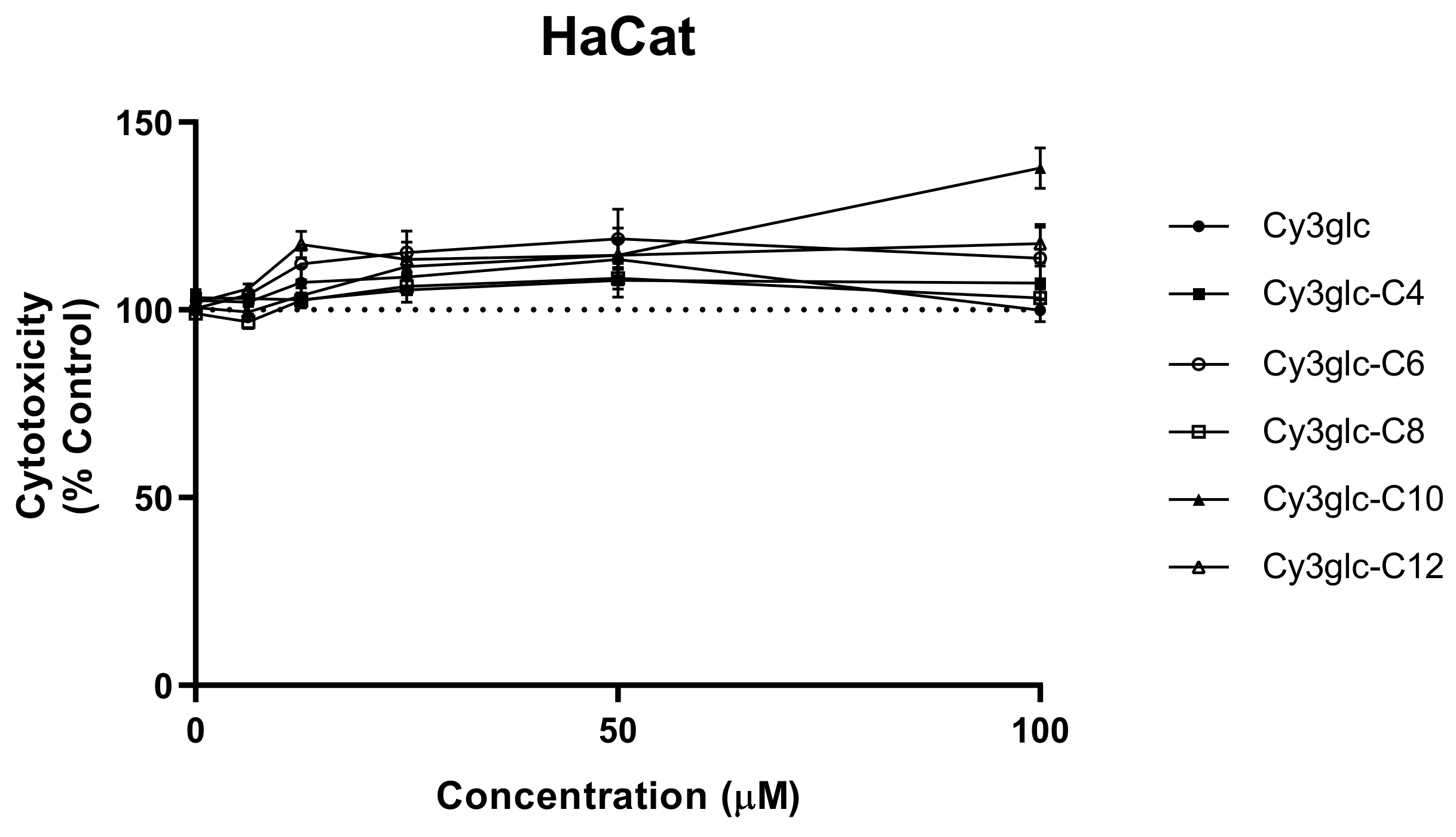
The stability tests suggested that the compounds have potential to exert their biological activities within the context of a cellular environment, maintaining their intact structure over a time period of several hours. Such observations led to the need to evaluate their cytotoxicity. For this reason, MTT assay was performed and it was possible to observe that none of the compounds presented significant cytotoxicity at concentrations below 100 μM (Figure 3). The results of cell survival for the higher concentrations are above 100%, which may be the result of false positives due to solubility issues of the more lipophilic compounds in aqueous solutions. Prior results with HaCat cells performed with different anthocyanins and extracts have demonstrated that Cy3glc has no significant cytotoxicity towards this cell line at concentrations below 100 μM [5].

3.3. ROS Production

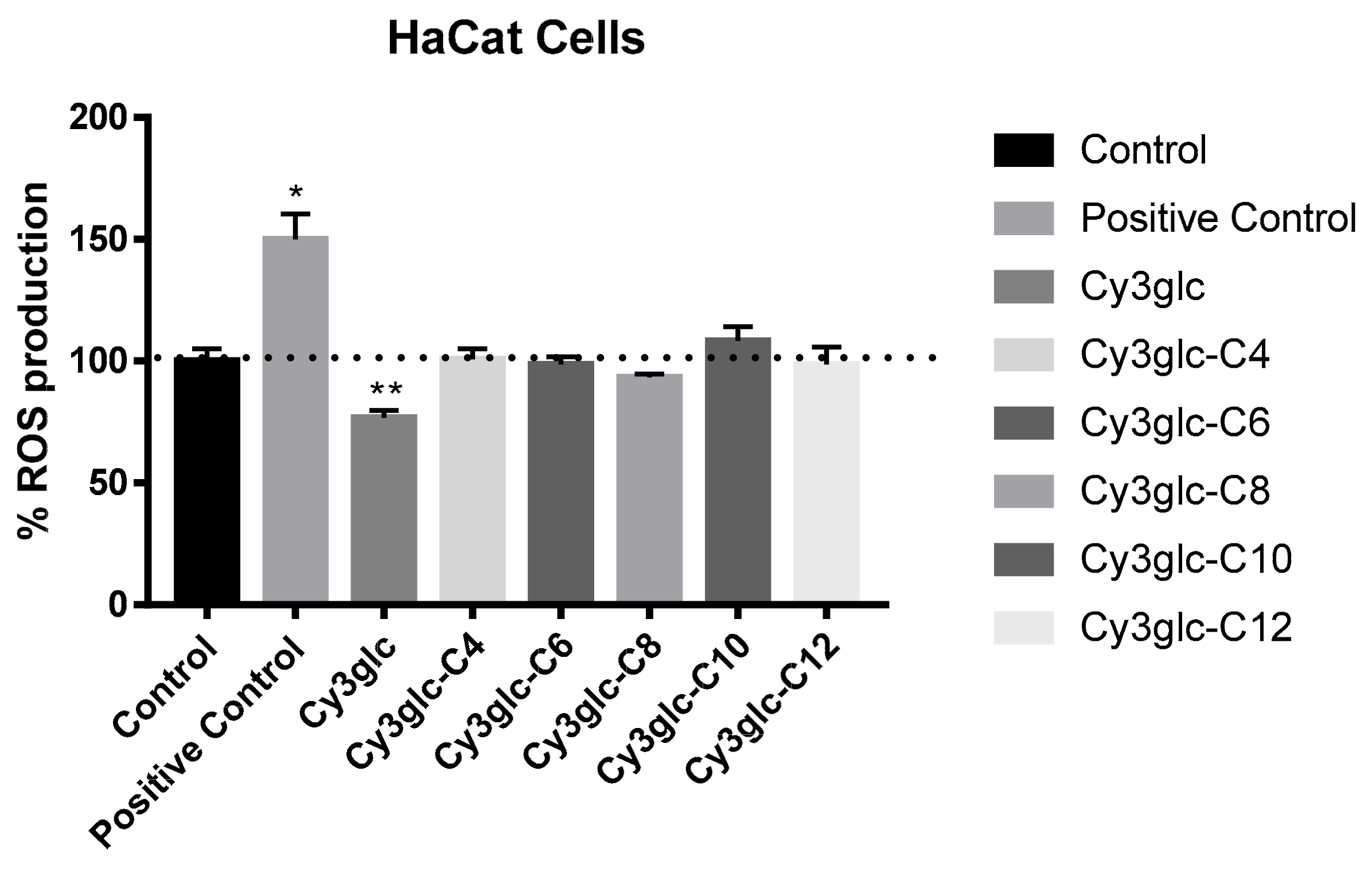
The production of reactive oxygen species is closely linked to inflammation processes within the cellular environment, which is normally exacerbated in a context of disease leading to large tissue injury [24]. In such perspective, the role of the lipophilic anthocyanin derivatives in ROS modulation within HaCat cells was evaluated. A concentration of 50 μM was used in order to ensure the best equilibrium between the potential modulation and solubility issues. The results showed that only Cy3glc had a significant reduction of ROS, 24 h after the incubation period when compared to the control (Figure 4).

3.4. Enzymatic Activity

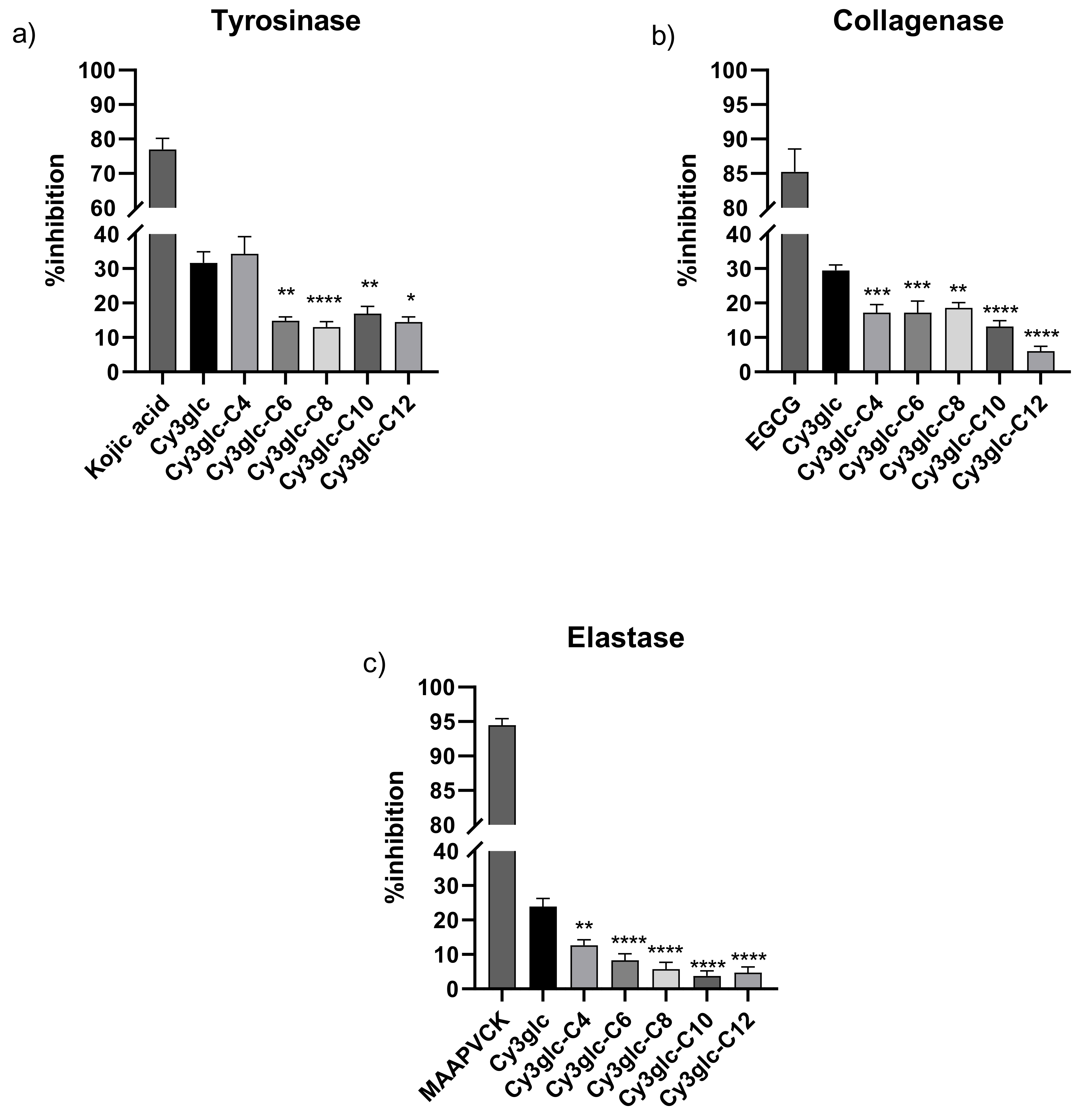
The effect of Cy3glc and lipophilic derivatives in the modulation of different enzymes involved in aging processes and skin diseases development was evaluated (Figure 5). Overall, the increment of the aliphatic chain resulted in a reduced inhibition of the enzymatic activity. In the case of tyrosinase, a significant reduction could be observed after the addition of a 6 carbons chain forward, with no significant effect on the inhibition ability of the parent compound upon the functionalization with a C4 aliphatic chain. However, in the case of collagenase, a readily decrease on the inhibition capacity was observed for all the functionalized derivatives, with Cy3glc-C12 showing the lowest inhibitory effect of collagenase activity. For elastase, the results clearly evidence the trend of inhibitory capacity reduction with the increment of the aliphatic chain. When comparing the results between the parent compound Cy3glc and the most aliphatic compound Cy3glc-C12, in all the enzymes the reductions on inhibitory capacity ranged several percentual units (a 17.1% reduction in the case of tyrosinase, 23.5% reduction in the case of collagenase and 20% in the case of elastase). Also, tyrosinase was shown to be the most sensible among the tested enzymes to the inhibitory effect of the compounds (with values ranging from 31.6% for Cy3glc to 14.5% for Cy3glc-C12).

3.5. Minimum Inhibitory Concentration (MIC) and Minimum Bactericidal Concentration (MBC) Values

The MIC and MBC values obtained against reference strains and against multidrug-resistant isolates are shown in Table 1 and Table 2, respectively. Cy3glc, Cy3glc-C4, and Cy3glc-C12 had no antibacterial activity. While for Cy3glc-C6, Cy3glc-C8, and Cy3glc-C10, MIC values could be obtained (except for Cy3glc-C6 against P. aeruginosa strain and isolates), being the antibacterial activity stronger against Gram-positives (lower MIC values) than Gram-negatives. Generally, the activity increased from Cy3glc-C6 to Cy3glc-C10. Thus, Cy3glc-C10 was the most active against all bacteria tested. Moreover, the three active compounds were bactericidal against P. aeruginosa, E. coli and S. aureus, since the MBC was equal or 2× the MIC. However, interestingly, those compounds were seemingly bacteriostatic against E. faecalis ATCC 29212.

4. Discussion

This work intended to ascertain the possible antimicrobial and antiaging effect of a library of anthocyanin-fatty acid esters. In this work, we described for the first time the possible dual effect of these derivatives and the importance of chain length in the studied activities.
Concerning the in vitro cell studies and having in mind the physiological target of these compounds, their stability in different conditions must be established. In vitro cell cultures must be maintained with crucial supplements such as serum. The results herein showed a significant interaction between the compounds and serum, which compromised their overall stability. However, in the presence of cells, the degradation rate observed previously was highly reduced, suggesting that these compounds may have the potential to interact with the cell membrane, by the mechanisms suggested before. It is also interesting to note for the case of cy3glc-C6, that the degradation resulted in the formation of the parent compound cy3glc within the first hours. This may constitute a strategic way of anthocyanin delivery to the cells. This type of methodology was already explored for other polyphenols like resveratrol with successful results [25].
Furthermore, at the concentrations tested in keratinocytes, none of the compounds showed significant toxicity. Such results are indicative of a potential role as modulatory effectors rather than apoptotic agents. These features can be useful for further potential applications in the cosmetics industry as preventive antiaging. In the case of the inflammatory state promoted by ROS, the lyophilization of the anthocyanin was shown to reduce its inhibitory potential. This may be explained by the fact that the acylation with the different fatty acids somehow reduces the scavenging capacity of the flavylium core, potentially through a hindrance of the reactive functional sites. Although, acylation could increase the transport efficiency, as observed in bacteria, thereby enabling the cytosolic action of esterase enzymes, resulting in a release of the precursor molecules. In this way the antioxidant potential is not lost, and the bioavailability is increased. Still, the use of saturated fatty acids linked may not be the most interesting ones for this type of mechanism. Previously, the antioxidant features of malvidin-3-glucoside–oleic acid ester derivative by means of DPPH, FRAP and lipid peroxidation assays were assessed, which confirmed that the structural modification of the genuine malvidin-3-glucoside into a more lipophilic compound did not compromise its antioxidant potential [16]. Besides, the possible breakdown of these type of molecules (anthocyanin-unsaturated fatty acid), may increase the antioxidant plasmatic protection.
Among others, the increase in the expression of skin matrix metalloproteinases (MMPs) resulting from these aggressions (especially UV radiation) is one of the key processes of skin aging and skin diseases [26]. Focusing on the development of strategies to inhibit such enzymes represents a wise strategy for the cosmetics industry. Consequently, herein, the role of the cy3glc lipophilic derivatives in the modulation of three main skin MMPs was assessed. For both tyrosinase, elastase, and collagenase the results suggested a reduction of the inhibitory efficiency with the functionalization revealing an inverse relation between the efficacy and the length of the fatty acid chain. Besides being the most widespread anthocyanin in nature, cy3glc is also the most studied. Several studies showed the inhibitory effect of this anthocyanin in the enzymatic activity of the enzymes tested herein [27,28,29]. This may be explained by the presence of an O-catechol group in the flavylium B ring structure of cy3glc. For example, tyrosinase contains a type 3 copper active center (binuclear copper, CuA and CuB) coordinated by three histidine residues. The diphenolase activity of the enzyme is achieved by the binding of two adjacent hydroxyl groups of the catechols (L-DOPA) to the two copper ions within the active site, which contains a hydrophobic ‘pocket’ that is sterically favorable to the binding of the catechol [30]. Indeed, previous studies with isolated anthocyanins (pelargonidin, cyanidin, delphinidin, pelargonidin-3-O-β-D-glucoside, cyanidin-3-O-β-D-glucoside, delphinidin-3-O-β-D-glucoside) showed that cy3glc had the greatest inhibitory capacity due to the presence of the O-catechol moiety [31]. In the case of elastase, a previous study performed with 40 phenolic compounds showed that strongest inhibition was demonstrated for the compounds bearing a O-catechol moiety [32]. In a recent study, catechol was shown to significantly reduce the relative levels of MMP-2 (collagenase type IV) mRNA expression in Huh7 and PLC/PRF/5 cells [33]. The fact that the derivatives showed a reduced inhibitory effect compared to the parent compound may be explained by the potential steric hindrance promoted by the acylation with the fatty acids, which may function as bulky groups blocking an efficient access to the active pocket of the respective enzymes, thereby causing a reduced inhibitory capacity. A similar effect was observed when comparing the activity of cyanidin aglycone and cy3glc, in which an reduction of inhibitory activity was observed in the presence of the glucose moiety [31]. Nevertheless, even with a fatty acid chain of 12 carbons, the derivatives showed a weak inhibitory action for the three enzymes (ranging from 5 to 15%).
Regarding the antibacterial activity, Cy3glc did not affect the bacterial growth of the several strains tested (MIC > 512 µg/mL). However, the insertion of a fatty acid chain sprouted the antibacterial activity. From the MIC results obtained, we observed that there was an optimum length for the fatty acid chain to affect the bacterial growth, since only Cy3glc-C6, Cy3glc-C8, and Cy3glc-C10 presented antibacterial effect, Cy3glc-C10 being the most effective one. The fatty chain in Cy3glc-C4 was too short to cause any antibacterial activity, while that in Cy3glc-C12 was too long. Thus, there is a relationship between lipophilicity and antibacterial activity of the tested compounds, which has been reported as well for other compounds, namely lipophilic N-acyldiamines [34]. It is likely that the side chain length of the compound affects its ability to permeate the bacterial membrane, thus influencing the antibacterial effect obtained. Moreover, all three active ester derivatives were more effective against Gram-positive bacteria than Gram-negative bacteria. In fact, it is well-known that differences in the cell wall composition between Gram-positives and Gram-negatives account for differences in the activity of many compounds [35,36]. In Gram-negative bacteria, the outer cell membrane is fairly impermeable to large molecules and hampers the diffusion of hydrophobic compounds through lipopolysaccharide layer [37].

5. Conclusions

This work was able to give new perceptions on the potential of cyanidin-3-glucoside-lipophilic conjugates to interact with cell membranes and in this way stabilize and protect these compounds from cellular degradation. Also, a strategic way to deliver anthocyanin to the cells was proposed by using enzymatic esterification reaction. There is no doubt that besides the previous advantage, this type of methodology of covalently linking a fatty acid to the anthocyanin glucose scaffold is also associated with a decrease in scavenging power of the original anthocyanin in the conjugated form. Also, an inverse relation between the inhibitory efficacy of skin enzymes capable of degrading extracellular matrix components and the length of the fatty acid chain was observed. Even though, in vivo, after enzymatic or chemical release, the original properties of both anthocyanin and fatty acid may be recovered, a remarkable antimicrobial effect was observed towards Gram-positive reference strains, including MRSA strains. The activity is improved by increasing lipophilicity, and the best results were obtained for compounds having an alkyl chain with 8 to 10 carbon atoms. The compounds were more active against Gram-positive bacteria, probably due do their surfactant type interaction with the bacterial membrane.

Author Contributions

Conceptualization, I.F., N.M. and L.C.; methodology, I.F., M.G., H.O., P.C., L.J.B., M.G., L.C.; investigation, I.F., H.O., P.C., L.J.B.; resources, N.M. and V.d.F.; writing—original draft preparation, I.F., L.J.B. and H.O.; writing—review and editing, I.F., L.C., N.M. and V.d.F.; supervision, L.C. and I.F.; project administration, N.M., P.G. and L.C.; funding acquisition, N.M., V.d.F. and L.C. All authors have read and agreed to the published version of the manuscript.

Funding

This work was supported by PTDC/OCE-ETA/31250/2017—“Novel Anthocyanin-Based Smart Sensors for Food Packaging”, supported by national funds by FCT / MCTES and co-supported by Fundo Europeu de Desenvolvimento Regional (FEDER) throughout COMPETE 2020—Programa Operacional Competitividade (POCI-01-0145-FEDER-031250). Processes 09 00340 i001

Acknowledgments

This work was supported by the Associate Laboratory for Sustainable Chemistry, Clean Processes and Technologies LAQV. The latter is financed by national funds from UIDB/50006/2020. This work was also supported by the project PTDC/AGR-TEC/3078/2014. L.C. and I.F. acknowledge their FCT research contracts. P.C. gratefully acknowledges her doctoral grants from FCT (SFRH/BD/150661/2020). H.O. would like to thank the FCT for his research contract (PTDC/QUI-OUT/29013/2017). This research is supported by AgriFood XXI I&D&I project (NORTE-01-0145-FEDER-000041) cofinanced by European Regional Development Fund (ERDF), through the NORTE 2020 (Programa Operacional Regional do Norte 2014/2020).

Conflicts of Interest

The authors declare no conflict of interest.

References

  1. Farage, M.A.; Miller, K.W.; Elsner, P.; Maibach, H.I. Intrinsic and extrinsic factors in skin ageing: A review. Int. J. Cosmet. Sci. 2008, 30, 87–95. [Google Scholar] [CrossRef] [PubMed]
  2. Baumann, L. Skin ageing and its treatment. J. Pathol. 2007, 211, 241–251. [Google Scholar] [CrossRef]
  3. Wang, Y.; Wang, L.; Wen, X.; Hao, D.; Zhang, N.; He, G.; Jiang, X. NF-κB signaling in skin aging. Mech. Ageing Dev. 2019, 184, 111–160. [Google Scholar] [CrossRef] [PubMed]
  4. Dunaway, S.; Odin, R.; Zhou, L.; Ji, L.; Zhang, Y.; Kadekaro, A.L. Natural Antioxidants: Multiple Mechanisms to Protect Skin From Solar Radiation. Front. Pharmacol. 2018, 9, 392. [Google Scholar] [CrossRef] [Green Version]
  5. Evora, A.; de Freitas, V.; Mateus, N.; Fernandes, I. The effect of anthocyanins from red wine and blackberry on the integrity of a keratinocyte model using ECIS. Food Funct. 2017, 8, 3989–3998. [Google Scholar] [CrossRef]
  6. Nanashima, N.; Horie, K.; Maeda, H.; Tomisawa, T.; Kitajima, M.; Nakamura, T. Blackcurrant Anthocyanins Increase the Levels of Collagen, Elastin, and Hyaluronic Acid in Human Skin Fibroblasts and Ovariectomized Rats. Nutrients 2018, 10, 495. [Google Scholar] [CrossRef] [PubMed] [Green Version]
  7. Oliveira, H.; Basílio, N.; Pina, F.; Fernandes, I.; De Freitas, V.; Mateus, N. Purple-Fleshed sweet potato acylated anthocyanins: Equilibrium network and photophysical properties. Food Chem. 2019, 288, 386–394. [Google Scholar] [CrossRef]
  8. Mattarei, A.; Biasutto, L.; Rastrelli, F.; Garbisa, S.; Marotta, E.; Zoratti, M.; Paradisi, C. Regioselective O-derivatization of quercetin via ester intermediates. An improved synthesis of rhamnetin and development of a new mitochondriotropic derivative. Molecules 2010, 15, 4722–4736. [Google Scholar] [CrossRef] [PubMed] [Green Version]
  9. Cruz, L.; Fernandes, V.C.; Araújo, P.; Mateus, N.; De Freitas, V. Synthesis, characterisation and antioxidant features of procyanidin B4 and malvidin-3-glucoside stearic acid derivatives. Food Chem. 2015, 174, 480–486. [Google Scholar] [CrossRef]
  10. Cruz, L.; Guimarães, M.; Araújo, P.; Évora, A.; de Freitas, V.; Mateus, N. Malvidin 3-Glucoside–Fatty Acid Conjugates: From Hydrophilic toward Novel Lipophilic Derivatives. J. Agric. Food. Chem. 2017, 65, 6513–6518. [Google Scholar] [CrossRef]
  11. Grajeda-Iglesias, C.; Salas, E.; Barouh, N.; Baréa, B.; Figueroa-Espinoza, M.C. Lipophilization and MS characterization of the main anthocyanins purified from hibiscus flowers. Food Chem. 2017, 230, 189–194. [Google Scholar] [CrossRef] [PubMed]
  12. Yang, W.; Kortesniemi, M.; Yang, B.; Zheng, J. Enzymatic acylation of anthocyanins isolated from alpine bearberry (Arctostaphylos alpina) and lipophilic properties, thermostability, and antioxidant capacity of the derivatives. J. Agric. Food Chem. 2018, 66, 2909–2916. [Google Scholar] [CrossRef]
  13. Cruz, L.; Benohoud, M.; Rayner, C.M.; Mateus, N.; de Freitas, V.; Blackburn, R.S. Selective enzymatic lipophilization of anthocyanin glucosides from blackcurrant (Ribes nigrum L.) skin extract and characterization of esterified anthocyanins. Food Chem. 2018, 266, 415–419. [Google Scholar] [CrossRef] [PubMed] [Green Version]
  14. Guimarães, M.; Pérez-Gregorio, M.; Mateus, N.; de Freitas, V.; Galinha, C.F.; Crespo, J.G.; Portugal, C.A.M.; Cruz, L. An efficient method for anthocyanins lipophilization based on enzyme retention in membrane systems. Food Chem. 2019, 300, 125167. [Google Scholar] [CrossRef] [PubMed]
  15. Fernandez-Aulis, F.; Torres, A.; Sanchez-Mendoza, E.; Cruz, L.; Navarro-Ocana, A. New acylated cyanidin glycosides extracted from underutilized potential sources: Enzymatic synthesis, antioxidant activity and thermostability. Food Chem. 2020, 309, 125796. [Google Scholar] [CrossRef]
  16. Cruz, L.; Fernandes, I.; Guimaraes, M.; de Freitas, V.; Mateus, N. Enzymatic synthesis, structural characterization and antioxidant capacity assessment of a new lipophilic malvidin-3-glucoside-oleic acid conjugate. Food Funct. 2016, 7, 2754–2762. [Google Scholar] [CrossRef]
  17. Guimarães, M.; Mateus, N.; de Freitas, V.; Branco, L.C.; Cruz, L. Microwave-Assisted Synthesis and Ionic Liquids: Green and Sustainable Alternatives toward Enzymatic Lipophilization of Anthocyanin Monoglucosides. J. Agric. Food. Chem. 2020, 68, 7387–7392. [Google Scholar] [CrossRef]
  18. Mendoza, J.; Pina, F.; Basílio, N.; Guimarães, M.; De Freitas, V.; Cruz, L. Extending the stability of red and blue colors of malvidin-3-glucoside-lipophilic derivatives in the presence of SDS micelles. Dyes Pigment. 2018, 151, 321–326. [Google Scholar] [CrossRef]
  19. Guimarães, M.; Mateus, N.; de Freitas, V.; Cruz, L. Improvement of the Color Stability of Cyanidin-3-glucoside by Fatty Acid Enzymatic Acylation. J. Agric. Food. Chem. 2018, 66, 10003–10010. [Google Scholar] [CrossRef]
  20. Marquez-Rodriguez, A.S.; Guimarães, M.; Mateus, N.; de Freitas, V.; Ballinas-Casarrubias, M.L.; Fuentes-Montero, M.E.; Salas, E.; Cruz, L. Disaccharide anthocyanin delphinidin 3-O-sambubioside from Hibiscus sabdariffa L.: Candida antarctica lipase B-catalyzed fatty acid acylation and study of its color properties. Food Chem. 2021, 344, 128603. [Google Scholar] [CrossRef]
  21. Clinical and Laboratory Standards Institute. M07-A10:Methods for Dilution of Antimicrobial Susceptibility Tests for Bacteria That Grow Aerobically; Approved Standard—10th Edition; Clinical and Laboratory Standards Institute: Wayne, PA, USA, 2015; Volume 35. [Google Scholar]
  22. Bessa, L.J.; Eaton, P.; Dematei, A.; Placido, A.; Vale, N.; Gomes, P.; Delerue-Matos, C.; Sa Leite, J.R.; Gameiro, P. Synergistic and antibiofilm properties of ocellatin peptides against multidrug-resistant Pseudomonas aeruginosa. Future Microbiol. 2018, 13, 151–163. [Google Scholar] [CrossRef] [PubMed] [Green Version]
  23. Phan, H.; Yoda, T.; Chahal, B.; Morita, M.; Tkagi, M.; Vestergaard, M. Structure-dependent interactions of polyphenols with a biomimetic membrane system. Biochim. Biophys. Acta (BBA) Biomembr. 2014, 1838, 2670–2677. [Google Scholar] [CrossRef] [Green Version]
  24. Mittal, M.; Siddiqui, M.; Tran, K.; Reddy, S.; Malik, A. Reactive Oxygen Species in Inflammation and Tissue Injury. Antioxid. Redox Signal. 2014, 20, 1126–1167. [Google Scholar] [CrossRef] [Green Version]
  25. Vercauteren, J. Compositions Comprising Stilbene Polyphenol Derivatives and Use Thereof for Combating the Ageing of Living Organisms and Diseases Affecting Same. U.S. Patent 2,0100,310,615 12 September 2007. [Google Scholar]
  26. Pittayapruek, P.; Meephansan, J.; Prapapan, O.; Komine, M.; Ohtsuki, M. Role of Matrix Metalloproteinases in Photoaging and Photocarcinogenesis. Int. J. Mol. Sci. 2016, 17, 868. [Google Scholar] [CrossRef] [Green Version]
  27. Santos, J.; La, V.D.; Bergeron, C.; Grenier, D. Inhibition of host- and bacteria-derived proteinases by natural anthocyanins. J. Periodontal Res. 2011, 46, 550–557. [Google Scholar] [CrossRef]
  28. Afaq, F.; Zaid, M.A.; Khan, N.; Dreher, M.; Mukhtar, H. Protective effect of pomegranate-derived products on UVB-mediated damage in human reconstituted skin. Exp. Dermatol. 2009, 18, 553–561. [Google Scholar] [CrossRef] [Green Version]
  29. Xue, Y.-L.; Miyakawa, T.; Hayashi, Y.; Okamoto, K.; Hu, F.; Mitani, N.; Furihata, K.; Sawano, Y.; Tanokura, M. Isolation and tyrosinase inhibitory effects of polyphenols from the leaves of persimmon, Diospyros kaki. J. Agric. Food. Chem. 2011, 59, 6011–6017. [Google Scholar] [CrossRef] [PubMed]
  30. Mukherjee, P.K.; Biswas, R.; Sharma, A.; Banerjee, S.; Biswas, S.; Katiyar, C.K. Validation of medicinal herbs for anti-tyrosinase potential. J. Herbal Med. 2018, 14, 1–16. [Google Scholar] [CrossRef]
  31. Tsuda, T.; Osawa, T. Inhibition of Tyrosinase Activity by the Anthocyanin Pigments Isolated from Phaseolus vulgaris L. Food Sci. Technol. Int. 1997, 3, 82–83. [Google Scholar] [CrossRef]
  32. Melzig, M.F.; Loser, B.; Ciesielski, S. Inhibition of neutrophil elastase activity by phenolic compounds from plants. Die Pharm. 2001, 56, 967–970. [Google Scholar]
  33. Lim, W.-C.; Kim, H.; Kim, Y.-J.; Jeon, B.-N.; Kang, H.-B.; Ko, H. Catechol inhibits epidermal growth factor-induced epithelial-to-mesenchymal transition and stem cell-like properties in hepatocellular carcinoma cells. Sci. Rep. 2020, 10, 7620. [Google Scholar] [CrossRef] [PubMed]
  34. De Almeida, C.G.; Garbois, G.D.; Amaral, L.M.; Diniz, C.C.; Le Hyaric, M. Relationship between structure and antibacterial activity of lipophilic N-acyldiamines. Biomed. Pharmacother. 2010, 64, 287–290. [Google Scholar] [CrossRef]
  35. Richter, M.F.; Hergenrother, P.J. The challenge of converting Gram-positive-only compounds into broad-spectrum antibiotics. Ann. N. Y. Acad. Sci. 2019, 1435, 18–38. [Google Scholar] [CrossRef] [PubMed] [Green Version]
  36. Tavares, T.D.; Antunes, J.C.; Padrão, J.; Ribeiro, A.I.; Zille, A.; Amorim, M.T.P.; Ferreira, F.; Felgueiras, H.P. Activity of Specialized Biomolecules against Gram-Positive and Gram-Negative Bacteria. Antibiotics 2020, 9, 314. [Google Scholar] [CrossRef] [PubMed]
  37. Reygaert, W.C. An overview of the antimicrobial resistance mechanisms of bacteria. AIMS Microbiol 2018, 4, 482–501. [Google Scholar] [CrossRef]
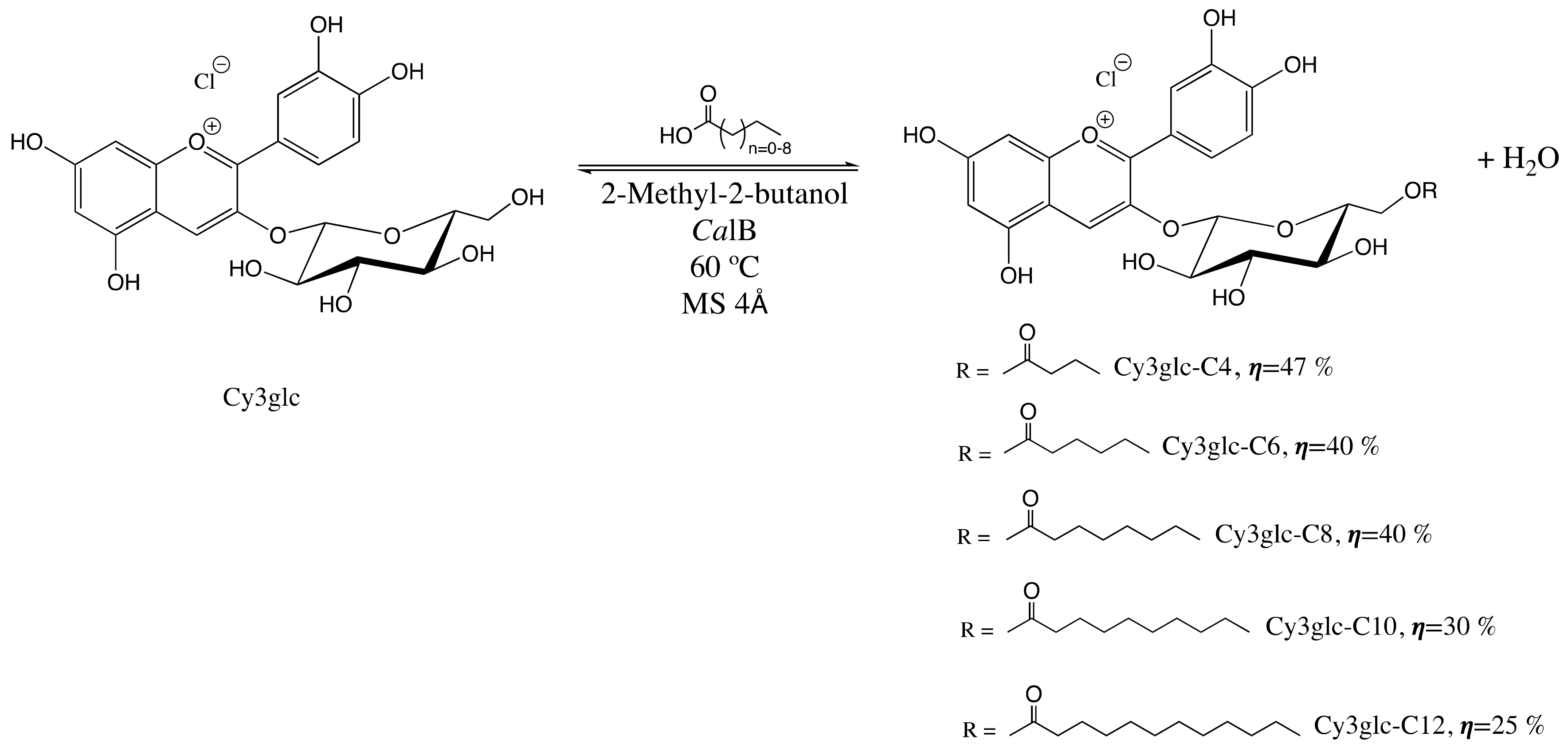
Scheme 1. CalB-catalyzed esterification reaction performed to achieve the different cy3glc-fatty acid derivatives.
Processes 09 00340 sch001
Figure 1. Physiological stability assays of Cy3glc-C4 were performed in DMEM (a) or HBSS (b), at pH 7.4 and 37 °C, during 5 h. Representative chromatograms at 520 nm.
Processes 09 00340 g001
Figure 2. Physiological stability of Cy3glc and Cy3glc-C6 was performed in DMEM 10% FBS in the presence (b,d) or absence (a,c) of HaCat cells, at pH 7.4 at 37 °C, during 5 and 24 h. Representative chromatograms at 520 nm.
Processes 09 00340 g002
Figure 3. Cytotoxicity activity of Cy3glc and Cy3glc-fatty acid derivatives in spontaneously transformed aneuploid immortal keratinocyte cell line from adult human skin (HaCat) evaluated by MTT assay.
Processes 09 00340 g003
Figure 4. Effect of Cy3glc and Cy3glc-fatty acid derivatives at 50 µM, on ROS production after 24 h in HaCat cells. Hydrogen peroxide was used as a positive control. Statistical comparisons were performed in comparison to control. * p < 0.05, p **< 0.01.
Processes 09 00340 g004
Figure 5. Inhibitory activity of lipophilic compounds at 50 µM against (a) tyrosinase, (b) collagenase (c) elastase enzymes. For each enzyme a positive control was included as indicated in Section 2. Statistical comparisons were performed using Cy3glc as control. * p < 0.05, ** p < 0.01, *** p < 0.001 and **** p < 0.0001.
Processes 09 00340 g005
Table 1. Minimum inhibitory concentrations (MICs) and minimum bactericidal concentration (MBCs), in µg/mL, of Cy3glc and Cy3glc-fatty acid derivatives against two Gram-negative and two Gram-positive reference strains.
CompoundP. aeruginosa ATCC 27853E. coli ATCC 25922S. aureus ATCC 29213E. faecalis ATCC 29212
MIC (MBC)
Cy3glc>512 (-)>512 (-)>512 (-)>512 (-)
Cy3glc-C4>512 (-)>512 (-)>512 (-)>512 (-)
Cy3glc-C6>512 (-)128 (128)8 (16)4 (>32)
Cy3glc-C8256 (256)32 (32)2 (4)2 (16)
Cy3glc-C10128 (128)16 (32)2 (2)1 (>8)
Cy3glc-C12>512 (-)>512 (-)>512 (-)>512 (-)
Table 2. Minimum inhibitory concentrations (MICs) and minimum bactericidal concentration (MBCs), in µg/mL of compounds against two multidrug-resistant P. aeruginosa isolates and two methicillin-resistant S. aureus (MRSA).
CompoundP. aeruginosa PA004P. aeruginosa PA002MRSA Sa1MRSA S007
MIC (MBC)
Cy3glc>512 (-)>512 (-)>512 (-)>512 (-)
Cy3glc-C4>512 (-)>512 (-)>512 (-)>512 (-)
Cy3glc-C6>512 (-)>512 (-)16 (64)16 (64)
Cy3glc-C8128 (128)>512 (-)8 (32)8 (8)
Cy3glc-C1064 (64)512 (512)8 (16)4 (8)
Cy3glc-C12>512 (-)>512 (-)>512 (-)>512 (-)
Publisher’s Note: MDPI stays neutral with regard to jurisdictional claims in published maps and institutional affiliations.