Article

Bioactive Chemical Constituents of Matthiola longipetala Extract Showed Antioxidant, Antibacterial, and Cytotoxic Potency

by
Yasser A. El-Amier
1,*,
Nouf S. Zaghloul
2 and
Ahmed M. Abd-ElGawad
3
1
Department of Botany, Faculty of Science, Mansoura University, Mansoura 35516, Egypt
2
Bristol Centre for Functional Nanomaterials, HH Wills Physics Laboratory, Tyndall Avenue, Bristol BS8 1FD, UK
3
Department of Plant Production, College of Food & Agriculture Sciences, King Saud University, P.O. Box 2460, Riyadh 11451, Saudi Arabia
*
Author to whom correspondence should be addressed.
Separations 2023, 10(1), 53; https://doi.org/10.3390/separations10010053
Submission received: 18 December 2022 / Revised: 8 January 2023 / Accepted: 12 January 2023 / Published: 13 January 2023

Abstract

:
The exploration of bioactive compounds from natural resources attracts the attention of researchers and scientists worldwide. M. longipetala is an annual aromatic herb that emits a pleasant odor during the night. Regarding the chemical composition and biological characteristics, M. longipetala extracts are poorly studied. The current study aimed to characterize the chemical composition of M. longipetala methanol extract using GC-MS and determine its biological potencies, including its capacity for cytotoxicity and antioxidant and antibacterial activities. In this approach, 37 components were identified, representing 99.98% of the total mass. The major chemical components can be classified as oxygenated hydrocarbons (19.15%), carbohydrates (10.21%), amines (4.85%), terpenoids (12.71%), fatty acids and lipids (50.8%), and steroids (2.26%). The major identified compounds were ascaridole epoxide (monoterpene, 12.71%) and methyl (E)-octadec-11-enoate (ester of fatty acid, 12.21%). The extract of M. longipetala showed substantial antioxidant activity. Based on the DPPH and ABTS scavenging, the antioxidant activity of the extracted components of M. longipetala revealed that leaf extract is the most effective with IC50 values of 31.47 and 28.94 mg/L, respectively. On the other hand, the extracted plant showed low antibacterial activities against diverse bacterial species, viz., Escherichia coli, Klebsiella pneumonia, Staphylococcus epidermidis, S. haemolyticus, and S. aureus. The most potent antibacterial results were documented for leaf and flower extracts against E. coli and S. aureus. Additionally, the extract’s effectiveness against HepG2 cells was evaluated in vitro using the measures of MTT, DNA fragmentation, and cell proliferation cycle, where it showed considerable activity. Therefore, we can conclude that M. longipetala extract displayed improvement in cytocompatibility and cell migration properties. In conclusion, M. longipetala could be considered a potential candidate for various bioactive compounds with promising biological activities. However, further characterization of the identified compounds, particularly the major compounds, is recommended to evaluate their efficacy, modes of action, and safety.

1. Introduction

Drugs derived from natural sources, particularly medicinal plants, attract the interest of scientists and researchers for the production of promising bioactive compounds that can be integrated into pharmaceutical applications [1]. Plants as natural resources provide people with various goods and services, such as food, fodder for animals, textiles, drugs, medicine, and dyes [2,3,4]. These confidential pharmaceutical drugs available from natural sources provided a reduction in side effects that were present with synthetic drugs [5,6,7]. Family Brassicaceae is an economically important family of flowering plants with 365 genera and 3250 species. These occur in all phytogeographic regions, notably the Mediterranean region, and are extensively dispersed [8]. In Egypt, Brassicaceae is one of the four largest families, represented by 103 species belonging to 53 genera [8]. Genus Matthiola incorporates nearly 55 species of herbs that are annual and perennial grown in the regions of Africa, Asia, and western Europe; in the flora of Egypt, four species are recorded [8]. The plants of genus Matthiola are grown and obtained throughout the year, particularly from January to October. The flowers of this genus are varied in color, i.e., yellow, red, white, or light purple [9]. Matthiola longipetala (Vent.) DC. is the most common and widespread member of this genus grown in the north of Africa and is commonly recognized as “Shigaara” or “Shoqaara”.
Recent research focused on the characterization of the active chemical components and biological applications of the extracted plants of the Matthiola genus. The different extracts of Matthiola species were stated to exhibit various biological activities such as antioxidant, neuroprotective, antimicrobial, antiparasitic, antiurolithiatic, insecticidal, anticancer, and allelopathic activities [10,11,12]. Chemically, many studies concerning the chemical constituents of the Matthiola genus revealed the presence of isothiocyanate, anthocyanins, glucosinolates, flavonoids, lipids, sterol glycosides, and volatile oils [11,13,14,15]. Subsequently, researchers prolonged the work on the chemical composition and biological diversity of the Matthiola genus [16,17,18].
According to the existing literature on the genus Matthiola, the chemical components of M. longipetala (evening stock) and its biological assessments are poorly studied. The chemical composition and insecticidal activity of the essential oil extracted from the Tunisian ecospecies of M. longipetala was reported by Hammami et al. [19]. Moreover, three phenolic and sterol glycosides were identified from M. longipetala growing in Tunisia [20]. In this context, the chemistry and antioxidant activity of the essential oil isolated from the Libyan ecospecies of M. longipetala were studied by Abdelshafeek et al. [18]. The fatty acid composition of a Turkish sample of M. longipetala was analyzed by Karaman et al. [16]. In order to investigate the biochemical elements responsible for the biological effects, the current study set out to characterize the chemical components of the Egyptian ecospecies of M. longipetala methanol extract by GC-MS, and evaluate the antioxidant, antibacterial, and anticancer activities of M. longipetala methanol extract.

2. Materials and Methods

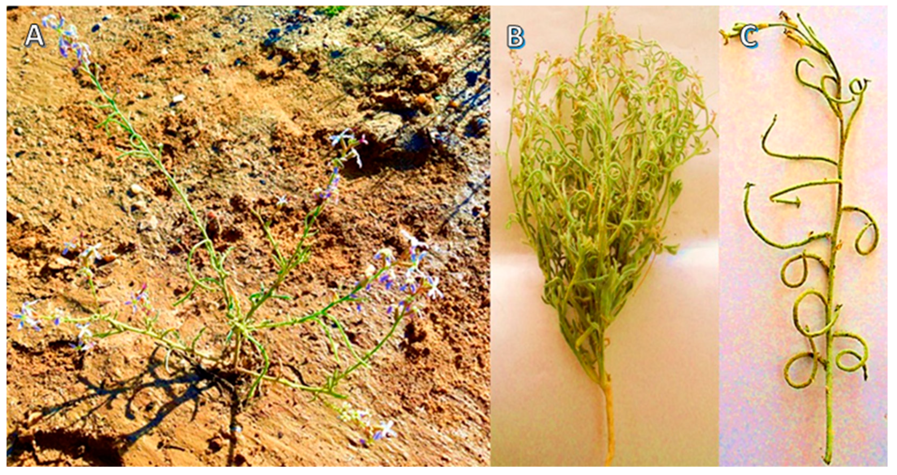
2.1. Plant Materials Collection, Preparation, and Extraction

The aboveground plant parts (Figure 1), including stems, leaves, flowers, and roots, were collected from populations of M. longipetala naturally grown in Wadi Ash-Sheikh, north Eastern Desert, Egypt (28°40′4.63″ N 31° 3′51.41″ E). The varied samples were cleaned of any impurities and left to dry for seven days in shady settings at ambient temperature (25 ± 3 °C). The plant specimen was identified by Dr. Yasser El-Amier (an author) following Boulos [8].
A voucher specimen was assembled, coded with Mans.0121312001, and deposited in the Herbarium of the Faculty of Science at Mansoura University, Mansoura, Egypt. All dried plant materials were combined and ground into a fine powder for chemical characterization. Ten grams of this mixture were placed in a conical flask (250 mL), covered with 150 mL of methanol, and shaken vigorously for two hours at room temperature in a horizontal water bath shaker (model Memmert WB14, Schwabach, Germany). Whatman filter sheets were used to filter the extract (no. 1, 125 mm, Sigma-Aldrich, Darmstadt, Germany). The residue was placed into glass vials and stored at 4 °C in the refrigerator while the extract was dried using a rotary evaporator [21].

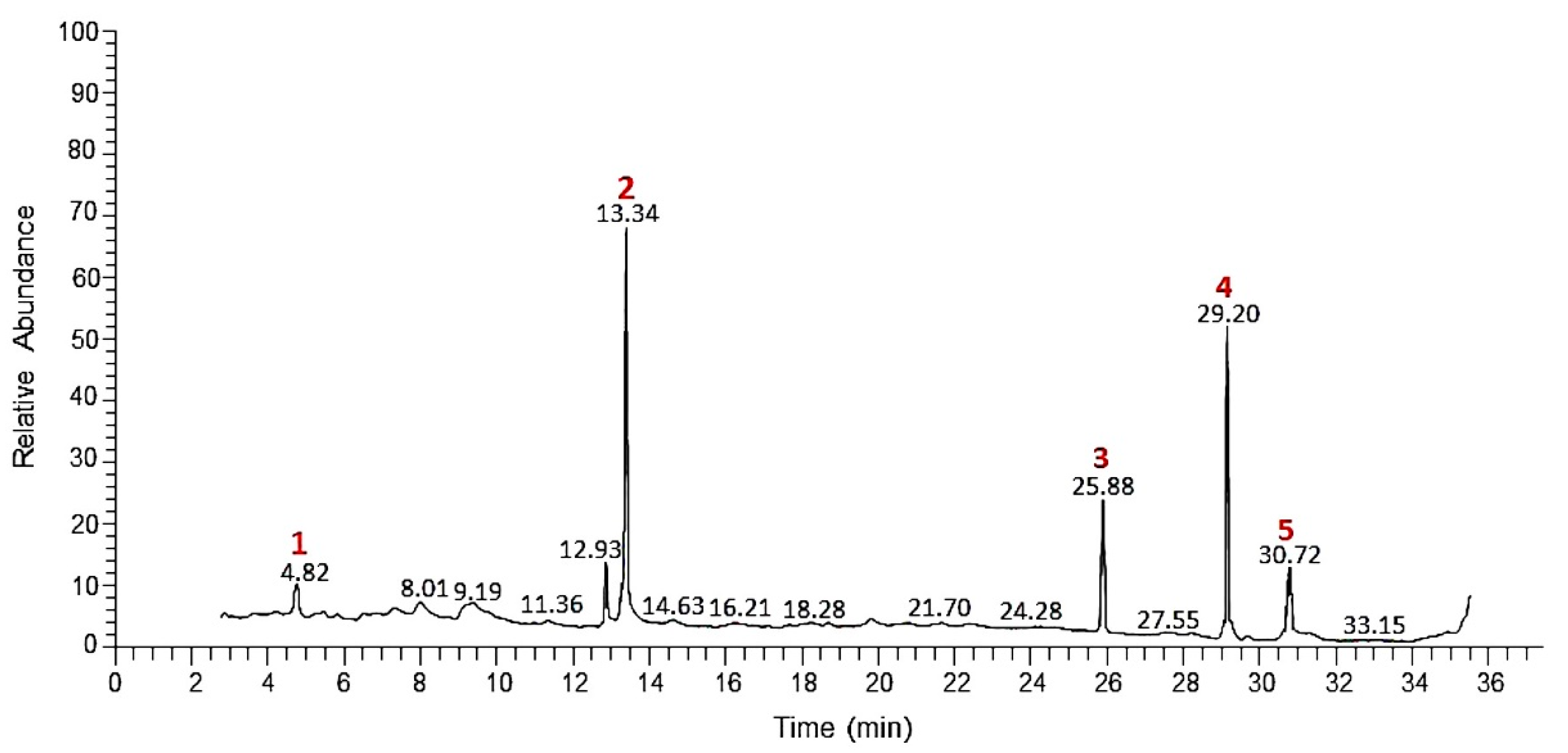
2.2. Gas Chromatography-Mass Spectrometry (GC-MS) of the Extract

A trace GC-TSQ mass spectrometer (Thermo Scientific, Austin, TX, USA) together with a TG-5MS capillary column with dimensions of 30 m × 0.25 mm and 0.25 m thickness were used to evaluate the chemical composition of the produced extract from M. longipetala aboveground sections. The temperature was first set at 50 °C and then it was programmed to rise by 5 °C/minute up to 250 °C and hold that temperature for 2 min before being raised by 30 °C/minute to 300 °C as the ultimate temperature, also to be held for 2 min. With a split ratio of 1:10, helium was used as the carrier gas while 0.2 µL of the sample (diluted in methanol, 1:10, v/v) was introduced into the apparatus. The EI mass spectra were plotted at 70 EV ionization voltage alongside the range of 50–500 for m/z. By comparing the mass spectra with those of the NIST 14 and WILEY 09 databases, the chemical authentication of the components in the M. longipetala extract was carried out and interpreted.

2.3. Antioxidant Activity Assay of M. longipetala Extracts

The methanol extract of different plant organs (root, stem, leaf, and flower) of M. longipetala were tested for their antioxidant activity over two assays: 2,2-diphenyl-1-picrylhydrazyl (DPPH) and 2,2-azinobis-(3-ethylbenzothiazoline-6-sulfonate) (ABTS).

2.3.1. DPPH Assay

The antioxidant activity was determined for each plant organ (root, stem, leaf, and flower) of M. longipetala according to Miguel [22]. Methanol extract of each plant organ was prepared by mixing 10 g of the plant powder with 150 mL methanol in a flask. At room temperature (25 ± 3 °C), the flasks were put in a horizontal water bath shaker (Memmert WB14, Schwabach, Germany) and shaken continuously for two hours. A rotary evaporator was used to filter and dry the methanol extract, and the dried residue was then collected in glass vials. Methanol was used to create concentrations of 5, 10, 20, 30, 40, and 50 mg/L from each residue. For assessment of antioxidant activity, equal volumes (1 mL) of either DPPH solution (0.135 Mm) or sample were homogenized well. The catechol was treated as the samples and considered a positive standard control. Following a half-hour of incubation in the dark at room temperature, the samples’ absorbance at =517 nm was measured using a UV/Vis spectrophotometer (model Spekol 11, Analytik Jena, Jena, Germany). The antioxidant scavenging activity was calculated and expressed as a percentage according to the following formula:
Scavenging   % = 100   × Absorbance control     Absorbance sample Absorbance control  
The experiment was designed with three replicas, and the inhibitive concentrations for 50% (IC50, mg/L) were calculated from the exponential curve between concentration and inhibition percentage.

2.3.2. ABTS Assay

According to Re et al. [23], the 2,2′azinobis(3-ethylbenzothiazoline-6-sulfonic acid) (ABTS, Sigma-Aldrich, Germany) radical cation decolorization test was also used to confirm the antioxidant activity. In order to determine the kinetic curves of the antioxidant activity of all samples, the decrease in absorbance was measured at room temperature after 0, 6, 12, 18, 24, and 30 min, and then every 10 min until the reaction reached a steady state or until the absorbance decreased less than 10% from the previous reading. The radical was created by combining 2.45 mM of K2S2O8 with 7 mM of ABTS (1/1, v/v) and homogenizing the mixture for a full 16 h at room temperature (25 ± 2 °C). After vigorously mixing 0.2 mL of each sample concentration (5, 10, 20, 30, 40, and 50 mg/L) with 2 mL of the ABTS solution in glass tubes, the absorbance at 734 nm was measured after 6 min at room temperature. The scavenging % and IC50 were computed as previously indicated in the DPPH test.

2.4. Antibacterial Activity Assay

The antibacterial activity of M. longipetala MeOH extract was evaluated using an agar well diffusion method [24]. Nutrient agar culture media, anon-selective medium containing beef extracts with peptone, yeast extracts, and NaCl to supply C, N, vitamins, and some trace constituents required for bacterial growth, was used. In a 2 L conical flask, 28 g of the nutrient agar media (Merck, Bangalore, India) was combined with 1000 mL of distilled water to create the nutrient medium. The medium was sterilized in the autoclave for 15 min at 121 °C under a pressure of 15 lbs. After cooling down to 45–50 °C, the medium was poured into sterilized Petri plates. The Cairo Microbiological Resources Centre (Cairo MIRCEN), Ain Shams University, Cairo, Egypt, provided eight microbial isolates. These isolates were four Gram-negative bacteria (Escherichia coli (NR_112558.1), Pseudomonas aeruginosa (CP050335.1), Salmonella typhimurium (NR_074910.1), and Klebsiella pneumoniae (NR_117683.1)) and four Gram-positive bacteria (Staphylococcus epidermidis (NR_116352.1), Staphylococcus aureus (NR_115606.1), Staphylococcus haemolyticus (NR_113345.1), and Staphylococcus xylosus (NR_113350.1)). Four standard antibiotics (tetracycline, cephradin, ampicillin, and azithromycin) were used.
The antibacterial effect was estimated by the agar well diffusion method with an inoculum of 1 × 10⁶ colony-forming units (CFU)/mL that was spread over a medium in the Petri plates. Within each Petri plate, four wells (5 mm each) were cut from the agar layer. A rotary evaporator was used to dry the methanol extract, and the dried residue was collected in glass vials. The residue was produced at a concentration of 10 mg/L using 1%, v/v dimethyl sulfoxide (DMSO). The standard antibiotics were also prepared with the same concentration (10 mg/L). About 50 μL of the methanol extract of each plant part was poured into the wells. As a negative control, DMSO was utilized; however, it showed no antibacterial action. The prepared plates spent 24 h in an incubator set at 37 °C. Measured along three axes, the inhibitory zone diameters in mm were reported as an average ± standard deviation.

2.5. Cytotoxicity and Cell Proliferation

The cytotoxicity of M. longipetala extract was evaluated via MTT assay [25]. The hepatocellular carcinoma, HePG-2, was chosen as a tumor cell line. We purchased the HePG-2 from ATCC via VACSERA (Cairo, Egypt). A stock solution containing 5 mg of MTT (3-(4,5-dimethyl-2-thiazolyl)-2,5-diphenyl-2H-tetrazolium bromide) was produced, vortexed well, filtered, and stored at −20 °C. The HepG2 were seeded onto 96-well plates with a density of 3 × 10³ cells/well suspended in 100 μL of complete medium. The plates were incubated in 5% CO2 at 37 °C for 24 h to promote adhesion and stabilization. Different quantities of the M. longipetala extract (31.3, 62.5, 125, 500, and 1000 g/mL) were made using DMSO (1% v/v) as the solvent. The cell lines were exposed to the extracts for 48 h. The media was discarded by aspiration, MTT (0.5 mg/mL) was supplied for cells, then the plates were incubated for four hours at 37 °C with 5%. About 100 µL of SDS (1%/0.01 M HCl) was poured into each well, and the growth of cells was measured at λmax = 570 nm using an absorbance microplate reader (Elx800, BioTek, Winooski, VT, USA). The results were presented as percentages with respect to the control.
A control of MTT solution without seeded cell lines was performed to avoid the effect of MTT cytotoxicity, which is also used to calculate cell viability using the formula below:
Cell   viability   % = 100   ×   A b s o r b a n c e S a m p l e   A b s o r b a n c e b l a n k A b s o r b a n c e c o n t r o l   A b s o r b a n c e b l a n k

2.6. Assay of Cell Motility

The motility of cell assay was performed to assess the wound healing effect in the form of cell migration. In the assay, the culture was imported into a six-well plate, seeded with cells, and grown to confluence. In order to make a wound, a monolayer of cells was scratched using a pipette tip, followed by twice washing using buffered salt solutions (PBS), and media to eliminate floating cells. Different inhibitory doses of M. longipetala extract were applied to the cells (IC5, IC10, IC25, and IC50). The cells were labelled using Hoechst (Sigma-Aldrich, Taufkirchen, Germany), which stains the nuclei of cells, at 0 time, following staining, and after 26 h. Images were collected at 0 time, immediately after staining, and 26 h after the wounds and were evaluated using phase-contrast microscopy on an inverted microscope [26].

2.7. Conventional PCR

In order to evaluate the mRNA expression of CD44 and MDR1, the PCR method was employed. The cells were treated with M. longipetala extract for 24 h and collected for the extraction of the cellular RNA. The total RNA was transferred to cDNA using (Qiagen, Germantown, MD, USA) 1 µL of cDNA in a total volume of 20 μL containing 10 μL Master Mix (Dream Taq Green PCR Master mix 2X, Thermo Fisher, Waltham, MA, USA), forward primer (0.5 μM), and reverse primer (0.5 μM) then the reaction volume was completed up to 20 μL using nuclease-free water. Conditions for the thermal cycle were set for denaturation at 95 °C for one minute, annealing at 72 °C for one minute, and a final extension of 10 min at 72 °C. The mRNA expression primer sequences were MDR1 at 58 °C: 5′-CCC ATC ATT GCA ATA GCA GG-3′ (forward), 5′-TGT TCA AAC TTC TGC TCC TGA-3′ (reverse), CD44 at 55 °C: 5′-TTT GCA TTG CAG TCA ACA GTC-3′ (forward), and 5′-TTA CAC CCC AAT CTT CAT GTC CAC-3′ (reverse).

2.8. Statistical Analysis

The cytotoxicity experiment was performed twice, whereas the antibacterial and antioxidant activity assays were conducted three times with three replications each. Using Costat software (CoHort Software, Monterey, CA, USA), the acquired data were put through a one-way ANOVA to determine the significance between samples.

3. Results and Discussion

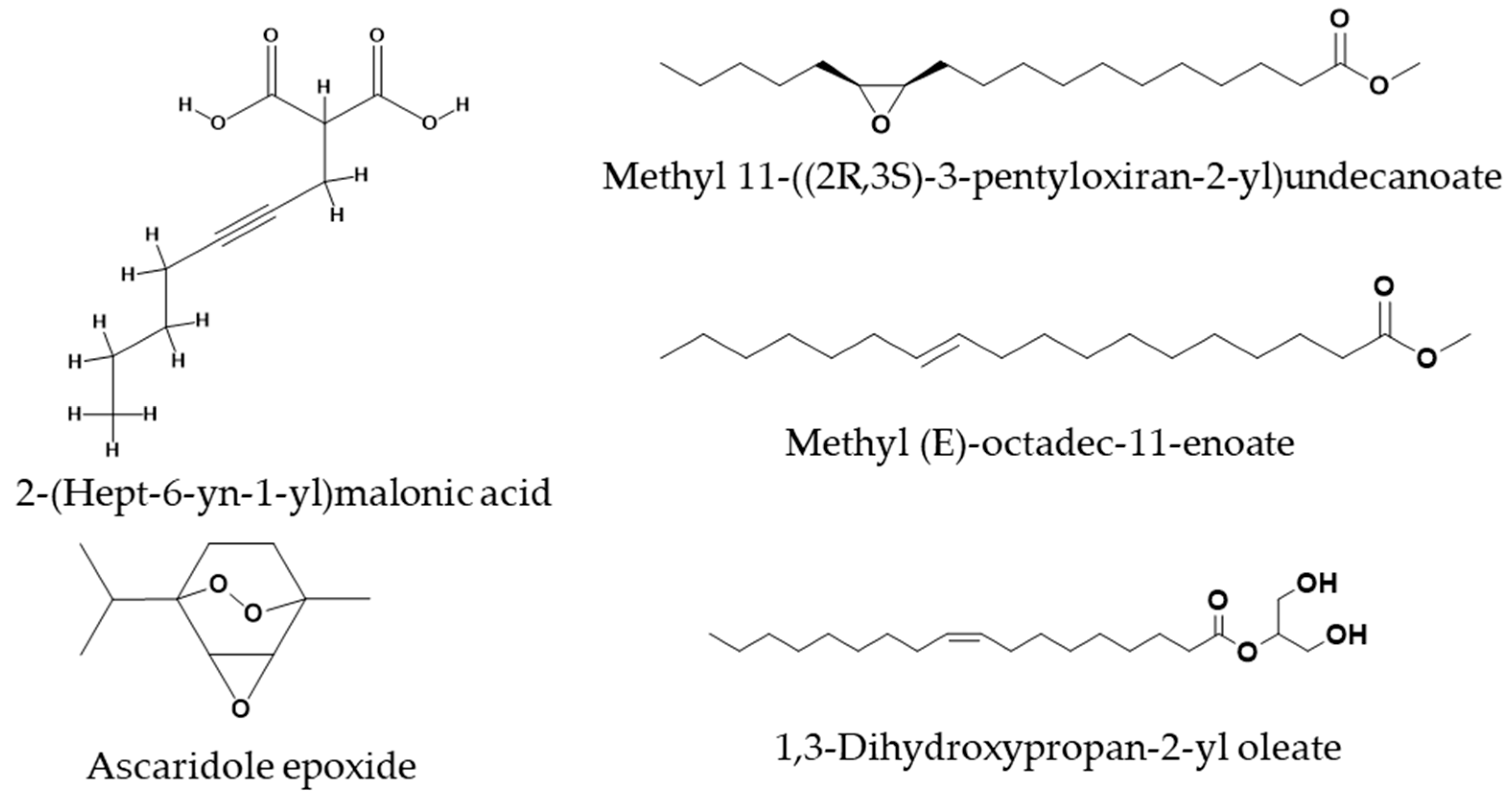
3.1. Chemical Characterization of M. longipetala Extract

The characterization of the chemical constituents of the methanol extract of M. longipetala aboveground parts was performed with gas chromatography-mass spectrometry (GC-MS) (Figure 2). The results demonstrated that the methanol extract has 37 compounds that are listed in detail in Table 1.
The M. longipetala extract had numerous main components (>2%), such as ascaridole epoxide, methyl (E)-octadec-11-enoate (12.21%), methyl 11-((2R,3S)-3-pentyloxiran-2-yl)undecanoate (7.51%), 1,3-Dihydroxypropan-2-yl oleate, 2-(hept-6-yn-1-yl)malonic acid (4.73%), 2-(acetylamino)-2-deoxyhexopyranose, 1-S-[(1E)-N-hydroxy-3-butenimidoyl]-1-thiohexopyranose (4.09%), (2Z,3E)-2-ethylidene-6-methylhepta-3,5-dienal (4.08%), 2-(((9Z,12Z)-Octadeca-9,12-dienoyl)oxy)propane-1,3-diyl diacetate, ethyl 2-hydroxycyclohexane-1-carboxylate (3.68%), (R,Z)-12-hydroxyoctadec-9-enoic acid, (3R,4S,5R)-3,4-dihydroxy-5-(1,2,3,4-tetrahydroxybutyl)dihydrofuran-2(3H)-one (2.64%), 4-amino-1,5-pentandioic acid (2.25%), and 2-bromotetradecanoic acid (2.17%) (Figure 3).
These compounds account for 63.50% of all recognized chemical substances (Table 1). The chemical composition of M. longipetala extract in the present study revealed variations to those reported for the Libyan ecospecies [27]. In addition, desulphosinigrin was identified as glucosides in the present study; however, Hammami et al. [20] and Hammami et al. [19] identified different glucosides and phenolics in the Tunisian ecospecies. Six categories may be established from the M. longipetala methanol extract’s chemical components (Figure 4) which are identified as oxygenated hydrocarbons (19.15%), carbohydrates (10.21%), amines (4.85%), terpenoids (12.71%), fatty acids and their derivatives (50.8%), and steroids (2.26%). Therefore, the fatty acids class and their derivatives “lipids” are the entire major constitutes with 50.8%, and only ascaridole epoxide as a monocyclic monoterpene compound was identified with 12.71% of the total percentage of the methanol extract chemical constitutes.
The abundant identified components of the M. longipetala methanol extract showed that it is oxygen-rich and related to diverse categories. Among these major compounds, ascaridole epoxide, methyl (E)-octadec-11-enoate, methyl 11-((2R,3S)-3-pentyloxiran-2-yl)undecanoate, and 1,3-dihydroxypropan-2-yl oleate represent 38.28% of the total composition of the M. longipetala methanol extract.

3.2. Biological Activities of the M. longipetala Extracts

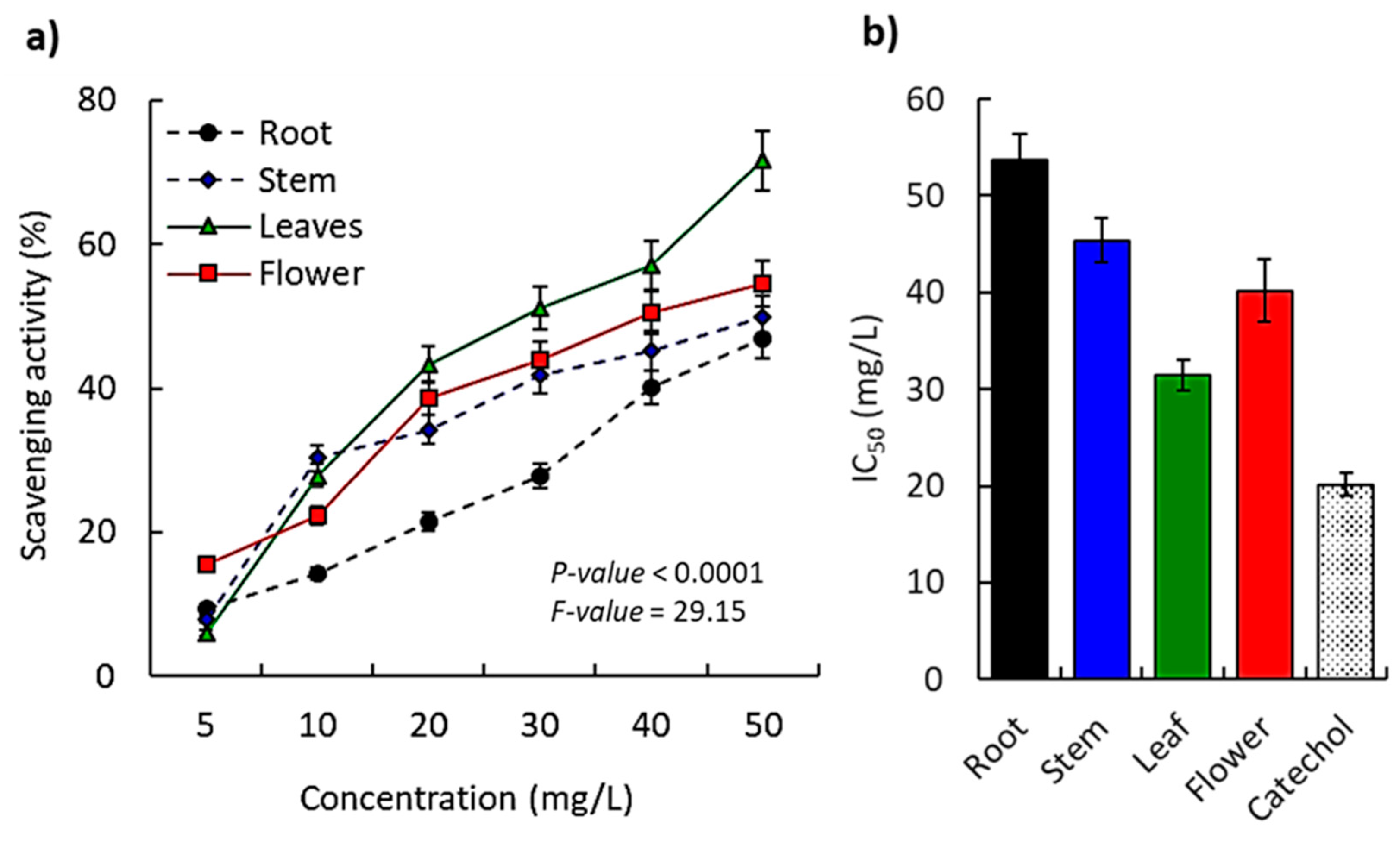
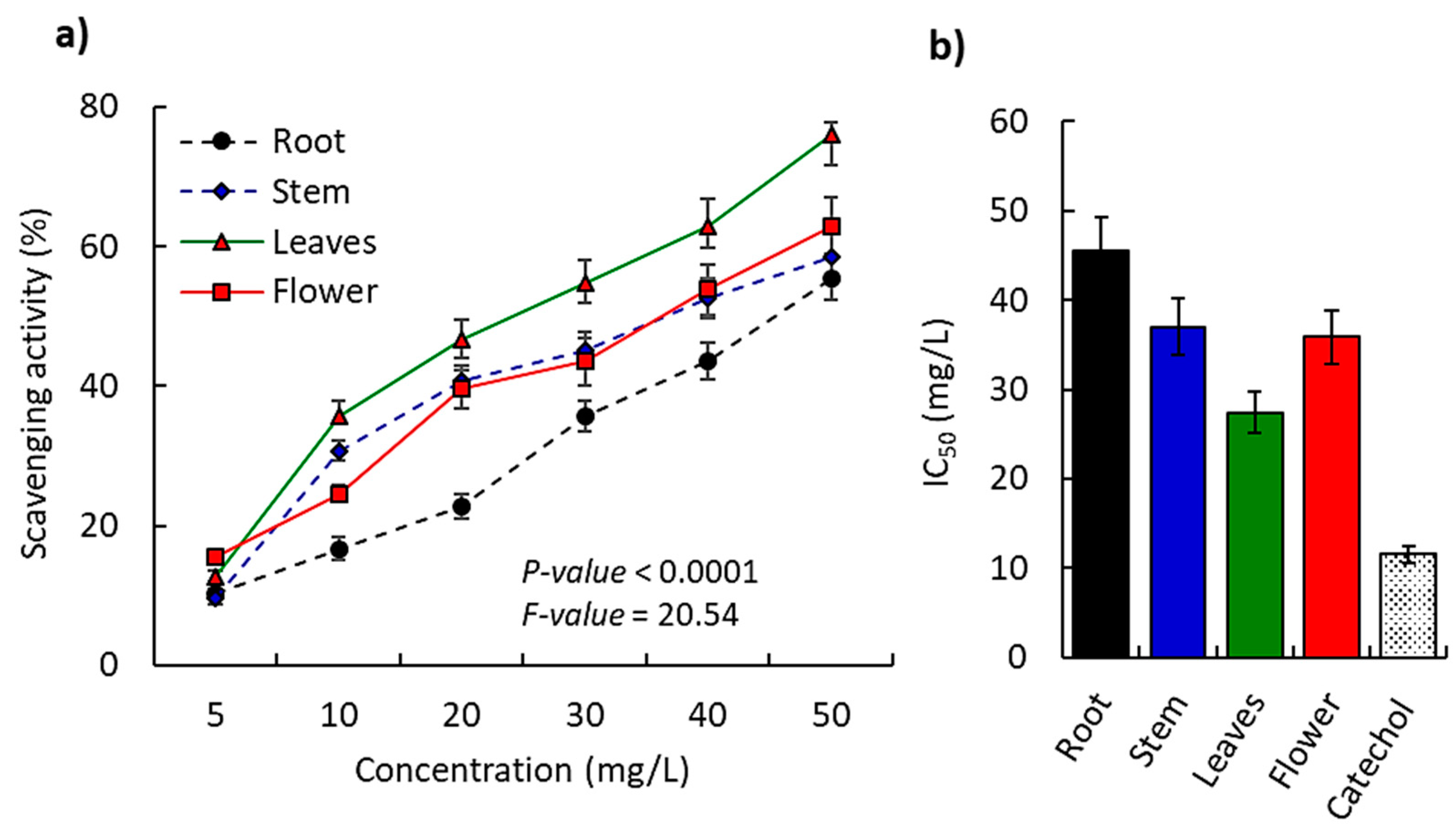
3.2.1. Antioxidant Activity

The antioxidant activity was appraised for the methanol extract of different parts of M. longipetala plant by DPPH and ABTS colorimetric assays. The assays showed substantial antioxidant activities compared to catechol as a reference standard. For the DPPH method, the scavenging activity of the extract was concentration-dependent, and a significant difference (p ˂ 0.0001) among the different organs was observed (Figure 5a).
At the lower dose (5 mg/L), flower extract showed the highest activity (15.65%); moreover, at the highest concentration of the extract (10 mg/L), the most potent scavenging activity percent (30.34%) was documented for leaf extract. Subsequently, and based on the calculations of the IC50 values, the results verified that leaf extract has the highest antioxidant scavenging activity (IC50 = 31.47 mg/L) compared to the other plant parts (Figure 5b). Furthermore, the flower extract revealed IC50 of 40.19 mg/L in the second order of the antioxidant potency followed by stem extract (IC50 = 45.4 mg/L), and finally root extract (IC50 = 53.76 mg/L).
On the other hand, the ABTS assay confirmed the data of the DPPH method, where the M. longipetala showed significant antioxidant activity in a concentration-dependent manner (Figure 6). Based on the IC50 value, results revealed that leaf extract had the highest antioxidant scavenging activity (IC50 = 28.94 mg/L), compared to the other plant parts. In the second, the flower extract revealed an IC50 value of 35.04 mg/L, followed by stem extract (IC50 = 41.56 mg/L), and finally root extract (IC50 = 50.23 mg/L).
Many studies on plant extracts showed that the leaf extracts have higher antioxidant activity compared to the other plant parts of the same plant [28,29]. The prevalence of oxygenated compounds in the methanol extract of M. longipetala could be the main cause for the substantial determined antioxidant activity of the present investigation [30,31]. It has been observed that terpenoid chemicals extracted from a variety of plants have a significant role as strong antioxidant agents [32,33], for instance, Deverra tortuosa [7], Salvia officinalis [34], Cleome amblyocarpa [31], Launaea species [35], Persicaria lapathifolia [31], Symphyotrichum squamatum [36], and Coriandrum sativum [37]. Predominantly, the major compounds are ascaridole epoxide (bicyclic monoterpenoid, 12.71%), methyl (E)-octadec-11-enoate (12.21%), methyl 11-((2R,3S)-3-pentyloxiran-2-yl)undecanoate (7.51%), and 1,3-dihydroxypropan-2-yl oleate (5.85%) as a category of fatty acids and their derivatives. Hence, ascaridole epoxide, and methyl (E)-octadec-11-enoate, as the major constituents of the methanol extract of M. longipetala, were reported as substantial antioxidant agents in many plants, for example, tea tree oil [38,39], and Chenopodium ambrosioides [40]. On the other hand, ascaridole activation was used recently as an effective step for skin sensitization [41]. Ascaridole epoxide, fatty acids, and lipids isolated from C. ambrosioides, and Euphorbia lathyrus revealed antioxidant activity for the free radicals [40,42].
Bioactive compounds such as phenolics, flavonoids, terpenes, or oxygenated hydrocarbons can scavenge free radicals because they contain active functional groups such as the OH group [43,44]. Herein, the leaf extract has more potent antioxidant activity than the flower, stem, and root extracts. The variation of the antioxidant activities between the extracted parts of M. longipetala is a result of the percentage of reactive oxygen species including the phenolic and flavonoid contents in each extract. Furthermore, the chemical components of plant extract have the propensity to combine with DPPH solution and subsequently stabilize free radicals [45]. The antioxidant results are in accordance with the literature that reported that the increased number of free hydroxy groups provided approach antioxidant characteristics [46,47].

3.2.2. Antibacterial Activity

To test the antibacterial efficacy of root, stem, leaf, and flower extracts from M. longipetala, we used an agar well diffusion experiment with four Gram-negative and four Gram-positive bacterial strains. The results demonstrated that the majority of the extracts considerably outperformed the tested conventional antibiotics in terms of their antibacterial activity against a variety of bacterial isolates with the exception of P. aeruginosa and S. typhimurium (Table 2). Regarding the effect on the Gram-negative bacterial isolates, leaf and flower extracts showed higher antibacterial activity against E. coli compared to root and stem extracts, while the four tested extracts (root, stem, leaf, and flower) showed comparable activity against K. pneumoniae (Table 2). However, all tested extracts were non-active against both P. aeruginosa and S. typhimurium. On the other hand, all tested extracts are comparable in their activity against the Gram-positive bacterial strains (S. epidermidis, S. aureus, and S. haemolyticus). However, only the flower extract of M. longipetala showed low antibacterial activity on S. xylosus, while the other extracts did not show activity against this strain.
The assessed antibacterial activity of the M. longipetala extract could be attributed to its contents of terpenoids and hydrocarbons, particularly the oxygenated compounds that have been reported to possess antimicrobial activity [48,49]. In addition, the major compound, ascaridole epoxide, as well as some fatty acids in the essential oils from several plants have been reported to possess antimicrobial characteristics [50,51].
It is important to note that some isolates of Gram-negative bacteria, such P. aeruginosa, showed resistance to the commonly used conventional antibiotics at a dosage of 10 mg L−1. Furthermore, neither ampicillin nor Zithromax had any effect on S. typhimurium (Table 2). However, none of the tested Gram-positive isolates showed any resistance to the used standard antibiotics. This result is in accordance with previously reported data [52].

3.2.3. Cytotoxicity and Cell Migration Analysis
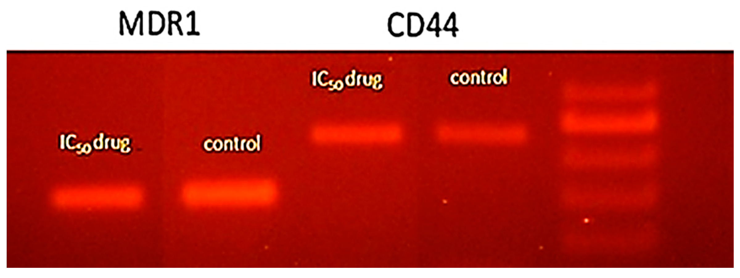
Scientists and researchers make their best effort to explore and develop new treatment protocols for using natural resources such as plants to control various types of cancer disease [53]. In comparison to chemotherapy medications, herbal extracts with specific therapeutic qualities are thought to be an efficient and safe resource for the treatment of illnesses [54]. In the current investigation, MTT (3-(4,5-dimethylthiazolyl-2)-2,5-diphenyltetrazolium bromide), which is used to determine cell viability, was employed to investigate M. longipetala extract for its efficacy as an anticancer agent. Additionally, the HepG2 cell line, a kind of hepatocellular carcinoma, was used to test the M. longipetala extract’s anticancer effectiveness.
The cell viability after the application of the M. longipetala extract revealed low activity as shown in Table 3. At the highest concentration of the extract (1000 µg/mL), the cell viability became 75.19%.
The mechanism of cytotoxicity, which is frequently reliant on the structure and nature of the extract’s bioactive chemical components, concentration, and the characteristics of the cancer cell line, might be attributed to the plant extract’s reported weak cytotoxic activity [55]. In addition, the cytotoxicity has been reported to affect according to the specification of the compound such as the morphology of the surface, volume, and condensation.
Cell migration is an important procedure that is integrated into many biological processes, such as tissue formation, development of the embryo, inflammation, immune defense, and cancer development [26]. After being treated with M. longipetala extract in the current study, HepG2 cell lines demonstrated wound healing activity in the cell migration test, where cells moved and covered the scratch’s center (Figure 7).
The healing of the wound was concentration-dependent, and the maximum healing was determined for the IC50 dose. Several bioactive compounds extracted from plants such as phenolics, volatile oils, and flavonoids were determined to possess wound-healing activity [56,57]. These compounds improve wound healing via epithelialization, stimulating fibroblasts, collagen deposition, angiogenesis, and reduced aggregation of the platelets [56]. The essential oil of Plectranthus tenuiflorus leaves has been reported to stimulate fibroblasts in vitro [58]. Moreover, terpenes of several plants have been reported to improve wound healing [59,60]. According to the observed substantial therapeutic activity of M. longipetala extract, it could be considered a biocompatible green material for wound healing; however, further study is recommended to determine its application in vivo.

DNA Fragmentation

DNA fragmentation is the crucial characteristic of apoptosis, and it was assessed by gel electrophoresis to characterize the cell death mediated by M. longipetala extract (Figure 8). The present results showed ladder pattern DNA fragmentation, where the densitometry analysis revealed a substantial increase in the DNA fragmentation with MDR1: 40.23% and CD44: 70.53% with respect to control.

The EC50 Value of M. longipetala Extract

In order to determine the EC50 value of the M. longipetala extract, a dose-dependent curve was performed as shown in Figure 9. The absorbance of the sample versus the log of extract dosages at various concentrations was plotted in order to determine the EC50 value of the M. longipetala extract. While high concentrations of the plant extract produce a maximum reaction, low dosages of the extract are insufficient to elicit a response [61]; the vertical point of the curve revealed the EC50 value. Regarding its cytotoxic impact on HepG2 cell lines, the extract of M. longipetala had an EC50 value of 2.36 g/mL.
Several Mentha species have been identified as possessing cytotoxic activity such as M. arvensis, M. piperita, M. longifolia, M. spicata [62], and M. piperita [63]. However, a further study in vivo is recommended for more assessment of the biological activities of M. longipetala for various applications. The observed cytotoxic activity of the presently studied M. longipetala could be attributed to the activity of the major compounds within the extract that could act either singularly or in a synergetic manner.

4. Conclusions

The present study revealed the presence of 37 chemical compounds of the Egyptian ecospecies M. longipetala extract. Most of the identified compounds were fatty acids and their derivatives. Ascaridole epoxide and methyl (E)-octadec-11-enoate are the major compounds. The methanol extracts of different organs of M. longipetala displayed auspicious biological activities, such as antioxidant, antibacterial, and anticancer activities. Specifically, leaf extract showed a higher antioxidant effect compared to other plant parts. The M. longipetala extract revealed considerable antibacterial activity. The obtained data demonstrated the ability of the M. longipetala extract to improve the proliferation and viability of hepatocellular carcinoma cells in a wound closure in vitro assay. The characterized chemical compounds and their significant biological activities from M. longipetala, particularly ascaridole epoxide, reinforced the opportunity for further research on this wild species for green eco-friendly drug discovery. Thus, more study is recommended for the further characterization of the major compounds, as well as for assessment of their mode(s) of action and safety.

Author Contributions

Conceptualization, A.M.A.-E. and Y.A.E.-A.; formal analysis, Y.A.E.-A.; investigation, A.M.A.-E. and Y.A.E.-A.; data curation, A.M.A.-E. and Y.A.E.-A.; writing—original draft preparation, A.M.A.-E. and Y.A.E.-A.; writing—review and editing, A.M.A.-E., Y.A.E.-A. and N.S.Z. All authors have read and agreed to the published version of the manuscript.

Funding

This research was supported by The Researchers Supporting Project number (RSPD2023R676) King Saud University, Riyadh, Saudi Arabia.

Institutional Review Board Statement

Not applicable.

Informed Consent Statement

Not applicable.

Data Availability Statement

Not applicable.

Acknowledgments

The authors extend their appreciation to The Researchers Supporting Project number (RSPD2023R676) King Saud University, Riyadh, Saudi Arabia.

Conflicts of Interest

The authors declare no conflict of interest.

References

  1. Shahrajabian, M.H.; Sun, W.; Cheng, Q. A review of ginseng species in different regions as a multipurpose herb in traditional Chinese medicine, modern herbology and pharmacological science. J. Med. Plants Res. 2019, 13, 213–226. [Google Scholar]
  2. Çelik Yılmaz, N.; Yılmaz, A.; Yılmaz, F. Coloring of Woolen Fabrics with Natural Resources and Investigating the Color Perceptions of Children on These Fabrics. J. Nat. Fibers 2023, 20, 2134269. [Google Scholar] [CrossRef]
  3. El-Amier, Y.A.; Abdelghany, A.M.; Abed Zaid, A. Green synthesis and antimicrobial activity of Senecio glaucus-mediated silver nanoparticles. Res. J. Pharm. Bio. Chem. Sci. 2014, 5, 631–642. [Google Scholar]
  4. Al-Rowaily, S.L.; Abd-ElGawad, A.M.; Assaeed, A.M.; Elgamal, A.M.; Gendy, A.E.-N.G.E.; Mohamed, T.A.; Dar, B.A.; Mohamed, T.K.; Elshamy, A.I. Essential oil of Calotropis procera: Comparative chemical profiles, antimicrobial activity, and allelopathic potential on weeds. Molecules 2020, 25, 5203. [Google Scholar] [CrossRef]
  5. Abd El-Gawad, A.M.; El-Amier, Y.A.; Bonanomi, G. Essential oil composition, antioxidant and allelopathic activities of Cleome droserifolia (Forssk.) Delile. Chem. Biodivers. 2018, 15, e1800392. [Google Scholar] [CrossRef]
  6. David, B.; Wolfender, J.-L.; Dias, D.A. The pharmaceutical industry and natural products: Historical status and new trends. Phytochem. Rev. 2015, 14, 299–315. [Google Scholar] [CrossRef]
  7. Fayed, E.M.; Abd-EIGawad, A.M.; Elshamy, A.I.; El-Halawany, E.S.F.; El-Amier, Y.A. Essential oil of Deverra tortuosa aerial parts: Detailed chemical profile, allelopathic, antimicrobial, and antioxidant activities. Chem. Biodivers. 2021, 18, e2000914. [Google Scholar] [CrossRef]
  8. Boulos, L. Flora of Egypt 1: Azollaceae–Oxalidaceae; Al Hadara Publishing: Cairo, Egypt, 1999. [Google Scholar]
  9. Jaén-Molina, R.; Caujapé-Castells, J.; Reyes-Betancort, J.A.; Akhani, H.; Fernández-Palacios, O.; de Paz, J.P.; Febles-Hernández, R.; Marrero-Rodríguez, Á. The molecular phylogeny of Matthiola R. Br. (Brassicaceae) inferred from ITS sequences, with special emphasis on the Macaronesian endemics. Mol. Phylogenetics Evol. 2009, 53, 972–981. [Google Scholar] [CrossRef]
  10. Canli, K.; Bozyel, M.E.; Benek, A.; Yetgin, A.; Senturan, M.; Altuner, E.M. Chemical composition and in vitro antimicrobial activity of Matthiola tricuspidata ethanol extract. Fresenius Environ. Bull. 2020, 29, 8863–8868. [Google Scholar]
  11. Miceli, N.; Cavò, E.; Ragusa, S.; Cacciola, F.; Dugo, P.; Mondello, L.; Marino, A.; Cincotta, F.; Condurso, C.; Taviano, M.F. Phytochemical characterization and biological activities of a hydroalcoholic extract obtained from the aerial parts of Matthiola incana (L.) R. Br. subsp. incana (Brassicaceae) growing wild in Sicily (Italy). Chem. Biodivers. 2019, 16, e1800677. [Google Scholar] [CrossRef]
  12. Miceli, N.; Cavò, E.; Spadaro, V.; Raimondo, F.M.; Ragusa, S.; Cacciola, F.; El Majdoub, Y.O.; Arena, K.; Mondello, L.; Condurso, C. Phytochemical profile and antioxidant activity of the aerial part extracts from Matthiola incana subsp. rupestris and subsp. pulchella (Brassicaceae) endemic to Sicily. Chem. Biodivers. 2021, 18, e2100167. [Google Scholar] [CrossRef] [PubMed]
  13. Schroder, H.S.; Dawood, S.; Yalch, M.M.; Donnellan, M.B.; Moser, J.S. The role of implicit theories in mental health symptoms, emotion regulation, and hypothetical treatment choices in college students. Cogn. Ther. Res. 2015, 39, 120–139. [Google Scholar] [CrossRef]
  14. Rasool, N.; Afzal, S.; Riaz, M.; Rashid, U.; Rizwan, K.; Zubair, M.; Ali, S.; Shahid, M. Evaluation of antioxidant activity, cytotoxic studies and GC-MS profiling of Matthiola incana (stock flower). Legume Res. 2013, 36, 21–32. [Google Scholar]
  15. Taviano, M.F.; Cavò, E.; Spadaro, V.; Raimondo, F.M.; Musolino, V.; Cacciola, F.; El Majdoub, Y.O.; Mondello, L.; Condurso, C.; Cincotta, F. Phytochemical constituents, antioxidant activity, and toxicity assessment of the aerial part extracts from the infraspecific taxa of Matthiola fruticulosa (Brassicaceae) endemic to Sicily. Molecules 2021, 26, 4114. [Google Scholar] [CrossRef] [PubMed]
  16. Karaman, S.; Gulseven, M.; Comlekcioglu, N.; Ilcim, A. Fatty acid composition of Matthiola longipetala ssp. bicornis from Turkey. Int. J. Agric. Biol. 2011, 13, 581–585. [Google Scholar]
  17. Tatsuzawa, F. Acylated anthocyanins in flowers of Matthiola tricuspidata (L.) R. Br. and Matthiola fruticulosa (L.) Maire (Brassicaceae). Biochem. Syst. Ecol. 2014, 100, 399–402. [Google Scholar] [CrossRef]
  18. Blažević, I.; Đulović, A.; Burčul, F.; Popović, M.; Montaut, S.; Bilušić, T.; Vrca, I.; Markić, J.; Ljubenkov, I.; Ruščić, M. Stability and bioaccessibility during ex vivo digestion of glucoraphenin and glucoraphasatin from Matthiola incana (L.) R. Br. J. Food Compos. Anal. 2020, 90, 103483. [Google Scholar] [CrossRef]
  19. Hammami, S.; Khoja, I.; Khoja, I.; Jannet, H.B.; Halima, M.B.; Mighri, Z. Flowers essential oil composition of Tunisian Matthiola longipetala and its bioactivity against Tribolium confusum insect. J. Essent. Oil Bear. Plants 2007, 10, 162–167. [Google Scholar] [CrossRef]
  20. Hammami, S.; Ciavatta, M.L.; Ben Jannet, H.; Cimino, G.; Mighria, Z. Three phenolic and a sterol glycosides identified for the first time in Matthiola longipetala growing in Tunisia. Croat. Chem. Acta 2006, 79, 215–218. [Google Scholar]
  21. Souza, M.M.; Silva, B.D.; Costa, C.S.; Badiale-Furlong, E. Free phenolic compounds extraction from Brazilian halophytes, soybean and rice bran by ultrasound-assisted and orbital shaker methods. An. Acad. Bras. Cienc. 2018, 90, 3363–3372. [Google Scholar] [CrossRef]
  22. Miguel, M.G. Antioxidant activity of medicinal and aromatic plants. Flavour Fragr. J. 2010, 25, 291–312. [Google Scholar] [CrossRef]
  23. Re, R.; Pellegrini, N.; Proteggente, A.; Pannala, A.; Yang, M.; Rice-Evans, C. Antioxidant activity applying an improved ABTS radical cation decolorization assay. Free. Radic. Biol. Med. 1999, 26, 1231–1237. [Google Scholar] [CrossRef]
  24. Murray, P.R.; Baroon, E.J.; Faller, M.A.; Tenover, F.C.; Yolken, R.H. Manual of Clinical Microbiology, 6th ed.; American Society for Microbiology: Washington, DC, USA, 1482. [Google Scholar]
  25. Zhu, Q.; Jiang, L.; Wang, X. The expression of Duffy antigen receptor for chemokines by epithelial ovarian cancer decreases growth potential. Oncol. Lett. 2017, 13, 4302–4306. [Google Scholar] [CrossRef] [Green Version]
  26. Pijuan, J.; Barceló, C.; Moreno, D.F.; Maiques, O.; Sisó, P.; Marti, R.M.; Macià, A.; Panosa, A. In vitro cell migration, invasion, and adhesion assays: From cell imaging to data analysis. Front. Cell Dev. Biol. 2019, 7, 107. [Google Scholar] [CrossRef] [Green Version]
  27. Abdelshafeek, K.A.; Abdelmohsen, M.M.; Hamed, A.; Shahat, A.A. Investigation of some chemical constituents and antioxidant activity extracts of Matthiola longipetala subsp. longipetala. Chem. Nat. Compd. 2013, 49, 539–543. [Google Scholar] [CrossRef]
  28. Beevi, S.S.; Narasu, M.L.; Gowda, B.B. Polyphenolics profile, antioxidant and radical scavenging activity of leaves and stem of Raphanus sativus L. Plant Foods Hum. Nutr. 2010, 65, 8–17. [Google Scholar] [CrossRef]
  29. Chinnarasu, C.; Montes, A.; Pereyra, C.; Casas, L.; Fernández-Ponce, M.T.; Mantell, C.; Pattabhi, S.; de la Ossa, E.M. Preparation of polyphenol fine particles potent antioxidants by a supercritical antisolvent process using different extracts of Olea europaea leaves. Korean J. Chem. Eng. 2016, 33, 594–602. [Google Scholar] [CrossRef]
  30. Assaeed, A.; Elshamy, A.; El Gendy, A.E.-N.; Dar, B.; Al-Rowaily, S.; Abd-ElGawad, A. Sesquiterpenes-rich essential oil from above ground parts of Pulicaria somalensis exhibited antioxidant activity and allelopathic effect on weeds. Agronomy 2020, 10, 399. [Google Scholar] [CrossRef] [Green Version]
  31. Abd-ElGawad, A.M.; Elgamal, A.M.; El-Amier, Y.A.; Mohamed, T.A.; El Gendy, A.E.-N.G.; Elshamy, A.I. Chemical composition, allelopathic, antioxidant, and anti-inflammatory activities of sesquiterpenes rich essential oil of Cleome amblyocarpa Barratte & Murb. Plants 2021, 10, 1294. [Google Scholar]
  32. Mbaveng, A.T.; Hamm, R.; Kuete, V. 19—Harmful and Protective Effects of Terpenoids from African Medicinal Plants. In Toxicological Survey of African Medicinal Plants; Kuete, V., Ed.; Elsevier: Waltham, MA, USA, 2014; pp. 557–576. [Google Scholar]
  33. Baccouri, B.; Rajhi, I. Terpenes and Terpenoids Recent Advances; IntechOpen: London, UK, 2021. [Google Scholar]
  34. Ghorbani, A.; Esmaeilizadeh, M. Pharmacological properties of Salvia officinalis and its components. J. Tradit. Complement. Med. 2017, 7, 433–440. [Google Scholar] [CrossRef]
  35. Elshamy, A.; Abd-ElGawad, A.M.; El-Amier, Y.A.; El Gendy, A.; Al-Rowaily, S. Interspecific variation, antioxidant and allelopathic activity of the essential oil from three Launaea species growing naturally in heterogeneous habitats in Egypt. Flavour Fragr. J. 2019, 34, 316–328. [Google Scholar] [CrossRef]
  36. Abd-ElGawad, A.M.; Elshamy, A.I.; El-Amier, Y.A.; El Gendy, A.E.-N.G.; Al-Barati, S.A.; Dar, B.A.; Al-Rowaily, S.L.; Assaeed, A.M. Chemical composition variations, allelopathic, and antioxidant activities of Symphyotrichum squamatum (Spreng.) Nesom essential oils growing in heterogeneous habitats. Arab. J. Chem. 2020, 13, 4237–4245. [Google Scholar] [CrossRef]
  37. Darughe, F.; Barzegar, M.; Sahari, M.A. Antioxidant and antifungal activity of coriander (Coriandrum sativum L.) essential oil in cake. Int. Food Res. J. 2012, 19, 1253–1260. [Google Scholar]
  38. Rudbäck, J.; Bergström, M.A.; Börje, A.; Nilsson, U.; Karlberg, A.T. α-Terpinene, an antioxidant in tea tree oil, autoxidizes rapidly to skin allergens on air exposure. Chem. Res. Toxicol. 2012, 25, 713–721. [Google Scholar] [CrossRef] [PubMed] [Green Version]
  39. Parveen, Z.; Nawaz, S.; Siddique, S.; Shahzad, K. Composition and antimicrobial activity of the essential oil from leaves of Curcuma longa L. Kasur variety. Indian J. Pharm. Sci. 2013, 75, 117–122. [Google Scholar] [CrossRef] [Green Version]
  40. Monzote, L.; Stamberg, W.; Staniek, K.; Gille, L. Toxic effects of carvacrol, caryophyllene oxide, and ascaridole from essential oil of Chenopodium ambrosioides on mitochondria. Toxicol. Appl. Pharmacol. 2009, 240, 337–347. [Google Scholar] [CrossRef]
  41. Chittiboyina, A.G.; Avonto, C.; Khan, I.A. What happens after activation of ascaridole? Reactive compounds and their implications for skin sensitization. Chem. Res. Toxicol. 2016, 29, 1488–1492. [Google Scholar] [CrossRef]
  42. Sosa, A.A.; Bagi, S.H.; Hameed, I.H. Analysis of bioactive chemical compounds of Euphorbia lathyrus using gas chromatography-mass spectrometry and fourier-transform infrared spectroscopy. J. Pharmacogn. Phytother. 2016, 8, 109–126. [Google Scholar]
  43. Al-Snafi, A.E. The medical benefit of Gnaphalium luteoalbum-A review. IOSR J. Pharm. 2019, 9, 40–44. [Google Scholar]
  44. Lu-Martínez, A.A.; Báez-González, J.G.; Castillo-Hernández, S.; Amaya-Guerra, C.; Rodríguez-Rodríguez, J.; García-Márquez, E. Studied of Prunus serotine oil extracted by cold pressing and antioxidant effect of P. longiflora essential oil. J. Food Sci. Technol. 2021, 58, 1420–1429. [Google Scholar] [CrossRef]
  45. Alam, M.N.; Bristi, N.J.; Rafiquzzaman, M. Review on in vivo and in vitro methods evaluation of antioxidant activity. Saudi Pharm. J. 2013, 21, 143–152. [Google Scholar] [CrossRef] [PubMed] [Green Version]
  46. Li, Y.X.; Li, Y.; Qian, Z.J.; Kim, M.M.; Kim, S.K. In vitro antioxidant activity of 5-HMF isolated from marine red alga Laurencia undulata in free radical mediated oxidative systems. J. Microbiol. Biotechnol. 2009, 19, 1319–1327. [Google Scholar] [CrossRef] [PubMed] [Green Version]
  47. Gu, F.; Kim, J.M.; Hayat, K.; Xia, S.; Feng, B.; Zhang, X. Characteristics and antioxidant activity of ultrafiltrated Maillard reaction products from a casein–glucose model system. Food Chem. 2009, 117, 48–54. [Google Scholar] [CrossRef]
  48. Salehi, B.; Krochmal-Marczak, B.; Skiba, D.; Patra, J.K.; Das, S.K.; Das, G.; Popović-Djordjević, J.B.; Kostić, A.Ž.; Anil Kumar, N.V.; Tripathi, A.; et al. Convolvulus plant-A comprehensive review from phytochemical composition to pharmacy. Phytother. Res. 2020, 34, 315–328. [Google Scholar] [CrossRef]
  49. Maccelli, A.; Vitanza, L.; Imbriano, A.; Fraschetti, C.; Filippi, A.; Goldoni, P.; Maurizi, L.; Ammendolia, M.G.; Crestoni, M.E.; Fornarini, S.; et al. Satureja montana L. essential oils: Chemical profiles/phytochemical screening, antimicrobial activity and o/w nanoemulsion formulations. Pharmaceutics 2020, 12, 7. [Google Scholar] [CrossRef] [PubMed] [Green Version]
  50. Brahim, M.A.S.; Fadli, M.; Hassani, L.; Boulay, B.; Markouk, M.; Bekkouche, K.; Abbad, A.; Ali, M.A.; Larhsini, M. Chenopodium ambrosioides var. ambrosioides used in Moroccan traditional medicine can enhance the antimicrobial activity of conventional antibiotics. Ind. Crops Prod. 2015, 71, 37–43. [Google Scholar] [CrossRef]
  51. Shareef, H.K.; Muhammed, H.J.; Hussein, H.M.; Hameed, I.H. Antibacterial effect of ginger (Zingiber officinale) roscoe and bioactive chemical analysis using gas chromatography mass spectrum. Orient. J. Chem. 2016, 32, 20–40. [Google Scholar]
  52. Abd El-Gawad, A.; El Gendy, A.; Elshamy, A.; Omer, E. Chemical composition of the essential oil of Trianthema portulacastrum L. Aerial parts and potential antimicrobial and phytotoxic activities of its extract. J. Essent. Oil Bear. Plants 2016, 19, 1684–1692. [Google Scholar] [CrossRef]
  53. Li, F.-S.; Weng, J.-K. Demystifying traditional herbal medicine with modern approach. Nat. Plants 2017, 3, 17109. [Google Scholar]
  54. Zaki, F.N. Field application of plant extracts against the aphid, B. brassicae and the whitefly, B. abaci and their side effects on their predators and parasites. Arch. Phytopathol. Plant Prot. 2008, 41, 462–466. [Google Scholar] [CrossRef]
  55. Khacha-ananda, S.; Tragoolpua, K.; Chantawannakul, P.; Tragoolpua, Y. Antioxidant and anti-cancer cell proliferation activity of propolis extracts from two extraction methods. Asian Pac. J. Cancer Prev. 2013, 14, 6991–6995. [Google Scholar] [CrossRef] [PubMed] [Green Version]
  56. Mazutti da Silva, S.M.; Rezende Costa, C.R.; Martins Gelfuso, G.; Silva Guerra, E.N.; De Medeiros Nóbrega, Y.K.; Gomes, S.M.; Pic-Taylor, A.; Fonseca-Bazzo, Y.M.; Silveira, D.; Magalhães, P.d.O. Wound healing effect of essential oil extracted from Eugenia dysenterica DC (Myrtaceae) leaves. Molecules 2019, 24, 2. [Google Scholar] [CrossRef] [PubMed]
  57. Elshamy, A.I.; Ammar, N.M.; Hassan, H.A.; El-Kashak, W.A.; Al-Rejaie, S.S.; Abd-ElGawad, A.M.; Farrag, A.H. Topical wound healing activity of myricetin isolated from Tecomaria capensis v. aurea. Molecules 2020, 25, 4870. [Google Scholar] [PubMed]
  58. Khorshid, F.; Ali, S.S.; Alsofyani, T.; Albar, H.A. Plectranthus tenuiflorus (Shara) promotes wound healing: In vitro and in vivo studies. Int. J. Bot. 2010, 6, 69–80. [Google Scholar] [CrossRef] [Green Version]
  59. Juneja, K.; Mishra, R.; Chauhan, S.; Gupta, S.; Roy, P.; Sircar, D. Metabolite profiling and wound-healing activity of Boerhavia diffusa leaf extracts using in vitro and in vivo models. J. Tradit. Complement. Med. 2020, 10, 52–59. [Google Scholar] [CrossRef]
  60. Salama, S.A.; Al-Faifi, Z.E.; Masood, M.F.; El-Amier, Y.A. Investigation and biological assessment of Rumex vesicarius L. extract: Characterization of the chemical components and antioxidant, antimicrobial, cytotoxic, and anti-dengue vector activity. Molecules 2022, 27, 3177. [Google Scholar] [CrossRef]
  61. Sundaram, V.; Mohammed, S.; Srinivasan, M.R.; Johnson, J.; Suepaul, R.; Pargass, I.; John, C.; Ramdhanie, D.; Lallack, S.; Daniel, E.; et al. Acute and subacute toxicity evaluation of hydroalcoholic extract from the stem bark of Bois Bande (Parinari campestris Aubl. 1772) in rats. BMC Pharmacol. Toxicol. 2021, 22, 51. [Google Scholar] [CrossRef] [PubMed]
  62. Hussain, A.I.; Anwar, F.; Nigam, P.S.; Ashraf, M.; Gilani, A.H. Seasonal variation in content, chemical composition and antimicrobial and cytotoxic activities of essential oils from four Mentha species. J. Sci. Food Agric. 2010, 90, 1827–1836. [Google Scholar]
  63. Jain, D.; Pathak, N.; Khan, S.; Raghuram, G.V.; Bhargava, A.; Samarth, R.; Mishra, P.K. Evaluation of cytotoxicity and anticarcinogenic potential of Mentha leaf extracts. Int. J. Toxicol. 2011, 30, 225–236. [Google Scholar]
Figure 1. Overview of Matthiola longipetala DC. (A), flowering plant (B), and fruiting branches with legumes (C).
Separations 10 00053 g001
Figure 2. Chromatogram of basic chemical components interpreted from the MeOH extract of M. longipetala aboveground parts by GC-MS. The major compounds peaks are numbered in red color from 1–5.
Separations 10 00053 g002
Figure 3. Main chemical components of the methanol extract of M. longipetala aboveground parts interpreted from GC-MS analysis.
Separations 10 00053 g003
Figure 4. Various classes of the characterized chemical compounds of M. longipetala extract derived via GC-MS analysis.
Separations 10 00053 g004
Figure 5. DPPH radical scavenging activity at different concentrations of the different parts of M. longipetala methanol extract (a), and IC50 values (b). Values are average (n = 3), and the bars represented the standard deviation.
Separations 10 00053 g005
Figure 6. ABTS radical scavenging activity at different concentrations of the different parts of M. longipetala methanol extract (a), and IC50 values (b). Values are average (n = 3), and the bars represented the standard deviation.
Separations 10 00053 g006
Figure 7. The impact of M. longipetala extracts on the cell cycle development of HepG2. (A) Microscopic reflection at 0 time of HepG2 cell line with 200× magnification, (B) Microscopic reflection after staining. (C) Microscopic reflection after 26 h. The photomicrographs referred to control group and treated groups at different doses of M. longipetala extracts.
Separations 10 00053 g007
Figure 8. The fragmentation analysis of DNA under the effect of M. longipetala extract. MDR1 and CD44 antibody pathways were characterized as targets of M. longipetala-dependent apoptosis.
Separations 10 00053 g008
Figure 9. Dose–response curves of M. longipetala extract cell growth inhibition as a percent of control against HepG2 cell lines. (a) Transform of the extracted M. longipetala, (b) Normalization of transform, OD is the absorbance value.
Separations 10 00053 g009
Table 1. Chemical characterization of the identified components in the aboveground parts of M. longipetala using GC-MS analysis.
No.RTConc. %Chemical NameClassificationMWMF
Oxygenated hydrocarbon
14.230.81 ± 0.02(E)-2-(1-(2-(2-methylpiperidine-1-carbonothioyl)hydrazono)ethyl)pyridine 1-oxideAryl hydrocarbon292.4C14H20N4OS
24.824.73 ± 0.032-(Hept-6-yn-1-yl)malonic acidAliphatic carboxylic acid453.44C16H15N5O7S2
35.160.89 ± 0.023-(2-Oxocyclohexyl)propanenitrileOxygenated hydrocarbon151.21C9H13NO
45.611.99 ± 0.01Methyl 3,5-dioxohexahydro-1H-pyrrolizine-2-carboxylateEster197.19C9H11NO4
59.322.46 ± 0.01(3R,4S,5R)-3,4-Dihydroxy-5-(1,2,3,4-tetrahydroxybutyl)dihydrofuran-2(3H)-oneOxygenated hydrocarbon238.19C8H14O8
69.43.68 ± 0.02Ethyl 2-hydroxycyclohexane-1-carboxylateEster172.22C9H16O3
712.934.08 ± 0.03(2Z,3E)-2-ethylidene-6-methylhepta-3,5-dienalOxygenated hydrocarbon150.22C10H14O
816.320.51 ± 0.029,10-Secocholesta-5,7,10(19)-triene-1,3-diol, 25-[(trimethylsilyl)oxy]-, (3á,5Z,7E)-Oxygenated hydrocarbon488.83C30H52O3Si
Carbohydrates
98.014.09 ± 0.021-S-[(1E)-N-Hydroxy-3-butenimidoyl]-1-thiohexopyranoseGlycoside279.31C10H17NO6S
108.720.33 ± 0.00α-D-Glucopyranoside, O-α-D-glucopyranosyl-(1.fwdarw.3)-α-D-fructofuranosylTrisaccharide504.44C18H32O16
118.770.38 ± 0.012,3,4,5,6,7,8-HeptahydroxyoctanamideGlycosyl amide255.22C8H17NO8
129.194.15 ± 0.022-(Acetylamino)-2-deoxyhexopyranoseCarbohydrate221.21C8H15NO6
1317.681.26 ± 0.01(2R,3S,4S,5R,6R)-2-(Aminomethyl)-6-(((2R,3S,4R,6S)-4,6-diamino-3-(((3R,4R,5R)-3,5-dihydroxy-5-methyl-4-(methylamino)tetrahydro-2H-pyran-2-yl)oxy)-2-hydroxycyclohexyl)oxy)tetrahydro-2H-pyran-3,4,5-triolAminoglycoside 482.53C19H38N4O10
Amines
144.151.71 ± 0.022-Ethylidene-1,5-dimethyl-3,3-diphenylpyrrolidineDiaryl cyclic amine277.41C20H23N
156.890.89 ± 0.011,3,5-Triazine-2,4-diamine,N,N’-bis(1-methylethyl)-6-(methylsulfonyl)-Hetryl amine273.36C10H19N5O2S
169.252.25 ± 0.024-Amino-1,5-pentandioic acidAmino acid175.18C7H13NO4
Terpenoids
1713.3412.71 ± 0.21Ascaridole epoxideBicyclic monoterpenoid184.24C10H16O3
Fatty acids and Lipids
184.380.53 ± 0.02Hexyl oleateFatty acid366.63C24H46O2
194.73.60 ± 0.012-(Hept-6-yn-1-yl)malonic acidOleic acid450.4C21H22O11
205.521.11 ± 0.01Palmitic acidFatty acid256.43C16H32O2
216.511.86 ± 0.02(E)-Hexadec-9-enoic acidFatty acid254.41C16H30O2
226.780.27 ± 0.003-(((9Z,12Z,15Z)-Octadeca-9,12,15-trienoyl)oxy)propane-1,2-diyl diacetateLipids 436.59C25H40O6
237.363.70 ± 0.022-(((9Z,12Z)-Octadeca-9,12-dienoyl)oxy)propane-1,3-diyl diacetateLipids438.61C25H42O6
249.850.77 ± 0.01Ethyl stearateLipids312.54C20H40O2
2511.351.90 ± 0.02(Z)-Hexadec-9-enoic acidLipids254.41C16H30O2
2612.431.13 ± 0.013-Hydroxydodecanoic acidLipids216.32C12H24O3
2714.612.52 ± 0.01(R,Z)-12-Hydroxyoctadec-9-enoic acidFatty acid298.47C18H34O3
2816.21.49 ± 0.018-((2R,3S)-3-Octyloxiran-2-yl)octanoic acidLipids298.47C18H34O3
2918.271.38 ± 0.00Oleic acidFatty acid282.47C18H34O2
3019.812.17 ± 0.022-Bromotetradecanoic acidFatty acid307.27C14H27BrO2
3122.361.63 ± 0.012,3-Dihydroxypropyl palmitateLipids330.51C19H38O4
3224.261.17 ± 0.012-Hydroxypropane-1,3-diyl dipalmitateLipids568.92C35H68O5
3325.887.51 ± 0.03Methyl 11-((2R,3S)-3-pentyloxiran-2-yl)undecanoateLipids312.49C19H36O3
3429.212.21 ± 0.37Methyl (E)-octadec-11-enoateLipids296.5C19H36O2
3530.725.85 ± 0.041,3-Dihydroxypropan-2-yl oleateLipids356.55C21H40O4
Steroids
3621.680.87 ± 0.01Estra-1,3,5(10)-trien-17β-olSteroid256.39C18H24O
3731.431.39 ± 0.01Ethyl 3,7,12-trihydroxycholan-24-oateSteroidal ester436.63C26H44O5
Total99.98
RT: Retention time, MW: Molecular Weight, MF: Molecular Formula.
Table 2. The antibacterial activity of the methanol extract (10 mg/L) of different parts of M. longipetala and antibiotics against various bacterial isolates.
MicrobesM. longipetala (10 mg/L)Standard Antibiotic (10 mg/L)
RootStemLeafFlowerAmpicillinAzithromycinCefotaximeTetracycline
Gram-negative bacteria
E. coli11 ± 0.51 a12 ± 0.2115 ± 0.5715 ± 0.3119 ± 0.4619 ± 0.3128 ± 0.7117 ± 0.58
P. aeruginosaNANANANANA14 ± 0.659 ± 0.45NA
S. typhimuriumNANANANANANA9 ± 0.229 ± 0.34
K. pneumoniae12 ± 0.4410 ± 0.5110 ± 0.6612 ± 0.706 ± 0.0811 ± 0.5118 ± 0.4319 ± 0.33
Gram-positive bacteria
S. epidermidis10 ± 0.2710 ± 0.3210 ± 0.0910 ± 0.689 ± 0.2021 ± 0.6218 ± 0.5518 ± 0.62
S. aureus15 ± 0.4112 ± 0.2015 ± 0.3315 ± 0.5027 ± 0.8718 ± 0.8120 ± 0.5318 ± 0.44
S. haemolyticus11 ± 0.2210 ± 0.1112 ± 0.4112 ± 0.6918 ± 0.7121 ± 0.536 ± 0.6621 ± 0.48
S. xylosusNANANA6 ± 0.5423 ± 0.3017 ± 0.5016 ± 0.3519 ± 0.53
p-value 0.05b˂0.001 ***˂0.001 ***˂0.001 ***˂0.001 ***˂0.001 ***˂0.001 ***˂0.001 ***˂0.001 ***
a value is an average of the inhibition zone diameter (mm) ± standard deviation, b the p-values were calculated based on factorial ANOVA at a probability level of 0.05, NA: no activity, *** p ˂ 0.001.
Table 3. Effect of M. longipetala extract at different concentrations on cell viability based on HepG2 cancer cell line.
Conc. (µg/mL)Cell Viability (%)Standard Deviation
R1R2Average
100076.4673.9275.191.79
50092.3192.3192.310.00
125115.38115.38115.380.00
62.5117.69118.46118.080.54
31.3119.23123.08121.152.72
0100.00100.00100.000.00
Disclaimer/Publisher’s Note: The statements, opinions and data contained in all publications are solely those of the individual author(s) and contributor(s) and not of MDPI and/or the editor(s). MDPI and/or the editor(s) disclaim responsibility for any injury to people or property resulting from any ideas, methods, instructions or products referred to in the content.

