Article

One-Step Chromatographic Approach for Purifying Peptides and Proteins from Venoms

1
Aix Marseille Univ, CNRS, Institut de Chimie Radicalaire UMR 7273, F-13390 Marseille, France
2
Latoxan, 26800 Portes-lès-Valence, France
3
Proteomics Platform, Marseille Proteomics (MaP), IMM, FR 3479, CNRS, F-13009 Marseille, France
*
Author to whom correspondence should be addressed.
Separations 2024, 11(6), 179; https://doi.org/10.3390/separations11060179
Submission received: 29 April 2024 / Revised: 20 May 2024 / Accepted: 28 May 2024 / Published: 6 June 2024
(This article belongs to the Section Chromatographic Separations)

Abstract

:
Animal venoms are intricate and teem with potential for groundbreaking medical advancements. Although traditional methods for purifying venom proteins are effective, they usually require complicated, multi-step processes that lead to lower yields. Our study introduces an efficient, one-step technique for extracting venom-derived proteins through reverse-phase high-performance liquid chromatography (RP-HPLC). We carefully optimized the RP-HPLC process, focusing on the gradient elution conditions and the strategic use of our columns’ stationary phase characteristics, to enhance the effectiveness of our separations. This enabled us to efficiently isolate six venom proteins: melittin (2.846 kDa) from Apis mellifera with a yield of 4.5% and homogeneity of 99%; α-cobratoxin (7.821 kDa) from Naja kaouthia with a yield of 15% and homogeneity of 99%; α-bungarotoxin (7.983 kDa) from Bungarus multicinctus with a yield of 7% and purity of 99%; calciseptine (7.035 kDa) from Dendroaspis polylepis with a yield of 6% and homogeneity of 95%; notexin (13.593 kDa) from Notechis scutatus with a yield of 10% and homogeneity of 95%; and CVFm (150 kDa) from Naja melanoleuca with a yield of 0.8% and homogeneity of 94%. These were all accomplished in one step. This breakthrough simplifies the purification of venom peptides and proteins, making the process more feasible and economical. It paves the way for developing new drugs and promising treatments that are both more effective and precisely targeted.

Graphical Abstract

1. Introduction

Venomous creatures, often feared for their potential lethality, have been extensively studied by scientists around the globe who have extracted valuable medicinal components from their venom. This research has led to significant breakthroughs, resulting in the development of drugs that offer substantial health benefits. As a result, several venom-derived compounds have been approved by the Food and Drug Administration (FDA) for treating various conditions. These include Captopril for managing hypertension, Ziconotide for treating severe chronic pain, and Exenatide for diabetes management [1]. Currently, peptides and proteins from animal venoms are contributing significantly across various fields including medicine, research [2,3,4], diagnostics [5], cosmetics [6], and agriculture [7]. They constitute 90–95% of the dry weight of many venoms and are especially valuable in drug development due to their small size, stability, potency, and specificity [7,8].
However, isolating these toxins is not a straightforward task. Typically found only in small quantities within venom’s complex mixtures, their purification poses significant challenges. According to the literature, multiple methods are often necessary to effectively purify these proteins. The process generally begins with an affinity chromatography step to separate the desired protein from the crude extract, followed by ion-exchange chromatography to eliminate contaminants and impurities. A final “polishing” step [9] involving size-exclusion chromatography is then employed to remove any remaining impurities, ensuring the final product is of high purity. Traditional purification processes involve numerous steps that not only reduce the yield but also incur significant costs and require considerable time. Consequently, in this study, we aimed to develop a more streamlined and rapid purification approach that enhances protein homogeneity and improves the accuracy of protein quantification. Our method primarily utilizes the well-established RP-HPLC technique, which is selected for its high-resolution capabilities. RP-HPLC is particularly effective at distinguishing between small and large polypeptides and often even between those with nearly identical sequences or differing by only a single amino acid (aa) residue, as shown by the separation of insulin variants from rabbits and humans that differ by only a single aa [10]. This technique offers several advantages including enhanced reproducibility, reduced costs, and accelerated analysis. RP-HPLC also uses less toxic mobile phases and features stable stationary phases and straightforward gradient elution, making it an optimal choice for protein purification.
One of the ongoing challenges in purification is maintaining the integrity of proteins or peptides during the process [11]. To address this, we have adhered to specific conditions such as choosing acetonitrile as the organic solvent, which is volatile and easily removed. Its low viscosity minimizes column back-pressure and therefore does not significantly affect the stability of proteins. Stability and activity can also be maintained by selecting the appropriate elution gradients and columns. Indeed, the denaturation of proteins on hydrophobic surfaces is kinetically slow, so choosing a reduced elution gradient reduces the residence time of the protein in the column [11]. Taking all these factors into consideration, venom peptides with molecular weights ranging from 2 kDa to 150 kDa were isolated: (i) The peptide α-Bungarotoxin (α-Bgtx), from Bungarus multicinctus, is a neurotoxin composed of 74 amino acids (aa), cross-linked by five disulfide bridges. This peptide acts as an antagonist to acetylcholine (ACh) at the nicotinic acetylcholine receptor (AChR), primarily binding to the α subunits of AChR with very high affinity and in an irreversible manner, leading to paralysis of striated muscles [12]. (ii) The peptide α-Cobratoxin (α-Cbtx) (71 aa cross-linked by five disulfide bridges) is the most lethal component of the venom of the Thai cobra (Naja kaouthia), found in many Southern Asian countries [13]. Its main mode of action is through the inhibition of nicotinic acetylcholine receptors (nAChRs) at the neuromuscular junction. (iii) Calciseptine, a 60 aa peptide reticulated with four disulfide bonds, functions as a specific inhibitor of L-type Ca2+ channels and constitutes 2.8% of the total Dendroaspis polylepis venom components [14]. (iv) Melittin, the predominant component of Apis mellifera bee venom, constitutes over 40% of its dry weight [15]. Melittin attaches to lipid bilayer membranes, adopts an amphipathic α-helical secondary structure, and alters the permeability barrier. This enables it to exhibit a broad spectrum of biological activities, including antibacterial, anticancer, and anti-inflammatory effects [16]. (v) Notexin is recognized as the most neurotoxic and myotoxic phospholipase A2 (PLA2) enzyme isolated from the venom of the Australian tiger snake, Notechis scutatus [17]. This protein comprises a single peptide chain of 119 aa, cross-linked by seven disulfide bridges [18]. (vi) Cobra Venom Factor (CVFm), from Naja melanoleuca, is an acidic glycoprotein with a typical MW ranging between 140 and 150 kDa. It is composed of three subunits designated as alpha, beta, and gamma, with MWs of approximately 70 kDa, 50 kDa, and 30 kDa, respectively [19].

2. Materials and Methods

2.1. RP-HPLC Purification

Solvents were purchased from Sigma-Aldrich (Saint-Quentin-Fallavier, France) and crude venoms were supplied by Latoxan (Portes lès Valence, France). To purify our molecules, venoms of Bungarus multicinctus, Dendroaspis polylepis polylepis, Apis Mellifera, Naja kaouthia, Naja melanoleuca, and Notechis scutatus (50 mg), solubilized in water at a concentration of 10 mg/mL, were filtered through 0.45-micron filters. The proteins were purified from the filtrates using RP-HPLC on RP columns and the fractions collected every 0.1 min (Table 1). Commercial proteins were used as references during the experiments. The co-elution of our purified molecules with the references allowed for quicker localization.
SDS-PAGE of CVFm was conducted using a 10% acrylamide gel under non-reducing and reducing (50 mM dithiothreitol) conditions following the method outlined by Laemmli [20].

2.2. Mass Spectrometry Analysis

The isolated peptides, with the exception of CVFm, were characterized by spectrometry MALDI–ToF using an α–cyanohydrocinnamic acid (HCCA) matrix (0.7–1 μL equal volumes of saturated solution at 10 μg/μL in MeCN or in 50% MeCN/0.3% TFA/H2O). Spectra recorded in linear positive method LP12 kDa (Bruker microflex) or HMASS 5–20 kDa were externally calibrated with suitable standards as Prot cal I of Brucker with a laser (λ = 337 nm, W = 75.2 μJ, 500 shots of υ = 10 Hz), and were analyzed by a Bruker Daltonics flex analysis software Flex Control 1.2 SP1 version 3.0 (2008) (Bruker, Palaiseau, France).
MALDI-ToF MS analysis was performed as previously described by Krayem et al. [21]. Proteomic analyses after trypsin digestion and tandem mass spectrometry (MS/MS) were performed as previously described by Kpebe et al. except for the searched taxonomy [22].

3. Results and Discussion

It is crucial to note that we initially used C18 100 Å columns for all short peptides except for notexin and CVFm. The outcomes were not very satisfactory for us. Consequently, we used a preparative column with larger pore silicas, specifically a C18 with 300 Å. Indeed, it is believed that materials with wider pores more effectively facilitate the entry of larger proteins into the porous matrix and enhance the partitioning between phases [10].

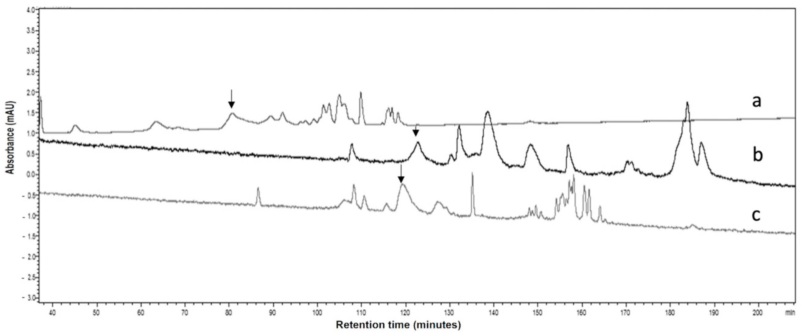
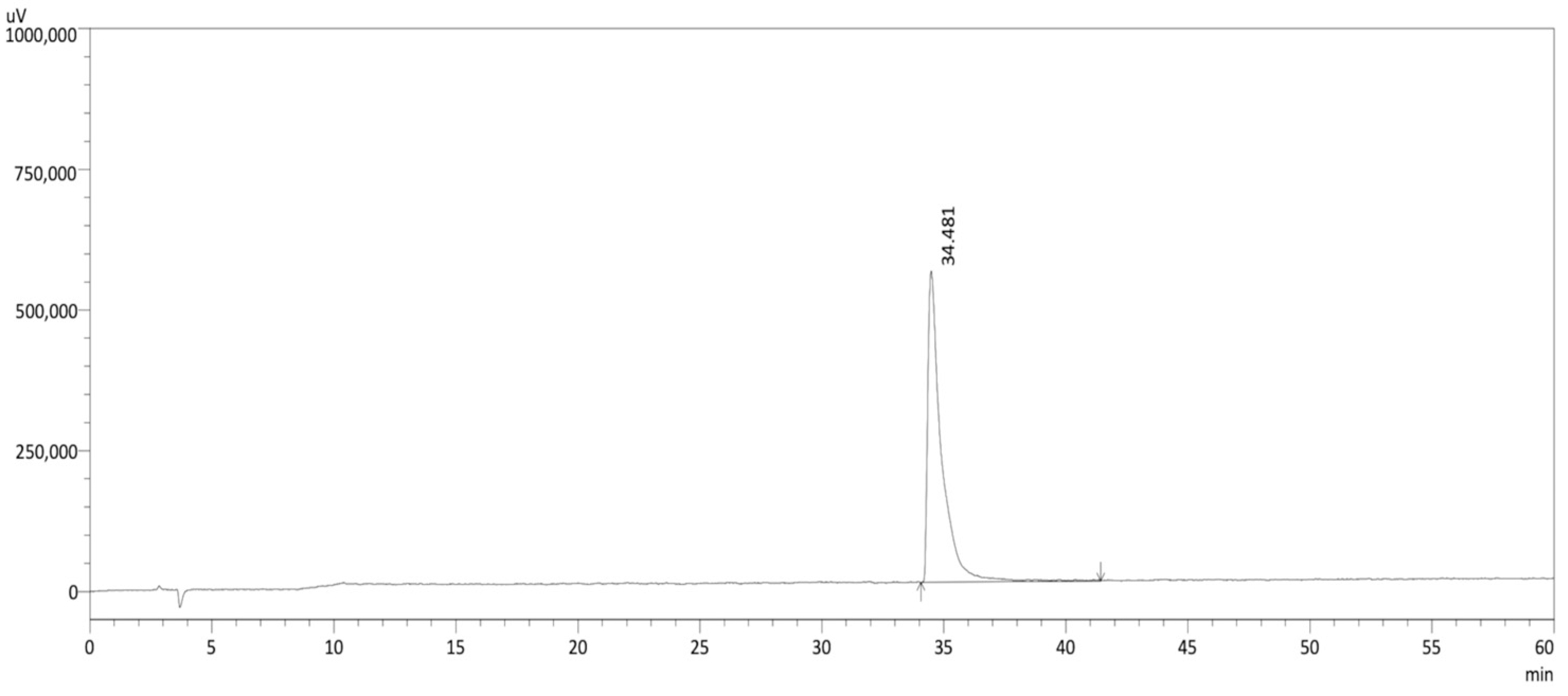
3.1. Purification of α-Bungarotoxin (α-Bgtx)

The purification of α-Bgtx was carried out in multiple stages. Eterović et al. (1975) and Hanley et al. (1977) used ion-exchange chromatography on a CM-Sephadex column combined with CM-cellulose chromatography [23] to achieve a yield of 27% [24] and a purity of 90% [25]. Similarly, Mebs et al. employed two CM-Sephadex C-25 or C-50 cation-exchange columns and two Sephadex G-25 columns for desalting the samples. With this method, the purified, desalted, and lyophilized toxin was obtained with a yield corresponding to about 15 to 20% of the weight of the lyophilized venom [26]. In our case, the focus was on purifying α-Bgtx using only a single chromatographic step to achieve higher homogeneity rates than those previously published. A comparative study of three different chromatographic columns (Figure 1), a Jupiter C18 300 Å 5 µm column (a), a Nucleosil C18 300 Å 5 µm column (b), and a ZORBAX C3 SB 300 Å 5 µm column (c), was conducted using analytical HPLC with an identical acetonitrile gradient A.
The comparison of the spectra revealed that the α-Bgtx peak obtained with the C3 ZORBAX SB 300 Å 5 µm column (Figure 1c), eluted at an intermediate time (120 min), was contaminated before and after. α-Bgtx is highly retained on the Nucleosil C18 Htec column (123 min) and exhibits contaminant peaks beforehand. The Jupiter C18 300 Å 5 µm column generates the following: (i) better separation and resolution of the peak corresponding to α-Bgtx, leading to (ii) higher purity and (iii) better yield. In addition, α-Bgtx eluted faster than on the two other columns (80 min). Although the Jupiter C18 300 Å and Nucleosil C18 300 Å columns have very similar characteristics and are end-capped, the chromatographic profile of α-Bgtx differs between them. These elution differences can be explained by several hypotheses: (i) the density of C18 grafting and the end-capping between the columns can influence how the molecule interacts with the stationary phase; (ii) variations in pore size, particle size, and particle shape between the columns can affect resolution and separation efficiency; and finally, (iii) the presence of uncapped silanols or other specific interactions at each column might also vary and influence the results. The Jupiter C18 300 column thus allowed us to purify the batch of venom from Bungarus multicinctus (Figure 2) with a yield of 7% and a homogeneity exceeding 99% (Figure 3). This purity was confirmed by MALDI-ToF mass spectrometry ([M + 1H] = 7.984 Da) (Figure 4).

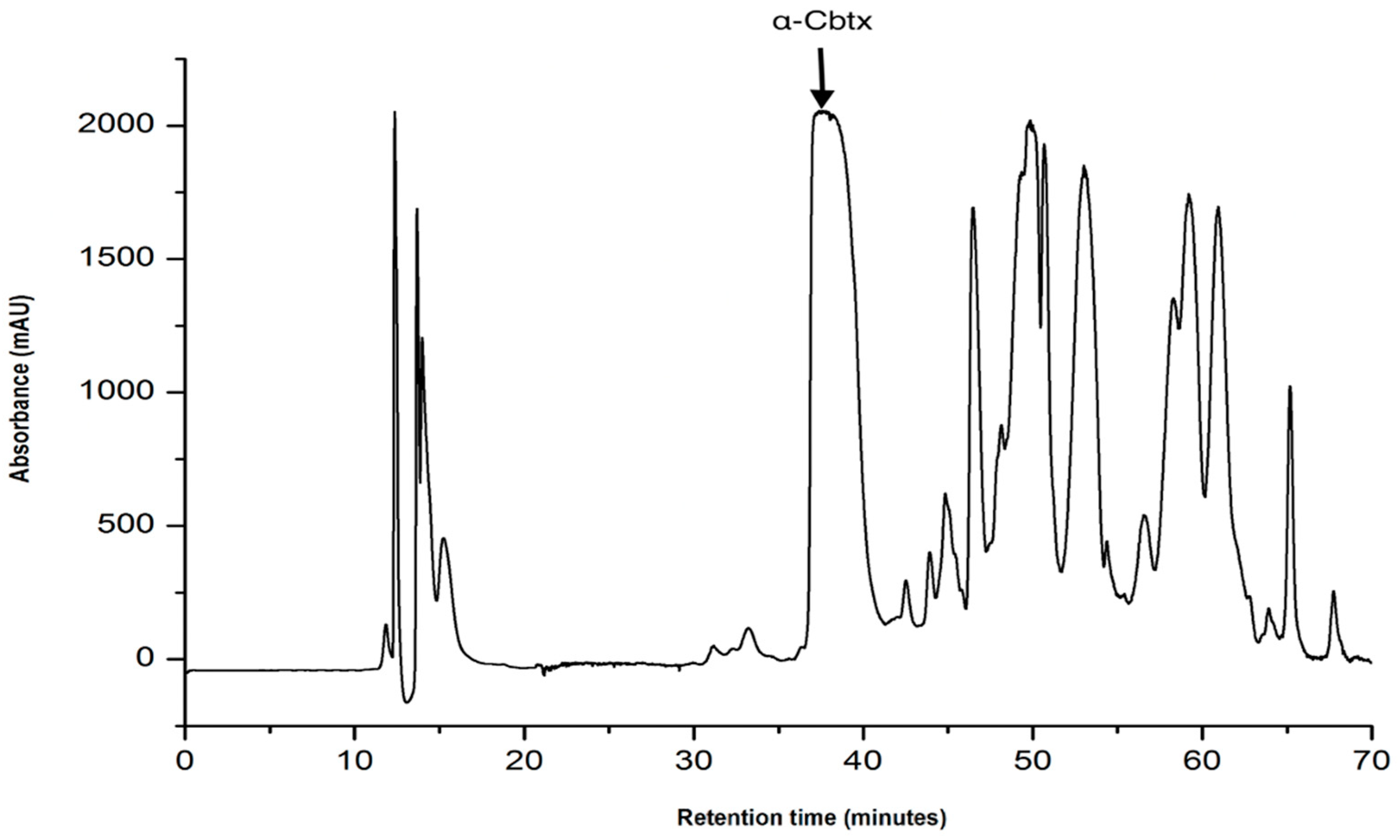
3.2. Purification of α-Cobratoxin (α-Cbtx)

Isolation procedures for this protein, as reported in the literature, are generally similar, varying only in the columns and solvents used. For example, Kukhtina et al. [27] utilized two methods to isolate α-Cbtx from Naja kaouthia’s venom. Initially, the venom was separated by gel filtration on a Sephadex G-50sf column. Fraction III, which contained all the proteins, was further purified by high-performance ion-exchange chromatography using a HEMA 1000BIO CM column. The molecular masses of the fractions were determined by MALDI-ToF MS analysis, and Fraction 3 was confirmed to be α-Cbtx. These fractions were ultimately purified using RP-HPLC on a Vydac C18 column. In the second method, crude venom was loaded onto a BioRex 70 cation exchanger equilibrated with ammonium acetate buffer. Separation was achieved using a gradient of increasing buffer molarity. The obtained fractions were analyzed by MS, and Fraction 4 was identified as α-Cbtx. The purity levels of these purification methods were not determined in the article [27].
Our group employed an efficient, one-step method. Naja kaouthia’s venom was purified exclusively on an RP-HPLC system. The MWs of the fractions from each peak were determined by MALDI-ToF (data not shown), and the peak, eluted at 37% of solvent B, shown in Figure 5, was assigned to α-Cbtx. Fractions from this peak were pooled, and a homogeneity of more than 99% was verified by analytical RP-HPLC (Figure 6).
This singular purification step, characterized by its simplicity, speed, and reproducibility, enabled the isolation of α-Cbtx with a yield of 15%.

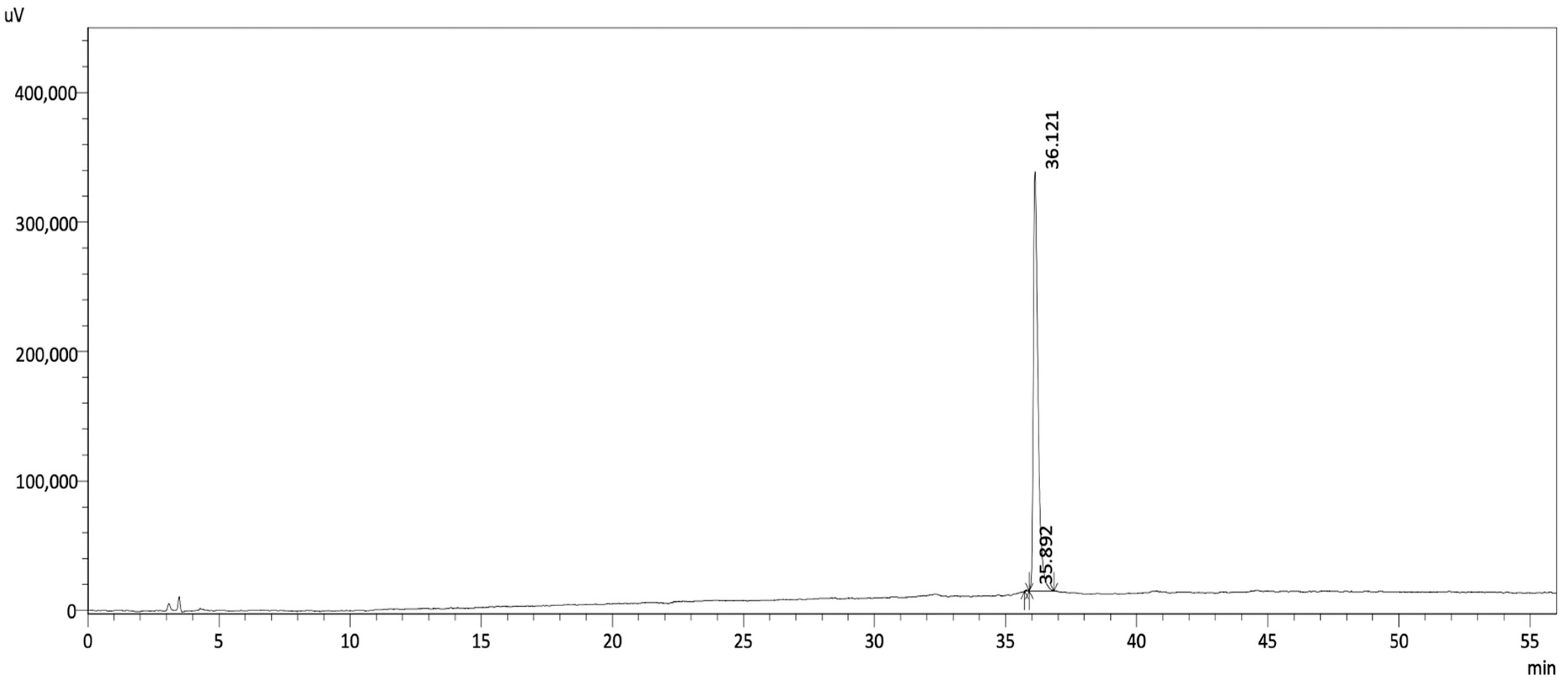
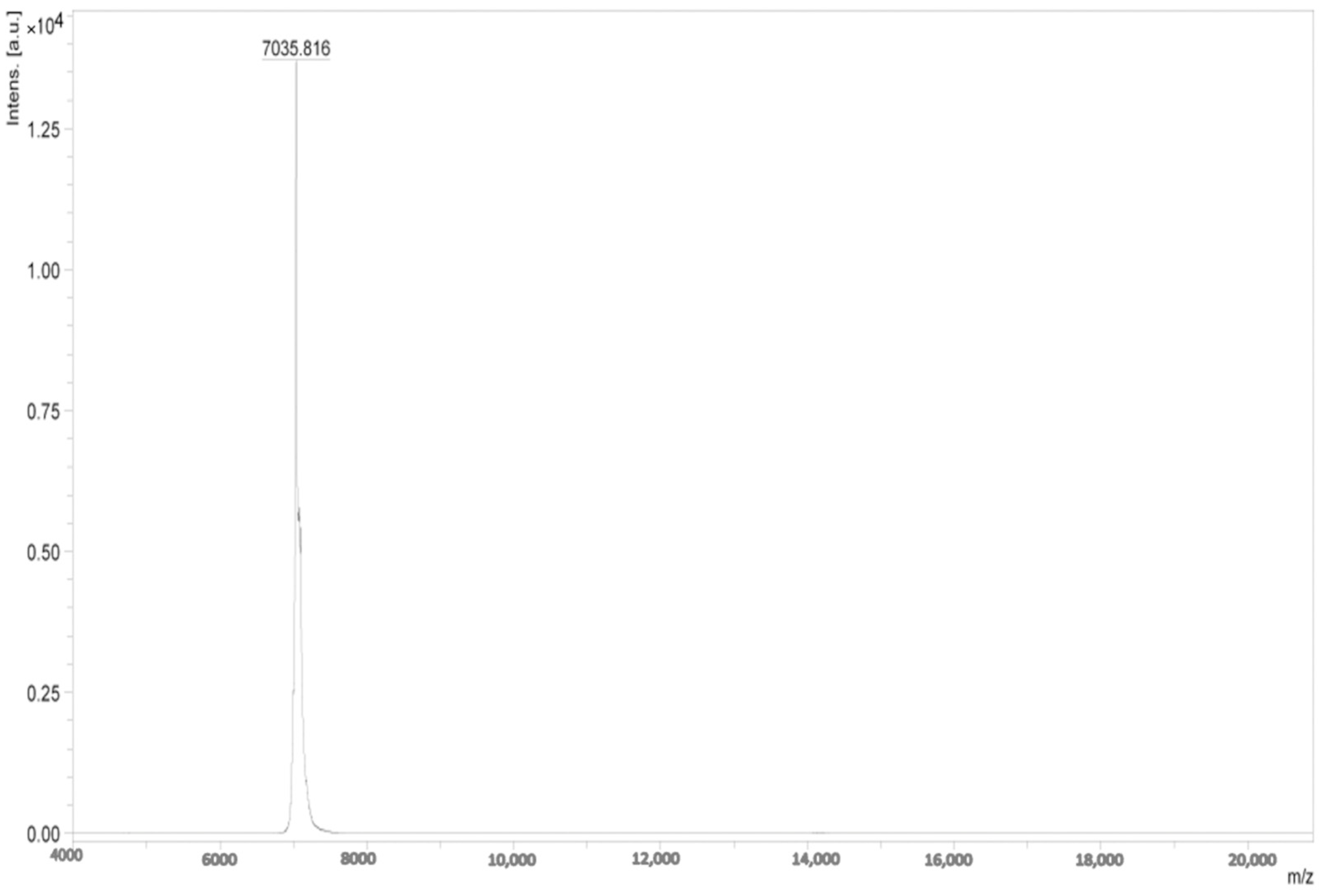
3.3. Purification of Calciseptine

The original purification process for calciseptine from the raw venom involved four crucial steps: (i) gel filtration on Sephadex 50, (ii) ion-exchange chromatography using TSK SP 5PW, (iii) HPLC on an RP-18 column, and (iv) desalination on a Trisacryl GF05 M column [28].
In our case, Dendroaspis polylepis polylepis’s venom was purified exclusively on the RP-HPLC system.
Using commercially available calciseptine as a reference, we identified and isolated the specific fraction containing calciseptine through preparative HPLC, with segmentation performed at 0.1 min intervals (Figure 7). This targeted fraction was eluted at 30% using a slope of 0.25% (gradient C). Calciseptine was obtained with a purity exceeding 95% and a yield of 6% (Figure 8).
The experimental mass obtained by MALDI-ToF ([M + 1H] = 7.036 Da) corresponds closely with the theoretical mass of calciseptine (7035 Da) (Figure 9).

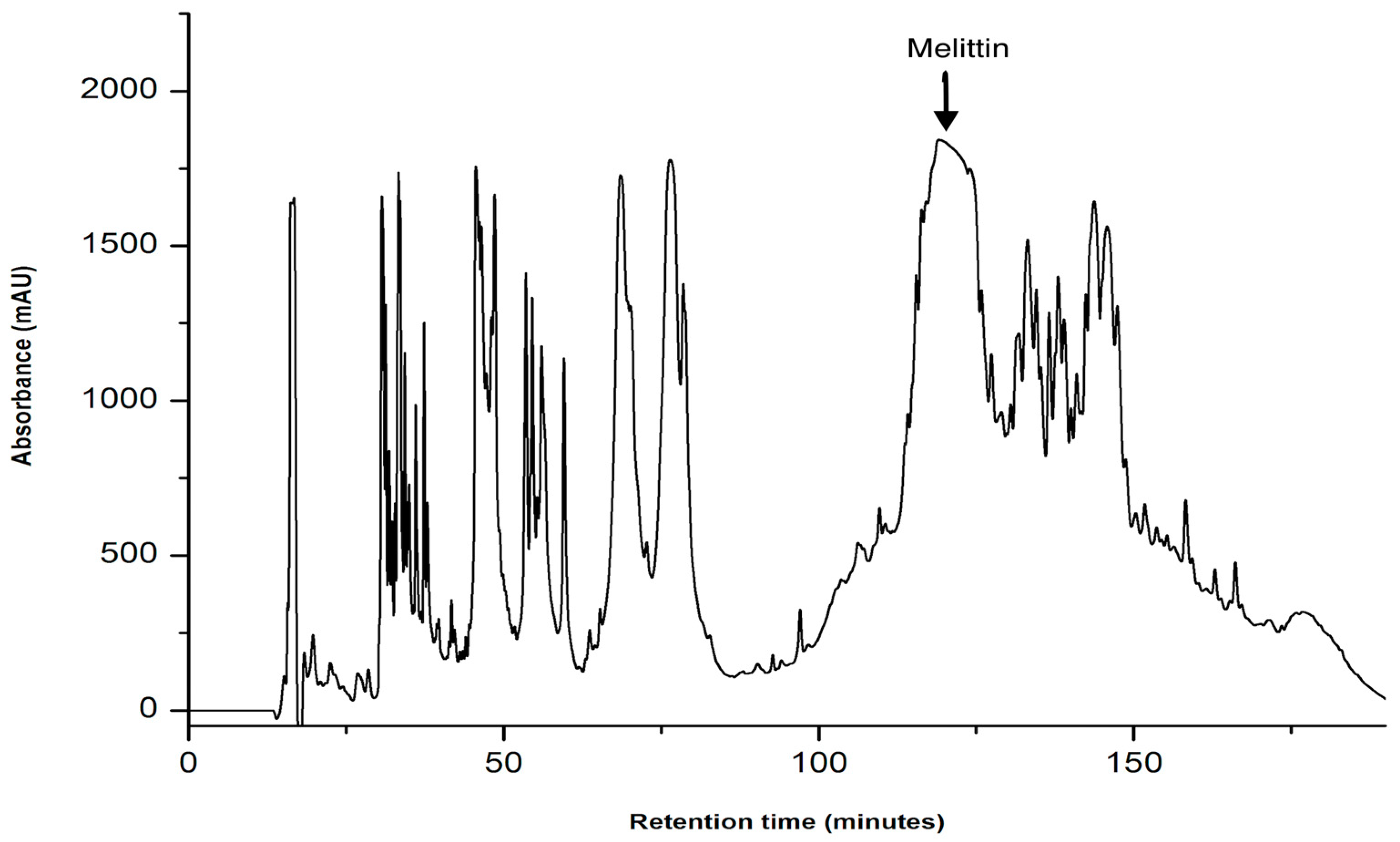
3.4. Purification of Melittin

Han et al. employed a multi-step purification process for melittin. Initially, the venom was fractionated using a Superdex peptide HE 10/30 column, followed by further purification on a PepRPC HR 10/10 column, and finally, the purified product was obtained through a Superdex 75 10/300 GL column. However, the description provided did not include data regarding yield and purity [15].
In contrast, Teoh et al. described a single-step purification method for melittin utilizing a HiTrap SP FF cation-exchange column. This approach achieved a purity of 93% for the peptide [29]. Nevertheless, this study omitted a desalting step, particularly considering the use of NaCl, which was not addressed in their report.
Melittin was purified with a yield of 4.5% on a Jupiter C18 300A column using gradient D with a slope of 0.25% B (Figure 10). Its homogeneity exceeding 99% was controlled using analytical RP-HPLC on a C18 column (Figure 11) and the purity was confirmed through MALDI-ToF [M + 1H] = 2845 Da (Figure 12).

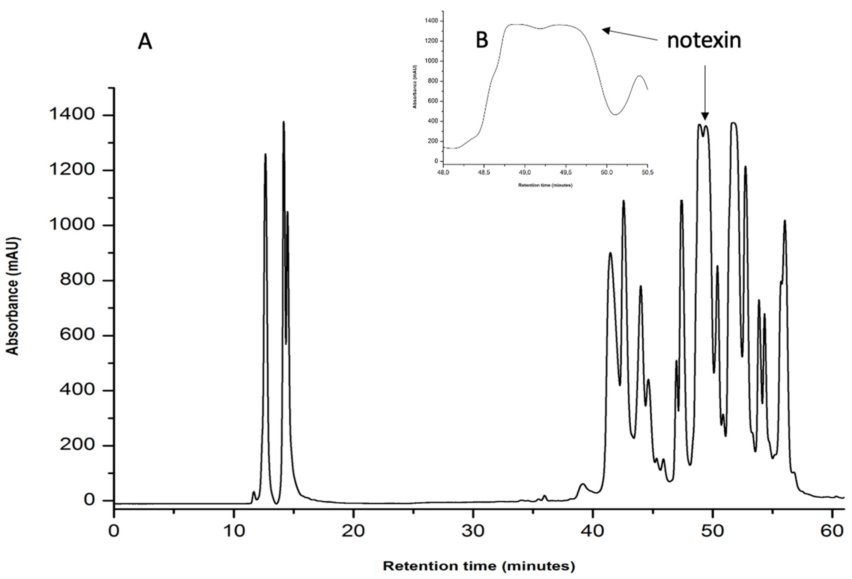
3.5. Purification of Notexin

Traditionally, isolation of notexin involved techniques such as gel filtration, high-performance ion exchange, and RP-HPLC. The protein was initially purified using cation-exchange chromatography on Bio-Rex 70 with an ammonium acetate gradient. The primary toxic component of the tiger snake venom was chromatographed on Sephadex G-50, and the principal peak, which was confirmed as notexin, showed a purity greater than 97% [18]. Chwetzoff et al. [30] noted that the ion-exchange fraction containing notexin comprised multiple components, leading to a reassessment of the initial purification method. Consequently, the fraction with notexin was further separated by RP-HPLC on a Nucleosil butyl large-pore column to distinguish between Notechis Np and Notechis Ns [30]. Indeed, Chwetzoff et al. demonstrated that Notechis Np and notexin share the same sequence, thus representing the same molecule. However, through Edman sequencing, they discovered that Notechis Ns differs by a single aa at position 16, where a lysine is replaced by an arginine. Nevertheless, this difference between the two isomers, Ns and Np, does not result in any variance in their presynaptic and myotoxic activities. The amount of Notechis Ns ranged from 38% to 6% of the total notexin content [30]. Although notexin can be isolated directly by ion-exchange chromatography of the crude venom, Halpert et al. first employed gel filtration on Sephadex G-75 to simplify its isolation, achieving a yield of 6.7% [31].
In a separate study, Yang and Chang [32] purified the toxin from tiger snake venom using SP-Sephadex C-25 instead of following the method of gel filtration on Sephadex G-25 and subsequent ion-exchange chromatography on Bio-Rex 70, as previously described by Halpert et al. [31]. Unlike the common use of these complex multi-step protocols, our approach enabled notexin Ns and Np to be purified in a single step using an RP-HPLC system (Figure 13 and Figure 14).
The homogeneity of notexin was evaluated using analytical RP-HPLC as illustrated in Figure 14 below: notexin Np was eluted at 12.6 min and represents 76%. As for notexin Ns, it was eluted at 12.9 min and represents 20%.
Notexin (Np and Ns) was obtained with a yield of 10% with a homogeneity exceeding 95%, compared to the 6.7% yield achieved by Halpert et al. [31].

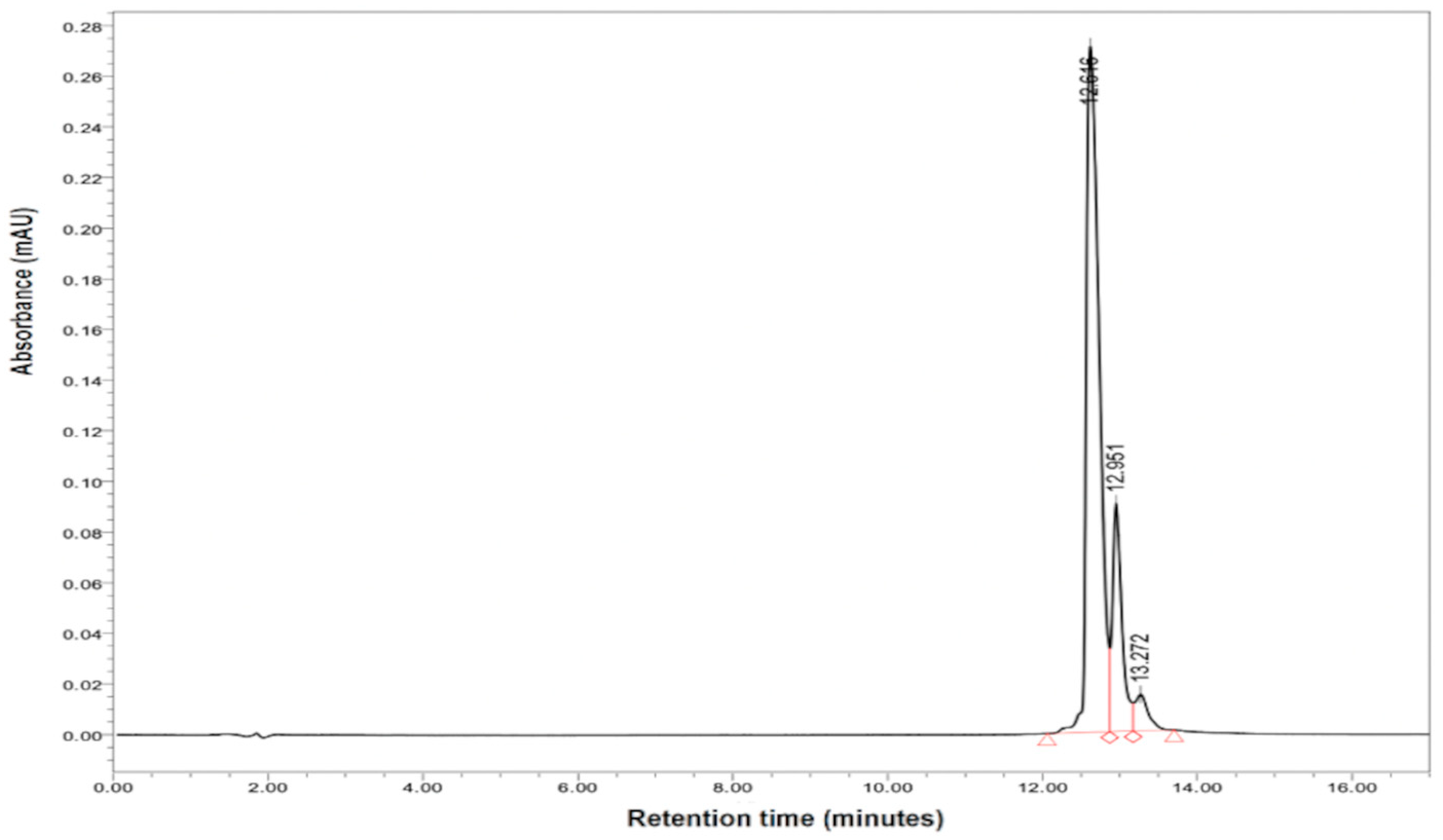
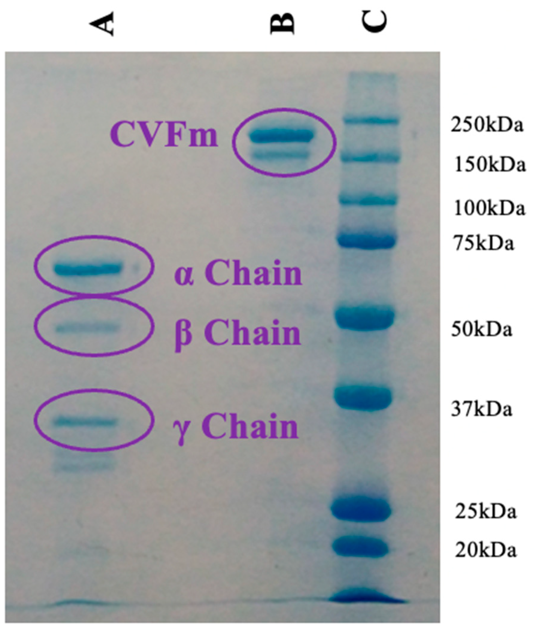
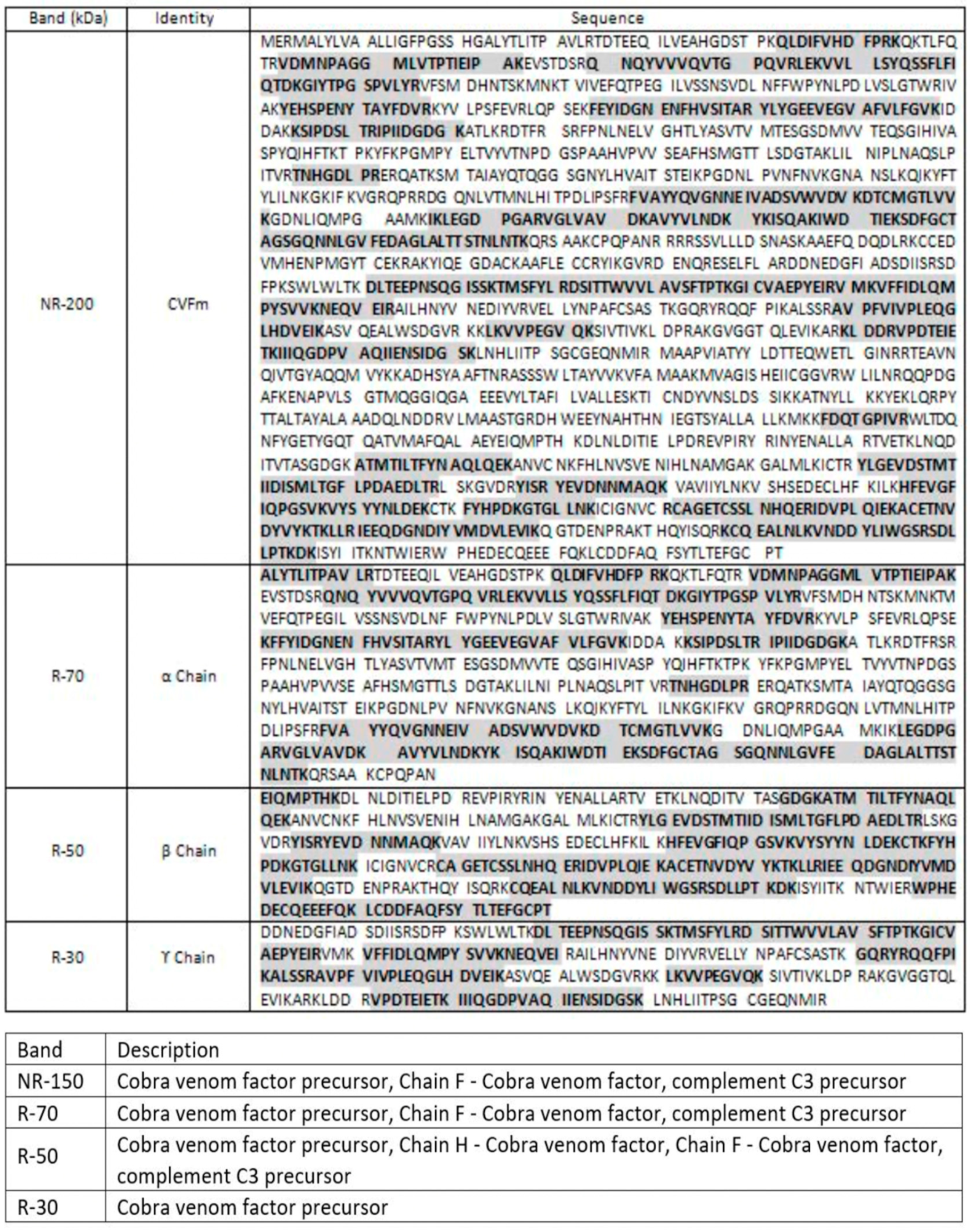
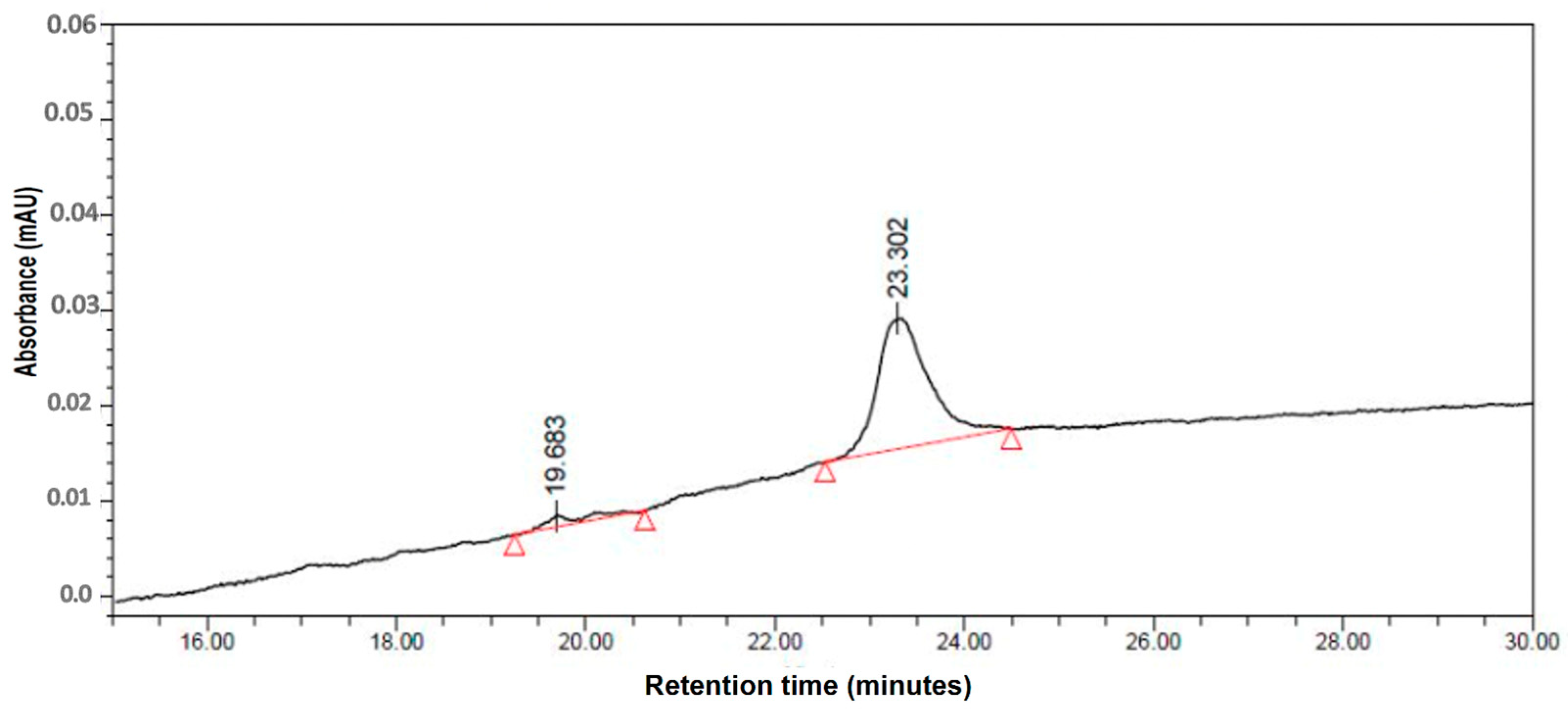
3.6. Purification of CVFm

V. Osipov et al. [19] extracted Cobra Venom Factor (CVF) from Naja melanoleuca venom using a multi-step purification process. The process began with the venom being dissolved and filtered through a Sephadex G50 superfine column. The fractions collected were then concentrated and further purified using a HEMA 1000BIO DEAE column. Following additional concentration and desalination, the samples underwent rechromatography on the same type of column but with a modified gradient. The final step involved desalting and drying the purified CVF for further analysis [19].
In our study, we successfully isolated this molecule in a single step using RP-HPLC on a Zorbax SB C3 300 Å column (Figure 15). Our choice was based on this column for two main reasons: (i) The larger pores of the Zorbax SB C3 300 Å allow better penetration and interaction of the protein with the stationary phase, which is crucial for proteins of size like 150 kDa. (ii) The C3 column utilizes propyl chains (C3), which offer lower hydrophobicity compared to C18 columns. This can be beneficial for proteins, as excessive hydrophobicity may lead to overly strong interactions and potentially cause issues with protein denaturation or incomplete recovery. The moderate hydrophobicity facilitates a gentler and more specific elution.
Subsequently, the purified protein is characterized using SDS-PAGE under both non-reduced and reduced conditions. The gel revealed a single band at approximately 150 kDa under non-reducing conditions and three major bands around 70, 50, and 30 kDa under reducing conditions, as illustrated in Figure 16. These findings align with those reported by V. Osipov et al. who observed bands at similar molecular weights (65–70, 50, and 30 kDa) [19]. Tryptic digestion of the isolated bands and their identification was performed as shown in Figure 16.
It should be noted that the sequence of CVFm has not been previously described, though it shows a similarity to that of CVFk, with a homology of 35% as identified by trypsin/LysC digestion and analyzed by ESI-Q-Exactive MS/MS (Figure 17).
Indeed, in column B, the band observed between 150 and 200 kDa represents CVFm. Meanwhile, in column A, the bands at approximately 70, 50, and 30 kDa correspond to the α, β, and γ subunits of CVFm, respectively. In summary, the CVFm isolated through a single RP-HPLC step and subsequently analyzed via analytical HPLC showed a purity exceeding 94% (Figure 18). The yield achieved was 0.8%, which slightly surpasses the results reported by V. Osipov et al. However, it should be noted that they did not provide purity levels in their study, making a direct comparison of yields between our research and theirs somewhat challenging. No loss of biological activity has been reported for CVFm, nor for the rest of the proteins (data not shown), contrary to what is sometimes described in the literature for large proteins.

4. Conclusions

Traditional methods for protein purification often involve intricate protocols and extensive preparation, which can significantly prolong the process and escalate costs. In contrast, our study underscores the effectiveness of employing a simplified single-step RP-HPLC method. We utilized straightforward and widely accessible solvent systems, markedly different from the more complex protocols documented in the literature that require intricate salt or pH gradients and labor-intensive setups such as FPLC. Our results suggest that the simplified single-step RP-HPLC method can be used for the rapid separation of proteins from complex mixtures prior to the integration of multiple separation methods. This approach offers a cost-effective and time-efficient alternative, highlighting the significance of our findings in protein purification protocols.

Author Contributions

Y.B.: investigation, methodology, writing, review, editing, and purifications of α-Bgtx, calciseptine, α-cobratoxin; S.A.: investigation, writing parent draft, purifications of CVFm, notexin, α-cobratoxin, SDS-PAGE; K.B.: purifications of melittin, α-cobratoxin, CVFm, notexin; A.C.: α-Bgtx, α-cobratoxin; S.B.: purification of calciseptine; T.B.: purification of α-Bgtx; P.M.: mass spectrometry and MS/MS; R.L.: mass spectrometry and review; D.G.: conceptualization and review; H.d.P.: conceptualization and review; K.M.: conceptualization and design of the experiments, writing, review, editing, and final approval. All authors have read and agreed to the published version of the manuscript.

Funding

This research received no external funding.

Data Availability Statement

The results of this study are fully documented and presented within the confines of this article.

Conflicts of Interest

We disclose that the students affiliated to latoxan, who are co-authors of this article, have received scholarships or grants (for thesis work or internships) from Latoxan as part of a collaboration on research projects. However, we affirm that Latoxan did not influence the study design, data collection, analysis, interpretation of data, manuscript writing, or the decision to submit the manuscript for publication.

References

  1. Morsy, M.A.; Gupta, S.; Dora, C.P.; Jhawat, V.; Dhanawat, M.; Mehta, D.; Gupta, K.; Nair, A.B.; El-Daly, M. Venoms classification and therapeutic uses: A narrative review. Eur. Rev. Med. Pharmacol. Sci. 2023, 27, 1633–1653. [Google Scholar] [CrossRef]
  2. Mabrouk, K.; Ram, N.; Boisseau, S.; Strappazzon, F.; Rehaim, A.; Sadoul, R.; Darbon, H.; Ronjat, M.; De Waard, M. Critical amino acid residues of maurocalcine involved in pharmacology, lipid interaction and cell penetration. Biochim. Biophys. Acta Biomembr. 2007, 1768, 2528–2540. [Google Scholar] [CrossRef] [PubMed]
  3. González García, M.C.; Radix, C.; Villard, C.; Breuzard, G.; Mansuelle, P.; Barbier, P.; Tsvetkov, P.O.; De Pomyers, H.; Gigmes, D.; Devred, F.; et al. Myotoxin-3 from the Pacific Rattlesnake Crotalus oreganus oreganus Venom Is a New Microtubule-Targeting Agent. Molecules 2022, 27, 8241. [Google Scholar] [CrossRef] [PubMed]
  4. Chalier, F.; Mugnier, L.; Tarbe, M.; Aboudou, S.; Villard, C.; Kovacic, H.; Gigmes, D.; Mansuelle, P.; de Pomyers, H.; Luis, J.; et al. Isolation of an anti–tumour disintegrin: Dabmaurin–1, a Peptide Lebein–1–Like, from Daboia mauritanica Venom. Toxins 2020, 12, 102. [Google Scholar] [CrossRef] [PubMed]
  5. Gosselin, R.C.; Douxfils, J. Ecarin based coagulation testing. Am. J. Hematol. 2020, 95, 863–869. [Google Scholar] [CrossRef] [PubMed]
  6. Verdoni, M.; Roudaut, H.; De Pomyers, H.; Gigmes, D.; Bertin, D.; Luis, J.; Mabrouk, K. ArgTX-636, a polyamine isolated from spider venom: A novel class of melanogenesis inhibitors. Bioorg. Med. Chem. 2016, 24, 5685–5692. [Google Scholar] [CrossRef] [PubMed]
  7. Smallwood, T.B.; Clark, R.J. Advances in venom peptide drug discovery: Where are we at and where are we heading? Expert Opin. Drug Discov. 2021, 16, 1163–1173. [Google Scholar] [CrossRef]
  8. King, G.F. Venoms as a platform for human drugs: Translating toxins into therapeutics. Expert Opin. Biol. Ther. 2011, 11, 1469–1484. [Google Scholar] [CrossRef]
  9. Thermo Fisher Scientific Inc. Successful Separations of Peptides, Proteins and Other Biomolecules HPLC Analysis of Biomolecules Technical Guide. Available online: www.thermoscientific.com/chromatography (accessed on 25 May 2010).
  10. Jean, R.; McClintock, R. Reversed-phase high-performance liquid chromatography of insulins from different species. J. Chromatogr. A 1983, 268, 112–119. [Google Scholar]
  11. Carr, D. The Handbook of Analysis and Purification of Peptides and Proteins by Reversed Phase HPLC; Grace Vydac: Hesperia, CA, USA, 2002; pp. 1–14. [Google Scholar]
  12. Brun, O.; Zoukimian, C.; Oliveira-Mendes, B.; Montnach, J.; Lauzier, B.; Ronjat, M.; Béroud, R.; Lesage, F.; Boturyn, D.; De Waard, M. Chemical Synthesis of a Functional Fluorescent-Tagged α-Bungarotoxin. Toxins 2022, 14, 79. [Google Scholar] [CrossRef]
  13. Wongtongkam, N.; Wilde, H.; Sitthi-Amorn, C.; Ratanabanangkoon, K. A Study of Thai Cobra (Naja kaouthia) Bites in Thailand. 2005. Available online: https://academic.oup.com/milmed/article/170/4/336/4577729 (accessed on 1 April 2005).
  14. de Weille, J.R.; Schweitz, H.; Maes, P.; Tartar, A.; Lazdunski, M. Calciseptine, a peptide isolated from black mamba venom, is a specific blocker of the L-type calcium channel. Proc. Natl. Acad. Sci. USA 1991, 88, 2437–2440. [Google Scholar] [CrossRef] [PubMed]
  15. Han, S.; Yeo, J.; Baek, H.; Lin, S.M.; Meyer, S.; Molan, P. Postantibiotic effect of purified melittin from honeybee (Apis mellifera) venom against Escherichia coli and Staphylococcus aureus. J. Asian Nat. Prod. Res. 2009, 11, 796–804. [Google Scholar] [CrossRef] [PubMed]
  16. Guha, S.; Ferrie, R.P.; Ghimire, J.; Ventura, C.R.; Wu, E.; Sun, L.; Kim, S.Y.; Wiedman, G.R.; Hristova, K.; Wimley, W.C. Applications and evolution of melittin, the quintessential membrane active peptide. Biochem. Pharmacol. 2021, 193, 114769. [Google Scholar] [CrossRef] [PubMed]
  17. Dixon, R.W.; Harris, J.B. Myotoxic activity of the toxic phospholipase, notexin, from the venom of the Australian tiger snake. J. Neuropathol. Exp. Neurol. 1996, 55, 1230–1237. [Google Scholar] [CrossRef] [PubMed]
  18. Karisson, E.; Eaker, D.; Rydén, L. Purification of a presynaptic neurotoxin from the venom of the Australian tiger snake Notechis scutatus scutatus. Toxicon 1972, 10, 405–413. [Google Scholar] [CrossRef] [PubMed]
  19. Osipov, A.V.; Mordvintsev, D.Y.; Starkov, V.G.; Galebskaya, L.V.; Ryumina, E.V.; Bel’tyukov, P.P.; Kozlov, L.V.; Romanov, S.V.; Doljansky, Y.; Tsetlin, V.I.; et al. Naja melanoleuca cobra venom contains two forms of complement-depleting factor (CVF). Toxicon 2005, 46, 394–403. [Google Scholar] [CrossRef] [PubMed]
  20. Laemmli, U.K. Cleavage of Structural Proteins during the Assembly of the Head of Bacteriophage T4. Nature 1970, 227, 680–685. [Google Scholar] [CrossRef] [PubMed]
  21. Krayem, N.; Parsiegla, G.; Gaussier, H.; Louati, H.; Jallouli, R.; Mansuelle, P.; Carrière, F.; Gargouri, Y. Functional characterization and FTIR-based 3D modeling of full length and truncated forms of Scorpio maurus venom phospholipase A2. Biochim. Biophys. Acta Gen. Subj. 2018, 1862, 1247–1261. [Google Scholar] [CrossRef] [PubMed]
  22. Kpebe, A.; Guendon, C.; Payne, N.; Ros, J.; Khelil Berbar, M.; Lebrun, R.; Baffert, C.; Shintu, L.; Brugna, M. An essential role of the reversible electron-bifurcating hydrogenase Hnd for ethanol oxidation in Solidesulfovibrio fructosivorans. Front. Microbiol. 2023, 14, 1139276. [Google Scholar] [CrossRef]
  23. Hanley, M.R.; Eterovic, V.A.; Hawkes, S.P.; Hebert, A.J.; Bennett, E.L. Neurotoxins of Bungarus multicinctus venom purification and partial characterization. Biochemistry 1977, 16, 5840–5849. [Google Scholar] [CrossRef]
  24. Eterovic, V.A.; Hebert, M.S.; Hanley, M.R.; Bennett, E.L. The lethality and spectroscopic properties of toxins from Bungarus multicinctus (Blyth) venom*. Toxicon 1975, 13, 37–48. [Google Scholar] [CrossRef] [PubMed]
  25. Eterović, V.A.; Aune, R.G.; Bennett, E.L. Radioactive Labeling of cx-Bungarotoxin with Tritium. Anal. Biochem. 1975, 68, 394–403. [Google Scholar] [CrossRef] [PubMed]
  26. Mebs, D.; Nan, K.; Iwanaga, S.; Samejima, Y.; Nan Lee, C. Purification, Properties and Amino Acid Sequence of a-Bungarotoxin from the Venom of Bungarus multicinctus. Biol. Chem. 1972, 353, 243–262. [Google Scholar]
  27. Kukhtina, V.V.; Weise, C.; Muranova, T.A.; Starkov, V.G.; Franke, P.; Hucho, F.; Wnendt, S.; Gillen, C.; Tsetlin, V.I.; Utkin, Y.N. Muscarinic toxin-like proteins from cobra venom. Eur. J. Biochem. 2000, 267, 6784–6789. [Google Scholar] [PubMed]
  28. Schweitz, H.; Bidard, J.-N.; Lazdunski, M. Purification and pharmacological characterization of peptide toxins from the black mamba (Dendroaspis polylepis) venom. Toxicon 1990, 28, 847–856. [Google Scholar] [CrossRef] [PubMed]
  29. Teoh, A.C.L.; Ryu, K.H.; Lee, E.G. One-step purification of melittin derived from Apis mellifera bee venom. J. Microbiol. Biotechnol. 2017, 27, 84–91. [Google Scholar] [CrossRef]
  30. Chwetzoff, S.; Mollier, P.; Bouet, F.; Rowan, E.C.; Harvey, A.L. On the purification of notexin Isolation of a single amino acid variant from the venom of Nutechis scutatus scutatus. FEBS Lett. 1990, 261, 226–230. [Google Scholar] [CrossRef] [PubMed]
  31. Halpert, J.; Eaker, D.; Karlsson, E. The role of phospholipase activity in the action of a presynaptic neurotoxin from the venom of Notechis scutatus scutatus (Australian tiger Snake). FEBS Lett. 1976, 61, 72–76. [Google Scholar] [CrossRef]
  32. Yang, C.-C.; Chang, L.-S. The N-terminal amino group essential for the biological activity of notexin from Notechis scutatus scutatus venom. Biochim. Biophys. Acta 1990, 1040, 35–42. [Google Scholar] [CrossRef]
Figure 1. Elution of Bungarus multicinctus venom on columns a, b, and c using 0% to 15%B for 30 min to 45% for 240 min gradient at 214 nm. Arrows indicate α-Bgtx.
Separations 11 00179 g001
Figure 2. α-Bgtx purification: preparative HPLC chromatogram of Bungarus multicinctus venom using gradient A at 214 nm.
Separations 11 00179 g002
Figure 3. Analytical HPLC profile of α-Bgtx on column c with a gradient G at 214 nm.
Separations 11 00179 g003
Figure 4. Mass spectrum of α-Bgtx using MALDI-ToF.
Separations 11 00179 g004
Figure 5. α-Cbtx purification: preparative HPLC chromatogram of Naja kaouthia venom using gradient B at 214 nm.
Separations 11 00179 g005
Figure 6. Analytical HPLC profile of α-Cbtx using gradient H at 214 nm.
Separations 11 00179 g006
Figure 7. Calciseptine purification: preparative HPLC profile of Dendroaspis polylepis polylepis using gradient C at 214 nm.
Separations 11 00179 g007
Figure 8. Analytical HPLC profile of calciseptine obtained with gradient G at 214 nm.
Separations 11 00179 g008
Figure 9. Mass spectrum of calciseptine using MALDI-ToF.
Separations 11 00179 g009
Figure 10. Melittin purification: preparative HPLC profile of Apis mellifera venom using gradient D at 214 nm.
Separations 11 00179 g010
Figure 11. Analytical HPLC profile of melittin using gradient G at 214 nm.
Separations 11 00179 g011
Figure 12. MALDI-ToF mass spectrum of melittin.
Separations 11 00179 g012
Figure 13. (A) Notexin purification: preparative HPLC profile of Notechis scutatus venom using gradient E at 214 nm. (B) Enlargement of the notexin elution region.
Separations 11 00179 g013
Figure 14. Analytical HPLC profile of notexin using a gradient I at 214 nm.
Separations 11 00179 g014
Figure 15. CVFm purification: preparative chromatogram of Naja melanoleuca venom using gradient F at 214 nm. The position of CVFm is indicated by an arrow.
Separations 11 00179 g015
Figure 16. SDS-PAGE of CVFm with three lanes labeled A, B, and C. Lane C is the molecular weight marker, as indicated by the distinct bands labeled with corresponding molecular weights (250 kDa down to 25 kDa). Lane B represents the sample under non-reducing conditions and Lane A under reducing conditions.
Separations 11 00179 g016
Figure 17. Enzymatic digestion results: trypsin/LysC and ESI-Q-Exactive (MS/MSMS).
Separations 11 00179 g017
Figure 18. Analytical HPLC profile of CVFm obtained with gradient J at 214 nm.
Separations 11 00179 g018
Table 1. Summary of the purification conditions for the 6 venom proteins. The solvents used were B (10% H2O, 0.1% TFA (v/v) in acetonitrile) in A (0.1% TFA (v/v) in water) at a flow rate of 6 mL/min. * Buffer B for α-Bgtx was 0.1% TFA (v/v) in acetonitrile. The absorbance was monitored by a UV–VIS detector at 214 and 280 nm.
Protein-MW (kDa)VenomPreparative
Conditions
Agilent 1260 Infinity
(Agilent Technologies, Santa Clara, CA, USA)
Analytical
Conditions
Shimadzu
LC-2010 HPLC (Shimadzu, Kyoto, Japan)
Retention Time (min)Homogeneity/
Yield
α-bungarotoxin
7.983
Bungarus multicinctus
-
Phenomenex Jupiter 300A 5 μm C18 250 mm × 21.2 mm
-
Gradient (A) 20–45% B * (200 min)
-
Phenomenex Jupiter® 5 µm C18 300 Å 250 × 4.6 mm
-
(G) 0–60% B (60 min)
3599%—7%
α-Cobratoxin-7.821Naja kaouthia
-
Phenomenex Jupiter 300A 5 μm C18 250 mm × 21.2 mm
-
(B) 22–50% B (60 min)
-
Purospher® STAR RP-18 250 × 4.6 mm (Merck, Darmstadt, Germany)
-
(H) 22–50% B (60 min)
24.599%—15%
Calciseptine-7.035Dendroaspis polylepis
polylepis
-
Phenomenex Jupiter 300A 5 μm C18 250 mm × 21.2 mm
-
(C) 20–60% B (160 min)
-
MACHEREY-NAGEL NUCLEOSIL 100-5 C18, 5 µm, 250 × 4.6 mm (Düren, Germany)
-
Gradient G
3695%—6%
Melittin-2.846Apis mellifera
-
Phenomenex Jupiter 300A 5 μm C18 250 mm × 21.2 mm
-
(D) 0–30% (30 min) to 60%B (120 min)
-
MACHEREY-NAGEL NUCLEOSIL 100-5 C18, 5 µm, 250 × 4.6 mm (Düren, Germany)
-
Gradient G
21.799%—4.5%
Notexin-13.593
(Np and Ns)
Notechis
scutatus
-
Phenomenex Jupiter 300A 5 μm C18 250 mm × 21.2 mm
-
(E) 20–40% B (40 min)
-
AdvanceBio RPmAb Diphenyl 150 × 4.6 mm (Agilent Technologies, Santa Clara, CA, USA)
-
(I) 20–40% B (20 min)
12.695%—10%
CVFm-150Naja
melanoleuca
-
Agilent ZORBAX 300SB-C3 250 × 21.2 mm 7μ (Agilent Technologies, Santa Clara, CA, USA)
-
(F) 0–100% B (100 min)
-
AdvanceBio RPmAb Diphenyl 150 × 4.6 mm
-
(J) 30–60% B (30 min)
2394%—0.8%
Disclaimer/Publisher’s Note: The statements, opinions and data contained in all publications are solely those of the individual author(s) and contributor(s) and not of MDPI and/or the editor(s). MDPI and/or the editor(s) disclaim responsibility for any injury to people or property resulting from any ideas, methods, instructions or products referred to in the content.

