Article

Characterization of Alginates of Sargassum from the Archipelago of Guadeloupe

by
Naika Marcin
1,2,
Valendy Thesnor
1,
Valentin Duvauchelle
3,
Alejandro Ponce-Mora
4,
Lucia Gimeno-Mallench
4,
Stacy Narayanin-Richenapin
1,
Laura Brelle
1,
Eloy Bejarano
4,
Christelle Yacou
1,
Muriel Sylvestre
1,
Cristel Onésippe-Potiron
1,
Patrick Meffre
3,
Zohra Benfodda
3 and
Gerardo Cebrian-Torrejon
1,*
1
COVACHIM-M2E Laboratory EA 3592, Department of Chemistry, University of the French West Indies, Fouillole Campus, UFR SEN, 97157 Pointe-à-Pitre Cedex, France
2
Ecole Nationale Supérieure de Chimie de Lille, ENSCL, Avenue Mendeleiev, 59650 Villeneuve-d’Ascq, France
3
UPR CHROME, Université de Nîmes, 30021 Nîmes, France
4
Department of Biomedical Sciences, School of Health Sciences and Veterinary School, Universidad Cardenal Herrera-CEU, CEU Universities, 46115 Valencia, Spain
*
Author to whom correspondence should be addressed.
Separations 2024, 11(8), 226; https://doi.org/10.3390/separations11080226
Submission received: 30 May 2024 / Revised: 16 July 2024 / Accepted: 17 July 2024 / Published: 25 July 2024
(This article belongs to the Section Analysis of Natural Products and Pharmaceuticals)

Abstract

:
Sargassum is a genus of brown algae that causes a major impact to marine ecosystems. Although accumulation of this invasive seaweed is considered an environmental problem, Sargassum can also be resource of valuable molecules such as alginates. Alginates are polysaccharides extracted from the cell walls of brown seaweeds with multiple applications in food, cosmetics, pharmaceuticals, and biotechnology. In this study, we explored four different extraction protocols to isolate alginates from Sargassum from the archipelago of Guadeloupe. H-NMR, FTIR, and SEM were carried out to characterize isolated samples and describe their chemical and morphological properties. Antioxidant capacity of the isolated alginates was tested in the sample. FRAP assay showed free radical scavenging activity, and cell viability assay showed that alginates keep protective capacity against oxidative stress in cell cultures. In sum, the isolation methods used in this study are effective extractive methods to obtain alginates from Sargassum, which highlight this seaweed as a potential source of bioactive compounds that may be exploited for industrial and biomedical purposes.

1. Introduction

Sargassum is a genus of brown algae that belongs to the family Sargassaceae, of the order Fucales. Sargassum, also known as gulfweed or sea holly, is considered one of the most complex genera within the class Phaeophyceae and comprises 536 species. Although primarily found in tropical and subtropical marine waters, Sargassum is distributed worldwide. The biomass of this invasive seaweed can be found around the world, off the coasts of countries such as Brazil, Mexico, India, and China [1,2].
Sargassum species typically have a highly branched thallus with hollow berry-like floats called pneumatocysts. The numerous fronds are generally small and leaf-like with toothed edges. Most species reproduce sexually, but the pelagic species reproduce by fragmentation. Some species of this brown seaweed can measure up to 12 m long. Sargassum can be fixed at the bottom by a thallus growing on rocky reefs, but most live exclusively by floating. These Sargassum are called holopelagics, and their entire life cycle occurs on the water surface. The Sargasso Sea in the western Atlantic Ocean is named after this seaweed due to massive floating masses of Sargassum natans and Sargassum fluitans. These brown algae have significant importance in the ecosystem since algae are the primary producer in the area, serving as substrate, shelter, and food for several marine animals such as fish, shrimp, crabs, birds, and turtles, acting as a focal point for marine biodiversity and productivity [3].
Although the ramifications of climate change on ecosystems remain highly uncertain, they seem to be profound for Sargassum. Sargassum overgrowth is linked to climate change, as warmer temperatures, increased nutrients, and altered currents and winds may favor the growth and distribution of the seaweed [4]. Since 2011, substantial volumes of pelagic Sargassum algae have been washing ashore along the coasts of the Caribbean, Gulf of Mexico, and West Africa, giving rise to events termed golden tides. These golden tides threaten coastal communities, native marine and coastal ecosystems, and the tourism industry. When Sargassum decomposes, it releases hydrogen sulfide, a toxic gas that can cause irritation and respiratory discomfort. Additionally, as Sargassum breaks down, it depletes the oxygen dissolved in the water, leading to reduced water quality and the formation of hypoxic zones (areas with low-oxygen levels), which can make it difficult for some marine species to survive. By 2015, Caribbean beaches witnessed the deposition of over 200,000 metric tons of Sargassum. Subsequent years, particularly 2018 and 2019, saw even more substantial influxes, necessitating the employment of over 9000 workers to remove more than one million metric tons of Sargassum from tourist-centric beaches. While algae removal can help alleviate the overall impact, it comes at a considerable cost. Therefore, methods aimed at valorizing this invasive Sargassum can offer essential economic relief while simultaneously addressing an environmental problem [4,5,6,7].
Despite the current detrimental environmental and economic impact of the spread of Sargasuum, this seaweed might be a valuable source of bioactive compounds. This brown seaweed has been widely used in traditional medicine and is known as a source of anti-inflammatory agents [8]. These seaweeds are composed of diverse metabolites, such as sugars of low molecular weight (i.e., D-mannitol), and other micronutrients, including fucoidans [9]. In addition, Sargassum algae exhibit a high concentration of alginates that represent between 4 to 40% of the dried weight of algae. These polysaccharides have multiple applications in food, cosmetics, pharmaceuticals, and biotechnology, and antioxidant properties were reported as potent biological properties in alginates isolated from Sargassum [10,11,12].
Alginate concentration and characteristics rely on different factors. Alginates are composed of two monomeric units, which are 1,4-linked-D-mannuronic and L-guluronic, and their distribution within the macromolecule varies according to the brown algae. On the coast of the West Indies, two species are present in this phenomenon of overgrowth and invasion: S. natan and S. fluitans. These species have high amounts of fiber, salts, complex carbohydrates, minerals, trace elements, amino acids, fatty acids, and bioactive compounds that could benefit animal health and soil improvement [13]. The ratio mannuronic acid/guluronic acid is dependent on the species, and it determines physical and chemical properties, such as viscosity, gel strength, solubility, and biocompatibility. The alginates from S. fluitans have a M/G ratio of 0.59 while the ones from S. natans display a ratio of 0.51 [14,15]. Unfortunately, toxic elements are found in Sargassum, limiting the use of seaweed extracts, but the isolation of these polysaccharides may be an alternative [16]. Alginate concentration isolated from the algae may depend on the environment, season of collection, and, importantly, also on the method of extraction [17]. Prior to the extraction process, a pre-treatment of brown algae is required in order to remove several metabolites that can interfere in the extraction (e.g., phenolic compounds or metals). The pre-treatment aims to improve the alginate solubilization as well as alginate purity, and pre-treatment strategies lead to an increase in yield [18,19].
In this study, we tested various alginate extraction protocols already described in the literature and applied them to Sargassum algae collected on the island of Guadeloupe (French West Indies, FWI). After isolating and characterizing them, the antioxidant capacity of alginates in vitro and in cell culture was assessed.

2. Materials and Methods

2.1. Extraction and Purification of Alginates

Sargassum algae were collected along the beaches of the island of Guadeloupe (French West Indies, FWI). Four different extraction protocols described in the literature were explored and applied to the algae collected in Guadeloupe. Depending on the extraction process chosen, alginic acid or sodium alginate (Figure 1) were obtained with significantly different yields. The four different protocols are described below.
Protocol 1. The first extraction method was carried out with Sargassum collected in October 2018 on the island of Les Saintes (part of Guadeloupe’s archipelago) and kept in seawater (changed regularly) until extractive work (4 days). The alginates were extracted by following the protocol described previously [20]. A total 29 g of wet algae were cut and soaked in 500 mL of 0.2 M H2SO4 for one hour at room temperature. Then, the Sargassum were washed with deionized water followed by 700 mL of sodium hypochlorite solution (0.315 wt% (weight percentage)). The sample was further washed with deionized water before the alginate extraction using a 2 wt% CaCO3 solution, where they were soaked for 24 h at room temperature. Then, the mixture was filtrated using a Büchner funnel to proceed to the precipitation of the alginic acid (Figure 1a) using a 6 M H2SO4 solution. The precipitate was finally dried at 80 °C for 2 days in a drying oven.
Algae collected on the beach of Sainte-Claire at Goyave (Guadeloupe) in February 2019 were used for procedures 2, 3, and 4 (see below). These algae were firstly cleaned with deionized water in order to remove sand and the maximum of salt. They were then dried to remove enough water and insects which may be present in the open air (for about 2 weeks), and finally stored in plastic bags until extraction operations.
Protocol 2. For the second extraction [20], 50 g of dried algae were first soaked in a bath of 1 M H2SO4 solution for 48 h. The excess of acid contained in algae residues was removed by rinsing with deionized water. Algae were subsequently put in a bath of 2 wt% Na2CO3 in a form of maceration for 48 h. After this step, a solid/liquid separation by filtration was made to recover the filtrate (solution of sodium alginate, Figure 1b). Then, alginic acid (Figure 1a) was precipitated with the addition of a 1M H2SO4 solution to pH < 2.
Protocol 3. We used a process previously published in [21,22]. A total of 29 g of dried algae were soaked for 24 h in 550 mL of formaldehyde to remove phenolic compounds and pigments. After washing with deionized water several times, algae were soaked for 5 days in 500 mL of 1M HCl. After swilling out the seaweed with water, they were plunged in 550 mL of a 3 wt% Na2CO3 solution for 3 days. A centrifugation step was carried out for 20 min at 5000 tr/min. The supernatant obtained was poured in 95% ethanol (volume equal to one-third of that of the supernatant). In order to have a suspension to facilitate the filtration at room temperature, this mixture was kept for 4 days in a 500 mL Erlenmeyer. The obtained precipitate was filtered using a Büchner funnel and washed with acetone, yielding the sodium alginate.
Protocol 4. The fourth process was reported for the first time by Mian and Percival [23] and adapted by Souchet and Rioux et al. [24,25]. For this last method, heating under reflux for 24 h is required. First, 29 g of algae were cut and soaked in 85% ethanol. Then, the algae’s residue was soaked in 500 mL of 2 wt% CaCl2 solution. After this step and filtration, the basification was made with a 2 wt% Na2CO3 solution. To precipitate the alginates, acetone was added (450 mL of acetone to 100 mL of solution). Finally, a filtration on a Büchner apparatus was carried out to obtain the sodium alginate.

2.2. Fourier Transform Infrared Spectroscopy (FT-IR Spectroscopy)

The infrared spectra of samples obtained from the 4 methods (acid alginic and sodium alginate) were recorded at room temperature in the wave-number range of 400–4000 cm−1 using a Jasco FT-IR 410 instrument (Jasco, Oklahoma, OK, USA) as a thin film on a NaCl disc, as stated; only structurally important peaks (ῡ) are presented in cm−1. A total of 16 scans were averaged for each sample with a digital step at 0.492 cm−1.

2.3. Nuclear Magnetic Resonance Spectroscopy (NMR Spectroscopy)

1H NMR spectra were recorded with a Bruker Avance 300 NMR spectrometer. Chemical shifts (δ) and coupling constants (J) are given in ppm and Hz, respectively, using residual solvent signals as reference for the 1H. 1H NMR analyses of alginate were performed in D2O and alginic acid in DMSO d6.

2.4. SEM/EDX Characterization and Elemental Composition

Both microstructural and chemical elemental analysis of samples were carried out using a scanning electron microscope (SEM) equipped with an energy dispersive X-ray (EDX) microanalyser (Hitachi S-2500, Hitachi, Tokyo, Japan). Acquisition parameters varied as follows: backscattered electron mode (BSE), electron beam voltage 15–20 kV, working distance 10.0–10.3 mm, spot size 3.5–5 nm. Prior to analysis, small pieces of the samples were mounted on aluminum stubs using carbon tape, and no metallic coating was required. EDX analysis was performed on several areas within the samples to determine their bulk elemental composition.

2.5. Ferric Reducing Antioxidant Power (FRAP) Assay

Alginates extracted using methods 1 and 2 were resuspended in sodium carbonate 7.5% (in water) until reaching a concentration of 1 mg/mL. Alginates 3 and 4 were diluted in water to reach a concentration of 1 mg/mL. The total antioxidant capacity of the solutions was measured using a commercial kit following the manufacturer’s instructions (ref. KF01003, BioQuoChem, Oviedo, Spain). The antioxidant activity is expressed as the FRAP value (μM Fe2+). The assay was carried out three times in triplicate for each alginate. DPPH (ref. KF01007, BioQuoChem, Spain) and ABTS (ref. KF01002, BioQuoChem, Spain) methods were used, but these techniques were not suitable due to the nature of the samples.

2.6. In Vitro Cellular Assay of Oxidative Stress

The antioxidant capacity of the alginates in cells was evaluated using mouse embryonic fibroblasts (MEFs). Cells were seeded in 96-well plates at a concentration of 10,000 cells/well. Cells at 80% of confluency were subjected to a 12 h treatment with different concentrations (0 µM, 75 µM, 125 µM, 200 µM, 500 µM, and 1 mM) of hydrogen peroxide (H2O2) (ref. HI0135100, Scharlab, Sentmenat, Spain). After the treatment, the cell viability was evaluated using the resazurin-based cell viability reagent AlamarBlue (ref. DAL110, Invitrogen, Thermofisher Scientific, Waltham, MA, USA). Plates were measured using the VICTOR X3 2030 Multilabel Reader (PerkinElmer, Waltham, MA, USA).

3. Results

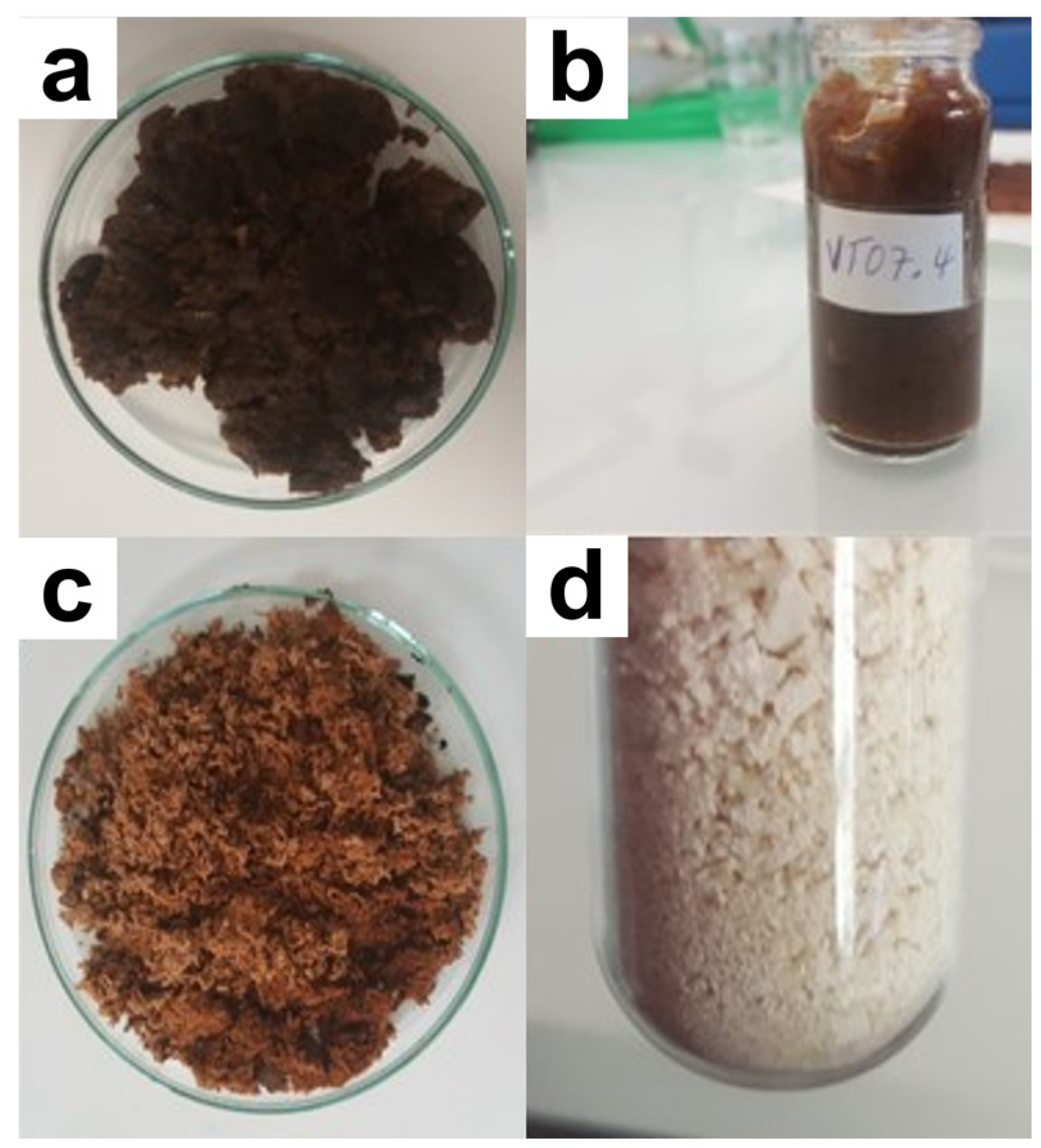
3.1. Yield, Visual Aspect, and Solubility of Extracts

According to the applied protocol, notable differences were observed in the extraction yield, getting the highest yield value using protocol 1. The use of sulfuric acid for precipitation in the last step of protocol 1 (yield of 44.7%) and 2 (yield of 37.6%) led to the formation of alginic acid, while the use of acetone in this last purification step led to sodium alginate for protocol 3 (yield of 9%) and protocol 4 (yield of 2.6%). After performing extractions from Sargassum algae, solubility tests were performed. As anticipated, alginic acid (Figure 1a) was insoluble in water but soluble in alkaline solutions, while sodium alginate (Figure 1b) was soluble in water. These solubility data are in alignment with the previous literature [23,24].
The precipitates obtained from the first three processes were brown (Figure 2a–c), while the sodium alginate obtained from the fourth extraction method was white (Figure 2d). Furthermore, the alginic acid samples presented a fibrous aspect (Figure 2a,b), and the sodium alginate samples had a dusty aspect (Figure 2c,d).

3.2. Spectroscopic Analyses

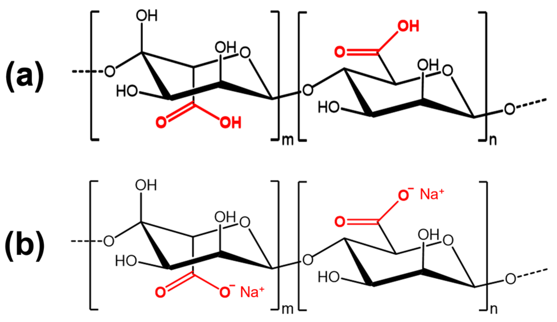
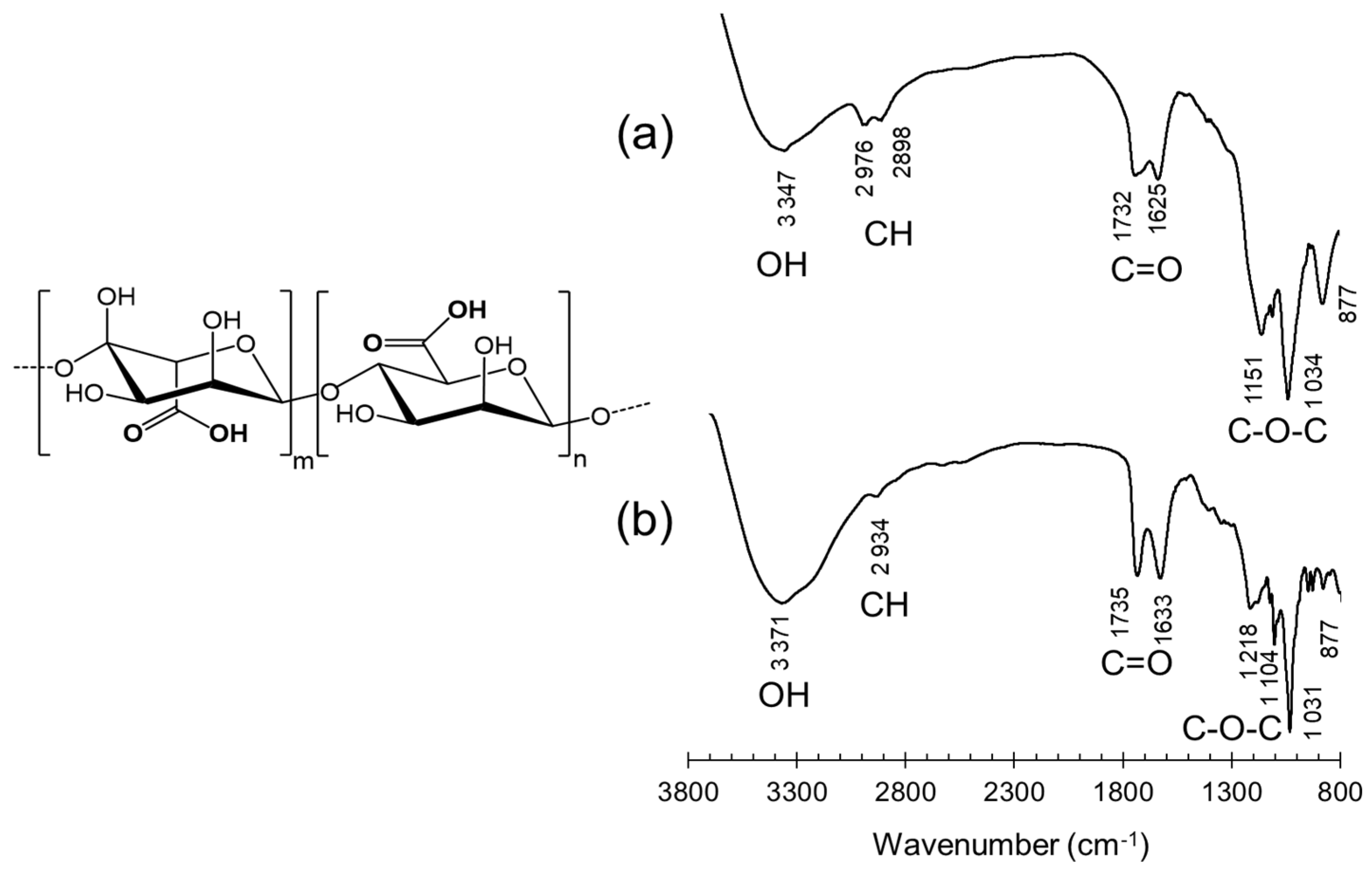
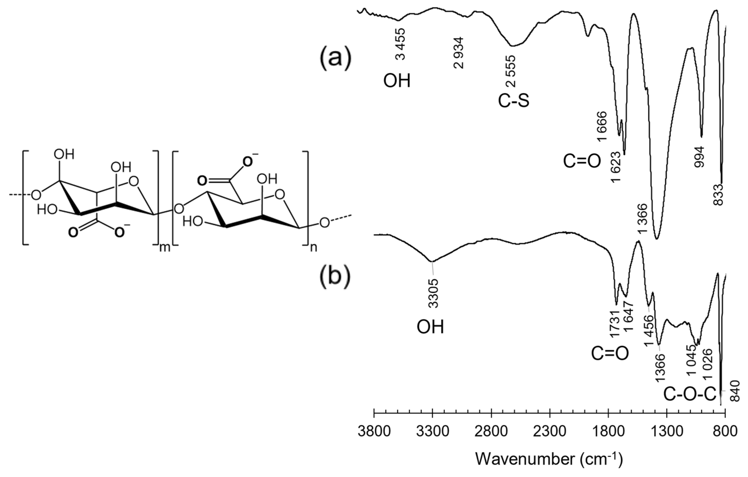
3.2.1. Infrared Spectroscopy

Protocol 1. The infrared spectrum of the alginic acid isolated from the Sargassum algae is presented in Figure 3a. A broad band at 3347 cm−1 was assigned to the hydrogen bond O-H from the alcohol stretching vibrations. This band is visible even if there is background noise. Two medium intensity bands (about 1732 and 1625 cm−1) were assigned to the O=C-O symmetric deformation vibration of the carboxylic acid. The bands measured at 1151, 1034, and 877 cm−1 were attributed to C-OH deformation of secondary alcohol, C-O deformation vibration of pyranose rings with a high intensity, and C-H rings of mannuronic acid residues, respectively.
Protocol 2. The infrared spectrum of the alginic acid extracted by the second extraction process is presented in Figure 3b. The infrared spectrum of the sample obtained from protocol 2 was similar to the alginic acid extracted in protocol 1 with a slight shift. An important band located at 3371 cm−1 was observed. This band was attributed to the O-H hydrogen band from the stretching vibrations of the alcohol. The band with a low intensity around 2934 cm−1 corresponds to the CHx stretching vibrations. Both bands in 1735 and 1633 cm−1 were attributed to the O=C-O asymmetric deformation vibration of the carboxylic acid. The two bands measured at 1104 and 1031 cm−1 could be assimilated to the C-O-C stretching vibration. The last band was detected at 877 cm−1 and was attributed to C-H mannuronic acid residues.
The FT-IR spectrum of sodium alginate was very different from alginic acid (Figure 4). Indeed, the strong bands corresponding to the alginic acid hydroxyl groups were weak in sodium alginate infrared spectra. The different results of the infrared analysis of sodium alginate are presented in detail below for protocols 3 and 4 [15,26,27,28].
Protocol 3. The infrared spectrum of the sodium alginate from the Sargassum algae is presented in Figure 4a. On this spectrum, a slight band at 3455 cm−1 was assigned to hydrogen band O-H from alcohol stretching vibrations. However, the broad band at 2555 cm−1 could be attributed to a C-S stretching vibration. The high-intensity bands were detected around 1666 and 1623 cm−1, which can correspond to the O-C-O asymmetric stretching vibration and C=O asymmetric stretching vibration of the carboxylate group. At 1366 cm−1, this high-intensity band can also correspond to C-C-H and O-C-H deformations. The bands measured at 994 and 833 cm−1 were attributed to the C-O stretching vibration of uronic acid and β-mannuronic acid or to the C-O-S stretching vibration. The bands at 2555 cm−1 and 833 cm−1 could indicate the presence of fucoidans in the sample [29,30,31].
Protocol 4. The infrared spectrum of the sodium alginate extracted by the fourth extraction process is presented in Figure 4b. A broad band at 3305 cm−1 is visible and was attributed to hydrogen band O-H from alcohol stretching vibrations. Other bands with a high intensity detected at 1731 and 1647 cm−1 were attributed to O=C-O the asymmetric deformation vibration of the carboxylate group. At 1456 cm−1, the band corresponds to the C-H or C-OH deformation. The band measured at 1366 cm−1 was a C-C-H or C-OH deformation. The bands measured at 1045, 1026 and 840 cm−1 were assimilated to C-OH deformation vibration with contribution of O=C-O symmetric deformation vibration of carboxylate group or C-H mannuronic acid residues, to C-H of α-L-glucuronic residues rings vibrations and to β-mannuronic acid vibration, respectively.
The characteristic bands of the sodium alginate and alginic acid groups were present in the analyses of the samples corresponding to the different protocols. This confirmed that the protocols established for the extraction of alginic acid are effective [32,33]. 1H NMR analyses were then performed to analyze the structure of the extraction products.

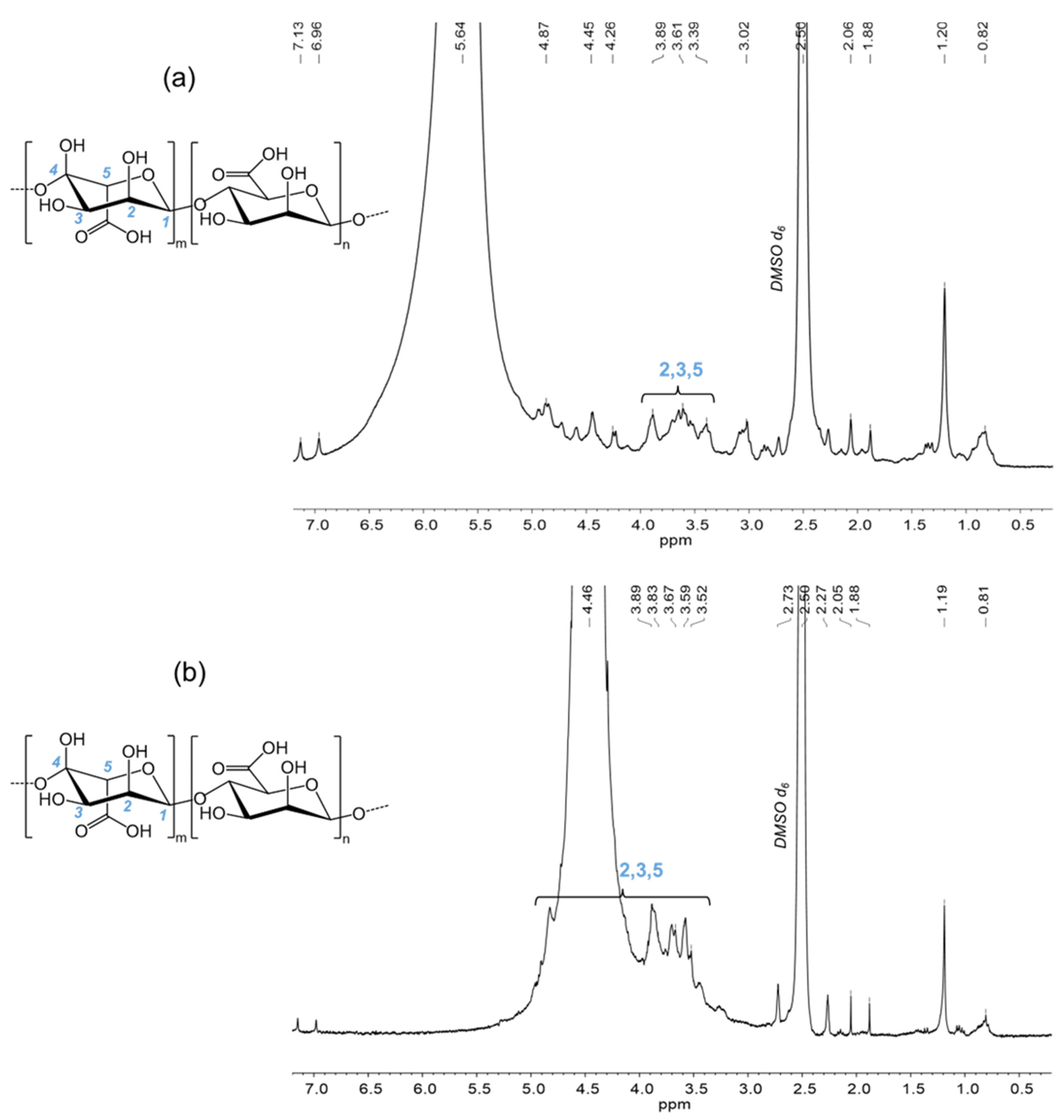
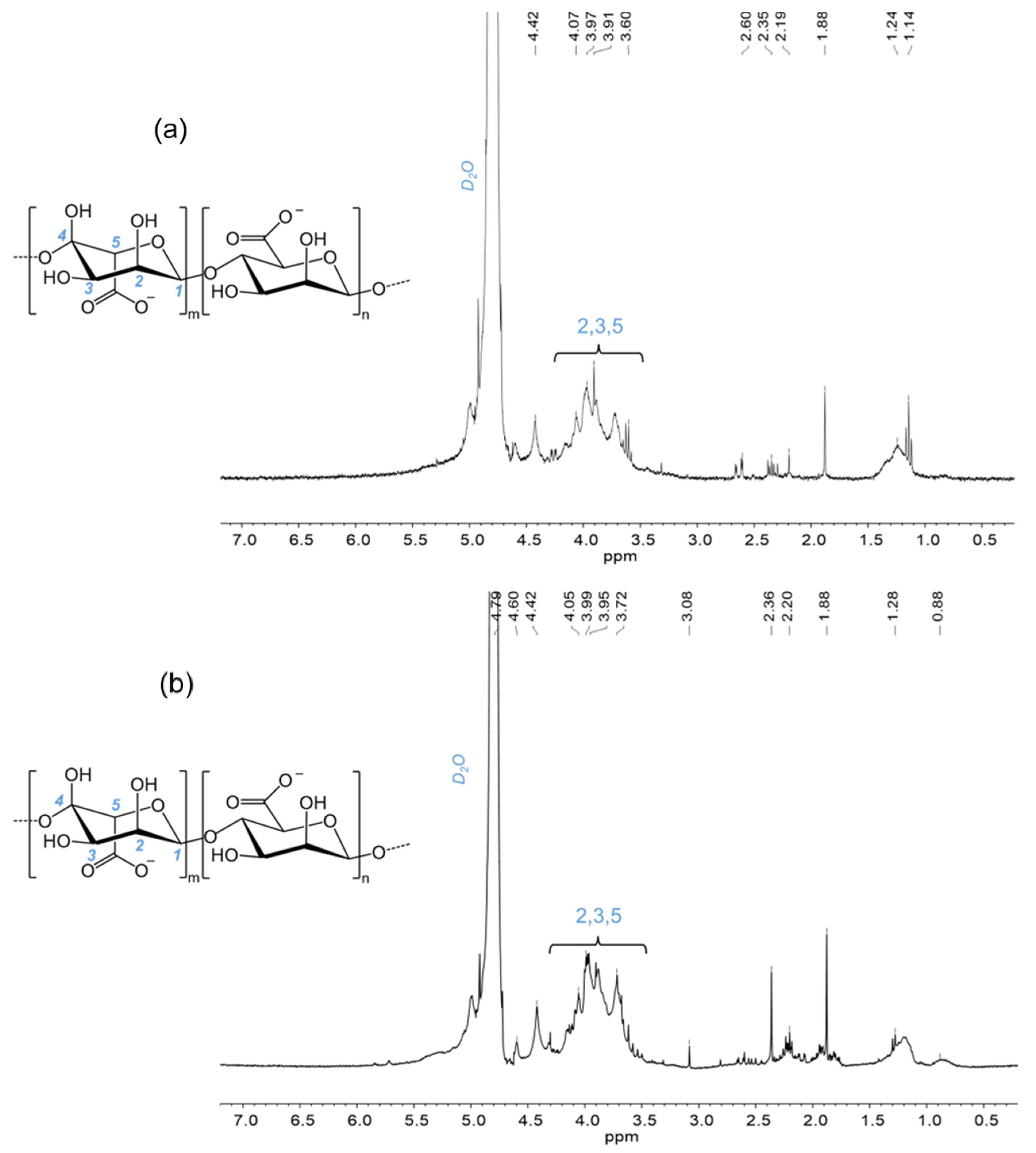
3.2.2. Nuclear Magnetic Resonance Spectroscopy (NMR)

Protocol 1. The 1H NMR spectrum (DMSO d6) of alginic acid is presented in Figure 5a. There were several characteristic signals, such as the singlet at 1.20 ppm, which corresponds to the three protons bound to the oxygen of alcohol groups. The massif signals present between 3.39 ppm and 3.89 ppm correspond to the protons bound to the carbons (C2, C3, and C5) of mannuronic and guluronic units.
Protocol 2. The 1H NMR spectrum (DMSO d6) of alginic acid is presented in Figure 5b. Several characteristic signals were observed, such as the singlet at 1.19 ppm that corresponds to the three protons bound to the oxygen of alcohol groups. The massif signals present from 3.52 ppm to 4.94 ppm are related to the protons bound to the carbons (C2, C3, and C5) of mannuronic and guluronic units.
Protocol 3. The 1H NMR spectrum (D2O) of sodium alginate is presented in Figure 6a. Several characteristic signals were identified, such as the massif signals present from 3.60 ppm to 4.15 ppm, and are attributed to the protons bound to the carbons (C2, C3, and C5) of mannuronic and guluronic units.
Protocol 4. The 1H NMR spectrum (D2O) of sodium alginate is presented in Figure 6b. Several characteristic signals were found, such as the massif signals present from 3.50 ppm to 4.16 ppm, and are attributed to the protons bound to the carbons (C2, C3, and C5) of mannuronic and guluronic units.

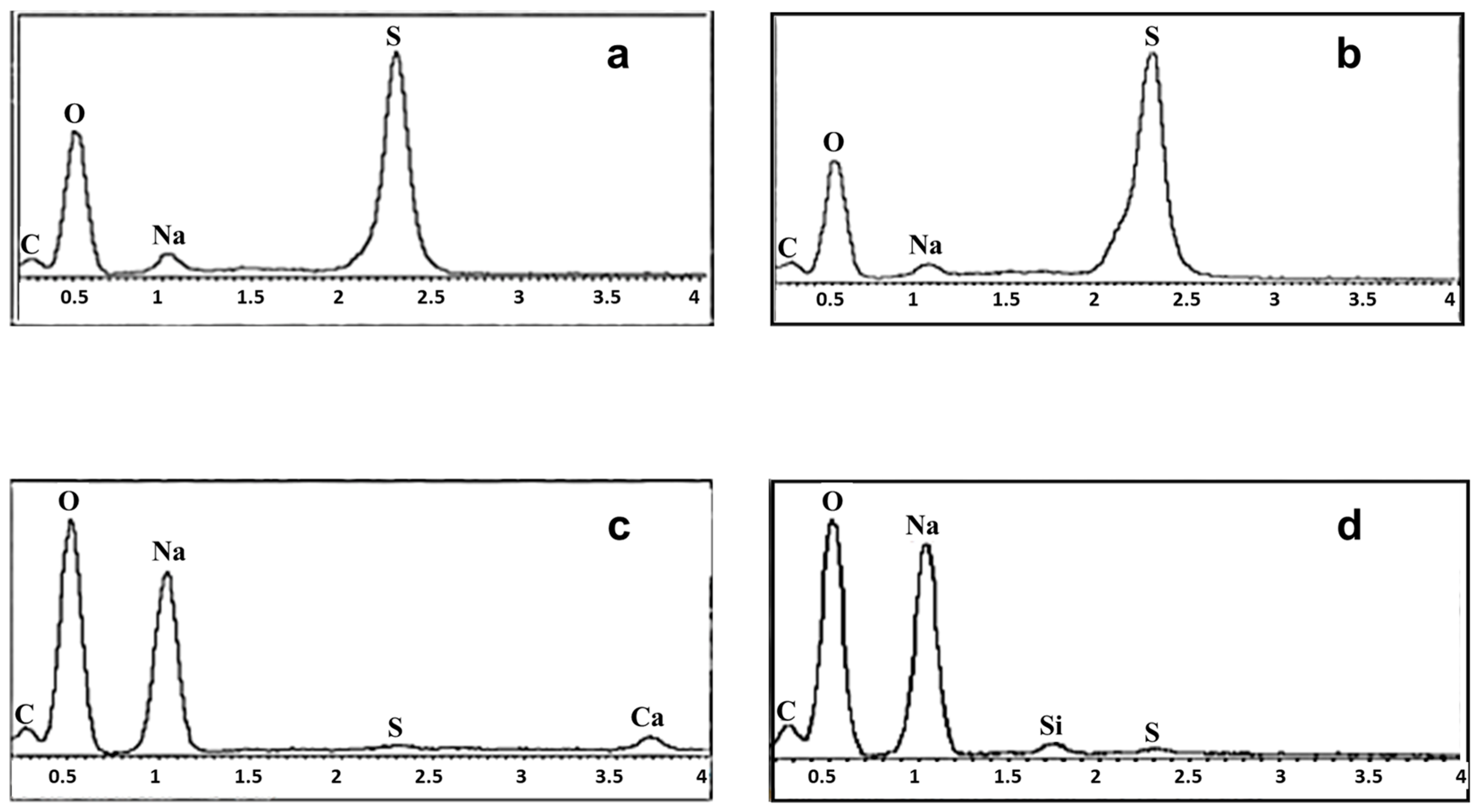
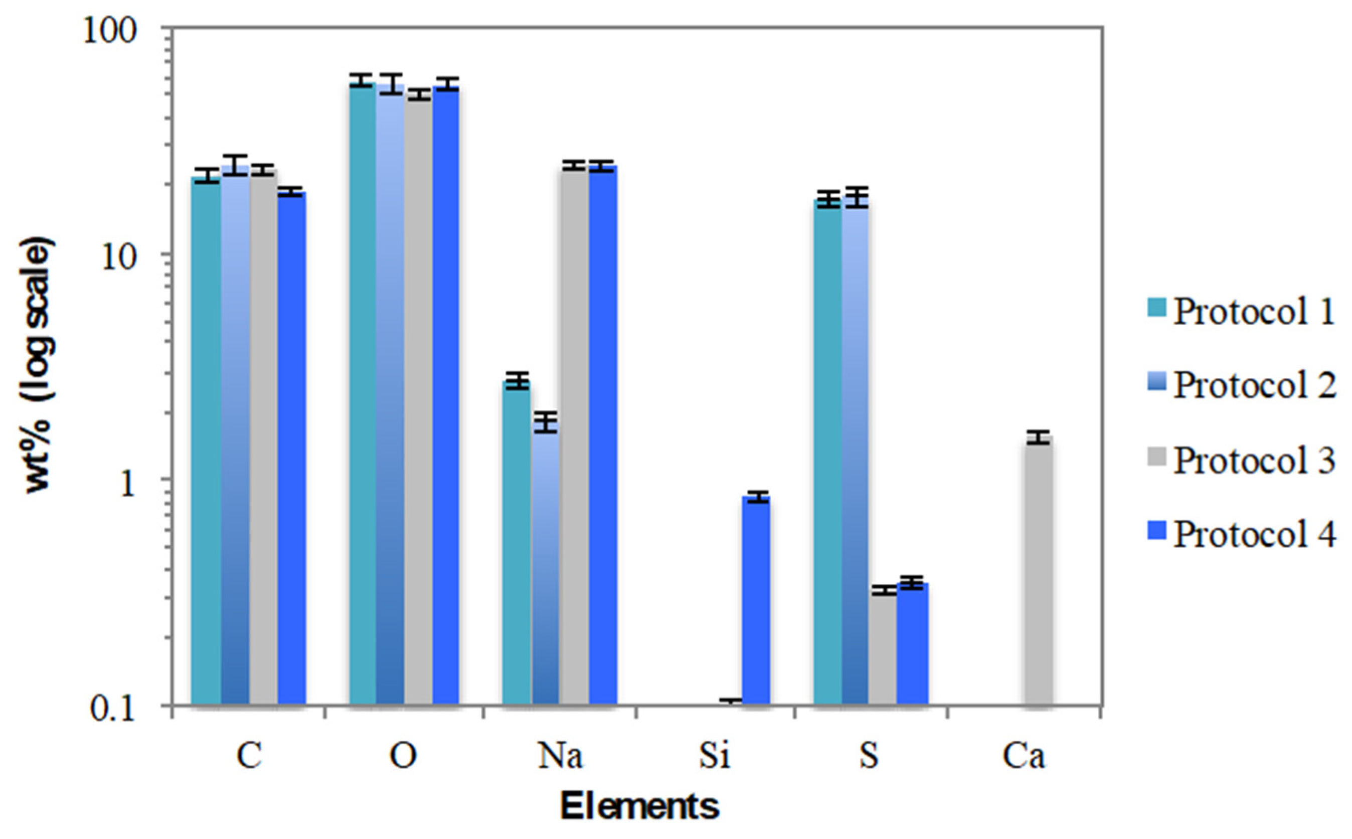
3.3. Morphological Analysis (SEM/EDX) and Elemental Composition

SEM micrographs for isolated alginates are illustrated with their corresponding energy dispersive X-ray (EDX) spectra in Figure 7 and Figure 8. The alginic acid extracted using protocols 1 and 2 exhibited similar morphologies with smooth and dense surfaces (Figure 7a,b), while sodium alginates isolated using protocols 3 and 4 had powdery and porous crystalline structures (Figure 7c,d). Such SEM observations are aligned with the visual appearance discussed above (Section 3.1).
The morphological appearance of the alginic acid extracted from protocols 1 and 2 was significantly different from the sodium alginate extracted from protocols 3 and 4, as shown in Figure 7. The EDX analyses reveal characteristic peaks of C, O, Na, S, Si, and Ca, all expected elements in the respective samples (Figure 8). The relative proportions of those main elements are shown in Figure 9. Equivalent C/O ratios were observed in all samples (approximately 0.4), suggesting homogenous and similar organic matrices. Sulphur was present in high concentrations in the alginic acid from protocols 1 and 2, which may be related to the use of H2SO4, whereas in protocols 3 and 4, the presence of lower amounts of sulfur could be attributed to the existence of fucoidan residues, a sulfated polysaccharide present in brown algae [19,25,26]. The intensity of the peaks corresponding to the sodium atom (Na) in the sodium alginate extracts synthesized using protocols 3 and 4 confirms the success of sodium alginate synthesis at the expense of alginic acid (Figure 8c,d and Figure 9).

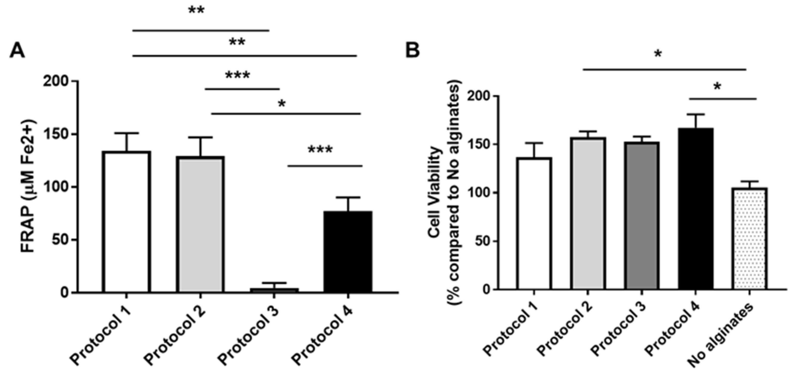
3.4. Antioxidant Capacity Analysis

In order to evaluate the antioxidant capacity of isolated alginates, we used ferric reducing antioxidant power assay (FRAP) that determinate antioxidant power as the reducing capacity of Fe+3 to Fe+2 [27]. Based on the FRAP test, the alginic acid synthesized from protocols 1 and 2 had a greater reductive power when compared to the sodium alginates from protocols 3 and 4. (Figure 10A).
Next, we evaluated if the isolated alginates protected fibroblast cells against oxidative stress-induced cell death. We found that cells treated with alginates extracted with different methods were less vulnerable against H2O2-derived toxicity. H2O2 induced cell death in fibroblasts in the absence of alginates (79.25 ± 4.82 compared to untreated cells). The rate of surveillance increased when cells were co-treated with alginates (protocol 1: 72.63 ± 4.24; protocol 2: 67.78 ± 6.44; protocol 3: 68.61 ± 6.84; protocol 4: 65.85 ± 6.68 compared to untreated conditions). We found that cell viability increased in alginate-treated cells at different levels (protocol 1: 36.8 ± 14.6; protocol 2: 57.6 ± 5.72; protocol 3: 52.6 ± 5.54; protocol 4: 67.3 ± 13.7) compared to cells unexposed to alginates (Figure 10B). These experiments indicate that the extraction methods preserved the antioxidant power in isolated alginates.

4. Discussion

The main results indicate the presence of alginates in Sargasso brown seaweed collected from various beaches in the Guadeloupe archipelago (French West Indies, FWI). The above-mentioned protocols were used to obtain yields of up to 45%. The yields of alginate extraction obtained from these processes were somewhat different. The first and second alginic acid extraction processes had higher extraction yields of 44.7% and 37.6%, respectively, while the third and fourth sodium alginate extraction processes had lower yields of 9.11% and 2.62%, respectively These yields are fully in line with those described in the literature and even slightly higher for protocols 1 and 2 [22,25,34,35,36,37,38].
The decrease in yield is closely related to the reagents used in the sodium alginate or alginic acid extraction process. Indeed, demineralization, but also removal of polyphenols before basification, can help to increase the chances of alginate extraction from brown algae. Even if the HCl used during the pre-treatment helps to coat the residues that are not needed, it can also lead to the degradation of the extracted alginate. The destruction of the ether bonds between two alginate monomers could explain this phenomenon [31]. The use of a hypochlorite solution for protocol 1 allowed the discoloration of the seaweed by removing polyphenols that can hinder the extraction of alginate because of their very strong affinity with the alginate contained in the brown seaweed [32,33]. Also, sulfuric acid in protocols 1 and 2 demineralized the brown algae, facilitating the alginate extraction process [20]. Sodium or calcium carbonate was used for the basification of the medium during the different extraction processes. These are a good alternatives because they are inexpensive and can confer a basic pH to the medium very quickly. Their presence at low concentration, i.e., 2% or 3% in the medium, maximizes the yield, as shown in a study by Mazumder et al. [34].
The four protocols developed offer the possibility to obtain alginate or alginic acid of different color and appearance while maintaining the integrity of the extracted polysaccharide. A study on the solubility of the two compounds in water had shown that when the alginic acid was dissolved in water, a viscous solution was obtained. On one hand, alginic acid has the necessary properties to form a gel in water, which allows it several applications in the alimentary area. On the other hand, alginate is much more soluble in water [39,40]. Regarding the four extraction methods, some differences were noticed in the color of the obtained products and their aspects. Particularly, the fourth extraction method is the only one in which a white dusty precipitate was obtained. With the other methods, the precipitates obtained presented a brown fibrous aspect. These color-associated features were described previously [40].
The chemical structures of both polymers were identified by 1H NMR, infrared analysis, and elemental analysis. The infrared analyses showed a significant difference between alginic acid and sodium alginate, although the peaks of basic functions such as hydroxyl, ketone, alkane, and ether groups were present in all the four samples. These results were compared with those presented in the literature [29,35,36,37], and some slight differences in the spectroscopic analyses were found, such as the peak displacements on the 1H NMR spectra or the peak intensities on the FT-IR spectra (e.g., for the bands around 3000 cm−1). These small differences could be related to the sample preparation. Despite the few differences between the four samples, the elemental analyses show a constant C/O ratio close to 0.4 for the four samples. The presence of more sodium in protocols 3 and 4 confirms the synthesis of sodium alginate.
Regarding antioxidant power, only alginate samples from protocols 1, 2, and 4 showed antioxidant properties in the FRAP analysis. Interestingly, all the samples made fibroblasts less vulnerable to oxidative stress. This suggests that, although the alginates from protocol 3 showed minimal reducing capacity of Fe+3 to Fe+2, they might be able to activate routes of protection against H2O2-induced cell death. This could be due to lower amounts of fucoidan residues in samples 3 and 4 capable of modulating the NRF2/HO)-1 signaling pathway to reduce oxidative stress [9,35,38,39].

5. Conclusions

In this study, four different alginic acid and sodium alginate extraction protocols already described in the literature were tested on Sargassum found in Guadeloupe, namely fluitans and natans. Yield, color, and solubility depended on the method used to extract the alginates. All the alginates exerted a protective role against oxidative stress in the cell cultures, although only the alginic acid and sodium alginate synthesized from protocols 1, 2, and 4 showed an antioxidant capacity in vitro. This opens up promising prospects for the use of Sargassum seaweed washed up in the French West Indies as a cheap source of alginates and as a bioresource containing bioactive molecules with antioxidant properties. Different extraction protocols could provide different alternatives for industrial, food, and pharmaceutical uses.

Author Contributions

Conceptualization, G.C.-T., L.B. and E.B.; methodology, G.C.-T., N.M., L.B., E.B., C.Y. and L.G.-M.; validation, G.C.-T., Z.B., L.B., M.S., E.B., C.Y. and L.G.-M.; formal analysis, G.C.-T., L.B., V.T., C.Y., Z.B., E.B. and L.G.-M.; investigation, N.M., V.T., S.N.-R., A.P.-M., V.D., L.G.-M. and C.Y.; resources, G.C.-T., M.S. and E.B.; data curation, G.C.-T., M.S., L.B., C.Y., S.N.-R., Z.B., V.D., E.B., L.G.-M. and A.P.-M.; writing—original draft preparation, N.M., V.T., L.B., G.C.-T., M.S., C.Y., C.O.-P., A.P.-M., E.B., L.G.-M., V.D., P.M. and Z.B.; writing—review and editing, L.B.; project administration, G.C.-T.; funding acquisition, G.C.-T., M.S., L.G.-M. and E.B. All authors have read and agreed to the published version of the manuscript.

Funding

This research was funded by the Interreg Caraïbes program (Projet SARGOOD), grant RYC2018-024434-I, and CEU-UCH IDOC23-06 (to L.G.-M.).

Data Availability Statement

The original contributions presented in the study are included in the article, further inquiries can be directed to the corresponding author.

Acknowledgments

Research project funded by the Interreg Caraïbes program (Project SARGOOD). This work was realized with the help of Patrick Daubias, working at the National School of Chemistry of Lille (ENSCL).

Conflicts of Interest

The authors declare no conflicts of interest.

References

  1. Rushdi, M.I.; Abdel-Rahman, I.A.M.; Saber, H.; Attia, E.Z.; Abdelraheem, W.M.; Madkour, H.A.; Hassan, H.M.; Elmaidomy, A.H.; Abdelmohsen, U.R. Pharmacological and natural products diversity of the brown algae genus Sargassum. RSC Adv. 2020, 10, 24951–24972. [Google Scholar] [CrossRef] [PubMed]
  2. Rosado-Espinosa, L.A.; Freile-Pelegrín, Y.; Hernández-Nuñez, E.; Robledo, D. A comparative study of Sargassum species from the Yucatan Peninsula coast: Morphological and chemical characterization. Phycologia 2020, 59, 261–271. [Google Scholar] [CrossRef]
  3. Rossignolo, J.A.; Felicio Peres Duran, A.J.; Bueno, C.; Martinelli Filho, J.E.; Savastano Junior, H.; Tonin, F.G. Algae application in civil construction: A review with focus on the potential uses of the pelagic Sargassum spp. Biomass. J. Environ. Manag. 2022, 303, 114258. [Google Scholar] [CrossRef]
  4. Marsh, R.; Kliris, N.S.; Tompkins, E.L.; Dash, J.; Dominguez Almela, V.; Tonon, T.; Oxenford, H.A.; Webber, M. Climate-sargassum interactions across scales in the tropical Atlantic. PLoS Clim. 2023, 2, e0000253. [Google Scholar] [CrossRef]
  5. Van Tussenbroek, B.I.; Arana, H.A.H.; Rodríguez-Martínez, R.E.; Espinoza-Avalos, J.; Canizales-Flores, H.M.; Gonzalez-Godoy, C.E.; Barba-Santos, M.G.; Vega-Zepeda, A.; Collado-Vides, L. Severe impacts of brown tides caused by Sargassum spp. on near-shore Caribbean seagrass communities. Mar. Pollut. Bull. 2017, 122, 272–281. [Google Scholar] [CrossRef]
  6. Resiere, D.; Valentino, R.; Nevière, R.; Banydeen, R.; Gueye, P.; Florentin, J.; Cabié, A.; Lebrun, T.; Mégarbane, B.; Guerrier, G.; et al. Sargassum seaweed on Caribbean islands: An international public health concern. Lancet 2018, 392, 2691. [Google Scholar] [CrossRef]
  7. Sissini, M.N.; de Barros Barreto, M.B.B.; Szechy, M.T.M.; de Lucena, M.B.; Oliveira, M.C.; Gower, J.; Liu, G.; de Oliveira Bastos, E.; Milstein, D.; Gusmao, F.; et al. The floating Sargassum (Phaeophyceae) of the South Atlantic Ocean—Likely scenarios. Phycologia 2017, 56, 321–328. [Google Scholar] [CrossRef]
  8. Saraswati; Giriwono, P.E.; Iskandriati, D.; Tan, C.P.; Andarwulan, N. Sargassum Seaweed as a Source of Anti-Inflammatory Substances and the Potential Insight of the Tropical Species: A Review. Mar. Drugs 2019, 17, 590. [Google Scholar] [CrossRef] [PubMed]
  9. Puhari, S.S.M.; Yuvaraj, S.; Vasudevan, V.; Ramprasath, T.; Arunkumar, K.; Amutha, C.; Selvam, G.S. Fucoidan from Sargassum wightii reduces oxidative stress through upregulating Nrf2/HO-1 signaling pathway in alloxan-induced diabetic cardiomyopathy rats. Mol. Biol. Rep. 2023, 50, 8855–8866. [Google Scholar] [CrossRef] [PubMed]
  10. Falkeborg, M.; Cheong, L.-Z.; Gianfico, C.; Sztukiel, K.M.; Kristensen, K.; Glasius, M.; Xu, X.; Guo, Z. Alginate oligosaccharides: Enzymatic preparation and antioxidant property evaluation. Food Chem. 2014, 164, 185–194. [Google Scholar] [CrossRef]
  11. Zhao, X.; Li, B.; Xue, C.; Sun, L. Effect of molecular weight on the antioxidant property of low molecular weight alginate from Laminaria japonica. J. Appl. Phycol. 2012, 24, 295–300. [Google Scholar] [CrossRef]
  12. Borazjani, N.J.; Tabarsa, M.; You, S.; Rezaei, M. Effects of extraction methods on molecular characteristics, antioxidant properties and immunomodulation of alginates from Sargassum angustifolium. Int. J. Biol. Macromol. 2017, 101, 703–711. [Google Scholar] [CrossRef]
  13. Nava-Jiménez, I.A.; Tejeda-Vega, S.; Cortina-Ramírez, G.E.; Zarazúa-Ortega, G.; Berriozabal-Islas, C.; Sánchez-Hernández, H. Macro and microelement analysis of Sargassum fluitans and Sargassum natans arriving in the coastal zone of Cancun, Quintana Roo, Mexico: Análisis de macro y microelementos de Sargassum fluitans y Sargassum natans que arriban a la zona costera de Cancún, Quintana Roo, México. Rev. Biol. Mar. Oceanogr. 2022, 57, 2022. [Google Scholar] [CrossRef]
  14. Darko, C.N.S.; Premarathna, A.D.; Humayun, S.; Agyei-Tuffour, B.; Goosen, N.J.; Tuvikene, R. Physico- and biochemical properties of alginates extracted from Ecklonia maxima and Sargassum fluitans using a simple cascade process. J. Appl. Phycol. 2023, 36, 661–674. [Google Scholar] [CrossRef]
  15. Mohammed, A.; Bissoon, R.; Bajnath, E.; Mohammed, K.; Lee, T.; Bissram, M.; John, M.N.; Jalsa, N.K.; Lee, K.Y. Ward Multistage extraction and purification of waste Sargassum natans to produce sodium alginate: An optimization approach. Carbohydr. Polym. 2018, 198, 109–118. [Google Scholar] [CrossRef]
  16. Rodríguez-Martínez, R.E.; Roy, P.D.; Torrescano-Valle, N.; Cabanillas-Terán, N.; Carrillo-Domínguez, S.; Collado-Vides, L.; García-Sánchez, M.; van Tussenbroek, B.I. Element concentrations in pelagic Sargassum along the Mexican Caribbean coast in 2018–2019. PeerJ 2020, 8, e8667. [Google Scholar] [CrossRef]
  17. Draget, K.I.; Bræk, G.S.; Smidsrød, O. Alginic acid gels: The effect of alginate chemical composition and molecular weight. Carbohydr. Polym. 1994, 25, 31–38. [Google Scholar] [CrossRef]
  18. Hernández-Carmona, G.; McHugh, D.J.; Arvizu-Higuera, D.L.; Rodríguez-Montesinos, Y.E. Pilot plant scale extraction of alginate from Macrocystis pyrifera. 1. Effect of pre-extraction treatments on yield and quality of alginate. J. Appl. Phycol. 1998, 10, 507–513. [Google Scholar] [CrossRef]
  19. Lorbeer, A.J.; Lahnstein, J.; Bulone, V.; Nguyen, T.; Zhang, W. Multiple-response optimization of the acidic treatment of the brown alga Ecklonia radiata for the sequential extraction of fucoidan and alginate. Bioresour. Technol. 2015, 197, 302–309. [Google Scholar] [CrossRef]
  20. Perez, R.; Kaas, R.; Campello, F.; Arbault, S.; Barbaroux, O. La Culture des Algues Marines Dans le Monde; Institut Français de Recherche pour l’Exploitation de la Mer, Ed.; IFREMER: Plouzané, France, 1992. [Google Scholar]
  21. Calumpong, H.P.; Maypa, A.P.; Magbanua, M. Population and alginate yield and quality assessment of four Sargassum species in Negros Island, central Philippines. In Proceedings of the Sixteenth International Seaweed Symposium, Cebu City, Philippines, 12–17 April 1998; Kain, J.M., Brown, M.T., Lahaye, M., Eds.; Springer: Dordrecht, The Netherlands, 1999; pp. 211–215. [Google Scholar] [CrossRef]
  22. Fenoradosoa, T.A.; Ali, G.; Delattre, C.; Laroche, C.; Petit, E.; Wadouachi, A.; Michaud, P. Extraction and characterization of an alginate from the brown seaweed Sargassum turbinarioides Grunow. J. Appl. Phycol. 2010, 22, 131–137. [Google Scholar] [CrossRef]
  23. Mian, A.J.; Percival, E. Carbohydrates of the brown seaweeds himanthalia lorea, bifurcaria bifurcata, and Padina pavonia. Carbohydr. Res. 1973, 26, 133–146. [Google Scholar] [CrossRef]
  24. Souchet, N. Etude de L’influence de la Periode de Recolte de L’algue Brune Laminaria longicruris sur sa Composition en Polysaccharides Bioactifs. Ph.D. Thesis, Université Laval, Quebec City, QC, Canada, 2004. [Google Scholar]
  25. Rioux, L.-E.; Turgeon, S.L.; Beaulieu, M. Characterization of polysaccharides extracted from brown seaweeds. Carbohydr. Polym. 2007, 69, 530–537. [Google Scholar] [CrossRef]
  26. Hu, H.; Xie, Y.; Li, Y.; Li, L.; Tian, N.; Zhu, W.; Yan, G.; Wu, C. Development of high drug-loading nanomicelles targeting steroids to the brain. IJN 2013, 9, 55. [Google Scholar] [CrossRef]
  27. Pereira, R.; Tojeira, A.; Vaz, D.C.; Mendes, A.; Bártolo, P. Preparation and Characterization of Films Based on Alginate and Aloe Vera. Int. J. Polym. Anal. Charact. 2011, 16, 449–464. [Google Scholar] [CrossRef]
  28. Daemi, H.; Barikani, M. Synthesis and characterization of calcium alginate nanoparticles, sodium homopolymannuronate salt and its calcium nanoparticles. Sci. Iran. 2012, 19, 2023–2028. [Google Scholar] [CrossRef]
  29. Chale-Dzul, J.; de Vaca, R.P.-C.; Quintal-Novelo, C.; Olivera-Castillo, L.; Moo-Puc, R. Hepatoprotective effect of a fucoidan extract from Sargassum fluitans Borgesen against CCl4-induced toxicity in rats. Int. J. Biol. Macromol. 2020, 145, 500–509. [Google Scholar] [CrossRef]
  30. Ptak, S.H.; Sanchez, L.; Fretté, X.; Kurouski, D. Complementarity of Raman and Infrared spectroscopy for rapid characterization of fucoidan extracts. Plant Methods 2021, 17, 130. [Google Scholar] [CrossRef] [PubMed]
  31. Soto-Vásquez, M.R.; Alvarado-García, P.A.A.; Youssef, F.S.; Ashour, M.L.; Bogari, H.A.; Elhady, S.S. FTIR Characterization of Sulfated Polysaccharides Obtained from Macrocystis integrifolia Algae and Verification of Their Antiangiogenic and Immunomodulatory Potency In Vitro and In Vivo. Mar. Drugs 2023, 21, 36. [Google Scholar] [CrossRef]
  32. Leal, D.; Matsuhiro, B.; Rossi, M.; Caruso, F. FT-IR spectra of alginic acid block fractions in three species of brown seaweeds. Carbohydr. Res. 2008, 343, 308–316. [Google Scholar] [CrossRef]
  33. Jauregui-Rincon, J.; Antonio, J.; Medina-Ramirez, I. Zimm-Bragg Model Applied to Sorption of Dyes by Biopolymers: Alginic Acid and Xanthan. In Biotechnology of Biopolymers; Elnashar, M., Ed.; InTech: London, UK, 2011. [Google Scholar] [CrossRef]
  34. Haug, A.; Larsen, B.; Samuelsson, B.; Sjövall, J.; Munch-Petersen, J. The Solubility of Alginate at Low pH. Acta Chem. Scand. 1963, 17, 1653–1662. [Google Scholar] [CrossRef]
  35. Myklestad, S.; Haug, A. Studies on the solubility of alginic acid from Ascophyllum Nodosum at low pH. In Proceedings of the Fifth International Seaweed Symposium, Halifax, UK, 25–28 August 1965; Elsevier: Amsterdam, The Netherlands, 1966; pp. 297–303. [Google Scholar] [CrossRef]
  36. Awad, H.; Aboul-Enein, H.Y. A Validated HPLC Assay Method for the Determination of Sodium Alginate in Pharmaceutical Formulations. J. Chromatogr. Sci. 2013, 51, 208–214. [Google Scholar] [CrossRef] [PubMed]
  37. Hanjabam, M.D.; Kumar, A.; Tejpal, C.S.; Krishnamoorthy, E.; Kishore, P.; Ashok Kumar, K. Isolation of crude fucoidan from Sargassum wightii using conventional and ultra-sonication extraction methods. Bioact. Carbohydr. Diet. Fibre 2019, 20, 100200. [Google Scholar] [CrossRef]
  38. Yuan, Y.; Macquarrie, D. Microwave assisted extraction of sulfated polysaccharides (fucoidan) from Ascophyllum nodosum and its antioxidant activity. Carbohydr. Polym. 2015, 129, 101–107. [Google Scholar] [CrossRef] [PubMed]
  39. Saji, S.; Hebden, A.; Goswami, P.; Du, C. A Brief Review on the Development of Alginate Extraction Process and Its Sustainability. Sustainability 2022, 14, 5181. [Google Scholar] [CrossRef]
  40. McHugh, D.J. A Guide to the Seaweed Industry; FAO Fisheries Technical Paper, No. 441; Food and Agriculture Organization of the United Nations: Rome, Italy, 2003. [Google Scholar]
Figure 1. Alginic acid (a) and sodium alginate (b).
Separations 11 00226 g001
Figure 2. Isolated alginic acid (a,b) and alginate (c,d).
Separations 11 00226 g002
Figure 3. Infrared spectra of alginic acid obtained from (a) protocol 1 and (b) protocol 2.
Separations 11 00226 g003
Figure 4. Infrared spectra of alginate samples obtained from (a) protocol 3 and (b) protocol 4.
Separations 11 00226 g004
Figure 5. 1H NMR spectra of alginic acid samples in DMSO d6 obtained from (a) protocol 1 and (b) protocol 2.
Separations 11 00226 g005
Figure 6. 1H NMR spectra of sodium alginate samples in D2O obtained from (a) protocol 3 and (b) protocol 4.
Separations 11 00226 g006
Figure 7. SEM micrographs of alginic acid extracted from (a) protocol 1 and (b) protocol 2, and alginate extracted from (c) protocol 3 and (d) protocol 4.
Separations 11 00226 g007
Figure 8. EDX of alginic acid (a,b) and sodium alginate (c,d).
Separations 11 00226 g008
Figure 9. Elemental composition of samples 1 to 4 of alginic acid (protocols 1 and 2) and sodium alginate (protocols 3 and 4) determined via EDX measurements. Analysis shows error bars with ±S.D. from the mean (n = 3).
Separations 11 00226 g009
Figure 10. In vitro antioxidant activity in isolated alginates of Sargassum. (A) FRAP and (B) cell viability analysis data are showed. The vertical bar represents the mean and standard error of the mean for each protocol in three independent experiments. * p < 0.05, ** p < 0.01, *** p < 0.001 (Student’s t test).
Separations 11 00226 g010
Disclaimer/Publisher’s Note: The statements, opinions and data contained in all publications are solely those of the individual author(s) and contributor(s) and not of MDPI and/or the editor(s). MDPI and/or the editor(s) disclaim responsibility for any injury to people or property resulting from any ideas, methods, instructions or products referred to in the content.

