Article

Rapid Determination of Rivaroxaban by Using Terahertz Metamaterial Biosensor

Shanghai Key Lab of Modern Optical System, Terahertz Spectrum and Imaging Technology National Cooperative Innovation Center, Terahertz Technology Innovation Research Institute, University of Shanghai for Science and Technology, Shanghai 200093, China
*
Author to whom correspondence should be addressed.
Photonics 2024, 11(9), 814; https://doi.org/10.3390/photonics11090814
Submission received: 13 August 2024 / Revised: 23 August 2024 / Accepted: 28 August 2024 / Published: 29 August 2024
(This article belongs to the Section Optical Interaction Science)

Abstract

:
Rivaroxaban, a direct oral anticoagulant, is widely used in the management and prevention of thrombotic conditions. Dose adjustments are necessary to optimize efficacy based on individual physiological differences. However, current analytical methods are impractical for clinical use due to complex sample preparation and lengthy detection times. This paper presents a terahertz (THz) metamaterial biosensor for the rapid determination of rivaroxaban within 10–15 min. The THz absorption peaks of rivaroxaban were first identified based on THz spectroscopy. Subsequently, a metamaterial structure with rotational symmetry was designed to resonate at the absorption peaks of rivaroxaban. Theoretical simulations and experimental measurements analyzed changes of the resonance peak at different rivaroxaban concentrations, including frequency shifts and amplitude variations. Based on these changes, rivaroxaban concentration can be quantified with the limits of detection (LODs) of 5.01 μmol/mL for peak shift and 1.067 μmol/mL for peak absorbance, respectively. This study presents a novel approach for the rapid determination of rivaroxaban, providing potential improvements in therapeutic drug monitoring and personalized medical treatment.

Graphical Abstract

1. Introduction

Oral anticoagulant rivaroxaban (C19H18ClN3O5S) can directly inhibit factor Xa, an important intersection in the coagulation process, and is responsible for converting plasminogen (factor II) into thrombin (factor IIa) [1,2]. With its high bind ability for factor Xa [3] (92–95%), rivaroxaban can effectively inhibit the activity of factor Xa and consequently curtails thrombin generation [4,5]. Compared with conventional oral anticoagulants (e.g., heparin and warfarin), rivaroxaban not only exhibits a potent anticoagulant effect [6,7] but also offers advantages including a fast absorption rate (reaching peak blood concentration within 2–4 h [8]), high tolerance, and safety [9]. These advantages have led to its wide application in the prevention and treatment of thromboembolisms [10], such as deep venous thrombosis, pulmonary embolism, and intra-atrial thrombus diseases [11]. However, the therapeutic efficacy of rivaroxaban is closely related to its blood concentration, necessitating the consideration of individual physiological differences [8]. Therefore, monitoring the rivaroxaban concentration is essential for assessing bleeding risk, as well as promoting the safe and effective use of the drug, especially for elderly patients with hepatic or renal insufficiency [12,13]. Given these needs, there is an urgent need for a rapid, accurate, and cost-effective method to detect rivaroxaban in clinical applications.
The detection methods of rivaroxaban are typically classified as indirect or direct. Indirect methods include the prothrombin time (PT) method [14,15] and the anti-Factor Xa assay [16,17]. The PT method indirectly reflects the blood rivaroxaban concentration by measuring the PT value [18] but may be affected by different prothrombin-activators [19,20] and systemic diseases (e.g., hepatic damage, vitamin K deficiency, etc.) [19]. The anti-Factor Xa assay indirectly measures blood rivaroxaban concentrations by evaluating the factor Xa activity. This method has good linearity [21] with a broad range (20–660 ng/mL) [22], but some studies have suggested a potential overestimation of results [8,23]. Direct methods include high-performance liquid chromatography (HPLC) [24,25] and liquid chromatography−mass spectrometry (LC-MS/MS) [26,27]. They are known for their high sensitivity and precision in determining rivaroxaban concentration [26,28], but they require complex sample preparation and a long analysis time (several hours) [29]. Therefore, there is a need for an accurate, simple, and cost-effective method to meet the demand for the rapid and efficient determination of rivaroxaban in clinical applications.
THz waves (0.1–10 THz) occupy a unique position in the electromagnetic spectrum, between microwaves and infrared waves [30,31,32]. They resonate with target analytes, resulting in characteristic THz absorption peaks [33]. Moreover, THz waves are less harmful to tissues and biomolecules compared to X-rays due to their lower photon energy [34] and non-ionizing reactions [35]. Therefore, the fingerprint properties and non-destructive nature of THz waves render it highly promising for biomedical detection applications [36,37,38,39,40]. However, THz detection technology faces challenges due to the mismatch between THz wavelength and target analyte size. This causes the absorption peak to become weaker, which limits the detection sensitivity in trace analysis [41,42,43]. Metamaterials offer a solution to this challenge by enabling surface plasmon resonance (SPR) through selecting suitable materials and designing periodic unit structures [44]. SPR can enhance the interaction between analytes and THz waves. The combination of THz technology with metamaterials has recently shown promise, particularly for the detection of trace drug molecules [45,46,47].
This paper introduces a novel method that combines the THz spectroscopy and metamaterial for a highly sensitive detection of rivaroxaban (a widely used anticoagulant). Firstly, we qualitatively examined the absorption peaks of rivaroxaban and used them as the basis for designing a metamaterial structure comprising periodic rotationally symmetric four split-ring resonator. Next, we fabricated the metamaterial biosensor through photolithography and observed that its experimental resonance peak at 2.34 THz closely matched the absorption peaks of rivaroxaban located at 2.28 THz and 2.47 THz. Subsequently, quantitative analysis of rivaroxaban solutions with varying concentrations revealed relationships between changes in the resonance peak (frequency and absorbance) and the concentration of rivaroxaban. The limit of detection (LOD) of this method was determined to be at the level of 1–5 µmol/mL, meeting clinical standards for rivaroxaban analysis (11.47–821 µmol/mL). The results show this method offers a highly sensitive and low-LOD alternative for trace drug analysis.

2. Materials and Methods

2.1. Materials

The rivaroxaban (purity ≥ 99%) used in this study was purchased from Sigma-Aldrich (St. Louis, MO, USA), and cyclic olefin copolymer (COC), with an average particle size below 60 µm, was purchased from the Shanghai Institute of Nuclear Research (Shanghai, China). To ensure sample stability, the rivaroxaban samples were stored under sealed conditions at 2–8 °C until use. Deionized water was obtained from an ELGA Purelab Classic ultrapure water system (ELBA LabWater, Lane End, UK).

2.2. THz Spectroscopy

2.2.1. Experimental Equipment

A THz spectroscopy system (TAS7400; ADVANTEST CORPORATION, Tokyo, Japan) was used to acquire THz spectra, with an effective frequency range of 0.5–4.5 THz, a signal dynamic range of 60 dB, and a spectral resolution of 1.9 GHz, and each spectrum was scanned 512 times (the integration time is 2–3 min). In the sample chamber, the temperature was maintained at 20 ± 2 °C by using an air conditioner, and the relative humidity was maintained at 3% by introducing dry nitrogen. Both the temperature and relative humidity were recorded using a commercially available temperature and humidity recorder.

2.2.2. Rivaroxaban Qualitative Measurement

Rivaroxaban powder was mixed with COC at a weight ratio of 10:70, and the mixture was compressed into a pellet with a 13 mm diameter and 0.7 ± 0.01 mm thickness under 8 tons. In this process, mass loss was controlled within 1%.
The resulting rivaroxaban pellets were qualitatively analyzed by a THz spectrometer over a spectral range of 0–5 THz. Each spectrum was scanned 512 times, taking 2–3 min. The measurement was repeated five times for each pellet.

2.2.3. Rivaroxaban Quantitative Measurement

For quantitative measurements, rivaroxaban solutions were prepared by dissolving varying weights of rivaroxaban in 500 μL of deionized water. The target concentration falls within the therapeutic range of 13.7–548.3 μmol/mL, as reported in the literature about venous thromboembolism patients receiving a 20 mg dose [12]. Therefore, the rivaroxaban concentrations were set at 0, 5, 7.5, 10, 12.5, 15, 20, 25, 30, 50, and 100 μmol/mL.
In the THz measurement, 10 µL of each solution was pipetted onto the surface of a metamaterial biosensor and then dried under vacuum (taking 5 min). The dried samples formed a uniform thin film on the biosensor, which was then measured in the THz spectrometer to acquire the spectra. Each spectrum was scanned 512 times, taking 2–3 min. The measurement was repeated five times for each sample.

2.3. Manufacturing and Microscopic Imaging for THz Metamaterial Biosensor

The THz metamaterial biosensor was fabricated via standard photolithography on a 500 μm thick silica substrate. The periodic unit pattern was etched onto the substrate, followed by the deposition of a 5 nm chromium adhesion layer and a 50 nm gold layer using magnetron sputtering. The biosensor was then treated with a dispergator at 80 °C for 2 h to remove the excess photoresist.
The surface morphology of the biosensor was observed and imaged using an Axio Imager M2 optical microscope (ZEISS, Oberkochen, Germany), employing a 100× objective lens and a 12 V 100 W halogen lamp as the illumination source.

2.4. Data Processing and Analysis

Data processing and analysis were performed using ORIGIN 2021b software and a Python algorithm. Firstly, the five spectra for each sample were averaged within the Python algorithm, with the error bar being in the order of magnitude of 10−2. The 0.9–4.5 THz range was chosen for analysis to mitigate noise in the 0.5–0.9 THz range. And there are 1889 data in the range of 0.9–4.5 THz. The algorithm also calculated the frequency shifts and absorbance changes of the resonance peak at various rivaroxaban concentrations. These results were imported into ORIGIN 2021b for nonlinear fitting to establish the relationships between frequency shift, absorbance, and concentration. The spectral data from the raw biosensor served as a baseline, subtracted from the sample spectra to isolate concentration-dependent effects.

3. Results and Discussion

3.1. Analysis of THz Absorption Spectrum of Rivaroxaban

The metamaterial biosensor presented in this study was designed based on the THz absorption peaks of rivaroxaban. The molecular structure of rivaroxaban—which consists of a benzene ring; an S-five-membered ring; an N, O-five-membered ring; and an N, O-six-membered ring—is shown in Figure 1a. The THz waves resonate with rivaroxaban molecules to generate absorption peaks. Figure 1b illustrates seven characteristic absorption peaks of rivaroxaban, located at 1.06 THz, 1.45 THz, 2.28 THz, 2.47 THz, 2.87 THz, 3.52 THz, and 4.11 THz, respectively. Combined with the above result and the relevant literature [8], we then designed a metamaterial to enhance the absorption peaks at 2.28 THz and 2.47 THz.

3.2. Design and Simulation of THz Metamaterial Biosensor

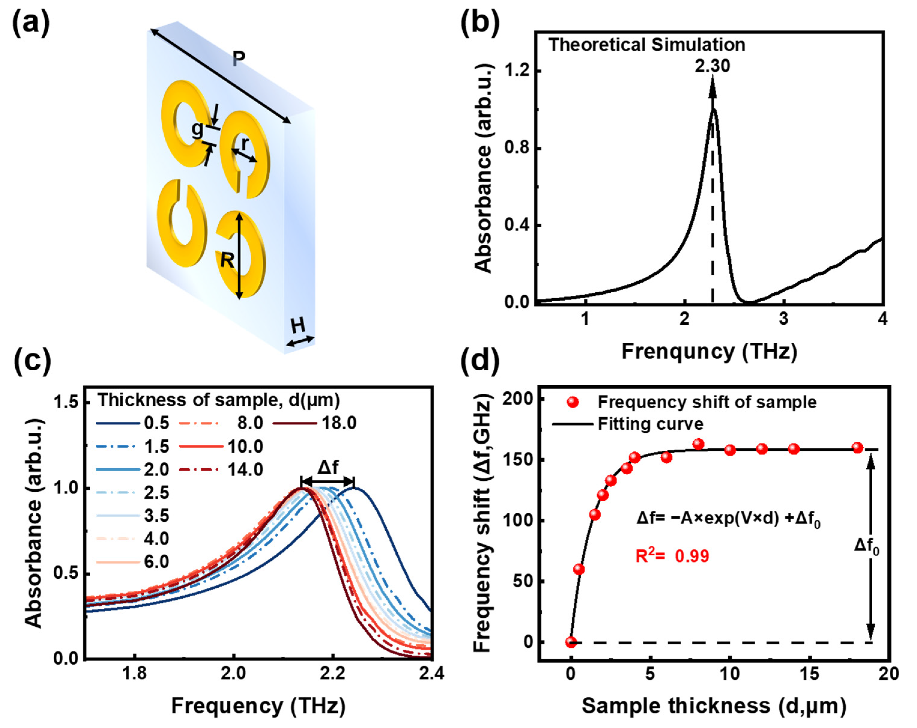
The metamaterial biosensor unit structure used was designed using COMSOL Multiphysics 5.4 software. A perfectly matched layer was applied to the top and bottom of the simulation model to absorb excitation modes and higher-order reflections. Floquet’s periodical boundary condition was used in both the x- and y-directions of the model, while an open boundary condition was used in the z-direction, parallel to the wave propagation direction. Figure 2a illustrates a 3D view of the metamaterial unit model, which comprises a silica substrate with a thickness (H) of 500.00 µm, and gold patterns with a period of 40.00 µm (P) and a thickness of 50 nm. Within the periodic unit, four split-ring resonators were designed with an inner diameter of 9.00 µm (r), an outer diameter of 16.60 µm (R), and a gap width of 3.00 µm (g). To improve computational efficiency, the gold was modeled as a perfect electrical conductor, and it would not cause the deformation of the target molecule for its stable chemical and physical properties. The theoretical spectrum of this metamaterial, shown in Figure 2b, peaks at 2.30 THz, aligning well with the absorption peaks of rivaroxaban at 2.28 THz and 2.47 THz. Consequently, it enables an enhancement in the interaction between rivaroxaban with THz waves at these frequencies.
Subsequently, we conducted simulations to study the effect of different sample thickness on the THz resonance peak. Sample thickness can represent sample concentration. According to the equation m = ρ V = ρ S d , when the sample density ( ρ ) and sample area ( S ) are constant, the sample mass ( m ) is directly proportional to the sample thickness ( d ). Experimentally, 10 µL of rivaroxaban solution was dropped onto the metamaterial surface and then dried to form a thin film. A thicker film contains more rivaroxaban from the same solution volume, equating to a higher concentration. On the other hand, the metamaterial biosensor is highly sensitive to dielectric constant changes near its surface [48]. When a rivaroxaban film forms on the biosensor surface, the dielectric constant remains unchanged, but concentration variations modify the dielectric environment in the longitudinal space near the surface. This modification affects the spatial distribution of the plasmon wave within the gold rings, consequently changing the intensity and frequency of the THz wave. Therefore, it allows us to sensitively detect the samples with different thickness. The simulation results are shown in Figure 2c. As the sample thickness increases from 0.5 µm to 18 µm, the resonance peak shifts toward lower frequencies. Additionally, we conducted a quantitative analysis of the frequency shift and further developed a theoretical nonlinear saturation model. This model correlates the frequency shift with sample thickness, providing a theoretical function as follows:
Δ f = A × exp ( V × d ) + Δ f 0
where d represents the sample thickness, ∆f is the frequency shift of the metamaterial, and ∆f0 is the difference between the maximum and minimum frequency shift. A is a scaling coefficient approximately equal to the absolute value of ∆f0, and V is the rate of curve saturation. A larger V indicates a quicker approach to saturation. The fitting quality is characterized using the determination coefficient R2.
Figure 2d displays the fitting curve for samples with different thickness. As sample thickness increases, the increment of the frequency shift gradually decreases, eventually approaching 0. By substituting the specific frequency shifts into Equation (1), the relationship between frequency shift and sample thickness is obtained as follows:
Δ f = ( 154.413 ± 3.806 ) × exp [ ( 0.73 ± 0.039 ) d ] + ( 158.54 ± 1.61958 ) , R 2 = 0.99
The fitting results show a determination coefficient of 0.99, indicating that the model has a high accuracy. By calculating the frequency shift ∆f, we can determine the sample thickness d, which subsequently enables the determination of the sample concentration. Therefore, THz spectroscopy can sensitively detect sample concentrations with the assistance of the metamaterial.

3.3. Quantitative Detection of Rivaroxaban Solutions Based on THz Metamaterial Biosensor

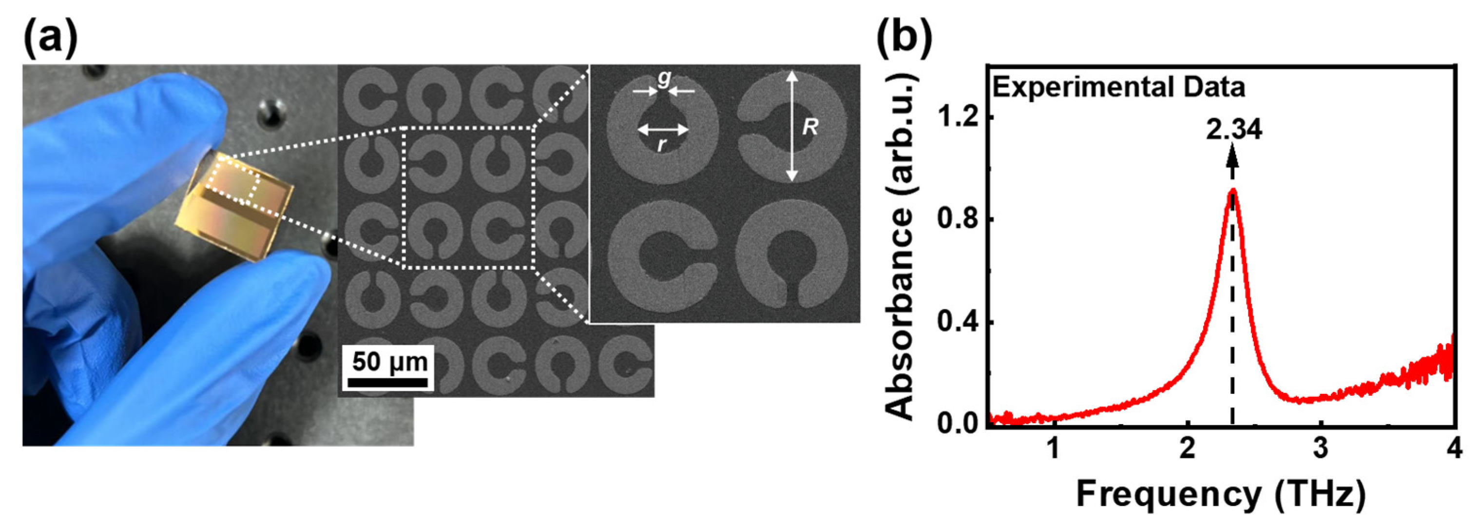
Subsequently, we used standard photolithography to fabricate the metamaterial model, as shown in Figure 2a. A 5 nm chromium layer was added as an adhesion layer between the gold split-ring resonators and the silica substrate. Figure 3a presents the digital photo and optical images of the THz metamaterial biosensor. For each resonator, the gap width is g = 2.53 µm, the inner diameter is r = 7.56 µm, and the outer diameter is R = 17.61 µm. The actual sizes differ from the theoretical sizes by a maximum of 6.1%. Figure 3b shows an experimental resonance peak at 2.34 THz, closely matching the theoretical result of 2.30 THz, with an error below 1.7%. This slight variation is contributed to by differences in actual and theoretical sizes, as well as the imperfect conductivity of the gold layer in experiments compared to theoretical simulations.
In quantitative experiments, rivaroxaban solutions of different concentrations were dropped onto the biosensor surface and dried to form a uniform film. This resulted in longitudinal distributions of the dielectric environment on the biosensor due to the presence of different amounts of rivaroxaban. THz spectroscopy was used for measurements, and the resulting THz spectra are shown in Figure 4a. To clearly demonstrate the spectral changes at different concentrations, we zoomed in on the spectra within the range of 2.1–2.4 THz, as shown in Figure 4b. It is obvious that as the sample concentration increases, the resonance peak of the biosensor shifts toward lower frequencies.
To further study the quantitative relationship between frequency shift and concentration, we extracted the frequency shifts at different rivaroxaban concentrations and performed an exponential fit according to Equation (1). The resulting fitting curve is shown in Figure 4c. The corresponding equation is as follows:
Δ f = ( 159.956 ± 11.471 ) × exp [ ( 0.021 ± 0.003 ) M ] + ( 141.814 ± 12.53 ) , R 2 = 0.99
where ∆f corresponds to the frequency shift, and M represents the rivaroxaban concentration. The results indicate that the frequency shift increases with the rivaroxaban concentration, stabilizing after reaching a certain threshold. This stabilization is attributed to the diminishing change in the overall equivalent capacitance at the biosensor surface as the sample distance increases, resulting in a slower frequency shift change. The determination coefficient R2 reaches 0.99, confirming the consistency of the frequency shift–rivaroxaban concentration relationship with the nonlinear saturation model.
Furthermore, to assess the detection properties of this method, we calculated the LOD for rivaroxaban with this biosensor. The LOD is calculated as Equation (4):
L O D = 3.3 × S D K
where SD refers to the response deviation, which is the standard deviation of the resonance peak for the raw metamaterial biosensor (0 mmol/L). K represents the maximum slope of the fitting curve. An SD value of 0.005 was obtained by averaging the spectra of the repeated tests with the raw biosensor. K was obtained from the maximum slope of the curve, with a value of 3.294 × 10⁻³. Consequently, the LOD is 5.01 μmol/mL based on the frequency shift.
Furthermore, we found that the peak absorbance changes with the rivaroxaban concentration, as depicted in Figure 4b. It is evident that as the concentration increases, the peak absorbance decreases. Similarly, we extracted the absorbance at different rivaroxaban concentrations to establish the relationship between concentration and absorbance, as shown in Figure 4d. The fitting equation is as follows:
Δ A = ( 0.161 ± 0.014 ) × exp [ ( 0.060 ± 0.009 ) M ] + ( 0.76 ± 0.011 ) , R 2 = 0.95
where ΔA corresponds to the changes in the peak absorbance, and M corresponds to the rivaroxaban concentration. The results indicate that the peak absorbance decreases with increasing concentration and stabilizes after reaching a certain level. Additionally, the LOD was calculated as 1.067 μmol/mL based on the absorbance–concentration relationship.
Based on the above analysis, quantitative studies of rivaroxaban can be conducted with the frequencies and absorbances measured at different concentrations. The limit of detection (LOD) can be as low as 1 µmol/mL, allowing for the accurate detection of samples at and above this concentration. By purifying the blood (taking 15–20 min), proteins and other components can be effectively removed, resulting into a sample for THz detection. Although there may still be traces of residual components in the treated solution, these residual components cause only a slight shift in the resonance frequency of the metamaterial. However, this frequency change does not significantly affect the amplitude of the frequency shift that reflects changes in rivaroxaban concentration. In fact, it introduces only an initial offset in the frequency shift without affecting the overall nonlinear model trend and analysis accuracy. This method meets the clinical requirement for the concentration detection of rivaroxaban, which is within the range of 11.47–821 µmol/mL. This method is fast (10–15 min per sample) and non-destructive. Therefore, the results demonstrate the feasibility of rapidly and accurately detecting drug concentration using the nonlinear saturation model of the metamaterial biosensor, indicating its potential application in clinical medicine.

4. Conclusions

This study proposes a novel detection method that combines a metamaterial biosensor with THz spectroscopy. Firstly, THz spectroscopy was applied to obtain the characteristic spectrum of rivaroxaban, identifying its absorption peaks. Subsequently, a metamaterial structure with periodic four split-ring resonators was designed. The theoretical resonance peak at 2.30 THz and the experimental peak at 2.34 THz, match well with the absorption peaks of rivaroxaban (2.28–2.47 THz). Theoretical simulations were conducted to analyze the peak frequency response to sample thickness, leading to the development of a nonlinear saturation model that describes this relationship. Finally, the biosensor was employed to detect rivaroxaban solutions of varying concentrations. The results revealed a saturated correlation between frequency shift and concentration, as well as between absorbance and concentration. The corresponding LODs were calculated to be 5.01 μmol/mL and 1.067 μmol/mL, respectively, meeting clinical requirements for rivaroxaban detection. Further optimization of the metamaterial structure is anticipated to enhance sensitivity and specificity. This study confirms that the combination of a metamaterial biosensor with THz spectroscopy is a promising approach for the concentration detection of drugs, providing a potential tool for future non-destructive, rapid, and quantitative analysis of blood rivaroxaban concentration.

Author Contributions

Methodology, X.W.; software, J.W. and X.H.; formal analysis, X.H.; investigation, J.W. and X.H.; resources, Y.P.; writing—original draft preparation, X.H.; writing—review and editing, X.W.; visualization, X.H.; supervision, Y.P. and X.W.; project administration, Y.P. and X.W.; funding acquisition, Y.P. All authors have read and agreed to the published version of the manuscript.

Funding

This work was supported by the National Natural Science Foundation of China (61805140, 62335012, 61988102) and the National Key Research and Development Program of China (2022YFA1404004, 2023YFF0719200).

Institutional Review Board Statement

Not applicable.

Informed Consent Statement

Not applicable.

Data Availability Statement

Data underlying the results presented in this paper are not publicly available at this time but may be obtained from the authors upon reasonable request.

Conflicts of Interest

There are no conflicts of interest to declare.

References

  1. Zhang, Y.-M.; Liu, J.-Y.; Sun, X.-D.; Zhang, M.; Liu, X.-G.; Chen, X.-L. Rivaroxaban improves hidden blood loss, blood transfusion rate and reduces swelling of the knee joint in knee osteoarthritis patients after total knee replacement. Medicine 2018, 97, e12630. [Google Scholar] [CrossRef] [PubMed]
  2. Mueck, W.; Schwers, S.; Stampfuss, J. Rivaroxaban and other novel oral anticoagulants: Pharmacokinetics in healthy subjects, specific patient populations and relevance of coagulation monitoring. Thromb. J. 2013, 11, 10. [Google Scholar] [CrossRef] [PubMed]
  3. Perzborn, E.; Roehrig, S.; Straub, A.; Kubitza, D.; Misselwitz, F. The discovery and development of rivaroxaban, an oral, direct factor Xa inhibitor. Nat. Rev. Drug Discov. 2011, 10, 61–75. [Google Scholar] [CrossRef]
  4. Becker, R.C. Factor Xa inhibitors: Critical considerations for clinical development and testing. J. Thromb. Thrombolysis 2021, 52, 397–402. [Google Scholar] [CrossRef]
  5. Jeong, J.-S.; Ha, E.-S.; Park, H.; Lee, S.-K.; Kim, J.-S.; Kim, M.-S. Measurement and correlation of solubility of rivaroxaban in dichloromethane and primary alcohol binary solvent mixtures at different temperatures. J. Mol. Liq. 2022, 357, 119064. [Google Scholar] [CrossRef]
  6. Kuuskne, M.; Dankoff, J. Oral rivaroxaban for the treatment of symptomatic pulmonary embolism: Are we ready? Can. J. Emerg. Med. 2014, 16, 155–157. [Google Scholar] [CrossRef] [PubMed]
  7. Mladentsev, D.Y.; Kuznetsova, E.N.; Skvortsova, M.N.; Dashkin, R.R. Review on Synthetic Approaches toward Rivaroxaban (Xarelto), an Anticoagulant Drug. Org. Process Res. Dev. 2022, 26, 2311–2329. [Google Scholar] [CrossRef]
  8. Wu, X.; Wang, L.P.; Peng, Y.; Wu, F.; Cao, J.M.; Chen, X.H.; Wu, W.W.; Yang, H.N.; Xing, M.M.; Zhu, Y.M.; et al. Quantitative analysis of direct oral anticoagulant rivaroxaban by terahertz spectroscopy. Analyst 2020, 145, 3909–3915. [Google Scholar] [CrossRef]
  9. Hassan, E.; Motwani, J. Real world experience of efficacy and safety of rivaroxaban in paediatric venous thromboembolism. Thromb. Res. 2023, 221, 92–96. [Google Scholar] [CrossRef]
  10. Tomić, N.; Anđić, V.; Ćurlik, D.; Čeko, J.; Tanović Avdić, A.; Mehić, M.; Šukalo, A.; Glamočlija, U. Therapy adherence, safety and efficacy of rivaroxaban in prevention of venous thromboembolism in patients with hip or knee endoprosthesis. Eur. J. Orthop. Surg. Traumatol. 2024, 34, 1435–1440. [Google Scholar] [CrossRef]
  11. Siegmund, H.U.; Burghaus, R.; Kubitza, D.; Coboeken, K. Contribution of rivaroxaban to the international normalized ratio when switching to warfarin for anticoagulation as determined by simulation studies. Br. J. Clin. Pharmacol. 2015, 79, 959–966. [Google Scholar] [CrossRef]
  12. Li, Y.; Du, L.-p.; Chen, Y.-x.; Mei, D. Pharmacokinetics and clinical monitoring of rivaroxaban. Clin. Medicat. J. 2018, 16, 80–85. [Google Scholar]
  13. Pengo, V.; Crippa, L.; Falanga, A.; Finazzi, G.; Marongiu, F.; Palareti, G.; Poli, D.; Testa, S.; Tiraferri, E.; Tosetto, A.; et al. Questions and answers on the use of dabigatran and perpectives on the use of other new oral anticoagulants in patients with atrial fibrillation A consensus document of the Italian Federation of Thrombosis Centers (FCSA). Thromb. Haemost. 2011, 106, 868–876. [Google Scholar] [CrossRef] [PubMed]
  14. Kim, B.; Jang, S.; Lee, Y.J.; Park, N.; Cho, Y.U.; Park, C.J. The rivaroxaban-adjusted normalized ratio: Use of the prothrombin time to monitor the therapeutic effect of rivaroxaban. Br. J. Biomed. Sci. 2019, 76, 122–128. [Google Scholar] [CrossRef]
  15. Tripodi, A. Which test to use to measure the anticoagulant effect of rivaroxaban: The prothrombin time test. J. Thromb. Haemost. 2013, 11, 576–578. [Google Scholar] [CrossRef]
  16. Samama, M.M. Which test to use to measure the anticoagulant effect of rivaroxaban: The anti-factor Xa assay. J. Thromb. Haemost. 2013, 11, 579–580. [Google Scholar] [CrossRef] [PubMed]
  17. Von Horn, H.; Rasmusson, A.; Söderblom, L.; Malmström, R.E.; Antovic, J. Using a low-molecular weight heparin-calibrated anti-factor Xa assay to assess the concentration of apixaban and rivaroxaban. Int. J. Lab. Hematol. 2022, 44, 163–167. [Google Scholar] [CrossRef]
  18. Turpie, A.G.G.; Kreutz, R.; Llau, J.; Norrving, B.; Haas, S. Management consensus guidance for the use of rivaroxaban—An oral, direct factor Xa inhibitor. Thromb. Haemost. 2012, 108, 876–886. [Google Scholar] [CrossRef]
  19. Barrett, Y.C.; Wang, Z.Q.; Frost, C.; Shenker, A. Clinical laboratory measurement of direct factor Xa inhibitors: Anti-Xa assay is preferable to prothrombin time assay. Thromb. Haemost. 2010, 104, 1263–1271. [Google Scholar] [CrossRef]
  20. Mueck, W.; Lensing, A.W.A.; Agnelli, G.; Decousus, H.; Prandoni, P.; Misselwitz, F. Rivaroxaban Population Pharmacokinetic Analyses in Patients Treated for Acute Deep-Vein Thrombosis and Exposure Simulations in Patients with Atrial Fibrillation Treated for Stroke Prevention. Clin. Pharmacokinet. 2011, 50, 675–686. [Google Scholar] [CrossRef]
  21. Testa, S.; Tripodi, A.; Legnani, C.; Pengo, V.; Abbate, R.; Dellanoce, C.; Carraro, P.; Salomone, L.; Paniccia, R.; Paoletti, O.; et al. Plasma levels of direct oral anticoagulants in real life patients with atrial fibrillation: Results observed in four anticoagulation clinics. Thromb. Res. 2016, 137, 178–183. [Google Scholar] [CrossRef] [PubMed]
  22. Samama, M.M.; Contant, G.; Spiro, T.E.; Perzborn, E.; Guinet, C.; Gourmelin, Y.; Le Flem, L.; Rohde, G.; Martinoli, J.L.; Rivaroxaban Anti Factor Xa, C. Evaluation of the anti-factor Xa chromogenic assay for the measurement of rivaroxaban plasma concentrations using calibrators and controls. Thromb. Haemost. 2012, 107, 379–387. [Google Scholar] [CrossRef] [PubMed]
  23. Bardy, G.; Fischer, F.; Appert, A.; Baldin, B.; Stève, M.; Spreux, A.; Lavrut, T.; Drici, M.D. Is anti-factor Xa chromogenic assay for Rivaroxaban appropriate in clinical practice? Advantages and comparative drawbacks. Thromb. Res. Int. J. Vasc. Obs. Hemorrhage Hemost. 2015, 136, 396–401. [Google Scholar] [CrossRef] [PubMed]
  24. Derogis, P.B.M.; Sanches, L.R.; de Aranda, V.F.; Colombini, M.P.; Mangueira, C.L.P.; Katz, M.; Faulhaber, A.C.L.; Mendes, C.E.A.; Ferreira, C.E.D.; França, C.N.; et al. Determination of rivaroxaban in patient’s plasma samples by anti-Xa chromogenic test associated to High Performance Liquid Chromatography tandem Mass Spectrometry (HPLC-MS/MS). PLoS ONE 2017, 12, e0171272. [Google Scholar] [CrossRef]
  25. Lagoutte-Renosi, J.; Le Poupon, J.; Girard, A.; Montange, D.; Davani, S. A simple and fast HPLC-MS/MS method for simultaneous determination of direct oral anticoagulants apixaban, dabigatran, rivaroxaban in human plasma. J. Chromatogr. B 2018, 1100–1101, 43–49. [Google Scholar] [CrossRef]
  26. De Oliveira, A.C.; Davanço, M.G.; de Campos, D.R.; Sanches, P.H.G.; Cirino, J.P.G.; Carvalho, P.D.; Antônio, M.A.; Coelho, E.C.; Porcari, A.M. Sensitive LC-MS/MS method for quantification of rivaroxaban in plasma: Application to pharmacokinetic studies. Biomed. Chromatogr. 2021, 35, e5147. [Google Scholar] [CrossRef]
  27. Douxfils, J.; Tamigniau, A.; Chatelain, B.; Chatelain, C.; Wallemacq, P.; Dogné, J.M.; Mullier, F. Comparison of calibrated chromogenic anti-Xa assay and PT tests with LC-MS/MS for the therapeutic monitoring of patients treated with rivaroxaban. Thromb. Haemost. 2013, 110, 723–731. [Google Scholar] [CrossRef]
  28. Hu, R.; Li, H.; Luo, L.; Tan, Y.; Xiao, Y.; Yu, G.; Zhang, J.; Liao, X. Method for Detecting Rivaroxaban Related Substance Content, Involves Utilizing Octadecyl Silane Bonded Silica Gel as Stationary Phase, Buffered Saline Solution and Acetonitrile as Mobile Phase and UV Detector and Carrying out HPLC Process. Patent CN105651871-A, 8 June 2016. p. 11. [Google Scholar]
  29. Zhang, L.; Ding, H.; Lin, L.; Wang, Y.; Guo, X.; Tian, H. Transmission versus reflection spectroscopy for discrimination of human and nonhuman blood. Infrared Phys. Technol. 2019, 99, 1–4. [Google Scholar] [CrossRef]
  30. Peng, Y.; Shi, C.J.; Zhu, Y.M.; Gu, M.; Zhuang, S.L. Terahertz spectroscopy in biomedical field: A review on signal-to-noise ratio improvement. Photonix 2020, 1, 12. [Google Scholar] [CrossRef]
  31. Hu, X.; Zhou, L.; Wu, X.; Peng, Y. Review on near-field detection technology in the biomedical field. Adv. Photonics Nexus 2023, 2, 044002. [Google Scholar] [CrossRef]
  32. Wei, X.; Wu, X.; Lu, X.; Wang, J.; Li, J.; Yang, Y.; Chu, X.; Wang, Q.; Jin, Z.; Peng, Y. Far Infrared Spectrum of Antithrombotic Drug Xarelto. Chin. J. Lasers 2023, 50, 1507207. [Google Scholar] [CrossRef]
  33. Hu, S.Y.; Sun, C.; Wu, X.; Peng, Y. Polarization-Independent Terahertz Surface Plasmon Resonance Biosensor for Species Identification of Panax and Paeonia. Photonics 2023, 10, 250. [Google Scholar] [CrossRef]
  34. Lu, S.; Zhang, X.; Zhang, Z.; Yang, Y.; Xiang, Y. Quantitative measurements of binary amino acids mixtures in yellow foxtail millet by terahertz time domain spectroscopy. Food Chem. 2016, 211, 494–501. [Google Scholar] [CrossRef] [PubMed]
  35. Wei, A.J.; Zhou, Z.H.; Liu, S.J.; Zuo, J.; Zhang, C.L. Analytical method for studying terahertz vibrations in different ginseng. In Proceedings of the International Conference on Optical Instruments and Technology-IRMMW-THz Technologies and Applications, Beijing, China, 26–28 October 2019. [Google Scholar]
  36. Ge, H.; Jiang, Y.; Lian, F.; Zhang, Y.; Xia, S. Quantitative determination of aflatoxin B1 concentration in acetonitrile by chemometric methods using terahertz spectroscopy. Food Chem. 2016, 209, 286–292. [Google Scholar] [CrossRef] [PubMed]
  37. Pawar, A.Y.; Sonawane, D.D.; Erande, K.B.; Derle, D.V. Terahertz technology and its applications. Drug Invent. Today 2013, 5, 157–163. [Google Scholar] [CrossRef]
  38. Fu, X.J.; Liu, Y.J.; Chen, Q.; Fu, Y.; Cui, T.J. Applications of Terahertz Spectroscopy in the Detection and Recognition of Substances. Front. Phys. 2022, 10, 869537. [Google Scholar] [CrossRef]
  39. Lin, J.; Xue, Y.; Wang, W.; Sun, M.; Shi, S.; Zhang, S.; Shi, Y. Enhancing Multi-Spectral Fingerprint Sensing for Trace Explosive Molecules with All-Silicon Metasurfaces. Nanomaterials 2024, 14, 738. [Google Scholar] [CrossRef] [PubMed]
  40. Xue, Y.; Zhang, S.; Lin, J.; Wang, W.; Chai, Z.; Sun, M.; Shi, Y.; Zhang, Y. Metasurface-based sensor with terahertz molecular fingerprint enhancement in trace additives identification. J. Phys. D Appl. Phys. 2024, 57, 235104. [Google Scholar] [CrossRef]
  41. Gu, H.Y.; Shi, C.J.; Wu, X.; Peng, Y. Molecular methylation detection based on terahertz metamaterial technology. Analyst 2020, 145, 6705–6712. [Google Scholar] [CrossRef]
  42. Liang, L.; Wen, L.; Jiang, C.; Chen, Q. Research progress of terahertz sensor based on artificial microstructure. Infrared Laser Eng. 2019, 48, 12–28. [Google Scholar] [CrossRef]
  43. Liu, B.W.; Peng, Y.; Hao, Y.F.; Zhu, Y.M.; Chang, S.J.; Zhuang, S.L. Ultra-wideband terahertz fingerprint enhancement sensing and inversion model supported by single-pixel reconfigurable graphene metasurface. Photonix 2024, 5, 10. [Google Scholar] [CrossRef]
  44. Singh, R.; Cao, W.; Al-Naib, I.; Cong, L.; Withayachumnankul, W.; Zhang, W. Ultrasensitive terahertz sensing with high-Q Fano resonances in metasurfaces. Appl. Phys. Lett. 2014, 105, 171101. [Google Scholar] [CrossRef]
  45. Li, W.; Zhao, W.; Cheng, S.; Zhang, H.; Yi, Z.; Sun, T.; Wu, P.; Zeng, Q.; Raza, R. Tunable metamaterial absorption device based on Fabry–Perot resonance as temperature and refractive index sensing. Opt. Lasers Eng. 2024, 181, 108368. [Google Scholar] [CrossRef]
  46. Li, W.; Liu, Y.; Ling, L.; Sheng, Z.; Cheng, S.; Yi, Z.; Wu, P.; Zeng, Q.; Tang, B.; Ahmad, S. The tunable absorber films of grating structure of AlCuFe quasicrystal with high Q and refractive index sensitivity. Surf. Interfaces 2024, 48, 104248. [Google Scholar] [CrossRef]
  47. Li, W.; Liu, M.; Cheng, S.; Zhang, H.; Yang, W.; Yi, Z.; Zeng, Q.; Tang, B.; Ahmad, S.; Sun, T. Polarization independent tunable bandwidth absorber based on single-layer graphene. Diam. Relat. Mater. 2024, 142, 110793. [Google Scholar] [CrossRef]
  48. Kushwaha, A.S.; Kumar, A.; Kumar, R.; Srivastava, S.K. A study of surface plasmon resonance (SPR) based biosensor with improved sensitivity. Photonics Nanostruct.-Fundam. Appl. 2018, 31, 99–106. [Google Scholar] [CrossRef]
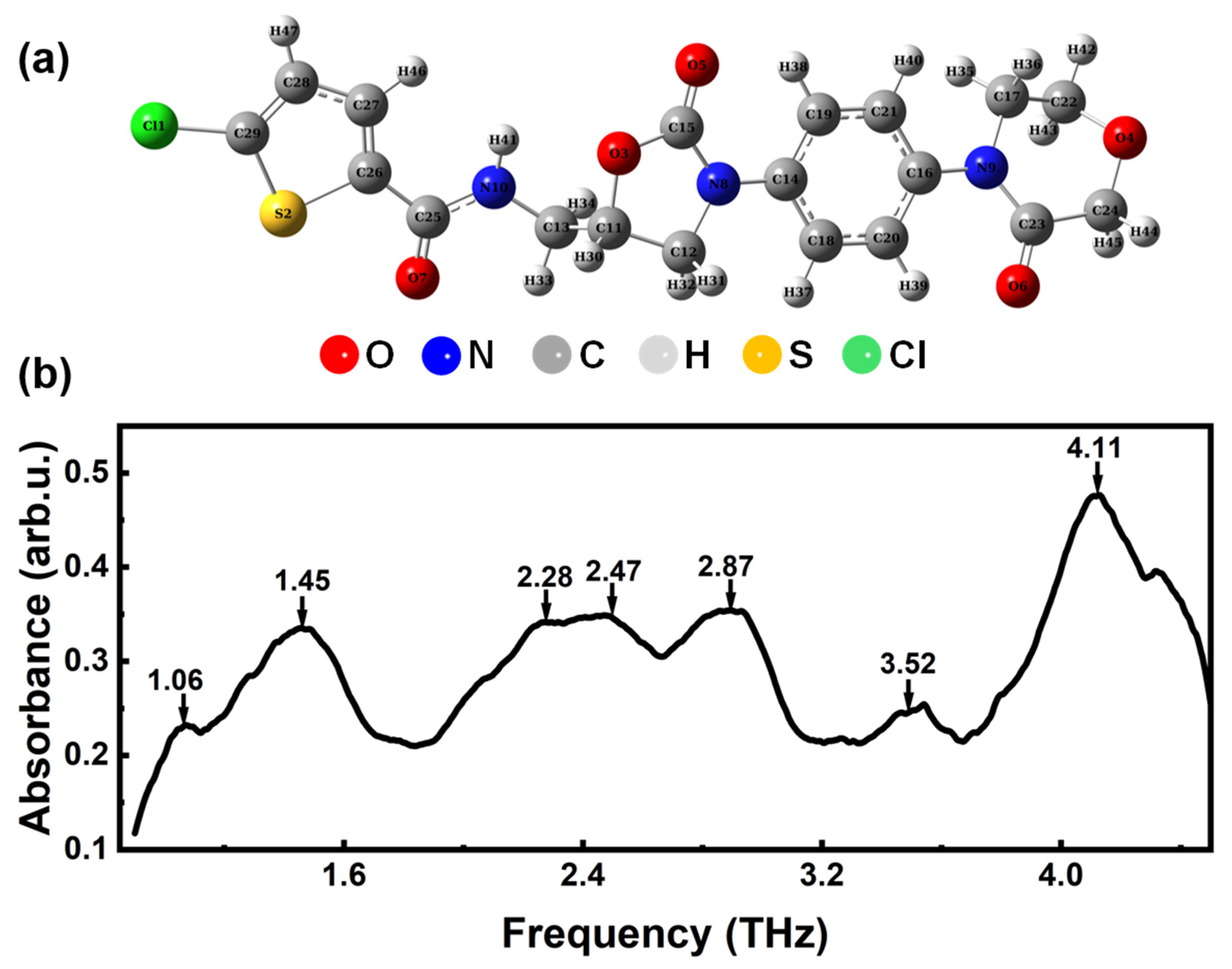
Figure 1. (a) The molecular structure of rivaroxaban. (b) Experimental THz absorption spectra of rivaroxaban in the range of 0.9–4.5 THz.
Photonics 11 00814 g001
Figure 2. (a) A 3D view of the metamaterial unit, where the period is P = 40.00 µm, the inner diameter is r = 9.00 µm, the outer diameter is R = 16.60 µm, and the gap width is g = 3.00 µm. (b) Theoretical absorption spectrum of the metamaterial. (c) Theoretical absorption spectra of the metamaterial for samples with different thickness. (d) Frequency shift as a function of sample thickness.
Photonics 11 00814 g002
Figure 3. (a) Digital photo and optical images of the THz metamaterial biosensor, where the inner diameter is r = 7.56 µm, the outer diameter is R = 17.61 µm, and the gap width is g = 2.53 µm. (b) THz experimental spectra of the raw biosensor.
Photonics 11 00814 g003
Figure 4. (a,b) The absorption spectra of rivaroxaban solutions with different concentrations. (c) The frequency shift as a function of the rivaroxaban concentration. (d) The absorbance changes as a function of the rivaroxaban concentration.
Photonics 11 00814 g004
Disclaimer/Publisher’s Note: The statements, opinions and data contained in all publications are solely those of the individual author(s) and contributor(s) and not of MDPI and/or the editor(s). MDPI and/or the editor(s) disclaim responsibility for any injury to people or property resulting from any ideas, methods, instructions or products referred to in the content.