Review

Multimodal Imaging in Stem Cell Therapy for Retinal Disease

1
Department of Ophthalmology, Wilmer Eye Institute, Johns Hopkins University, Baltimore, MD 21287, USA
2
Department of Ophthalmology, Fuzhou University Affiliated Provincial Hospital, Fuzhou 350001, China
3
Department of Ophthalmology, Provincial Clinical Medical College of Fujian Medical University, Fuzhou 350001, China
4
Department of Biomedical Engineering, Johns Hopkins University, Baltimore, MD 21287, USA
*
Author to whom correspondence should be addressed.
Photonics 2025, 12(5), 413; https://doi.org/10.3390/photonics12050413
Submission received: 26 February 2025 / Revised: 12 April 2025 / Accepted: 21 April 2025 / Published: 24 April 2025

Abstract

:
Stem cell therapy has emerged as a promising approach for treating various retinal diseases, particularly degenerative retinal diseases such as geographic atrophy in age-related macular degeneration (AMD), retinitis pigmentosa (RP), and Stargardt disease. A wide variety of imaging techniques have been employed in both preclinical and clinical settings to assess the efficacy and safety of stem cell therapy for retinal diseases. These techniques can be classified into two categories: methods for imaging stem cells and those for the overall morphology and function of the retina. The techniques employed for stem cell imaging include optical imaging, magnetic resonance imaging (MRI), and radionuclide imaging. Additional imaging techniques include fundus photography, fluorescein angiography, and fundus autofluorescence. Each technique has its own advantages and disadvantages, and thus, the use of multimodal imaging can help to overcome the shortcomings and achieve a more comprehensive evaluation of stem cell therapy in retinal disease. This review discusses the characteristics of the main techniques and cell-labeling techniques applied in stem cell therapy, with a particular focus on the applications of multimodal imaging. Furthermore, this review discusses the challenges and prospects of multimodal imaging in stem cell therapy for retinal disease.

Graphical Abstract

1. Introduction

As a technique undergoing rapid development, stem cell therapy offers a promising potential to treat many vision-threatening diseases that are challenging to manage with conventional therapeutic approaches, especially degenerative retinal diseases. The objective of stem cell therapy is to restore vision or slow down the further degeneration of the retina by replacing the dysfunctional retinal cells or rescuing retinal cells by releasing trophic factors [1].
As the field of stem cell-based therapy is rapidly developing, numerous studies have been conducted to investigate the potential of this approach in the treatment of retinal diseases, with a particular focus on retinal degenerative conditions such as geographic atrophy (GA) in age-related macular degeneration (AMD), retinitis pigmentosa (RP), diabetic retinopathy, glaucoma, recessive Stargardt macular degeneration (STGD1), and retinal degenerations caused by other factors [2,3]. A variety of stem cell types have been employed in the treatment of retinal diseases, including embryonic stem cells (ESCs), induced pluripotent stem cells (iPSCs), mesenchymal stem cells (MSCs), and progenitor cells [2]. The use of stem cell therapy for the treatment of retinal diseases has shown promising results in animal models [4,5,6]. Furthermore, several clinical trials also demonstrated an acceptable safety profile and some functional improvements in patients with AMD, RP, and STGD1 using stem cell-based therapy, both in the short and long term [7,8,9,10,11,12,13,14,15,16,17,18,19,20,21,22,23,24]. Despite the advancements made in the current clinical trials, there are still considerable obstacles to overcome that impede the speed of the clinical translation of stem cell therapy.
The traditional methods of assessing the efficacy and safety of treatments, such as visual acuity, electroretinogram, and imaging modalities for structural changes, like fundus photography (FP) and optical coherence tomography (OCT), are insufficient for capturing the complexities of retinal alterations at the cellular level. The success of stem cell therapy is contingent on many factors, including the method of delivery, cell differentiation, distribution, survival, and function. Following the successful delivery of stem cells to the target area, the transplanted cells must correctly engraft into the targeted tissue, differentiate into the desired cells, and maintain their long-term biological effect to achieve therapeutically optimal results. The histological method represents the gold standard for identifying cell type and distribution. However, histology is a one-time examination, which can only be applied at the endpoint of an experiment and is destructive. Consequently, it is not applicable in the context of a clinical trial. Furthermore, in the case of an animal experiment, the application of a histological examination to assess stem cell therapy may necessitate the use of a large number of animals, as well as a considerable investment of time and financial resources. As a result, the non-invasive monitoring of these processes at the cellular or even molecular level in real time has become a pivotal concern in the evaluation of biosafety and the efficacy of stem cell-based therapy.
A wide range of imaging techniques have been utilized throughout the various stages of stem cell therapy for retinal diseases, enabling the accurate delivery of the stem cells and observation of the complicated processes that occur following implantation. These techniques can be classified into two principal categories: methods for tracking stem cells and those for the overall morphology and function of the retina.
Currently, there are three common techniques for the in vivo tracking of stem cells: optical imaging, magnetic resonance imaging (MRI), and radionuclide imaging [25]. Among them, optical imaging is the most prevalent in the field of ophthalmology due to the optical transparency of the anterior portion of the eye. Nuclear imaging will not be addressed in this paper as its low resolution restricts its use in ophthalmic cellular imaging. Besides cell imaging, other imaging techniques, including FP, fluorescein angiography (FA), and fundus autofluorescence (FAF), can also provide valuable insights into the morphology, circulation, and metabolic changes in the retina [26]. As each technique has its own advantages and disadvantages, researchers attempt to combine two or more techniques to achieve the most optimal outcome, which is known as multimodal imaging.
This review provides an overview of the principal imaging techniques employed in the in vivo study of stem cell therapy for retinal diseases, categorizing them into preclinical methods for cellular tracking and clinical tools for structural and functional assessment in the following sections. Furthermore, it will focus on the application of multimodal imaging in stem cell therapy for retinal diseases and its role in enhancing treatment outcomes. This review will also discuss the challenges and prospects of using multimodal imaging to assist stem cell therapy for retinal diseases.

2. Imaging Techniques for Retinal Stem Cell Therapy

Current imaging approaches for retinal stem cell therapy face a critical issue: no cell-labeled agents are yet approved for clinical tracking, limiting all stem cell-specific imaging to preclinical research. Conversely, clinical imaging relies on unlabeled techniques, primarily OCT, FA, and FAF, to assess the structural integration and functional outcomes of transplanted cells. This section systematically examines both domains: (i) experimental methods allowing stem cell tracking in animal models and (ii) clinical tools assessing therapeutic efficacy in trials, highlighting multimodal strategies to bridge this translational gap.

2.1. Preclinical Imaging Techniques

2.1.1. Cell Labeling

Cell labeling is usually required to differentiate transplanted stem cells from host cells, irrespective of the imaging method employed. In general, stem cell labeling can be conducted via two principal methods: direct and indirect labeling methods [27]. Direct labeling entails the introduction of labeling agents into cells prior to transplantation. This approach is straightforward and relatively safe, as it circumvents the necessity for genetic modification. However, it is constrained by the limitations of short-term monitoring, due to the dilution of signals resulting from the radioactive decay of the labeled agents or the cell division and dispersion [27]. Furthermore, the processes of the phagocytosis of dead labeled cells by macrophages and the exocytosis of intracellular labels have the potential to yield false positives [25]. The indirect labeling method involves the integration of reporter genes into stem cells through genetic modification, enabling stable expression over time. While this method is complex and time-consuming, it is particularly well suited to longitudinal studies [28].
A variety of cell-labeling agents are employed in eye research, including fluorescent dyes, reporter genes, magnetic particles, and nanoparticles [29,30]. Some cell-labeling agents can enhance signals in multiple imaging modalities, thereby facilitating the application of multimodal imaging.
Fluorescent dyes like indocyanine green (ICG), 5-(and-6)-carboxyfluorescein diacetate succinimidyl ester (CFSE), DiI-linked acetylated low-density lipoprotein (DiI-AcLDL), Alexa Fluor 488-HA, Alexa Fluor 568, Qtracker 655, and tdTomato have been employed as cell-labeling agents in fluorescence imaging (FI) to monitor the transplanted stem cells in the retina [31,32,33,34,35]. Among these, ICG stands out as a U.S. Food and Drug Administration (FDA)-approved fluorescent dye that has been extensively used for cell labeling in many research studies [36]. Since ICG operates in the near-infrared (NIR) spectrum, it allows for deeper imaging with less background autofluorescence from ocular tissues [37]. It can also be employed as a cell-labeling agent in cell tracing by both optical imaging and photoacoustic imaging (PAI) in its molecular form [37], making it a suitable candidate for multimodal imaging. However, its use for cell labeling remains preclinical and is not approved for clinical retinal stem cell tracking, in contrast to its established diagnostic role in angiography.
Fluorescent dye-based imaging is confronted with challenges such as potential photobleaching and toxicity, particularly in long-term studies. The degradation of fluorescence is observed in studies using fluorescent dyes as cell-labeling agents; for example, when ICG is used to label transplanted cells, the fluorescence of ICG normally disappears within 4 weeks [35,38]. While ICG is not typically phototoxic under standard imaging conditions, prolonged intracellular retention may contribute to cell toxicity [37], affecting both the accuracy and safety of long-term studies [39]. In addition, the autofluorescence of the eye must be taken into account when applying fluorescent cell-labeling agents, as it has the potential to interfere with the detection of labeled cells, thereby reducing image clarity and accuracy [40,41].
Using a reporter gene that can encode a fluorescent protein as a cell-labeling agent can provide a more stable and long-lasting fluorescence signal compared to fluorescent dyes. Among all the fluorescent proteins, green fluorescent protein (GFP) has been used with the greatest frequency. GFP is a type of red-shifted fluorescent protein (FP). This type of FP is advantageous for providing better contrast with reduced autofluorescence at longer wavelengths [42]. Additionally, at far-red and NIR wavelengths, the absorption of light is much less than visible light, facilitating a higher sensitivity with lower toxicity [43]. Shi et al. [31] compared three fluorescent probes for the in vivo tracing of endothelial progenitor cells (EPCs) in a mouse model of laser-induced retinal injury. The results demonstrated a decline in fluorescence over time in the CFSE- and Dil-AcLDL-labeled group, where direct labeling was employed. In contrast, the GFP-labeled group, which employed indirect labeling, demonstrated an increase in fluorescence over the course of the experiment. One potential explanation for this phenomenon is the continuous expression of the GFP gene, which may have resulted in the accumulation of the fluorescent protein within the cells. This indicated that direct labeling may be more suitable for short-term studies, whereas indirect labeling may help to prevent photobleaching and thus make it more ideal for long-term studies. The above-mentioned advantages of GFP render it a preferred choice in a multitude of studies, as stem cell therapy research often necessitates prolonged periods of observation. It is currently being employed to monitor the activities of a range of transplanted grafts in vivo, including human embryonic stem cell (hESC)-derived retinal cells [44], allogeneic iPSC-derived retinal pigment epithelium (RPE) cells [45], and human iPSC retinas [46] with a long-lasting GFP signal for at least three months [44] following implantation.
Despite the prevalent utilization of GFP in the preclinical investigation of stem cell therapy for retinal disorders, this approach remains exclusively limited to research settings and has not been adopted clinically. There are certain disadvantages that impede its clinical translation. Some researchers [47,48] posit that the introduction of GFP may elicit an immune response in the host. Such an immune reaction has the potential to result in the rejection of the transplanted cells or the onset of inflammation, which may ultimately compromise the therapeutic outcomes. Additional barriers include the risks of toxic effects [49], genome alteration and gene silencing [50], and the complexity and high cost of the production processes, all of which collectively preclude the current clinical application of reporter gene-based tracking methods.
Compared to traditional cell-labeling agents, nanoparticles (NPs) exhibit unique physical and chemical properties [51], coupled with good biocompatibility [52,53]. A distinctive feature of NPs is their ease of synthesis, coupled with the straightforward manipulation of their molecular structures, facilitated by the presence of accessible functional groups [54]. By modifying their structure and size, it is possible to alter their optical properties to optimize their use for specific wavelengths [55,56]. In addition, the conjugation of NPs with other agents enables the application of NPs in disease diagnosis, drug delivery, biocatalysis, and tissue engineering, as well as biomedical imaging [57].
Given these characteristics of NPs, they have been employed as a contrast agent across a variety of imaging modalities in recent years, including MRI, FI, PAI, ultrasound imaging, and computed tomography (CT) [56]. For the in vivo imaging of stem cells, NPs are frequently conjugated with fluorescent dyes, thereby enabling detection by both fluorescent and non-fluorescent imaging modalities. The robust and sizable nanoparticle structures ensure prolonged fluorescent labeling with high photostability, while the process and cost of labeling are significantly reduced when compared with the use of reporter genes [39]. The combination of these features makes NP-based cell labeling an optimal candidate for multimodal imaging. Gold nanoparticles (GNPs) represent a promising class of cell-labeling agents for ophthalmic imaging with minimal toxicity [53,58]. Some gold-based nanoparticle drug delivery systems have been approved by the FDA and are being employed to enhance healthcare and disease management [59]. It has been demonstrated by several studies that GNPs can enhance a variety of ophthalmic imaging techniques, including OCT and PAI [60,61,62,63,64,65,66,67]. Furthermore, they are capable of long-term tracking of stem cells in retinal replacement therapy with minimal toxicity [38,56,68]. Additionally, GNPs have antiangiogenic and anti-inflammatory properties [69,70], making them valuable for cell labeling in stem cell therapy.
While nanoparticles offer significant advantages for tracking stem cells in retinal therapies, challenges such as potential toxicity [71], impact on cell function [72], and exocytosis [73] must be addressed. Additionally, in cases where transplanted stem cells die, NPs may persist with the potential to generate false-positive signals [74]. Furthermore, the limited understanding of the accumulation of NPs in the body engenders uncertainty regarding potential long-term side effects.
Table 1 presents a comparison of various cell-labeling agents utilized in stem cell therapy for retinal diseases.

2.1.2. Optical Imaging

Optical imaging represents the most prevalent approach for the in vivo assessment of the efficacy and safety of stem cell therapy targeting retinal diseases. The transparent structure of the anterior segment of the eye facilitates the penetration of light, thereby enabling optimal optical imaging. Optical imaging includes a number of different techniques, such as FI, OCT, bioluminescence imaging, and PAI. We will not discuss bioluminescence imaging in this context due to its relatively low resolution, which has limited its use in ophthalmology. For an effective visualization of transplanted stem cells, imaging depth must typically reach the subretinal space, where cells are commonly implanted to replace or support the RPE and photoreceptors. The choice of wavelength critically influences imaging depth, contrast, and compatibility with cell-labeling agents: shorter wavelengths enhance contrast for superficial layers like the inner retina and match dyes like CFSE or DiI [31], while longer near-infrared wavelengths improve penetration to deeper structures like the subretinal space and choroid, aligning with agents like ICG for optimal signal detection [35,75]. In addition, optical imaging offers the potential for real-time structural and functional imaging at both cellular and subcellular levels with high resolution and minimal invasiveness [76]. Furthermore, optical imaging is relatively inexpensive compared to other imaging modalities like MRI and nuclear medicine, making it more accessible for widespread clinical use. While optical imaging offers many significant advantages, certain limitations do exist, like depth penetration and tissue scattering. The absorption of light in the visible spectrum by hemoglobin and other molecules may result in a reduction in optical signals by approximately 10-fold per centimeter of tissue [77]. Any opacity along the optical path, which may be attributed to the potential adverse effects of stem cell therapy, may lower the image quality. Such opacities may include vitreous opacity resulting from endophthalmitis or hemorrhage, or lens opacity resulting from injury during the cell transplantation process. FI is a technique that detects fluorescence light emitted by excited fluorophores when irradiated with a light source with specific wavelengths from ultraviolet (UV) to NIR [78]. It relies on the use of fluorescent agents to label specific cells, thereby generating light signals under external excitation. These signals can be detected by optical instruments and reflect the crucial processes of post-stem cell transplantation, including cell proliferation, differentiation, and migration [25,43]. The optical instruments that have been applied for retinal FI tracing transplanted stem cell include a fundus fluorescein angiography (FFA) camera [35], scanning laser ophthalmoscopy (SLO) [79], fluorescence adaptive optics scanning laser ophthalmoscopy (FAOSLO) [34], and fluorescence confocal scanning laser ophthalmoscopy (fcSLO) [33].
The advantages of FI include a high resolution, real-time monitoring, and multi-fluorescence detection ability when multiple fluorescence agents are included [39]. Furthermore, some fluorescent agents can also be detected by other retinal imaging modalities, thus facilitating the integration of FI into multimodal imaging systems, which can provide a more comprehensive understanding of the intricate processes involved in stem cell therapy [35].
One disadvantage of FI is that it can only provide two-dimensional (2D) information, which may impede the evaluation of how effectively transplanted cells integrate into the existing retinal architecture. To address this limitation, researchers are increasingly employing multimodal imaging techniques to integrate FI with OCT or PA imaging, thereby obtaining both cellular-level fluorescence data and cross-sectional structural information of the retina [33,35,38,80]. Most of the other disadvantages of FI come from the fluorescent cell-labeling agents, such as photobleaching, phototoxicity, and background from autofluorescence. These limitations may be rectified through the enhancement of cell-labeling agents.
PAI is a novel and promising non-ionizing imaging modality that allows for the acquisition of three-dimensional images [81]. This technique is based on simultaneous optical excitation and ultrasound detection via optical absorption-induced thermoelastic expansion [57]. It combines the advantages of optical and ultrasound imaging to provide high-resolution, high-sensitivity, and high-contrast images at greater imaging depths [25]. Optimization of the PAI system enables satisfactory image quality at merely 1% of the American National Standards Institute (ANSI) safety limit for laser fluence, with no observed retinal damage [82]. When combined with specific contrast agents, this technology enables the acquisition of not only structural but also functional and molecular information. Non-invasive PAI can provide oxygen saturation levels [81]. Furthermore, PAI systems can facilitate more accurate cell delivery to the target area by providing real-time ultrasound guidance [83]. PAI can be enhanced by a variety of cell-labeling agents, rendering it an appropriate modality for multimodal imaging. However, despite its capacity for three-dimensional imaging, one limitation of PAI is its inability to discern the distinct layers of the retina, a capability that distinguishes it from OCT. An additional concern with PAI is its safety, as it typically uses a nanosecond pulsed laser for excitation. Although photoacoustic imaging of the retinal and choroidal vasculature in rabbit eyes has demonstrated an acceptable safety profile [84], further comprehensive long-term safety studies are required for clinical translation. Furthermore, most PAI systems currently require a transducer to contact the eyelid, coupled with ultrasound gel or directly contacting the conjunctiva, which can cause damage, abrasion, infection, and discomfort [85]. In addition, due to the inherent point-scanning mechanism of PAI (a one-dimensional depth-resolved signal per laser pulse) [86], both scanning speed and lateral coverage are limited, typically requiring >1 min to complete a 3 × 3 mm2 to 7 × 7 mm2 scan in larger animal models such as rabbits [82,87]. Other potential drawbacks of this modality include the complexity of setting up the system, technical challenges in raising the signal-to-noise ratio (SNR), and avoiding artifacts [86].

2.1.3. Magnetic Resonance Imaging (MRI)

MRI provides high-resolution imaging through a non-invasive and radiation-free method that is not subject to depth limitations. By combining contrast agents and reporter genes, MRI can trace stem cells through the alteration of the relaxation time of water protons, which produces distinct signals that differentiate transplanted cells from host cells [25]. In contrast to optical imaging, MRI is capable of penetrating opaque tissues, thereby enabling imaging of structures with potential opacity in the cornea, lens, or vitreous. Ma et al. [29] demonstrated that photoreceptor precursors derived from hESCs can be labeled with superparamagnetic iron oxide nanoparticles (SPIONs) and effectively tracked using MRI in the subretinal space. The hypointense signals corresponding to the labeled cells were visible up to 12 weeks post-transplantation.
Despite the successful utilization of MRI to track stem cells by Ma et al., the employment of MRI in the investigation of retinal diseases using stem cell therapy remains relatively limited. One reason is that although MRI has high resolution (an in-plane spatial resolution of 100 µm and a voxel resolution of 200 μm × 200 μm × 400 μm can be achieved in in vivo eye imaging at 7.0 T within clinically acceptable scan times [88]), this is insufficient to cope with the complex and intricate structure of the posterior segment [30]. Moreover, the use of specific contrast agents, such as SPIONs, may present potential toxicity risks, necessitating the development of safer alternatives [89]. The degradation of signals from the contrast agents may also impede the long-term cell-tracking ability of MRI [29]. Furthermore, it is important to acknowledge that MRI is a complex and costly imaging modality, which may restrict its accessibility and widespread use in clinical settings. Additionally, this approach is not applicable in patients with contraindications to MRI.

2.1.4. Emerging Imaging Techniques with Potential for Retinal Stem Cell Therapy

As research progresses, new methods are emerging that might have the potential to advance the field of stem cell therapy imaging. Although these technologies have not yet been used for in vivo imaging in retinal stem cell therapy, they might help overcome current limitations in resolution, specificity, or functional assessment.
FLIO is an emerging imaging technique for measuring the lifetime of endogenous retinal fluorophores in vivo, with a better ability to detect weaker endogenous fluorophores, a wide working spectral range, and reduced background interference associated with typical FI [90]. While FLIO has not yet been applied to the in vivo tracking of transplanted stem cells in the retina, its sensitivity to detect fluorophores suggests its potential for improving the visualization of fluorescently labeled stem cells and pigments in transplanted cells, complementing FI-based imaging. Additionally, FLIO can assess metabolic states by quantifying changes in fluorescence lifetime, which has been used to observe metabolic trajectories in retinal organoids to reflect cellular viability and function [91]. With further developments, FLIO may help to improve the precision of imaging for retinal stem cell therapies.
Molecular imaging offers the potential to visualize specific biomarkers associated with retinal diseases beyond traditional structural and functional imaging techniques. For instance, CD44—a cell surface glycoprotein—is significantly upregulated in retinal conditions such as retinitis pigmentosa (RP) [92] and choroidal neovascularization (CNV) [93]. Although direct in vivo imaging of CD44 in retinal diseases has not been extensively reported, molecular imaging probes targeting CD44 have been developed for other pathologies like cancer [94]. Adapting these probes for ocular use might provide valuable insights into disease progression and response to therapeutic interventions, including those involving stem cell transplantation, by monitoring host tissue responses.

2.2. Clinical Imaging Techniques

Clinical imaging techniques are widely adopted in clinical trials and practice, focusing on the structural and functional evaluation of the retina rather than direct cellular tracking.

2.2.1. Optical Coherence Tomography (OCT)

OCT is a non-invasive imaging technique that provides high-resolution, cross-sectional images of the retina, choroid, and part of the sclera with minimal invasiveness. OCT allows for the in vivo visualization of retinal structures with a resolution almost comparable to that achieved through histopathology, thus making it a powerful tool for diagnosing and monitoring retinal diseases. Common spectral-domain OCT (SD-OCT) and swept-source OCT (SS-OCT) are capable of detecting low-reflectivity structures with high resolution exceeding 100 dB, and this is attributed to the high scan rate [75]. The high scanning speed also facilitates the generation of a 3D structure of the posterior segment of the eye in a short time [30]. Advanced OCT techniques such as adaptive optics OCT (AO-OCT) have further improved resolution to the cellular level [95]. This technique has been applied to patients undergoing iPSC-derived retinal organoid transplantation, and a detailed visualization of cellular structures can be obtained [96]. Given the long-standing and widespread use of OCT in both clinical and experimental contexts, as well as its high acceptance among patients and researchers, it is a readily applicable tool in the field of stem cell therapy for retinal diseases. Additionally, the non-invasive and well-tolerated nature of OCT imaging allows for repeated monitoring of stem cell behavior in vivo over time, further enhancing its utility in long-term stem cell therapy research. And since contrast agents are not required, it is extremely useful for clinical trials [8,23,97,98,99] or animal studies [5] that involve large volumes and require long-term follow-up.
The transplanted stem cell can be directly observed in real-time in OCT images as hyperreflective signals [6,34], which can not only provide valuable information for monitoring the grafts after transplantation but also facilitate the transplantation process itself. Li and colleagues successfully employed real-time OCT guidance for subretinal injection in rabbits [100]. Similarly, Nguyen et al. used a comparable method to successfully deliver human retinal pigment epithelium (ARPE-19) cells to the subretinal space [35]. At the same time, an intraoperative OCT was also employed to facilitate the subretinal implantation of a graft (California Project to Cure Blindness Retinal Pigment Epithelium, CPCB-RPE1) during surgery (NCT02590692) [101].
As one of the optical imaging methods, OCT is also affected by opaque ocular media. Furthermore, the small size and diffuse distribution of the graft may also impede visualization, as the transplanted cells share similar characteristics with host tissue, thereby making them difficult to distinguish from the background [56]. A further potential drawback of OCT imaging is the presence of motion artifacts. In a clinical setting, the use of commercially available OCT systems with rapid image acquisition times may assist in the reduction in the impact of motion artifacts [75]. On the other hand, laboratory-made OCTs for animal experiments are often subjected to a slower scanning speed. In order to obtain high-quality imaging, it is essential that the animal is adequately anesthetized and positioned correctly.

2.2.2. Other Related Techniques

Besides those imaging techniques used for in vivo stem cell tracking, other ocular imaging techniques, such as fundus photography (FP), scanning laser ophthalmoscopy (SLO), fundus autofluorescence (FAF), indocyanine green angiography (ICGA), and fluorescein angiography (FA), can also facilitate stem cell delivery as well as contribute to the assessment of the efficacy and safety of stem cell therapy.
FP, SLO, and FAF offer a valuable two-dimensional visualization of the retina’s general morphology from an en-face perspective in real time. Both imaging modalities are non-invasive, fast, readily available, and inexpensive. The equipment used for FP and SLO can also be used for FI. Compared to FP, SLO can combine images from multiple wavelengths to provide better image quality and more information at different depths and over a larger area [102]. FAF allows the intrinsic fluorescence emitted by molecules such as lipofuscin in the retinal pigment epithelium (RPE) to be detected when excited by specific wavelengths of light, thereby providing a valuable means of visualizing metabolic changes in the RPE [103]. Furthermore, the devices utilized for FP and FAF may also be capable of obtaining fluorescence imaging signals with specific filters and excitation light.
FA and ICGA are valuable tools that are commonly employed in clinical and experimental settings for the assessment of retinal and choroidal vasculature [104,105]. They are effective in observing morphological and functional changes in the retinal and choroidal vasculature, thus indicating the physiopathological changes in the retina and choroid, such as neovascularization and inflammation, to some extent.
These imaging modalities have been in clinical use for a considerable period, and their safety for long-term use has been well documented. In such instances, when compared to other novel imaging techniques, they have been more frequently employed in clinical trials that utilize stem cell therapy for the treatment of retinal diseases [83,106]. However, these techniques have an inherent limitation: they do not permit direct observation of the transplanted stem cells at the cellular level. Consequently, in investigations of stem cell therapy for retinal diseases, they are typically employed as part of a multimodal imaging system or in combination with histological examinations to facilitate the monitoring of cell survival and integration within the host retina.

3. Multimodal Imaging

The previously mentioned imaging techniques have distinct advantages for the evaluation of stem cell therapy for retinal diseases. However, when employed alone, their inherent limitations may lead to insufficient or unsafe practices for long-term monitoring (Table 2). Therefore, combining different imaging tools that complement each other is an effective approach for researchers and clinicians to obtain more comprehensive insights into the tracking, therapeutic efficacy, and safety of stem cells in vivo.
The combination of two or more optical imaging modalities is the most commonly used approach in multimodal imaging due to the convenience and relatively low cost of optical imaging. Among them, combined OCT and other 2D imaging techniques, such as FP, FFA, and FAF, are the most easily achievable and cost-effective. The combination of 2D and 3D imaging techniques can facilitate a more comprehensive understanding of the distribution and integration of implanted cells. Moreover, the integration of OCT into a multimodal imaging system can facilitate cell delivery, as OCT enables the real-time guidance of implantation [35,107]. For clinical application, these imaging techniques have been employed in clinical practice for an extended period, with favorable safety profiles, making them suitable for long-term follow-up. Additionally, other advanced cell-tracking imaging techniques that include cell-labeling agents have not been approved by the FDA for application in the eye. Consequently, this is the only type of multimodal imaging employed in clinical trials of stem cell therapy for retinal diseases [9,15,23,97,98,99,108,109,110]. Without the help of cellular markers, it is challenging for clinicians to obtain information at the cellular level, which impedes the clinical translation of stem cell therapy.
Although there are restricted options for imaging tools in clinical trials due to safety concerns, a variety of advanced imaging modalities with various cell-labeling agents have been investigated in animal experiments, highlighting the potential benefits of multimodal imaging.
Laver et al. combined OCT and fcSLO to enhance the real-time early assessment of transplanted hESC-derived photoreceptor precursor cells (PPCs) labeled with intracellular fluorescent quantum dots (Qtracker 655, QT) [33]. By measuring the QT signal changes from fcSLO and OCT, this bimodal imaging technique was able to achieve a more accurate detection of graft dynamics and potential leakage from the subretinal space following injection with a cloudy vitreous. Li et al. [80] utilized a multimodal imaging system integrating OCT, fluorescence microscopy (FM), and lasing emission to track the 3D migration trajectories of individual ARPE-19 cells labeled with CdS nanowires in rabbit eyes for 28 days. The CdS nanowires served as cell-labeling agents, providing 3D information of individual cells (Figure 1). These results indicate that the capacity of multimodal imaging to track implanted stem cells is significantly enhanced by cell labeling, which can be achieved at the cellular level with the appropriate labeling agents.
Moreover, the prolonged observation periods that are required to evaluate the efficacy and safety of stem cell therapy highlight the need to optimize cell-labeling techniques that can safely facilitate long-term multimodal imaging enhancement.
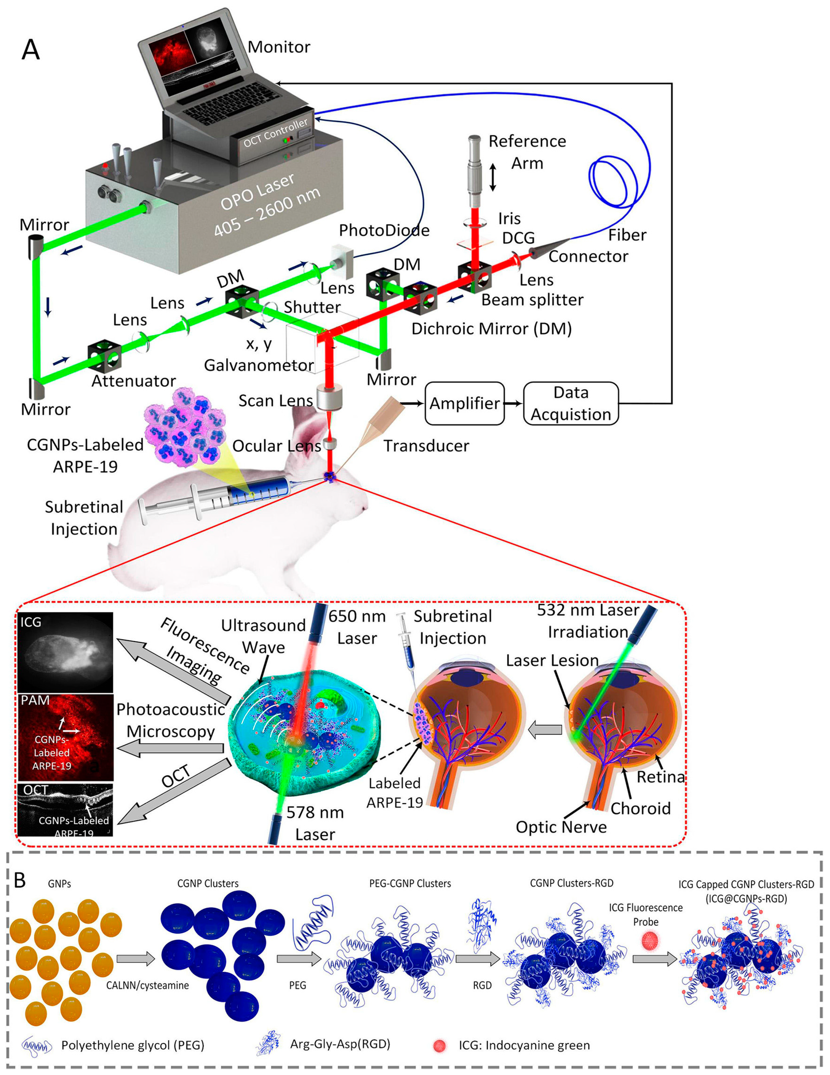
In studies conducted by Nguyen et al. [35,38,68,111], a non-invasive, high-spatial-resolution, triple-modality imaging system (Figure 2) was developed by combining OCT, PAI, and FI to monitor transplanted cells in rabbits. By using different cell-labeling agents, different degrees of imaging enhancement were achieved. When human retinal pigment epithelium cells (ARPE-19) were labeled with ICG [35], the FI signals were enhanced up to 37-fold, and the cells were able to be tracked for 21 days after transplantation. The combination of ICG with chain-like GNP (CGNP) conjugated with RGD peptides to form an ICG-CGNP-RGD cell-labeling agent can enhance signals in FI, OCT, and PAI [35]. The PAI signals can be detected for up to 56 days following transplantation, exhibiting an increasing intensity of up to 30-fold, thereby facilitating the long-term tracking of stem cells in vivo. The ICG fluorescence signal was observed to persist for a maximum of 7 days, which may be attributed to the accelerated photobleaching of ICG, potentially induced by repeated optical examinations.
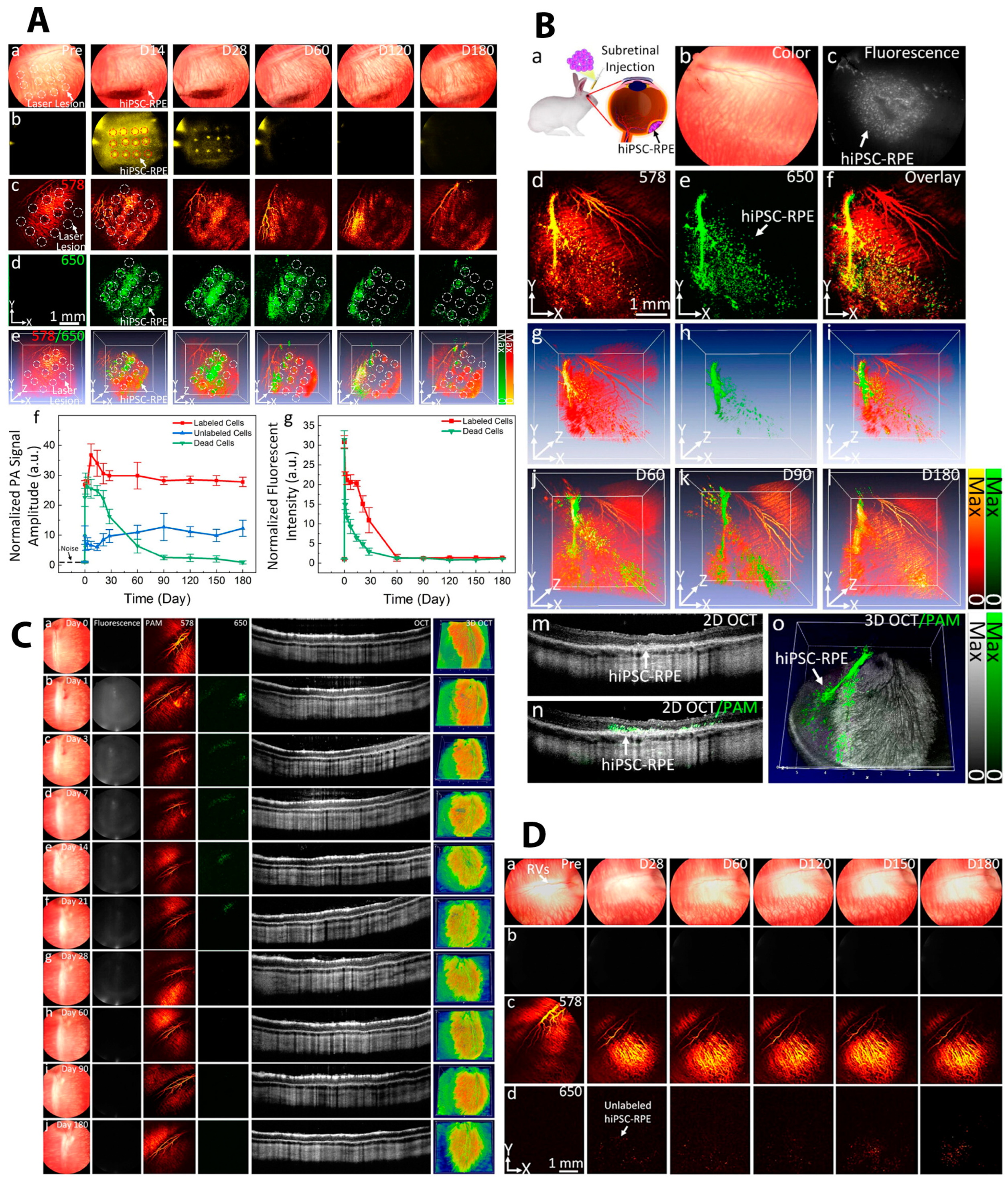
In subsequent experiments, Nguyen et al. further enhanced the signal of the multimodal imaging system through the additional optimization of cellular markers. An ultraminiature chain-like gold nanoparticle cluster (GNC) nanosensor (GNC-RGD-ICGs) with an optical absorption peak in the near-infrared regime and a diameter of approximately 7–8 nm was developed to label retinal pigment epithelium (hiPSC-RPE) cells differentiated from human-induced pluripotent stem cells [38]. In this experiment, the PAI signals were enhanced up to 37-fold, while the signal from OCT was enhanced up to 195%, and the fluorescent signal from ICG was enhanced up to 31-fold. Meanwhile, the PAI signal demonstrated enhancement for up to 6 months following transplantation, while the ICG signal exhibited observable characteristics for up to 28 days. Additionally, Nguyen et al. [65,66,93] observed, through multimodal imaging at a three-dimensional level, that in models of laser-induced RPE injury, transplanted stem cells exhibited a tendency to aggregate in the injured area, seeking to repair the damage. In contrast, in a normal retina, they demonstrated the capacity to form a monolayer distribution. The PA signal generated by dead cells was noted to rapidly decrease over time. When the dead hiPSC-RPE cells are labeled with the same GNC-RGD-ICG, the PA signal generated by GNCs decreases rapidly after transplantation. This can help identify cell survival in vivo [38] (Figure 3). It is noteworthy that no substantial toxic effects resulting from cell labeling or imaging were discerned in the course of these studies. These studies demonstrate that multimodal imaging, enhanced by cellular labeling, can provide non-invasive, high-resolution, and three-dimensional observations of stem cell distribution, survival, and integration in vivo with minimal risk over an extended period of time.
Other imaging modalities have been poorly studied as a tool for evaluating stem cell therapy in retinal diseases. This is primarily due to several factors. The inadequate resolution of these imaging tools to effectively visualize intricate retinal structures represents a significant obstacle to their utilization in the assessment of retinal diseases. The unique structural characteristics of the eye necessitate high-resolution imaging of the retina while requiring less tissue penetration than other organs. This makes optical imaging the most suitable imaging modality for the eye. Consequently, techniques other than optical imaging are also less frequently considered when developing multimodal imaging setups.

4. Challenges and Prospects

A variety of reliable methods for in vivo monitoring of stem cells have been established, and an increasing number of researchers are turning to multimodal imaging approaches to minimize the limitations of each imaging technique. The development of cell-labeling techniques has also facilitated the achievement of enhanced imaging for long-term use. However, significant challenges remain.
A more profound comprehension of the mechanisms underlying stem cell therapy requires the imaging of cellular and even molecular structures. However, the majority of current imaging techniques are unable to achieve the requisite resolution without the use of exogenous contrast agents. Although multimodal imaging enhances the capacity of imaging across spatial dimensions, the intrinsic resolution remains constrained by the imaging instruments employed. As previously discussed, numerous studies have demonstrated that advanced imaging techniques and cell labeling can facilitate more effective observation of transplanted stem cells in vivo. However, the majority of these techniques, including PAI and NPs, have yet to receive approval for clinical use in the eye. Concurrently, the approval process for novel imaging modalities and cell-labeling agents is both time-consuming and labor-intensive.
Safety remains a critical challenge in advancing multimodal imaging for stem cell therapy in retinal diseases. For instance, PAI typically employs nanosecond pulsed lasers, which need to adhere to ANSI safety limits to prevent retinal burns; exceeding these limits could pose significant risks [82]. Although current PAI systems can operate safely at only 1% of ANSI limits with no observed retinal damage (Section 2.1.2), long-term safety studies and non-contact transducer designs are still unmet needs. Furthermore, the interaction between contrast agents or cell-labeling agents required for different imaging modalities has the potential to result in adverse effects, including toxicity, immune responses, and unpredictable side effects (see Section 2.1). In addition, fluorescent dyes may cause toxicity, reporter genes may trigger immune rejection, and nanoparticles, despite their promise, carry risks of cellular dysfunction or accumulation in the body [35,38,71]. The development of multifunctional, biocompatible cell-labeling agents and the optimization of imaging protocols are therefore essential to ensure safety and facilitate the clinical translation of multimodal imaging.
Another issue that requires attention is image registration. The use of multimodal imaging frequently necessitates the overlaying of images of the same region captured by different imaging modalities, thereby generating a more comprehensive understanding of the targeted area. The alignment of images from different modalities, which exhibit a range of characteristics including varying resolutions, field-of-view sizes, and physical properties, presents a significant challenge [112,113].
The acquisition of both functional and structural information represents another significant challenge for in vivo multimodal imaging, particularly in the context of clinical practice. In clinical trials, patients who require stem cell therapy often present with very poor visual acuity, which can make it challenging to assess visual function using conventional methods. Functional evaluation tools such as visual evoked potential (VEP) and an electroretinogram (ERG) may not be fully adequate for discerning the subtle changes that result from the treatment. Moreover, the survival of cells after transplantation into the eye is difficult to determine.
Despite the numerous challenges that it faces, multimodal imaging remains a highly promising solution to address the limitations and combine the strengths of each imaging modality. The advancement of existing imaging techniques, coupled with the development of innovative techniques such as PAI, will facilitate the development and advancement of multimodal imaging.
The integration of adaptive optics (AO) technology into existing optical systems can increase the resolution to 2 μm by correcting optical wavefront aberrations [114]. Given its compatibility with a wide range of retinal imaging devices, including fundus cameras, SLO, and OCT, AO holds significant potential to enhance the resolution of these devices to the level of individual cells, thereby expanding the application of these devices in stem cell imaging.
Recently, a novel imaging technique, dynamic full-field optical coherence tomography (D-FFOCT), has demonstrated considerable potential for label-free and non-invasive imaging of retinal cells. D-FFOCT captures and processes time-varying signal fluctuations caused by the motions of microstructures attributed to cellular activities by repeated OCT scans [115]. It exhibited a noteworthy capacity to unveil the dynamic structures of different layers of retinal explants [116]. Moreover, D-FFOCT demonstrated the capability to discern specific cell types, including dead cells, in retinal organoids [117]. These characteristics render D-FFOCT a potentially viable method for facilitating label-free in vivo stem cell tracking.
Furthermore, the ongoing evolution of cell-labeling technology offers a promising avenue for the development of multimodal imaging. Emerging cell-labeling agents, such as GNPs, can be utilized for imaging purposes and also exhibit therapeutic effects, including anti-inflammatory and anti-neovascularization properties [118]. Additionally, the capacity of NPs to deliver drugs into the eye makes them a potential treatment for controlling the immune response following transplantation. And with proper modification, NPs might help with cell fate analysis [57].
In addition, further development of 3D real-time imaging technologies such as OCT and PAI may also provide additional assistance during the transplantation process. The prospective developments will facilitate the utilization of multimodal imaging for the purposes of not only acquiring imaging information but also functional parameters, thereby aiding the treatment of disease.
In conclusion, multimodal imaging can facilitate a more comprehensive understanding of the process of stem cell therapy in the retina. It can also contribute to the long-term safety and effective assessment of the therapy, as well as to the success of the transplantation and the reduction in adverse effects. Multimodal imaging-guided stem cell therapy for retinal diseases will not only achieve precise treatment but also improve efficacy, which has a promising chance for clinical translation.

Author Contributions

Conceptualization, Y.M.P.; methodology, M.Z. and Y.M.P.; formal analysis, M.Z. and Y.M.P.; investigation, M.Z. and Y.M.P.; resources, Y.M.P.; writing—original draft preparation, M.Z. and Y.M.P.; writing—review and editing, M.Z. and Y.M.P.; visualization, Y.M.P.; supervision, Y.M.P.; project administration, Y.M.P.; funding acquisition, Y.M.P. All authors have read and agreed to the published version of the manuscript.

Funding

This research was funded by grants from the National Eye Institute (YMP: 1R01EY033000 and 1R01EY034325), as well as the Fight for Sight-International Retinal Research Foundation (YMP: FFSGIA16002), the Alcon Research Institute Young Investigator Grant (YMP), and unrestricted departmental funding from Research to Prevent Blindness. Additional support came from the Dr. Jonas Friedenwald Professorship in Ophthalmology (YMP).

Institutional Review Board Statement

Not applicable.

Informed Consent Statement

Not applicable.

Data Availability Statement

The original contributions presented in this study are included in the article. Further inquiries can be directed to the corresponding author.

Conflicts of Interest

The authors declare no conflicts of interest.

Abbreviations

The following abbreviations are used in this manuscript:
AMDAge-related macular degeneration
RPRetinitis pigmentosa
STGD1Stargardt macular degeneration
ESCsEmbryonic stem cells
iPSCsInduced pluripotent stem cells
MSCMesenchymal stem cell
OCTOptical coherence tomography
ICGIndocyanine green
CFSE5-(and-6)-carboxyfluorescein diacetate succinimidyl ester
DiI-AcLDL1,1′-dilinoleyl-3,3,3′,3′-tetramethylindo-carbocyanine perchlorate-linked acetylated low-density lipoprotein
FIFluorescence imaging
FDAFood and Drug Administration
NIRNear-infrared
PAIPhotoacoustic imaging
GFPGreen fluorescent protein
FPFluorescent protein
EPCEndothelial progenitor cell
hESCHuman embryonic stem cell
NPNanoparticle
MRIMagnetic resonance imaging
CTComputed tomography
GNPGold nanoparticle
SLOScanning laser ophthalmoscopy
FAOSLOFluorescence adaptive optics scanning laser ophthalmoscopy
fcSLOFluorescence confocal scanning laser ophthalmoscopy
PAMPhotoacoustic microscopy
2DTwo-dimensional
3DThree-dimensional
SD-OCTSpectral-domain OCT
SS-OCTSwept-source OCT
PAIPhotoacoustic imaging
SPIONsSuperparamagnetic iron oxide nanoparticles
FPFundus photography
FAFFundus autofluorescence
ICGAIndocyanine green angiography
FAFluorescein angiography
RPERetinal pigment epithelium
PPCPhotoreceptor precursor cell
FMFluorescence microscopy
CGNPChain-like GNP
GNCChain-like gold nanoparticle cluster
VEPVisual evoked potential
ERGElectroretinogram
D-FFOCTDynamic full-field optical coherence tomography

References

  1. Voisin, A.; Pénaguin, A.; Gaillard, A.; Leveziel, N. Stem cell therapy in retinal diseases. Neural Regen. Res. 2023, 18, 1478–1485. [Google Scholar] [CrossRef]
  2. Wu, K.Y.; Dhaliwal, J.K.; Sasitharan, A.; Kalevar, A. Cell Therapy for Retinal Degenerative Diseases: Progress and Prospects. Pharmaceutics 2024, 16, 1299. [Google Scholar] [CrossRef]
  3. Radu, M.; Brănișteanu, D.C.; Pirvulescu, R.A.; Dumitrescu, O.M.; Ionescu, M.A.; Zemba, M. Exploring Stem-Cell-Based Therapies for Retinal Regeneration. Life 2024, 14, 668. [Google Scholar] [CrossRef] [PubMed]
  4. Coffey, P.J.; Girman, S.V.; Wang, S.; Hetherington, L.; Keegan, D.J.; Adamson, P.; Greenwood, J.; Lund, R.D.; Lund, R.D. Long-term preservation of cortically dependent visual function in RCS rats by transplantation. Nat. Neurosci. 2002, 5, 53–56. [Google Scholar] [CrossRef] [PubMed]
  5. Yang, J.M.; Chung, S.J.; Yun, K.; Kim, B.; So, S.; Kang, S.; Kang, E.; Lee, J.Y. Long-term effects of human induced pluripotent stem cell-derived retinal cell transplantation in Pde6b knockout rats. Exp. Mol. Med. 2021, 53, 631–642. [Google Scholar] [CrossRef] [PubMed]
  6. Ben M’Barek, K.; Habeler, W.; Plancheron, A.; Jarraya, M.; Regent, F.; Terray, A.; Yang, Y.; Chatrousse, L.; Domingues, S.; Masson, Y.; et al. Human ESC–derived retinal epithelial cell sheets potentiate rescue of photoreceptor cell loss in rats with retinal degeneration. Sci. Transl. Med. 2017, 9, eaai7471. [Google Scholar] [CrossRef]
  7. Schwartz, S.D.; Hubschman, J.P.; Heilwell, G.; Franco-Cardenas, V.; Pan, C.K.; Ostrick, R.M.; Mickunas, E.; Gay, R.; Klimanskaya, I.; Lanza, R. Embryonic stem cell trials for macular degeneration: A preliminary report. Lancet 2012, 379, 713–720. [Google Scholar] [CrossRef]
  8. Song, W.K.; Park, K.M.; Kim, H.J.; Lee, J.H.; Choi, J.; Chong, S.Y.; Shim, S.H.; Del Priore, L.V.; Lanza, R. Treatment of Macular Degeneration Using Embryonic Stem Cell-Derived Retinal Pigment Epithelium: Preliminary Results in Asian Patients. Stem Cell Rep. 2015, 4, 860–872. [Google Scholar] [CrossRef]
  9. Mehat, M.S.; Sundaram, V.; Ripamonti, C.; Robson, A.G.; Smith, A.J.; Borooah, S.; Robinson, M.; Rosenthal, A.N.; Innes, W.; Weleber, R.G.; et al. Transplantation of Human Embryonic Stem Cell-Derived Retinal Pigment Epithelial Cells in Macular Degeneration. Ophthalmology 2018, 125, 1765–1775. [Google Scholar] [CrossRef]
  10. Li, S.-Y.; Liu, Y.; Wang, L.; Wang, F.; Zhao, T.-T.; Li, Q.-Y.; Xu, H.-W.; Meng, X.-H.; Hao, J.; Zhou, Q.; et al. A phase I clinical trial of human embryonic stem cell-derived retinal pigment epithelial cells for early-stage Stargardt macular degeneration: 5-years’ follow-up. Cell Prolif. 2021, 54, e13100. [Google Scholar] [CrossRef]
  11. Monville, C.; Bertin, S.; Devisme, C.; Brazhnikova, E.; Jaillard, C.; Walter, H.; Plancheron, A.; Jarraya, M.; Bejanariu, A.; Abbas, S. Phase I/II open-label study of implantation into one eye of hESC-derived RPE in patients with retinitis pigmentosa due to monogenic mutation: First safety results. Investig. Ophthalmol. Vis. Sci. 2023, 64, 3829. [Google Scholar]
  12. Humayun, M.S.; Clegg, D.O.; Dayan, M.S.; Kashani, A.H.; Rahhal, F.M.; Avery, R.L.; Salehi-Had, H.; Chen, S.; Chan, C.; Palejwala, N.; et al. Long-term Follow-up of a Phase 1/2a Clinical Trial of a Stem Cell-Derived Bioengineered Retinal Pigment Epithelium Implant for Geographic Atrophy. Ophthalmology 2024, 131, 682–691. [Google Scholar] [CrossRef]
  13. Mandai, M.; Watanabe, A.; Kurimoto, Y.; Hirami, Y.; Morinaga, C.; Daimon, T.; Fujihara, M.; Akimaru, H.; Sakai, N.; Shibata, Y.; et al. Autologous Induced Stem-Cell-Derived Retinal Cells for Macular Degeneration. N. Engl. J. Med. 2017, 376, 1038–1046. [Google Scholar] [CrossRef] [PubMed]
  14. Takagi, S.; Mandai, M.; Gocho, K.; Hirami, Y.; Yamamoto, M.; Fujihara, M.; Sugita, S.; Kurimoto, Y.; Takahashi, M. Evaluation of Transplanted Autologous Induced Pluripotent Stem Cell-Derived Retinal Pigment Epithelium in Exudative Age-Related Macular Degeneration. Ophthalmol. Retin. 2019, 3, 850–859. [Google Scholar] [CrossRef] [PubMed]
  15. Park, S.S.; Bauer, G.; Abedi, M.; Pontow, S.; Panorgias, A.; Jonnal, R.; Zawadzki, R.J.; Werner, J.S.; Nolta, J. Intravitreal Autologous Bone Marrow CD34+ Cell Therapy for Ischemic and Degenerative Retinal Disorders: Preliminary Phase 1 Clinical Trial Findings. Investig. Ophthalmol. Vis. Sci. 2015, 56, 81–89. [Google Scholar] [CrossRef] [PubMed]
  16. Siqueira, R.C.; Messias, A.; Voltarelli, J.C.; Scott, I.U.; Jorge, R. Intravitreal Injection of Autologous Bone Marrow–Derived Mononuclear Cells for Hereditary Retinal Dystrophy: A Phase I Trial. Retina 2011, 31, 1207–1214. [Google Scholar] [CrossRef]
  17. Park, S.S.; Bauer, G.; Fury, B.; Abedi, M.; Perotti, N.; Colead-Bergum, D.; Nolta, J.A. Phase I Study of Intravitreal Injection of Autologous CD34+ Stem Cells from Bone Marrow in Eyes with Vision Loss from Retinitis Pigmentosa. Ophthalmol. Sci. 2025, 5, 100589. [Google Scholar] [CrossRef]
  18. Siqueira, R.C.; Messias, A.; Messias, K.; Arcieri, R.S.; Ruiz, M.A.; Souza, N.F.; Martins, L.C.; Jorge, R. Quality of life in patients with retinitis pigmentosa submitted to intravitreal use of bone marrow-derived stem cells (Reticell-clinical trial). Stem Cell Res. Ther. 2015, 6, 29. [Google Scholar] [CrossRef]
  19. Kashani, A.H.; Lebkowski, J.S.; Rahhal, F.M.; Avery, R.L.; Salehi-Had, H.; Dang, W.; Lin, C.-M.; Mitra, D.; Zhu, D.; Thomas, B.B.; et al. A bioengineered retinal pigment epithelial monolayer for advanced, dry age-related macular degeneration. Sci. Transl. Med. 2018, 10, eaao4097. [Google Scholar] [CrossRef]
  20. Sugita, S.; Mandai, M.; Hirami, Y.; Takagi, S.; Maeda, T.; Fujihara, M.; Matsuzaki, M.; Yamamoto, M.; Iseki, K.; Hayashi, N.; et al. HLA-Matched Allogeneic iPS Cells-Derived RPE Transplantation for Macular Degeneration. J. Clin. Med. 2020, 9, 2217. [Google Scholar] [CrossRef]
  21. Wiącek, M.P.; Gosławski, W.; Grabowicz, A.; Sobuś, A.; Kawa, M.P.; Baumert, B.; Paczkowska, E.; Milczarek, S.; Osękowska, B.; Safranow, K.; et al. Long-Term Effects of Adjuvant Intravitreal Treatment with Autologous Bone Marrow-Derived Lineage-Negative Cells in Retinitis Pigmentosa. Stem Cells Int. 2021, 2021, 6631921. [Google Scholar] [CrossRef] [PubMed]
  22. Das, T.; del Cerro, M.; Jalali, S.; Rao, V.S.; Gullapalli, V.K.; Little, C.; Loreto, D.A.D.; Sharma, S.; Sreedharan, A.; del Cerro, C.; et al. The Transplantation of Human Fetal Neuroretinal Cells in Advanced Retinitis Pigmentosa Patients: Results of a Long-Term Safety Study. Exp. Neurol. 1999, 157, 58–68. [Google Scholar] [CrossRef] [PubMed]
  23. Liu, Y.; Chen, S.J.; Li, S.Y.; Qu, L.H.; Meng, X.H.; Wang, Y.; Xu, H.W.; Liang, Z.Q.; Yin, Z.Q. Long-term safety of human retinal progenitor cell transplantation in retinitis pigmentosa patients. Stem Cell Res. Ther. 2017, 8, 209. [Google Scholar] [CrossRef]
  24. Kuppermann, B.D.; Boyer, D.S.; Mills, B.; Yang, J.; Klassen, H.J. Safety and Activity of a Single, Intravitreal Injection of Human Retinal Progenitor Cells (jCell) for Treatment of Retinitis Pigmentosa (RP). Investig. Ophthalmol. Vis. Sci. 2018, 59, 2987. [Google Scholar]
  25. Yang, J.; Yan, M.; Wang, Z.; Zhang, C.; Guan, M.; Sun, Z. Optical and MRI Multimodal Tracing of Stem Cells In Vivo. Mol. Imaging 2023, 2023, 4223485. [Google Scholar] [CrossRef]
  26. Park, S.; Nguyen, V.P.; Wang, X.; Paulus, Y.M. Gold Nanoparticles for Retinal Molecular Optical Imaging. Int. J. Mol. Sci. 2024, 25, 9315. [Google Scholar] [CrossRef]
  27. Kim, M.H.; Lee, Y.J.; Kang, J.H. Stem Cell Monitoring with a Direct or Indirect Labeling Method. Nucl. Med. Mol. Imaging 2016, 50, 275–283. [Google Scholar] [CrossRef]
  28. Kircher, M.F.; Gambhir, S.S.; Grimm, J. Noninvasive cell-tracking methods. Nat. Rev. Clin. Oncol. 2011, 8, 677–688. [Google Scholar] [CrossRef]
  29. Ma, D.J.; Lim, M.-S.; Park, U.C.; Park, J.-B.; Ji, S.Y.; Yu, H.G. Magnetic iron oxide nanoparticle labeling of photoreceptor precursors for magnetic resonance imaging. Tissue Eng. Part C Methods 2019, 25, 532–542. [Google Scholar] [CrossRef]
  30. Nguyen, V.P.; Zhe, J.; Hu, J.; Ahmed, U.; Paulus, Y.M. Molecular and cellular imaging of the eye. Biomed. Opt. Express 2023, 15, 360–386. [Google Scholar] [CrossRef]
  31. Shi, H.; Wang, X.-R.; Bi, M.-C.; Yang, W.; Wang, D.; Liu, H.-L.; Liang, L.-L.; Li, X.-H.; Hao, Q.; Cui, Z.-H. Comparison of three fluorescence labeling and tracking methods of endothelial progenitor cells in laser-injured retina. Int. J. Ophthalmol. 2018, 11, 580. [Google Scholar]
  32. Shi, H.; Yang, W.; Cui, Z.H.; Lu, C.W.; Li, X.H.; Liang, L.L.; Song, E. Tracking of CFSE-labeled endothelial progenitor cells in laser-injured mouse retina. Chin. Med. J. 2011, 124, 751–757. [Google Scholar]
  33. Laver, C.R.J.; Metcalfe, A.L.; Szczygiel, L.; Yanai, A.; Sarunic, M.V.; Gregory-Evans, K. Bimodal in vivo imaging provides early assessment of stem-cell-based photoreceptor engraftment. Eye 2015, 29, 681–690. [Google Scholar] [CrossRef] [PubMed]
  34. Aboualizadeh, E.; Phillips, M.J.; McGregor, J.E.; DiLoreto, D.A., Jr.; Strazzeri, J.M.; Dhakal, K.R.; Bateman, B.; Jager, L.D.; Nilles, K.L.; Stuedemann, S.A.; et al. Imaging Transplanted Photoreceptors in Living Nonhuman Primates with Single-Cell Resolution. Stem Cell Rep. 2020, 15, 482–497. [Google Scholar] [CrossRef] [PubMed]
  35. Nguyen, V.P.; Li, Y.; Henry, J.; Qian, T.; Zhang, W.; Wang, X.; Paulus, Y.M. In Vivo Subretinal ARPE-19 Cell Tracking Using Indocyanine Green Contrast-Enhanced Multimodality Photoacoustic Microscopy, Optical Coherence Tomography, and Fluorescence Imaging for Regenerative Medicine. Transl. Vis. Sci. Technol. 2021, 10, 10. [Google Scholar] [CrossRef] [PubMed]
  36. Desmettre, T.; Devoisselle, J.; Mordon, S. Fluorescence properties and metabolic features of indocyanine green (ICG) as related to angiography. Surv. Ophthalmol. 2000, 45, 15–27. [Google Scholar] [CrossRef]
  37. Filippi, M.; Garello, F.; Pasquino, C.; Arena, F.; Giustetto, P.; Antico, F.; Terreno, E. Indocyanine green labeling for optical and photoacoustic imaging of mesenchymal stem cells after in vivo transplantation. J. Biophotonics 2019, 12, e201800035. [Google Scholar] [CrossRef]
  38. Nguyen, V.P.; Karoukis, A.J.; Qian, W.; Chen, L.; Perera, N.D.; Yang, D.; Zhang, Q.; Zhe, J.; Henry, J.; Liu, B.; et al. Multimodal Imaging-Guided Stem Cell Ocular Treatment. ACS Nano 2024, 18, 14893–14906. [Google Scholar] [CrossRef]
  39. Yun, W.S.; Cho, H.; Jeon, S.I.; Lim, D.K.; Kim, K. Fluorescence-Based Mono- and Multimodal Imaging for In Vivo Tracking of Mesenchymal Stem Cells. Biomolecules 2023, 13, 1787. [Google Scholar] [CrossRef]
  40. Chen, G.; Zhang, Y.; Li, C.; Huang, D.; Wang, Q.; Wang, Q. Recent Advances in Tracking the Transplanted Stem Cells Using Near-Infrared Fluorescent Nanoprobes: Turning from the First to the Second Near-Infrared Window. Adv. Healthc. Mater. 2018, 7, 1800497. [Google Scholar] [CrossRef]
  41. Lee, J.Y.; Hwang, Y.; Kim, J.H.; Kim, Y.S.; Jung, B.K.; Kim, P.; Lee, H. In Vivo Fluorescence Retinal Imaging Following AAV2-Mediated Gene Delivery in the Rat Retina. Investig. Ophthalmol. Vis. Sci. 2016, 57, 3390–3396. [Google Scholar] [CrossRef]
  42. Müller, M.G.; Georgakoudi, I.; Zhang, Q.; Wu, J.; Feld, M.S. Intrinsic fluorescence spectroscopy in turbid media: Disentangling effects of scattering and absorption. Appl. Opt. 2001, 40, 4633–4646. [Google Scholar] [CrossRef] [PubMed]
  43. Ntziachristos, V. Fluorescence molecular imaging. Annu. Rev. Biomed. Eng. 2006, 8, 1–33. [Google Scholar] [CrossRef] [PubMed]
  44. Chao, J.R.; Lamba, D.A.; Klesert, T.R.; Torre, A.L.; Hoshino, A.; Taylor, R.J.; Jayabalu, A.; Engel, A.L.; Khuu, T.H.; Wang, R.K.; et al. Transplantation of Human Embryonic Stem Cell-Derived Retinal Cells into the Subretinal Space of a Non-Human Primate. Transl. Vis. Sci. Technol. 2017, 6, 4. [Google Scholar] [CrossRef] [PubMed]
  45. McGill, T.J.; Stoddard, J.; Renner, L.M.; Messaoudi, I.; Bharti, K.; Mitalipov, S.; Lauer, A.; Wilson, D.J.; Neuringer, M. Allogeneic iPSC-Derived RPE Cell Graft Failure Following Transplantation Into the Subretinal Space in Nonhuman Primates. Investig. Ophthalmol. Vis. Sci. 2018, 59, 1374–1383. [Google Scholar] [CrossRef]
  46. Tu, H.-Y.; Watanabe, T.; Shirai, H.; Yamasaki, S.; Kinoshita, M.; Matsushita, K.; Hashiguchi, T.; Onoe, H.; Matsuyama, T.; Kuwahara, A.; et al. Medium- to long-term survival and functional examination of human iPSC-derived retinas in rat and primate models of retinal degeneration. EBioMedicine 2019, 39, 562–574. [Google Scholar] [CrossRef]
  47. Chung, S.H.; Mollhoff, I.N.; Mishra, A.; Sin, T.N.; Ngo, T.; Ciulla, T.; Sieving, P.; Thomasy, S.M.; Yiu, G. Host Immune Responses after Suprachoroidal Delivery of AAV8 in Nonhuman Primate Eyes. Hum. Gene Ther. 2021, 32, 682–693. [Google Scholar] [CrossRef]
  48. Vandenberghe, L.H.; Bell, P.; Maguire, A.M.; Cearley, C.N.; Xiao, R.; Calcedo, R.; Wang, L.; Castle, M.J.; Maguire, A.C.; Grant, R.; et al. Dosage thresholds for AAV2 and AAV8 photoreceptor gene therapy in monkey. Sci. Transl. Med. 2011, 3, 88ra54. [Google Scholar] [CrossRef]
  49. Jurgielewicz, P.; Harmsen, S.; Wei, E.; Bachmann, M.H.; Ting, R.; Aras, O. New imaging probes to track cell fate: Reporter genes in stem cell research. Cell. Mol. Life Sci. 2017, 74, 4455–4469. [Google Scholar] [CrossRef]
  50. Shatos, M.A.; Mizumoto, K.; Mizumoto, H.; Kurimoto, Y.; Klassen, H.; Young, M.J. Multipotent Stem Cells from the Brain and Retina of Green Mice. e-biomed J. Regen. Med. 2001, 2, 13–15. [Google Scholar] [CrossRef]
  51. Yeh, Y.-C.; Creran, B.; Rotello, V.M. Gold nanoparticles: Preparation, properties, and applications in bionanotechnology. Nanoscale 2012, 4, 1871–1880. [Google Scholar] [CrossRef]
  52. Fan, J.; Hung, W.; Li, W.; Yeh, J. Biocompatibility study of gold nanoparticles to human cells. In Proceedings of the 13th International Conference on Biomedical Engineering: ICBME 2008, Singapore, 3–6 December 2008; pp. 870–873. [Google Scholar]
  53. Betzer, O.; Meir, R.; Dreifuss, T.; Shamalov, K.; Motiei, M.; Shwartz, A.; Baranes, K.; Cohen, C.J.; Shraga-Heled, N.; Ofir, R.; et al. In-vitro Optimization of Nanoparticle-Cell Labeling Protocols for In-vivo Cell Tracking Applications. Sci. Rep. 2015, 5, 15400. [Google Scholar] [CrossRef] [PubMed]
  54. Adijanto, J.; Naash, M.I. Nanoparticle-based technologies for retinal gene therapy. Eur. J. Pharm. Biopharm. 2015, 95, 353–367. [Google Scholar] [CrossRef]
  55. Afrooz, A.N.; Sivalapalan, S.T.; Murphy, C.J.; Hussain, S.M.; Schlager, J.J.; Saleh, N.B. Spheres vs. rods: The shape of gold nanoparticles influences aggregation and deposition behavior. Chemosphere 2013, 91, 93–98. [Google Scholar] [CrossRef] [PubMed]
  56. Chemla, Y.; Betzer, O.; Markus, A.; Farah, N.; Motiei, M.; Popovtzer, R.; Mandel, Y. Gold nanoparticles for multimodal high-resolution imaging of transplanted cells for retinal replacement therapy. Nanomedicine 2019, 14, 1857–1871. [Google Scholar] [CrossRef] [PubMed]
  57. Ma, M.; Shu, Y.; Tang, Y.; Chen, H. Multifaceted application of nanoparticle-based labeling strategies for stem cell therapy. Nano Today 2020, 34, 100897. [Google Scholar] [CrossRef]
  58. Meir, R.; Popovtzer, R. Cell tracking using gold nanoparticles and computed tomography imaging. Wiley Interdiscip. Rev. Nanomed. Nanobiotechnol. 2018, 10, e1480. [Google Scholar] [CrossRef]
  59. Dunpall, R.; Revaprasadu, N. Gold Fabricated Core–Shell Nanoparticles as Innovative Cancer Therapeutic Strategies to Improve Drug Delivery. 2017. Available online: https://books.rsc.org/books/edited-volume/1496/chapter-abstract/956312/Gold-fabricated-core-shell-nanoparticles-as?redirectedFrom=fulltext (accessed on 1 December 2024).
  60. Chen, F.; Si, P.; de la Zerda, A.; Jokerst, J.V.; Myung, D. Gold nanoparticles to enhance ophthalmic imaging. Biomater. Sci. 2021, 9, 367–390. [Google Scholar] [CrossRef]
  61. Agrawal, A.; Huang, S.; Wei Haw Lin, A.; Lee, M.H.; Barton, J.K.; Drezek, R.A.; Pfefer, T.J. Quantitative evaluation of optical coherence tomography signal enhancement with gold nanoshells. J. Biomed. Opt. 2006, 11, 041121. [Google Scholar] [CrossRef]
  62. Zagaynova, E.V.; Shirmanova, M.V.; Kirillin, M.Y.; Khlebtsov, B.N.; Orlova, A.G.; Balalaeva, I.V.; Sirotkina, M.A.; Bugrova, M.L.; Agrba, P.D.; Kamensky, V.A. Contrasting properties of gold nanoparticles for optical coherence tomography: Phantom, in vivo studies and Monte Carlo simulation. Phys. Med. Biol. 2008, 53, 4995–5009. [Google Scholar] [CrossRef]
  63. Liba, O.; SoRelle, E.D.; Sen, D.; de la Zerda, A. Contrast-enhanced optical coherence tomography with picomolar sensitivity for functional in vivo imaging. Sci. Rep. 2016, 6, 23337. [Google Scholar] [CrossRef] [PubMed]
  64. Nguyen, V.P.; Li, Y.; Zhang, W.; Wang, X.; Paulus, Y.M. High-resolution multimodal photoacoustic microscopy and optical coherence tomography image-guided laser induced branch retinal vein occlusion in living rabbits. Sci. Rep. 2019, 9, 10560. [Google Scholar] [CrossRef] [PubMed]
  65. Raveendran, S.; Lim, H.T.; Maekawa, T.; Vadakke Matham, M.; Sakthi Kumar, D. Gold nanocages entering into the realm of high-contrast photoacoustic ocular imaging. Nanoscale 2018, 10, 13959–13968. [Google Scholar] [CrossRef]
  66. Nguyen, V.P.; Qian, W.; Zhe, J.; Henry, J.; Wang, M.; Liu, B.; Zhang, W.; Wang, X.; Paulus, Y.M. Renally Clearable Ultraminiature Chain-Like Gold Nanoparticle Clusters for Multimodal Molecular Imaging of Choroidal Neovascularization. Adv. Mater. 2023, 35, e2302069. [Google Scholar] [CrossRef]
  67. Nguyen, V.-P.; Li, Y.; Henry, J.; Zhang, W.; Wang, X.; Paulus, Y.M. Gold Nanorod Enhanced Photoacoustic Microscopy and Optical Coherence Tomography of Choroidal Neovascularization. ACS Appl. Mater. Interfaces 2021, 13, 40214–40228. [Google Scholar] [CrossRef] [PubMed]
  68. Nguyen, V.P.; Fan, W.; Zhu, T.; Qian, W.; Li, Y.; Liu, B.; Zhang, W.; Henry, J.; Yuan, S.; Wang, X.; et al. Long-Term, Noninvasive In Vivo Tracking of Progenitor Cells Using Multimodality Photoacoustic, Optical Coherence Tomography, and Fluorescence Imaging. ACS Nano 2021, 15, 13289–13306. [Google Scholar] [CrossRef]
  69. Cho, W.K.; Kang, S.; Choi, H.; Rho, C.R. Topically administered gold nanoparticles inhibit experimental corneal neovascularization in mice. Cornea 2015, 34, 456–459. [Google Scholar] [CrossRef]
  70. Roh, Y.J.; Rho, C.R.; Cho, W.K.; Kang, S. The Antiangiogenic Effects of Gold Nanoparticles on Experimental Choroidal Neovascularization in Mice. Investig. Ophthalmol. Vis. Sci. 2016, 57, 6561–6567. [Google Scholar] [CrossRef]
  71. Yang, C.; Yang, J.; Lu, A.; Gong, J.; Yang, Y.; Lin, X.; Li, M.; Xu, H. Nanoparticles in ocular applications and their potential toxicity. Front. Mol. Biosci. 2022, 9, 931759. [Google Scholar] [CrossRef]
  72. Elkhenany, H.; Abd Elkodous, M.; Ghoneim, N.I.; Ahmed, T.A.; Ahmed, S.M.; Mohamed, I.K.; El-Badri, N. Comparison of different uncoated and starch-coated superparamagnetic iron oxide nanoparticles: Implications for stem cell tracking. Int. J. Biol. Macromol. 2020, 143, 763–774. [Google Scholar] [CrossRef]
  73. Oberländer, J.; Ayerbe, R.; Cabellos, J.; da Costa Marques, R.; Li, B.; Günday-Türeli, N.; Türeli, A.E.; Ofir, R.; Shalom, E.I.; Mailänder, V. Higher Loading of Gold Nanoparticles in PAD Mesenchymal-like Stromal Cells Leads to a Decreased Exocytosis. Cells 2022, 11, 2323. [Google Scholar] [CrossRef]
  74. Sun, Y.; Lu, Y.; Yin, L.; Liu, Z. The Roles of Nanoparticles in Stem Cell-Based Therapy for Cardiovascular Disease. Front. Bioeng. Biotechnol. 2020, 8, 947. [Google Scholar] [CrossRef]
  75. Aumann, S.; Donner, S.; Fischer, J.; Müller, F. Optical Coherence Tomography (OCT): Principle and Technical Realization. In High Resolution Imaging in Microscopy and Ophthalmology: New Frontiers in Biomedical Optics; Bille, J.F., Ed.; Springer International Publishing: Cham, Switzerland, 2019; pp. 59–85. [Google Scholar]
  76. Morgan, J.; Huckfeldt, R.; Wong, R.O.L. Imaging techniques in retinal research. Exp. Eye Res. 2005, 80, 297–306. [Google Scholar] [CrossRef]
  77. Contag, C.H.; Contag, P.R.; Mullins, J.I.; Spilman, S.D.; Stevenson, D.K.; Benaron, D.A. Photonic detection of bacterial pathogens in living hosts. Mol. Microbiol. 1995, 18, 593–603. [Google Scholar] [CrossRef]
  78. Mao, W.; Bui, H.-T.D.; Cho, W.; Yoo, H.S. Spectroscopic techniques for monitoring stem cell and organoid proliferation in 3D environments for therapeutic development. Adv. Drug Deliv. Rev. 2023, 201, 115074. [Google Scholar] [CrossRef]
  79. Yazdanyar, A.; Zhang, P.; Dolf, C.; Smit-McBride, Z.; Cary, W.; Nolta, J.A.; Zawadzki, R.J.; Marsh-Armstrong, N.; Park, S.S. Effects of intravitreal injection of human CD34+ bone marrow stem cells in a murine model of diabetic retinopathy. Exp. Eye Res. 2020, 190, 107865. [Google Scholar] [CrossRef]
  80. Li, X.; Zhang, W.; Li, Y.; Wu, X.; Wang, M.; Tan, X.; Paulus, Y.M.; Fan, X.; Wang, X. In vivo tracking of individual stem cells labeled with nanowire lasers using multimodality imaging. Biomed. Opt. Express 2022, 13, 4706–4717. [Google Scholar] [CrossRef]
  81. Nguyen, V.P.; Paulus, Y.M. Photoacoustic ophthalmoscopy: Principle, application, and future directions. J. Imaging 2018, 4, 149. [Google Scholar] [CrossRef]
  82. Zhang, W.; Li, Y.; Nguyen, V.P.; Derouin, K.; Xia, X.; Paulus, Y.; Wang, X. Ultralow energy photoacoustic microscopy for ocular imaging in vivo. J. Biomed. Opt. 2020, 25, 066003. [Google Scholar] [CrossRef]
  83. Donnelly, E.M.; Kubelick, K.P.; Dumani, D.S.; Emelianov, S.Y. Photoacoustic image-guided delivery of plasmonic-nanoparticle-labeled mesenchymal stem cells to the spinal cord. Nano Lett. 2018, 18, 6625–6632. [Google Scholar] [CrossRef]
  84. Li, Y.; Zhang, W.; Nguyen, V.P.; Khan, N.W.; Xia, X.; Wang, X.; Paulus, Y.M. Retinal safety evaluation of photoacoustic microscopy. Exp. Eye Res. 2021, 202, 108368. [Google Scholar] [CrossRef]
  85. Hosseinaee, Z.; Le, M.; Bell, K.; Reza, P.H. Towards non-contact photoacoustic imaging [review]. Photoacoustics 2020, 20, 100207. [Google Scholar] [CrossRef]
  86. Jeon, S.; Kim, J.; Lee, D.; Baik, J.W.; Kim, C. Review on practical photoacoustic microscopy. Photoacoustics 2019, 15, 100141. [Google Scholar] [CrossRef]
  87. Tian, C.; Zhang, W.; Mordovanakis, A.; Wang, X.; Paulus, Y.M. Noninvasive chorioretinal imaging in living rabbits using integrated photoacoustic microscopy and optical coherence tomography. Opt. Express 2017, 25, 15947–15955. [Google Scholar] [CrossRef]
  88. Niendorf, T.; Beenakker, J.W.M.; Langner, S.; Erb-Eigner, K.; Bach Cuadra, M.; Beller, E.; Millward, J.M.; Niendorf, T.M.; Stachs, O. Ophthalmic Magnetic Resonance Imaging: Where Are We (Heading To)? Curr. Eye Res. 2021, 46, 1251–1270. [Google Scholar] [CrossRef]
  89. Li, L.; Jiang, W.; Luo, K.; Song, H.; Lan, F.; Wu, Y.; Gu, Z. Superparamagnetic iron oxide nanoparticles as MRI contrast agents for non-invasive stem cell labeling and tracking. Theranostics 2013, 3, 595. [Google Scholar] [CrossRef]
  90. Dysli, C.; Wolf, S.; Berezin, M.Y.; Sauer, L.; Hammer, M.; Zinkernagel, M.S. Fluorescence lifetime imaging ophthalmoscopy. Prog. Retin. Eye Res. 2017, 60, 120–143. [Google Scholar] [CrossRef]
  91. Xue, Y.; Browne, A.W.; Tang, W.C.; Delgado, J.; McLelland, B.T.; Nistor, G.; Chen, J.T.; Chew, K.; Lee, N.; Keirstead, H.S.; et al. Retinal Organoids Long-Term Functional Characterization Using Two-Photon Fluorescence Lifetime and Hyperspectral Microscopy. Front. Cell. Neurosci. 2021, 15, 796903. [Google Scholar] [CrossRef]
  92. Ayten, M.; Straub, T.; Kaplan, L.; Hauck, S.M.; Grosche, A.; Koch, S.F. CD44 signaling in Müller cells impacts photoreceptor function and survival in healthy and diseased retinas. J. Neuroinflamm. 2024, 21, 190. [Google Scholar] [CrossRef]
  93. Mochimaru, H.; Takahashi, E.; Tsukamoto, N.; Miyazaki, J.; Yaguchi, T.; Koto, T.; Kurihara, T.; Noda, K.; Ozawa, Y.; Ishimoto, T.; et al. Involvement of Hyaluronan and Its Receptor CD44 with Choroidal Neovascularization. Investig. Ophthalmol. Vis. Sci. 2009, 50, 4410–4415. [Google Scholar] [CrossRef]
  94. Karakoçak, B.B.; Laradji, A.; Primeau, T.; Berezin, M.Y.; Li, S.; Ravi, N. Hyaluronan-Conjugated Carbon Quantum Dots for Bioimaging Use. ACS Appl. Mater. Interfaces 2021, 13, 277–286. [Google Scholar] [CrossRef]
  95. Ishikura, M.; Muraoka, Y.; Kadomoto, S.; Nishigori, N.; Kogo, T.; Numa, S.; Nakano, E.; Hata, M.; Ishihara, K.; Ooto, S.; et al. Evaluation of Foveal Cone and Muller Cells in Epiretinal Membrane using Adaptive Optics OCT. Ophthalmol. Sci. 2024, 4, 100362. [Google Scholar] [CrossRef]
  96. Ishikura, M.; Muraoka, Y.; Hirami, Y.; Tu, H.-Y.; Mandai, M. Adaptive Optics Optical Coherence Tomography Analysis of Induced Pluripotent Stem Cell-Derived Retinal Organoid Transplantation in Retinitis Pigmentosa. Cureus 2024, 16, e64962. [Google Scholar] [CrossRef]
  97. .Liu, Y.; Xu, H.W.; Wang, L.; Li, S.Y.; Zhao, C.J.; Hao, J.; Li, Q.Y.; Zhao, T.T.; Wu, W.; Wang, Y.; et al. Human embryonic stem cell-derived retinal pigment epithelium transplants as a potential treatment for wet age-related macular degeneration. Cell Discov. 2018, 4, 50. [Google Scholar] [CrossRef]
  98. Schwartz, S.D.; Regillo, C.D.; Lam, B.L.; Eliott, D.; Rosenfeld, P.J.; Gregori, N.Z.; Hubschman, J.-P.; Davis, J.L.; Heilwell, G.; Spirn, M. Human embryonic stem cell-derived retinal pigment epithelium in patients with age-related macular degeneration and Stargardt’s macular dystrophy: Follow-up of two open-label phase 1/2 studies. Lancet 2015, 385, 509–516. [Google Scholar] [CrossRef]
  99. Tuekprakhon, A.; Sangkitporn, S.; Trinavarat, A.; Pawestri, A.R.; Vamvanij, V.; Ruangchainikom, M.; Luksanapruksa, P.; Pongpaksupasin, P.; Khorchai, A.; Dambua, A. Intravitreal autologous mesenchymal stem cell transplantation: A non-randomized phase I clinical trial in patients with retinitis pigmentosa. Stem Cell Res. Ther. 2021, 12, 52. [Google Scholar] [CrossRef]
  100. Li, Y.; Zhang, W.; Nguyen, V.P.; Rosen, R.; Wang, X.; Xia, X.; Paulus, Y.M. Real-time OCT guidance and multimodal imaging monitoring of subretinal injection induced choroidal neovascularization in rabbit eyes. Exp. Eye Res. 2019, 186, 107714. [Google Scholar] [CrossRef]
  101. Kashani, A.H.; Uang, J.; Mert, M.; Rahhal, F.; Chan, C.; Avery, R.L.; Dugel, P.; Chen, S.; Lebkowski, J.; Clegg, D.O.; et al. Surgical Method for Implantation of a Biosynthetic Retinal Pigment Epithelium Monolayer for Geographic Atrophy: Experience from a Phase 1/2a Study. Ophthalmol. Retin. 2020, 4, 264–273. [Google Scholar] [CrossRef]
  102. Terasaki, H.; Sonoda, S.; Tomita, M.; Sakamoto, T. Recent Advances and Clinical Application of Color Scanning Laser Ophthalmoscope. J. Clin. Med. 2021, 10, 718. [Google Scholar] [CrossRef]
  103. Schmitz-Valckenberg, S.; Pfau, M.; Fleckenstein, M.; Staurenghi, G.; Sparrow, J.R.; Bindewald-Wittich, A.; Spaide, R.F.; Wolf, S.; Sadda, S.R.; Holz, F.G. Fundus autofluorescence imaging. Prog. Retin. Eye Res. 2021, 81, 100893. [Google Scholar] [CrossRef]
  104. Slakter, J.S.; Yannuzzi, L.A.; Guyer, D.R.; Sorenson, J.A.; Orlock, D.A. Indocyanine-green angiography. Curr. Opin. Ophthalmol. 1995, 6, 25–32. [Google Scholar] [CrossRef]
  105. Tsang, S.H.; Sharma, T. Fluorescein angiography. In Atlas of Inherited Retinal Diseases; Springer: Berlin/Heidelberg, Germany, 2018; pp. 7–10. [Google Scholar]
  106. Ahmed, I.; Johnston Jr, R.J.; Singh, M.S. Pluripotent stem cell therapy for retinal diseases. Ann. Transl. Med. 2021, 9, 1279. [Google Scholar] [CrossRef]
  107. Kashani, A.H.; Lebkowski, J.S.; Rahhal, F.M.; Avery, R.L.; Salehi-Had, H.; Chen, S.; Chan, C.; Palejwala, N.; Ingram, A.; Dang, W. One-year follow-up in a phase 1/2a clinical trial of an allogeneic RPE cell bioengineered implant for advanced dry age-related macular degeneration. Transl. Vis. Sci. Technol. 2021, 10, 13. [Google Scholar] [CrossRef]
  108. da Cruz, L.; Fynes, K.; Georgiadis, O.; Kerby, J.; Luo, Y.H.; Ahmado, A.; Vernon, A.; Daniels, J.T.; Nommiste, B.; Hasan, S.M. Phase 1 clinical study of an embryonic stem cell–derived retinal pigment epithelium patch in age-related macular degeneration. Nat. Biotechnol. 2018, 36, 328–337. [Google Scholar] [CrossRef]
  109. Oner, A.; Gonen, Z.B.; Sinim, N.; Cetin, M.; Ozkul, Y. Subretinal adipose tissue-derived mesenchymal stem cell implantation in advanced stage retinitis pigmentosa: A phase I clinical safety study. Stem Cell Res. Ther. 2016, 7, 178. [Google Scholar] [CrossRef]
  110. Nishida, M.; Tanaka, Y.; Tanaka, Y.; Amaya, S.; Tanaka, N.; Uyama, H.; Masuda, T.; Onishi, A.; Sho, J.; Yokota, S.; et al. Human iPS cell derived RPE strips for secure delivery of graft cells at a target place with minimal surgical invasion. Sci. Rep. 2021, 11, 21421. [Google Scholar] [CrossRef]
  111. Nguyen, V.P.; Karoukis, A.J.; Hu, J.; Wei, Z.; Yang, D.; Fahim, A.T.; Wang, X.; Paulus, Y.M. Selective nanosecond laser removal of retinal pigment epithelium for cell therapy. Sci. Rep. 2024, 14, 19457. [Google Scholar] [CrossRef]
  112. Bischof, J.; Fletcher, G.; Verkade, P.; Kuntner, C.; Fernandez-Rodriguez, J.; Chaabane, L.; Rose, L.A.; Walter, A.; Vandenbosch, M.; van Zandvoort, M.A.M.J.; et al. Multimodal bioimaging across disciplines and scales: Challenges, opportunities and breaking down barriers. npj Imaging 2024, 2, 5. [Google Scholar] [CrossRef]
  113. Jahnavi, G.S.; Sivaswamy, J. Multimodal Registration of Retinal Images. In Proceedings of the Computer Vision, Pattern Recognition, Image Processing, and Graphics, Singapore, 16–19 December 2017; Springer: Singapore, 2018; pp. 314–324. [Google Scholar]
  114. Szewczuk, A.; Zaleska-Żmijewska, A.; Dziedziak, J.; Szaflik, J.P. Clinical Application of Adaptive Optics Imaging in Diagnosis, Management, and Monitoring of Ophthalmological Diseases: A Narrative Review. Med. Sci. Monit. Int. Med. J. Exp. Clin. Res. 2023, 29, e941926. [Google Scholar] [CrossRef]
  115. Ren, C.; Hao, S.; Wang, F.; Matt, A.; Amaral, M.M.; Yang, D.; Wang, L.; Zhou, C. Dynamic contrast optical coherence tomography (DyC-OCT) for label-free live cell imaging. Commun. Biol. 2024, 7, 278. [Google Scholar] [CrossRef]
  116. Scholler, J.; Mazlin, V.; Thouvenin, O.; Groux, K.; Xiao, P.; Sahel, J.-A.; Fink, M.; Boccara, C.; Grieve, K. Probing dynamic processes in the eye at multiple spatial and temporal scales with multimodal full field OCT. Biomed. Opt. Express 2019, 10, 731–746. [Google Scholar] [CrossRef] [PubMed]
  117. Scholler, J.; Groux, K.; Goureau, O.; Sahel, J.-A.; Fink, M.; Reichman, S.; Boccara, C.; Grieve, K. Dynamic full-field optical coherence tomography: 3D live-imaging of retinal organoids. Light Sci. Appl. 2020, 9, 140. [Google Scholar] [CrossRef] [PubMed]
  118. Masse, F.; Ouellette, M.; Lamoureux, G.; Boisselier, É. Gold nanoparticles in ophthalmology. Med. Res. Rev. 2019, 39, 302–327. [Google Scholar] [CrossRef]
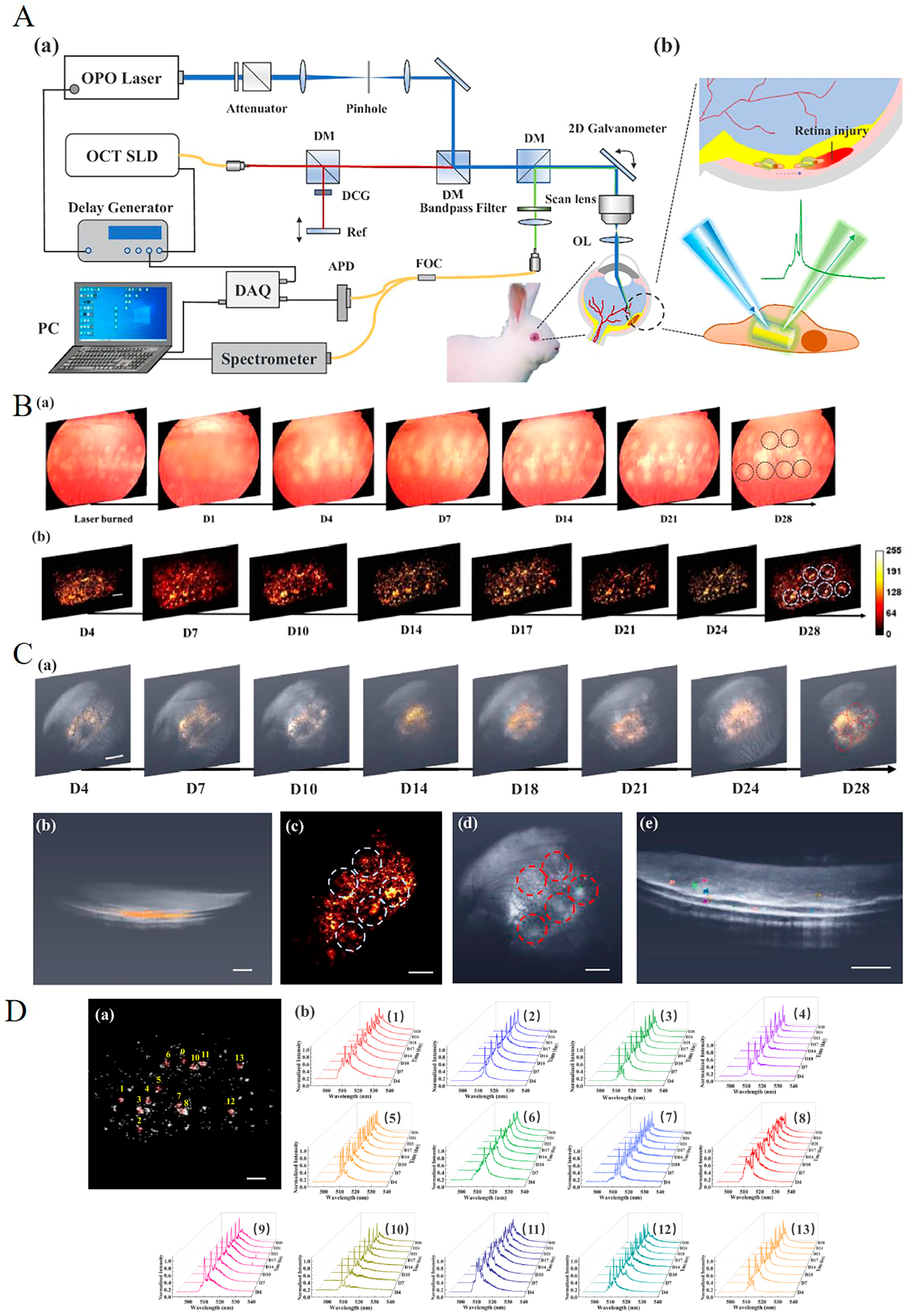
Figure 1. (Aa) Schematic of an optical coherence tomography (OCT), fluorescence microscopy (FM), and lasing emission nanowire multimodality imaging system. APD: avalanche photodiode; DAQ: data acquisition card; DCG: dispersion-compensation glass; FOC: fiber optic coupler; SLD: superluminescent diode; DM: dichroic mirror; OL: ophthalmic lens; OPO: optical parametric oscillator. (Ab) Illustration of CdS nanowire-labeled ARPE-19 cells migrating in the subretinal layer and generating lasing emission for tracking identification. (Ba) A series of color fundus images of the experimental retina region with laser-induced injuries. The images were acquired at different time points over a duration of 28 days. CdS nanowire-labeled ARPE-19 cells were injected on day 1 and were periodically observed starting from day 4 after the injected fluid was fully resorbed. (Bb) A series of FM images collected from the same experimental retinal region as in (Ba) at different time points after the retina region was clear to observe (from day 4). Scale bar: 200 µm. The black circles and blue circles in (Ba) and (Bb), respectively, highlight the locations of laser-induced injury spots. (Ca) A series of OCT X-Y plane projection images of the same region as in (B) over the period of 28 days. The overall retinal structure is rendered in grayscale, and nanowire-labeled ARPE-19 cells after thresholding are highlighted in yellow. (Cb) The side view of the 3D rendering of D21 shows that the extracted cells are mostly distributed in the subretinal space. Scale bar: 800 µm. (Cc) The corresponding FM image of the same region. The distribution pattern of the fluorescence signal was used to cross-validate the extracted OCT pattern in (Ca), which confirmed the location of CdS nanowire lasers. Scale bar: 800 µm. (Cd) The labeled cells corresponding to (Da) were highlighted with various colors in the OCT 3D volume rendering. The exact spatial coordinates were extracted from OCT. Scale bar: 800 µm. The gray and red circles in (Ca), (Cc), and (Cd), respectively, highlighted the corresponding laser-induced retina injury spots. (Ce) A side view of the 3D rendering of the same cells in (Cc). Scale bar: 500 µm. (Da) The instantaneous 2D locations of CdS nanowire-labeled ARPE-19 cells extracted from the FM image on D4. In total, 13 cells were tracked in this result. (Db) Normalized lasing spectra from the 13 identified ARPE-19 cells acquired at different time points during the period of 28 days. Each cell identity was presented by a unique lasing spectral waveform which was stable over the entire observation period. Reprinted (adapted) with permission from [80]. Licensed under CC BY 4.0.
Photonics 12 00413 g001
Figure 2. Custom-built photoacoustic microscope (PAM), OCT, and fluorescence multimodality imaging system, stem cell transplantation procedure, and synthesis procedure of functionalized chainlike gold nanoparticle clusters: (A) Schematic diagram of the imaging system and in vivo experiment protocol. Green laser photocoagulation was carried out to induce RPE and retinal injury. Then, multimodality PAM and OCT imaging were used to monitor the labeled ARPE-19 cells injected into the subretinal space for 90 days postinjection. Two different excitation wavelengths of 578 and 650 nm were applied to acquire PAM images. (B) Schematic diagram of ICG@CGNP cluster–RGD synthesis. Adapted with permission from [68]. Copyright {2024} American Chemical Society.
Photonics 12 00413 g002
Figure 3. (A) hiPSC-RPE cell migration and RPE replacement assessment: (Aa) Color fundus photography obtained in vivo before and after cell transplantation. Ddotted circles indicate the regions of the RPE injury caused by laser photocoagulation. The black area on the fundus images exhibits the distribution of hiPSC-RPE cells after transplantation. (Ab) Fluorescent images of the hiPSC-RPE cells labeled with ICG-GNC-RGD. (Ac,Ad) Location and migration pattern of the transplanted hiPSC-RPE cells were observed by capturing PAM images at 578 nm, enabling visualization of both the morphology of native retinal vessels and the transplanted stem cell differentiated to RPE (Ac), and at 650 nm to distinguish the transplanted stem cell differentiated to RPE from the vasculature (Ad). Pseudogreen color depicts the migration pattern of the transplanted hiPSC-RPE cells. (Ae) Combined 3D PAM images at 578 and 650 nm. (Af) Quantitative normalized PA signal amplitudes were obtained at multiple time points for three different treatment groups: hiPSC-RPE cells labeled with GNCs, unlabeled hiPSC-RPE cells, and dead hiPSC-RPE cells. (Ag) Quantitative normalized fluorescent intensities were obtained at multiple time points for two different treatment groups: hiPSC-RPE cells labeled with GNCs and dead hiPSC-RPE cells. Data are shown as mean ± SD (n = 3). (B) In vivo multimodal imaging of the transplanted hiPSC-RPE cells: (Ba) Schematic of subretinal transplantation. (Bb) Color fundus photo showing major retinal vessels. (Bc) Fluorescent image of the transplanted hiPSC-RPE cells labeled with ICG-GNC-RGD. The white color indicates the distribution of the transplanted hiPSC-RPE cells. (Bd,Be) Two-dimensional PAM images obtained at 578 nm (Bd) and 650 nm (Be), respectively. (Bf) Combined 2D PAM and OCT image. (BgBi) Three-dimensional volumetric PAM images. (BjBl) Combined 3D PAM and OCT images obtained at different time points: day 60 (Bj), day 90 (Bk), and day 180 (Bl), and post-transplantation. The green color shows the migration pattern of the transplanted hiPSC-RPE cells. (Bm) Selected 2D OCT image. (Bn) Combined 2D OCT and PAM image. (Bo) Combined 3D OCT and PAM image. (C) In vivo multimodal imaging of transplanted dead hiPSC-RPE cells: (CaCj) Color fundus photography, fluorescence, PAM images obtained at 578 and 650 nm, and 2D and 3D OCT images acquired before and after transplantation at different time points: 1, 3, 7, 14, 21, 28, 60, 90, and 180 days. (D) In vivo multimodal imaging of unlabeled hiPSC-RPE cells: (Da) Color fundus photographs. (Db) Fluorescent images. (Dc) PAM images obtained at 578 nm. (Dd) PAM images acquired at 650 nm. White arrows demonstrate the distribution of hiPSC-RPE cells. Reprinted (adapted) with permission from [38]. Copyright {2024} American Chemical Society.
Photonics 12 00413 g003
Table 1. Cell-labeling agents in retinal stem cell therapy.
Labeling AgentTypical Labeling AgentsAdvantagesDisadvantagesImaging Techniques
DirectFluorescent dyeICGSimple; applicable for multiple imaging modalitiesPhotobleaching; phototoxicity; not suitable for long-term monitoring; autofluorescence interference; false positivesFI
NanoparticleGNPsRelatively simple; easy to synthesize and modify to achieve different optical properties; easy to combine with other labeling agents; can be used for a variety of imaging techniques; potential for long-term imaging; GNPs have antiangiogenic and anti-inflammatory propertiesPotential toxicity and impact on cell function; exocytosis; false positives; potential long-term side effectsOCT
IndirectReporter geneSPIONsMore stable and long-lasting imaging signalsComplexity; high cost; delayed signal expression; immune response; toxicity; genome alteration and gene silencingFI; PAI
Indocyanine green (ICG); fluorescence imaging (FI); optical coherence tomography (OCT); photoacoustic imaging (PAI); green fluorescent protein (GFP); superparamagnetic iron oxide nanoparticles (SPIONs); gold nanoparticles (GNPs); superparamagnetic iron oxide nanoparticles (SPIONs); magnetic resonance imaging (MRI).
Table 2. Imaging techniques in retinal stem cell therapy.
Imaging TechniquesAdvantagesDisadvantagesApplications in Retinal Stem Cell Therapy
PreclinicalFIHigh resolution; real-time imaging; multi-fluorescence detection ability2D imagingTracking stem cells
PAIHigh resolution; high sensitivity; greater imaging depth; 3D imagingDifficult to distinguish transplanted cells from the background; artifactsTracking stem cells
MRI3D imaging; no depth limitation; capable of penetrating opaque tissues; long-term imagingLimited sectional structure of the retina; small scanning range; high cost and complexity of setting up the systemTracking stem cells
ClinicalOCTHigh resolution; real-time imaging; cross-sectional imaging; 3D imaging; non-invasive; widely used in clinical trialsPhotobleaching; phototoxicity; autofluorescence interferenceMonitoring morphology change; assisting cell delivery
Others
(FP, SLO, FAF, ICGA, FA)
Widely used in clinical trials; safe for long-term monitoringRelatively low resolution for retinal imaging; advantages come from cell-labeling agents (toxicity, degradation); complex and costly; contraindications of MRIAssessing efficacy and safety of stem cell therapy
Fluorescence imaging (FI); optical coherence tomography (OCT); photoacoustic imaging (PAI); two-dimensional (2D); three-dimensional (3D); fundus photography (FP); scanning laser ophthalmoscopy (SLO); fundus autofluorescence (FAF); indocyanine green angiography (ICGA); fluorescein angiography (FA).
Disclaimer/Publisher’s Note: The statements, opinions and data contained in all publications are solely those of the individual author(s) and contributor(s) and not of MDPI and/or the editor(s). MDPI and/or the editor(s) disclaim responsibility for any injury to people or property resulting from any ideas, methods, instructions or products referred to in the content.