Article

Dark Noise Suppression of NIR Response Enhanced Si-CMOS Sensor

Beijing Institute of Space Mechanics & Electricity, Beijing 100094, China
*
Author to whom correspondence should be addressed.
Photonics 2022, 9(5), 307; https://doi.org/10.3390/photonics9050307
Submission received: 17 March 2022 / Revised: 28 April 2022 / Accepted: 28 April 2022 / Published: 30 April 2022
(This article belongs to the Section Lasers, Light Sources and Sensors)

Abstract

:
We studied the effect of laser fluence on the dark noise performance of a laser-microstructured Si-based CMOS image sensor. The absorption characteristics and crystal properties of the microstructured sensor fabricated under different process conditions were investigated. Furthermore, a short-time etching method capable of improving the electrical performance of the laser-microstructured sensor was proposed. By removing amorphous silicon (a-Si) containing a large number of defects in the photosensitive surface of the microstructured Si-based CMOS image sensor, the etching method can effectively suppress the dark noise of the laser-microstructured Si-photodetector while maintaining the near-infrared response enhancement effect of the Si-photodetector irradiated by fs-laser. The results of the near-infrared imaging test show that on the basis of imaging brightness enhancement, the contrast ratio of the image formed by the CMOS image sensor in the microstructured region etched by RIE under short exposure time is significantly improved.

1. Introduction

Expanding the photoresponse spectrum of silicon-based detectors to the near-infrared band for applications in aerospace, optical communications, and autonomous driving is desired [1,2,3,4,5,6,7,8,9]. Silicon (Si), one of the familiar semiconductor materials, has become the cornerstone of the semiconductor industry with its abundant reserves, low cost, and mature technology. Because of the limitation of the indirect band gap of 1.12 eV, the absorption coefficient of silicon is extremely low at the near-infrared (NIR) band [10]. Femtosecond laser (fs-laser) irradiation is an effective means to extend the absorption limit of silicon to the near-infrared region by inducing an anti-reflection microstructure [11] on the surface of Si and incorporating chalcogenide hyperdoping [12,13,14] into the lattice of Si. Owing to the absorption of the laser-hyperdoping Si in both visible and near-infrared bands being extremely high, the microstructured material is also called black silicon.
In recent years, researchers have conducted considerable studies focusing on the near-infrared (NIR) photoresponse enhanced Si-photodetector fabricated by femtosecond (fs) laser irradiation [15,16,17,18,19,20]. Unfortunately, research into NIR laser-microstructured Si-photodetectors still faces some problems that need to be solved urgently. One of the thorniest issues is that the dark noise performance of the laser-microstructured device will be extremely deteriorated while the near-infrared response is enhanced. During fs-laser microstructuring, amorphous silicon-containing high-density structural defects [21,22,23,24,25] are introduced into the surface layer of sulfur-doped black silicon, which can cause the dark noise of the microstructured device to increase sharply. The direct result of this unfavorable factor is that the contrast ratio of the image formed by the microstructured imaging sensor decreases rapidly as the integration time increases. The poor noise performance of the laser-microstructured Si-photodetector severely limits the application of sulfur-hyperdoped black silicon in near-infrared photodetection.
In this article, the effect of laser fluence on the optical and electrical properties of a microstructured Si-sensor is mainly studied. Firstly, we explored the influence of laser fluence on the dark noise of the laser-microstructured Si-based CMOS image sensor. We then investigated the absorption characteristics and crystal properties of the untreated, laser-microstructured, and etched laser-microstructured CMOS image sensor. Furthermore, we proposed a short-time etching method capable of improving the electrical performance of the laser-microstructured sensor. By removing amorphous silicon (a-Si) containing a large number of defects in the photosensitive surface of the microstructured Si-based CMOS image sensor, the etching method can effectively suppress the dark noise of the laser-microstructured Si-photodetector while maintaining the near-infrared response enhancement effect of the Si-photodetector irradiated by fs-laser. The imaging test shows that the near-infrared imaging ability of the microstructured imaging sensor has been greatly improved after short-time etching treatment.

2. Materials and Methods

Commercial optical CMOS image sensors based on silicon were irradiated with Ti:sapphire fs-laser pulses (800 nm wavelength, 100 fs pulse duration, 150 μm spot size) at normal incidence in an SF6 ambiance with a pressure of 7 × 10⁴ Pa. After fs-laser processing, the microstructured sensor was etched by reactive ion etching (RIE) using SF6 plasma under 35 W power and 20 mTorr pressure to study its optical and electrical properties.
The surface morphology of samples was characterized by using scanning electron microscopy (SEM). To investigate the absorption characteristics of microstructured Si-devices, the integrated reflectance (R) and transmittance (T) spectra between 950 and 1100 nm were obtained by a Lambda-1050 spectrometer (PerkinElmer, Waltham, MA, USA) equipped with a 160 mm integrating sphere. Then the integrated absorptance (A) spectra were extracted through A = 1 − R − T. The confocal Raman spectroscopy with a HeNe laser of 632 nm excitation wavelength was used to measure the crystallinity of the laser-microstructured sensor before and after the etching treatment. In order to characterize the electrical performance of the samples, the laser-microstructured sensors were placed in a dark environment, and their dark noise performance was tested by reading the gray value of the output signal of the device at a specific integration time. In addition, the detector spectral responsivity measurement system (DSR 100) was used to characterize the spectral response of the sample and further calculate the quantum efficiency of the sensor.

3. Results and Discussion

In order to study the effect of laser energy on the electrical performance of the device, sulfur-hyperdoped silicon-based microstructures were fabricated on the surface of a commercial optical CMOS image sensor by using fs-laser irradiation with different fluence. A microstructured Si-based sensor is shown in Figure 1a. We can clearly observe that the microstructured surface of the detector has two distinctive features: a quasi-periodic micron-cone array and nanoscale granular structures covering the surface of the microconical spikes. On the one hand, the generation of this special surface morphology is caused by nonlinear optical effects such as self-focusing caused by the interaction between the femtosecond laser and silicon. On the other hand, under the action of ultra-high intensity laser, the fluorine atoms generated by SF6 multi-photon photodissociation can react with silicon to generate volatile products, which plays a role in etching, making the microstructure of sulfur supersaturated doping so sharp. Areas 1 and 2 are microstructured regions fabricated by the fs-laser with a fluence of 0.3 J/cm2 and 0.35 J/cm2, respectively. The gray value measurements of the laser-microstructured areas (marked as Area 1 and Area 2) and unprocessed area (marked as Normal) of the CMOS image sensor are shown in Figure 1b. Since the sensor is placed in a dark environment for gray value reading, the obvious increase in the gray value of the processing area means a sharp increase in the dark noise of these areas after femtosecond laser processing. The ultra-high dark noise makes the imaging effect of the microstructured sensor extremely poor, as shown in Figure 1c.
After a single crystalline silicon is ablated by a femtosecond laser, a laser-induced periodic surface structure (LIPSS) will be generated on the surface of the excited region of the material. The top layers at the top of this kind of microstructure are occupied by a large number of amorphous and polymorphic states [25], and these characteristic lattice defects are able to introduce trap energy levels in forbidden bands to form effective high-density recombination centers. This kind of deep level as the composite center will randomly capture or emit the electron–hole pair, resulting in generation-composite noise. Therefore, the dark noise performance of the microstructured regions deteriorates sharply after femtosecond laser irradiation. For the microstructured Si fabricated by ultrafast laser, the defects introduced during processing cannot be avoided. For the shape-specific microstructure of LIPSS, we speculate that reactive ion etching (RIE) can uniformly remove the disordered layer at the top of the micrometer-sized conical spike without destroying the morphology of the anti-reflection structure. The fabrication process of femtosecond laser hyperdoped microstructured Si-CMOS image sensor chip is explored. The gray value test results of the devices prepared by different processes at room temperature are shown in Figure 2a and Table 1.
The gray value in the microstructured area of the sensor becomes larger as the laser fluence increases, which means an increase in dark noise in the laser-processing region of the device. It is not difficult to see from Table 1 that continuous reactive ion etching in a short time can effectively reduce the intensity of the generated-composite noise, thereby improving the electrical properties of the device. We will next discuss in detail the physical reasons behind the generation of this phenomenon.
The inset of Figure 2a shows the SEM characterization results of the microstructured surface before (Area 6) and after etching (Area 7). Compared with the samples without etching treatment, the morphology of microstructured silicon changed significantly after 50 s etching: A large number of nanostructures covering the surface of the spikes were removed, and the average height of the periodically distributed microarray with sharper shape decreased to 2 μm. Under the action of high-energy ion bombardment, the microstructure is gradually peeled in a more uniform proportion in all directions except the bottom. We believe that this phenomenon is mainly caused by the following factors: the plasma concentration distribution is quite different in the etching process, so the removal amount near the top of the microstructure is significantly greater than that at the bottom; moreover, a large number of secondary structures such as nanoparticles covered on the surface of the microstructure can prevent the bombardment of high-energy ions on the bottom of the cone to a certain extent. The combined effect of these factors makes the microstructure maintains an antireflective conical structure after short-time etching.
The NIR absorption spectra of microstructured areas and the normal area are shown in Figure 3. Due to the limitation of the indirect band gap of 1.12 eV, the average absorptance of the commercial optical CMOS image sensor at wavelengths from 950 to 1100 nm is 26%. After laser processing, the NIR average absorptance of the Si-sensor increased to 88% at a fluence of 0.2 KJ/m2 (Area 6). With the laser fluence rising to 0.95 KJ/m2 (Area 4), the average NIR absorptance in the microstructured region increases to 97%. Unfortunately, although the average absorptance of the microstructured device increased by 10% in the wavelength ranges 950–1100 nm, the noise performance of the device deteriorated significantly. For the best electrical performance of the Si-photodetector in the near-infrared band, we needed to find a balance between the NIR light responsiveness and dark noise of the Si-device by controlling the etching time. Areas 4 and 5, Areas 6 and 7, and Areas 8 and 9 are three sets of comparative areas to investigate the effect of etching treatment on the optoelectronic properties of the laser-microstructured sensor. By comparing the NIR absorptance of the comparative areas, we found that a short etching time did not result in a significant drop in NIR absorptance of the microstructured regions. The NIR absorption spectral characteristics indicate that the hyperdoped layer covering the top of the conical microstructure, which dominates the ultra-high NIR absorptance of the fs-laser hyperdoped Si, is removed by a very small amount. More strikingly, the Raman spectra of microstructured Areas 6 and 7 illustrate that the crystal quality of the laser-irradiated area was significantly improved after short-time etching, as shown in Figure 3.
Compared with the Raman mode of the monocrystalline silicon, a disorder layer containing a large amount of amorphous silicon (a-Si) was formed on the surface of the sensor after fs-laser irradiation, which shows that an ordered–disordered phase transition occurs in the excited region of the Si-surface during the ultrafast interaction between the fs-laser and single crystalline silicon. Therefore, the hyperdoped black silicon showed several a-Si peaks (wide peaks near 150 cm−1 and 460–490 cm−1) in the Raman spectra. It is remarkable that the Raman peak of a-Si basically disappears after RIE etching; that is, the disorder layer on the device surface is effectively removed. The Raman mode of the black Si-sensor etched by RIE is close to the test results of conventional crystalline silicon (C-Si). This means that the a-Si generated by femtosecond laser irradiation on the surface layer of the laser-irradiated material is converted into monocrystalline silicon. The Raman characterization serves as strong evidence that the laser-induced damage layer induced by femtosecond laser irradiation is effectively removed through RIE treatment, which is in agreement with the dark noise measurement results, as shown in Table 1.
In addition, we would like to further discuss the difference in crystal structure damage between RIE and fs-laser ablation treatment. According to the paper of LEE et al. [26], commonly, the thickness of the RIE-damaged layer varies from a few nanometers to tens of nanometers depending on the etching conditions. In contrast, the thickness of the disordered layer induced by femtosecond laser ablation varies from a few hundred nanometers to a few micrometers from the TEM images, according to a study by Mazur et al. [27]. Our Raman measurements also show that the Si crystallinity only treated by RIE is close to that without any treatment. The Raman measurements illustrate that the crystal damage caused by RIE is much smaller than that caused by femtosecond laser irradiation. This is why the short-time RIE etching can effectively repair the fs-laser-irradiated damage by peeling off the laser-induced disorder layer containing a large amount of amorphous silicon.
Based on the study of optical and electrical properties of the microstructured Si-photodetector, a novel microstructured CMOS image sensor is prepared by fs-laser irradiation combined with short-time RIE etching, as shown in Figure 4a. At room temperature, the gray value test results of the microstructured device placed in the darkroom are shown in Figure 4b,c and Table 2. There is no obvious difference in dark noise performance between the microstructured area etched by RIE and the untreated area in a short integration time. Although the dark noise in the laser-microstructured region increases slightly when the integration time is increased to 100 ms, it is negligible compared with the test results in Figure 1 (the integration time is 41 ms). As shown in Figure 4d, the near-infrared average absorptance of the microstructured image sensor at wavelengths from 950 to 1100 nm decreased from 84% to 80% after a short-time etching. NIR absorption spectra show that the laser-microstructured device etched by RIE still has high near-infrared optical responsiveness.
The ratio of the quantum efficiency of the black silicon CMOS image sensor before and after etching in the range of 950–1100 nm is shown in Figure 4e. The ratio of the quantum efficiency, Rη, can be calculated from the following formula:
R η = η η 0 1
where η is the quantum efficiency of laser-microstructured CMOS image sensor before and after etching, and η0 is the quantum efficiency of the untreated commercial silicon CMOS image sensor.
As we expected, the slight drop in NIR absorptance of the laser-microstructured image sensor etched by RIE did not adversely affect the NIR response of the microstructured photodetector. Figure 4e shows that in the near-infrared region from 950 to 1100 nm, the quantum efficiency of the novel black silicon CMOS image sensor is still significantly improved (nearly 30%) compared with the commercial silicon CMOS image sensor without processing. More importantly, it can be seen from Table 2 that the gray value of the microstructured Si-detector at an integration time of 100 ms dropped by 37% after short-time etching treatment. The NIR quantum efficiency and the measurement results of gray value indicate that short-time continuous etching treatment can effectively suppress the dark noise of the laser-microstructured Si-photodetector while maintaining the near-infrared response enhancement effect of the Si-photodetector irradiated by fs-laser.
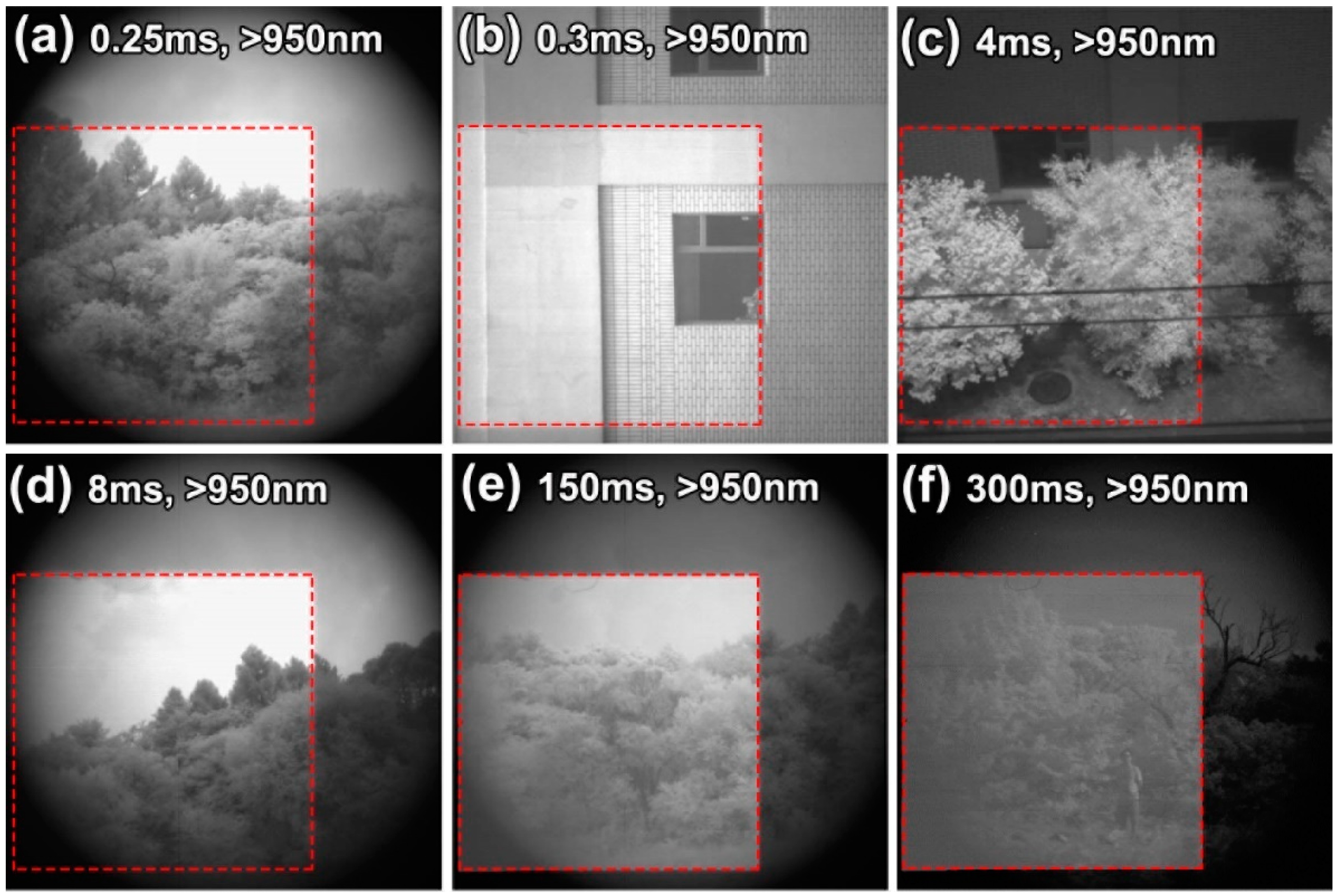
In order to verify the near-infrared detection ability of the laser-microstructured Si-CMOS image sensor after etching, the imaging performance of the device in the outdoor environment was tested. The camera imaging wavelength is greater than 950 nm. The test results are shown in Figure 5. The results of the near-infrared imaging test show that on the basis of imaging brightness enhancement, the contrast ratio of the image formed by the CMOS image sensor in the microstructured region etched by RIE under short exposure time is significantly improved. This indicates that the near-infrared imaging ability of the CMOS image sensor has been significantly improved after fs-laser irradiation combined with RIE etching.

4. Conclusions

In conclusion, the influence of fs-laser fluence on the dark noise of a laser-microstructured Si-based CMOS image sensor is demonstrated. We found that the electrical performance of the fs-laser-microstructured CMOS sensor was improved by RIE etching. The Raman characterization results show that short-time etching treatment improves the crystal quality of the photosensitive surface layer by peeling off the top disorder layer containing a large amount of amorphous silicon. The measurement results of gray value and NIR quantum efficiency indicate that the dark noise performance of the laser-microstructured CMOS sensor has been greatly improved while maintaining the enhanced NIR response. The NIR imaging test results show that on the basis of imaging brightness enhancement, the contrast ratio of the image formed by the CMOS image sensor in the microstructured region etched by RIE under a short exposure time is considerably increased. The method of fs-laser irradiation combined with short-time RIE treatment is very valuable for the manufacture of NIR-Si-CMOS sensors.

Author Contributions

Conceptualization, K.W.; methodology, K.W. and C.P.; software, K.W.; formal analysis, K.W. and Z.H.; supervision, Z.H.; writing, K.W. and C.P. All authors have read and agreed to the published version of the manuscript.

Funding

This research received no external funding.

Data Availability Statement

Not applicable.

Conflicts of Interest

The authors declare no conflict of interest.

References

  1. Knight, M.W.; Sobhani, H.; Nordlander, P.; Halas, N.J. Photodetection with active optical antennas. Science 2011, 332, 702–704. [Google Scholar] [CrossRef] [PubMed]
  2. Nazirzadeh, M.A.; Atar, F.B.; Turgut, B.B.; Okyay, A.K. Random sized plasmonic nanoantennas on silicon for low-cost broad-band near-infrared photodetection. Sci. Rep. 2014, 4, 7103. [Google Scholar] [CrossRef] [PubMed] [Green Version]
  3. Lin, K.T.; Chen, H.L.; Lai, Y.S.; Yu, C.C. Silicon-based broadband antenna for high responsivity and polarization-insensitive photodetection at telecommunication wavelengths. Nat. Commun. 2014, 5, 3288. [Google Scholar] [CrossRef] [PubMed] [Green Version]
  4. Mark, W.K.; Yumin, W.; Alexander, S.U.; Ali, S.; Bob, Y.Z.; Peter, N. Embedding Plasmonic Nanostructure Diodes Enhances Hot Electron Emission. J. Nano Lett. 2013, 13, 1687–1692. [Google Scholar]
  5. Kang, Y.; Liu, H.D.; Morse, M.; Paniccia, M.; Zadka, M.; Litski, S.; Sarid, G.; Pauchard, A.; Kuo, Y.H.; Chen, H.W.; et al. Monolithic germaniumsilicon avalanche photodiodes with 340 GHz gain-bandwidth product. J. Nat. Photonics 2009, 3, 59–63. [Google Scholar] [CrossRef]
  6. Sobhani, A.; Knight, M.W.; Wang, Y.; Zheng, B.; King, N.S.; Brown, L.V. Narrowband photodetection in the near-infrared with a plasmon-induced hot electron device. J. Nat. Commun. 2013, 4, 1643. [Google Scholar] [CrossRef] [PubMed] [Green Version]
  7. Chandra, A.; Giri, S.; Das, B.; Ghosh, S.; Chattopadhyay, K.K. NIR photodetector based on p-silicon nanowires/n-cadmium sulfide nanoscale junctions. J. Appl. Surf. Sci. 2021, 548, 149256. [Google Scholar] [CrossRef]
  8. Ajiki, Y.; Kan, T.; Yahiro, M.; Hamada, A.; Adachi, J.; Adachi, C.; Matsumoto, K.; Shimoyama, I. Silicon based near infrared photodetector using self-assembled organic crystalline nano-pillars. J. Appl. Phys. Lett. 2016, 108, 151102. [Google Scholar] [CrossRef]
  9. Casalino, M.; Coppola, G.; Sirleto, L.; Iodice, M.; Gioffre, M.; Rendina, I.; Sassi, U.; Lombardo, A.; Milana, S.; Sundaram, R.S. NIR silicon Schottky photodetector: From metal to graphene. In Proceedings of the 2014 Fotonica AEIT Italian Conference on Photonics Technologies, Naples, Italy, 12–14 May 2014; IEEE: Piscataway, NJ, USA, 2014; pp. 1–4. [Google Scholar]
  10. Miskovsky, N.M.; Cutler, P.H.; Mayer, A.; Weiss, B.L.; Brian, W.; Sullivan, T.E.; Lerner, P.B. Nanoscale devices for rectification of high frequency radiation from the infrared through the visible: A new approach. J. Nanotechnol. 2012, 2012, 512379. [Google Scholar] [CrossRef] [Green Version]
  11. Bonse, J.; Krüger, J.; Höhm, S.; Rosenfeld, A. Femtosecond laser-induced periodic surface structures. J. Laser Appl. 2012, 24, 042006. [Google Scholar] [CrossRef]
  12. Her, T.H.; Finlay, R.J.; Wu, C.; Deliwala, S.; Mazur, E. Microstructuring of silicon with femtosecond laser pulses. Appl. Phys. Lett. 1998, 73, 1673–1675. [Google Scholar] [CrossRef]
  13. Her, T.H.; Finlay, R.J.; Wu, C.; Mazur, E. Femtosecond laser-induced formation of spikes on silicon. Appl. Phys. A 2000, 70, 383–385. [Google Scholar] [CrossRef]
  14. Wu, C.; Crouch, C.H.; Zhao, L.; Carey, J.E.; Younkin, R.; Levinson, J.A.; Mazur, E. Near-unity below-band-gap absorption by microstructured silicon. Appl. Phys. Lett. 2001, 78, 1850. [Google Scholar] [CrossRef]
  15. Carey, J.E.; Crouch, C.H.; Shen, M.; Mazur, E. Visible and near-infrared responsivity of femtosecond-laser microstructured silicon photodiodes. Opt. Lett. 2005, 30, 1773. [Google Scholar] [CrossRef]
  16. Myers, R.A.; Farrell, R.; Karger, A.M.; Carey, J.E.; Mazur, E. Enhancing near-infrared avalanche photodiode performance by femtosecond laser microstructuring. Appl. Opt. 2006, 45, 8825. [Google Scholar] [CrossRef]
  17. Huang, Z.; Carey, J.E.; Liu, M.; Guo, X.; Mazur, E.; Campbell, J.C. Microstructured silicon photodetector. Appl. Phys. Lett. 2006, 89, 516. [Google Scholar] [CrossRef]
  18. Wang, X.; Huang, Y.; Liu, D.; Zhu, X.; Wang, B.; Zhu, H. Fabrication of tellurium doped silicon detector by femtosecond laser and excimer laser. Chin. J. Lasers 2013, 40, 0302001. [Google Scholar] [CrossRef]
  19. Du, L.; Wu, Z.; Rui, L.; Fei, T.; Jiang, Y. Near-infrared photoresponse of femtosecond laser processed Se-doped silicon n+ − n photodiodes. Opt. Lett. 2016, 41, 5031. [Google Scholar] [CrossRef]
  20. Li, C.H.; Zhao, J.H.; Yu, X.Y.; Chen, Q.D.; Feng, J.; Han, P.D.; Sun, H.B. Sulfur-Doped Silicon Photodiode by Ion Implantation and Femtosecond Laser Annealing. IEEE Sens. J. 2017, 17, 2367. [Google Scholar] [CrossRef]
  21. Mo, Y.; Bazant, M.Z.; Kaxiras, E. Sulfur point defects in crystalline and amorphous silicon. Phys. Rev. B 2004, 70, 3352. [Google Scholar] [CrossRef]
  22. Dong, X.; Li, N.; Liang, C.; Sun, H.; Feng, G.; Zhu, Z.; Shao, H.; Rong, X.; Zhao, L.; Zhuang, J. Strong Mid-Infrared Absorption and High Crystallinity of Microstructured Silicon Formed by Femtosecond Laser Irradiation in NF3 Atmosphere. Appl. Phys. Express 2013, 6, 1301. [Google Scholar] [CrossRef]
  23. Smith, M.J.; Sher, M.J.; Franta, B.; Lin, Y.T.; Mazur, E.; Gradecak, S. The origins of pressure-induced phase transformations during the surface texturing of silicon using femtosecond laser irradiation. J. Appl. Phys. 2012, 112, 083518. [Google Scholar] [CrossRef]
  24. Smith, M.J.; Lin, Y.T.; Sher, M.J.; Winkler, M.T.; Mazur, E.; Gradecak, S. Pressure-induced phase transformations during femtosecond-laser doping of silicon. J. Appl. Phys. 2011, 110, 053524. [Google Scholar] [CrossRef]
  25. Schade, M.; Varlamova, O.; Reif, J.; Blumtritt, H.; Erfurth, W.; Leipner, H.S. High-resolution investigations of ripple structures formed by femtosecond laser irradiation of silicon. Anal. Bioanal. Chem. 2010, 396, 1905. [Google Scholar] [CrossRef] [PubMed] [Green Version]
  26. Lee, Y.H.; Oehrlein, G.S.; Ransom, C. RIE-induced damage and contamination in silicon. Radiat. Eff. Defects Solids 1989, 111–112, 221–232. [Google Scholar] [CrossRef]
  27. Crouch, C.H.; Carey, J.E.; Shen, M.; Mazur, E.; Genin, F.Y. Infrared absorption by sulfur-doped silicon formed by femtosecond laser irradiation. Appl. Phys. A 2004, 79, 1635–1641. [Google Scholar] [CrossRef]
Figure 1. (a) Photograph of CMOS image sensor irradiated by fs-laser. (b) Gray value measurements of laser-microstructured areas (marked as Area 1 and Area 2) and unprocessed area (marked as Normal) of CMOS image sensor at an integration time of 41 ms. (c) Comparison of near-infrared imaging performance between microstructured area and unprocessed area of CMOS image sensor at an integration time of 41 ms. The imaging conditions (shooting time, weather, location, and light intensity) are 9 pm, cloudy, outdoor, and weak light intensity.
Photonics 09 00307 g001
Figure 2. (a) Gray value measurements of microstructured areas (irradiated with different laser fluences) and unprocessed area of CMOS image sensor at an integration time of 82 ms. The size of the photosensitive surface of the sensor is 22.5 × 22.5 mm. Inset: Side view SEM images of Area 6 and Area 7. (b) NIR absorption spectra of microstructured areas of CMOS image sensor. The NIR absorptance of an untreated normal area of CMOS image sensor is included for comparison.
Photonics 09 00307 g002
Figure 3. Raman spectra of laser-microstructured Areas 6 and 7. A Si only treated by RIE and a monocrystalline Si (C-Si) are given as a comparison.
Photonics 09 00307 g003
Figure 4. (a) Photograph of microstructured CMOS image sensor, microstructured area are marked with a red dashed line. (b,c) Gray value measurements of microstructured area and unprocessed area on the surface of microstructured CMOS image sensor at different integration times: (b) 4 ms; (c) 100 ms. (d) NIR absorption spectra of microstructured area of CMOS image sensor before and after short-time etching. The NIR absorptance of an untreated normal area of CMOS image sensor is included for comparison. (e) Quantum efficiency ratio versus wavelength for microstructured CMOS image sensor before and after short-time etching. The ratio of an untreated commercial silicon CMOS image sensor is included for reference.
Photonics 09 00307 g004
Figure 5. Comparison of near-infrared imaging performance between microstructured area and unprocessed area of CMOS image sensor at different integration times. The imaging conditions (shooting time, weather, location, light intensity, and integration time) corresponding to (ae) are (a) 7 am, cloudy, outdoor, low light intensity, 0.25 ms; (b) 7 am, sunny, indoor, strong light intensity, 0.3 ms; (c) 7 am, sunny, outdoor, strong light intensity, 4 ms; (d) 7 pm, cloudy, outdoor, low light intensity, 8 ms; (e) 8 pm, cloudy, outdoor, weak light intensity, 150 ms; (f) 9 pm, cloudy, outdoor, weak light intensity, 300 ms, respectively.
Photonics 09 00307 g005
Table 1. Gray value measurements of microstructured areas (fabricated under different process conditions) and unprocessed area of CMOS image sensor at an integration time of 82 ms.
Influence (kJ/m2)Etching Time (s)Gray Value
Normal--260
Area 10.3-3100
Area 20.35-3200
Area 30.45-3600
Area 40.95-4100
Area 50.952003500
Area 60.2-2800
Area 70.2502300
Area 80.38-3300
Area 90.38502900
Table 2. Gray value measurements of microstructured area and unprocessed area of CMOS image sensor at an integration time of 100 ms.
Influence (kJ/m2)Etching Time (s)Gray Value
Unprocessed Area--290
Microstructured Area0.13-1900
Microstructured Area0.13501200
Publisher’s Note: MDPI stays neutral with regard to jurisdictional claims in published maps and institutional affiliations.