Article

Pyridine-2,6-Dicarboxylic Acid Esters (pydicR2) as O,N,O-Pincer Ligands in CuII Complexes

by
Katharina Butsch
1,
Aaron Sandleben
1,
Maryam Heydari Dokoohaki
2,
Amin Reza Zolghadr
2,* and
Axel Klein
1,2,*
1
Department für Chemie, Institut für Anorganische Chemie, Universität zu Köln, Greinstraße 6, D-50939 Köln, Germany
2
Chemistry Department Faculty of Science, Shiraz University, Shiraz 71454, Iran
*
Authors to whom correspondence should be addressed.
Inorganics 2019, 7(4), 53; https://doi.org/10.3390/inorganics7040053
Submission received: 15 February 2019 / Revised: 31 March 2019 / Accepted: 9 April 2019 / Published: 14 April 2019
(This article belongs to the Special Issue First-Row Transition Metal Complexes)

Abstract

:
The pyridine-2,6-carboxylic esters pydicR2 with R = Me or Ph form the unprecedented mononuclear CuII complexes [Cu(pydicR2)Cl3] in one-pot reactions starting from pyridine-2,6-carboxychloride pydicCl2, CuII chloride, and NEt3 in MeOH or PhOH solution under non-aqueous conditions. The triethylammonium salts (HNEt3)[Cu(pydicR2)Cl3] were isolated. The methyl derivative could be crystallized to allow a XRD structure determination. Both structures were optimized using DFT calculations in various surroundings ranging from gas phase and the non-coordinating solvent CH2Cl2 to the weakly coordinating acetone and well-coordinating solvents acetonitrile (MeCN) or dimethylformamide (DMF), while detailed calculation showed the charge distribution, dipole moments, and HOMO–LUMO gap energies changing upon solvation. According to these calculations, the ion pairs and the anionic CuII complexes were stable, which shows only Cu–Cl bond elongation and weakening of the charge transfer between the anionic complex and the cation as solvents become polar. Synthesis attempts in the presence of water yielded the CuII complexes [Cu(pydic)(OH2)2]n and [Cu(OH2)6][{Cu(pydic)}2(µ-Cl)2], which results from pydicCl2 hydrolysis. Alternatively, the new pydic(IPh)2 (IPh = 2-iodo-phenyl) ester ligand was synthesized and reacted with anhydrous CuCl2, which yields the new binuclear complex [{Cu(pydic(IPh)2)Cl}2(µ-Cl)2]. EPR spectroscopy of the solid compounds reveals typical axial spectra in line with the observed and DFT calculated geometries. Cyclic voltammetry and UV–vis absorption spectroscopy in solution are in line with un-dissociated complex species [Cu(pydicR2)Cl3].

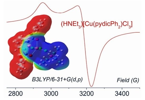
Graphical Abstract

1. Introduction

Pyridine-2,6-dicarboxylic esters are interesting oxido pincer ligands with an O,N,O donor set. They are derived from the potentially O,N,O tridentate binding 2,6-dicarboxylic acid (pydicH2) (Scheme 1). Pyridine-2-6-dicarboxylate (pydic2−) is a frequently used ligand and coordinates are usually in a deprotonated tridentate mode. The coordinates use the N donor atom and the two carboxylate O donor functions, including the possibility of bridging (Scheme 1) [1,2,3,4,5,6,7,8,9,10,11,12,13,14]. In contrast to this, the pydic ester ligands are neutral and have three options of tris-chelate O,N,O binding, which gives rise to three different isomers Ocarbonyl–M–Ocarbonyl (CC isomer), Ocarbonyl–M–Oalkoxy (CA isomer), and Oalkoxy–M–Oalkoxy (AA isomer) (Scheme 1) while a bridging mode observed frequently for pydic2− is less probable [15,16,17].
Pyridine-2,6-dicarboxylic alkyl- or arylester derivatives pydicR2 coordinating to metal ions have been reported with R = methyl [15,16,17,18,19,20,21], ethyl [18,20,22,23,24,25,26], iso-propyl [18], n-butyl [26], and benzyl [18], with further complexes containing related non-symmetric or macrocyclic esters RpydicR’ [23,24,27,28,29,30]. Aryl substituted systems are less frequent [27]. In most of these examples, the ligands coordinate in a tridentate mode. However, bidentate N,O coordination or monodentate N coordination has been observed [15,16,20,21,28]. Recently, Hakimi et al. found in the Cambridge Crystallographic Database that, from the complexes showing tridentate O,N,O coordination, roughly 42% of the submitted structures show Ocarbonyl–M–Ocarbonyl coordination (CC isomers, Scheme 1) [15], while AA (Oalkoxy–M–Oalkoxy) and CA (Ocarbonyl–M–Oalkoxy) isomers make up about 29% each. The CC isomers are expected to be superior to the CA and AA isomers since the carbonyl function provides less steric strain and potentially π back-bonding options in addition to the σ coordination of the O lone pairs. Quite generally, both Ocarbonyl and Oalkoxy coordination is considered to be relatively weak, e.g., compared to the carboxylate ligand function of pydic2−. Ester functions in polydentate ligands have been applied as hemilabile ligand functions in catalysis [31,32,33,34,35].
In this contribution, we will report on our investigations about the coordination of CuII toward the pyridine-2-6-dicarboxylic ester pydicR2 ligands with R = Me, Ph, and 2-iodo-phenyl (IPh). We report their synthesis and compare their molecular structures in the solid using single crystal XRD with the structures under various environments (gas phase, solution) using quantum chemical calculations based on the density functional theory (DFT). In these calculations, we were probing for their structural variability in terms of isomers, ion pair stabilization, or ligand replacement by potentially coordinating solvents such as MeCN and DMF. Detailed DFT calculations also revealed the influence of increasing solvent polarity on the dipole moments and charge distribution in the ion pairs. Since CuII (d9, S = ½) provides very useful spectroscopic (d–d bands, EPR) and electrochemical (CuII/CuI couple) patterns to study geometries and the strength of M–ligand bonds, we added EPR spectroscopy in the solid and solution to this study, together with UV–vis absorption, cyclic voltammetry, and UV–vis spectroelectrochemistry of selected compounds in the solution.

2. Results and Discussion

2.1. Synthesis and Molecular Structures of pydicR2 Ligands and their CuII Complexes

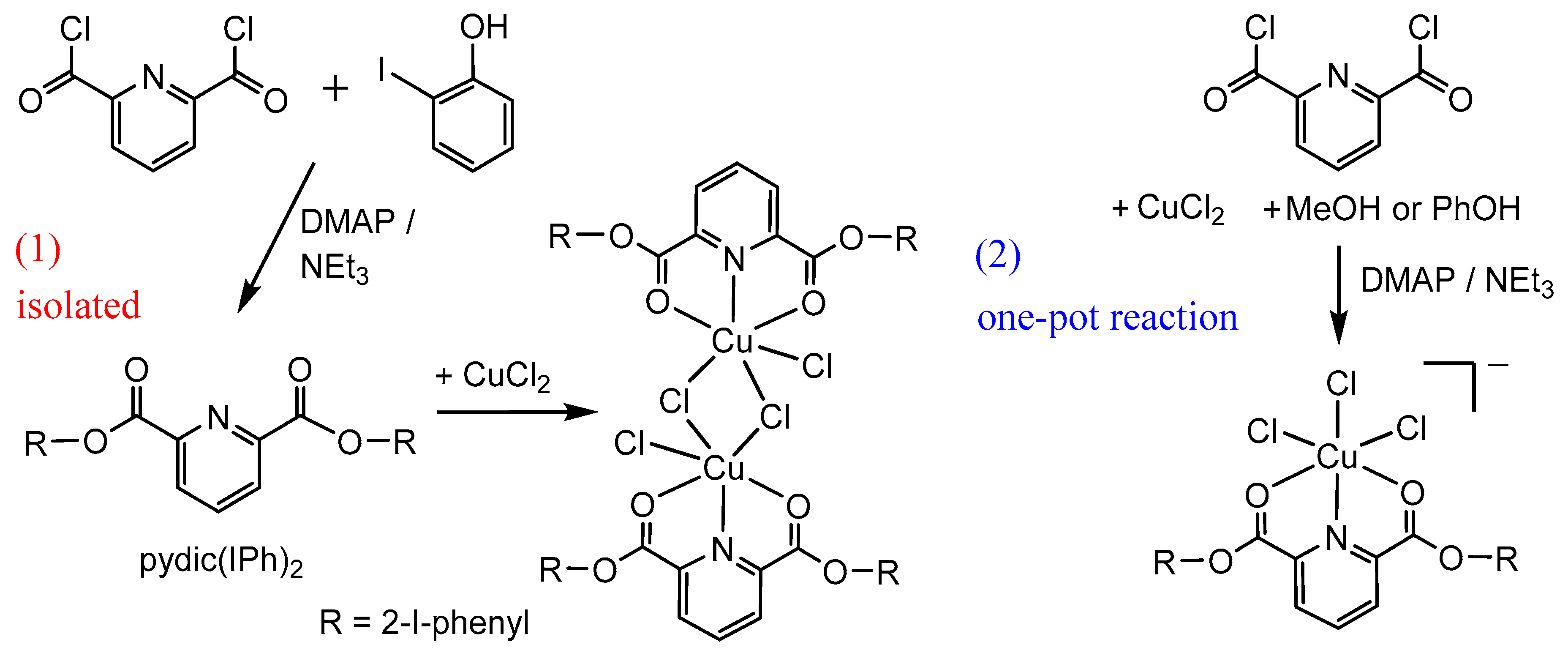
Two different reaction strategies were used to synthesize CuII complexes (Scheme 2). The first route has been previously used, e.g., for the formation of pydic ester complexes [{Cu(pydicR2)Cl}2(µ-Cl)2] (R = Me, Et, iPr) [17,22] and consists of two subsequent steps, the synthesis of the pydicR2 ligand, and its isolation followed by the complex formation reaction (1).
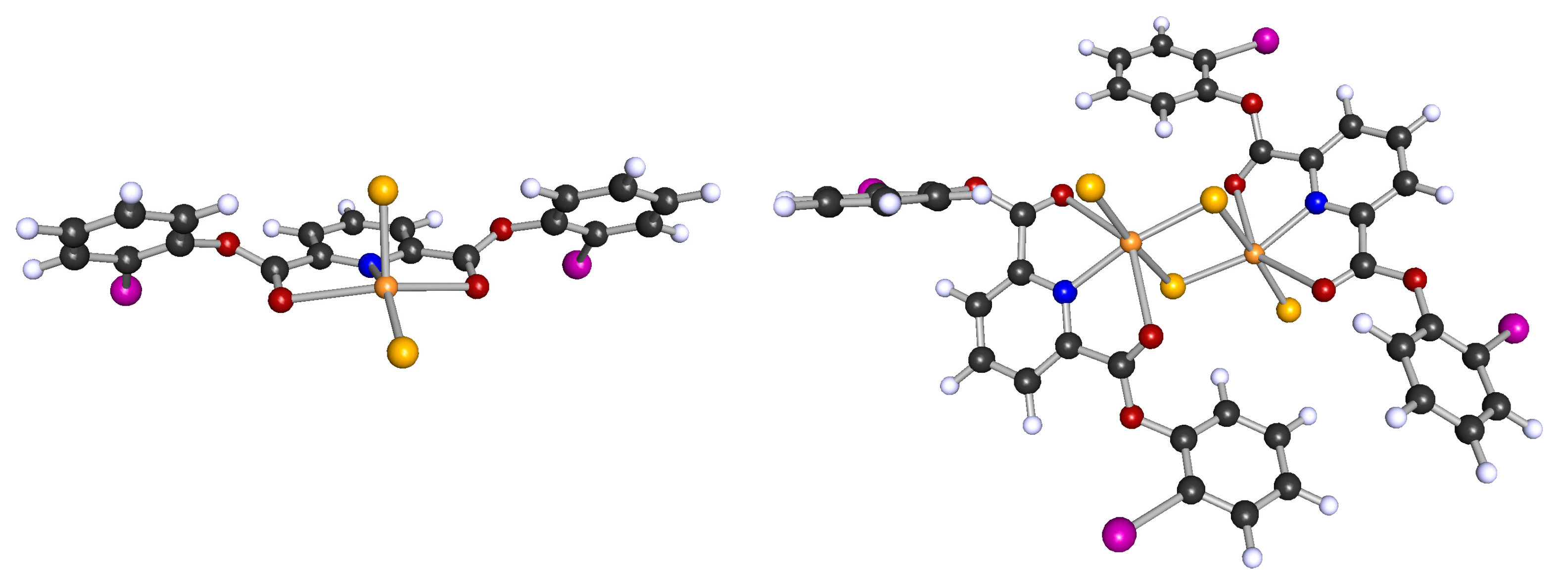
This method allowed the synthesis of the new ligand pydic(IPh)2 (IPh = 2-iodo-phenyl) in 82% yield (spectroscopic characterization, see Experimental Section). Single crystals suitable for XRD were obtained from acetone solutions by slow evaporation and the structure was solved in the orthorhombic space group Pbca (further details, full data, and Figure S1 in the Supplementary Materials). Reacting pydic(IPh)2 with anhydrous CuCl2 gave a new complex as a brown powder in 67% yield with a ligand:Cu ratio of 1:1 from elemental analysis. Similar reactions using pydicMe2, pydicEt2, or pydiciPr2 have been reported, which yielded binuclear complexes [{Cu(pydicR2)Cl}2(µ-Cl)2] [17,22,23]. We assume a binuclear structure for [{Cu(pydic(IPh)2)Cl}2(µ-Cl)2]. DFT-calculated optimized geometries confirmed this with the binuclear structure being more stable by about 75 kJ/mol compared with the mononuclear [Cu(pydic(IPh)2)Cl2] (Figure 1). Importantly, both the mononuclear and the binuclear complex show the CC isomeric structure (Scheme 1) with the two carbonyl O atoms binding to CuII in the two markedly elongated axial positions of a distorted trigonal pyramidal (mononuclear) or pseudo-octahedral (binuclear) coordination. Comparison of the calculated coordination modes CC, CA, and AA shows that the CC isomer is energetically favored.
In addition to this, we had the idea to set up a one-pot reaction using pyridine-2,6-dicarbonyl dichloride (pydicCl2), a CuII source, and the corresponding alcohol (methanol or phenol) to form the ligand in the presence of the coordinating metal ion and a base (2). The base was quite similar to what has been reported recently for the formation of lanthanide coordinated IRMOF-3-PY through a post-synthetic modification [5].
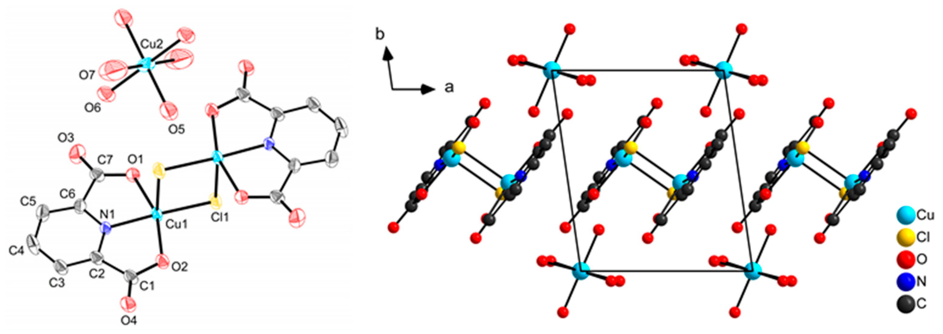
In initial experiments, we reacted CuCl2·2H2O and not carefully dried Cu(OAc)2, methanol or phenol, and pydicCl2 in a 1:1 ratio. In low yields of 12% and 24%, respectively, we could isolate [Cu(OH2)6][{Cu(pydic)}2(µ-Cl)2] and [Cu(pydic)(OH2)2]n (pydic2− = pyridine-2-6-dicarboxylate). Both compounds resulted from the hydrolysis of pydicCl2 to pydic2− in the presence of water while no ester was formed. The crystal structure (monoclinic P21/c) of [Cu(pydic)(OH2)2]n was reported before [36,37]. XRD suitable crystals of [Cu(OH2)6][{Cu(pydic)}2(µ-Cl)2] were obtained by slow evaporation of a methanol solution. The crystal structure was solved and refined in the triclinic space group P 1 ¯ (Figure 2, further details and full data in the Supplementary Materials). The structure reveals the complex cation [Cu(OH2)6]2+ and a centrosymmetric binuclear µ-chlorido bridged dianionic complex [{Cu(pydic)}2(µ-Cl)2]2−. [Cu(OH2)6]2+ can be described as an axially elongated octahedron (Cu2–O5 = 1.979(9) Å, Cu2–O6 = 1.965(9) Å, and Cu2–O11 = 2.53(2) Å), which is in line with previous reports [38,39]. The di-anion [{Cu(pydic)}2(µ-Cl)2]2− shows a distorted square pyramidal geometry around each copper atom with a short equatorial bond Cu–Cleq = 2.212(4) Å, long axial bond Cu–Clax = 2.692(3) Å, and a rather short equatorial Cu–N bond (1.93(1) Å). Very similar bond parameters have been reported for the dianion [{Cu(pydic)}2(µ-Cl)2]2− in the compounds [Cu(MeOH)(H2O)4][Cu2(pydic)2(µ-Cl)2]·H2O [40] and [Cu(Pz)2(H2O)4][Cu2(pydic)2(µ-Cl)2] (Pz = pyrazole) [41].
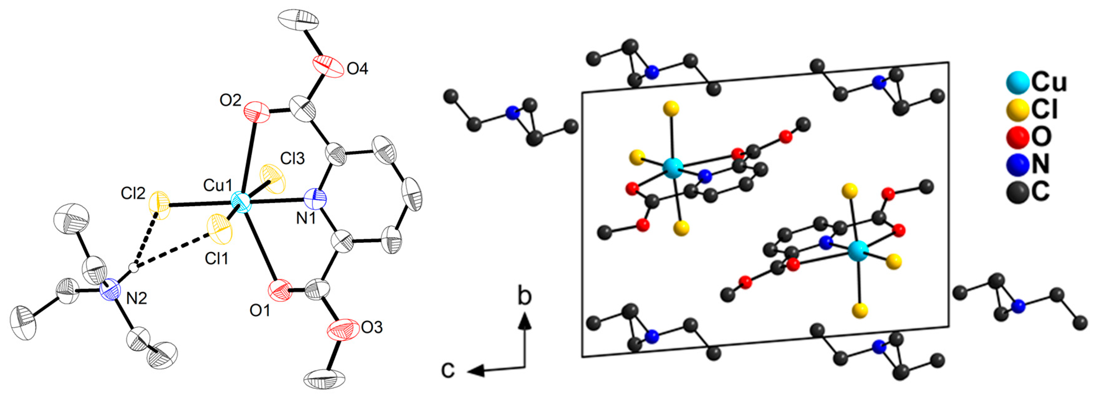
In further synthesis experiments using freshly distilled starting materials and anhydrous CuCl2, we obtained yellow-green crystalline materials. Repeated re-crystallisation from methanol was necessary but lowered the yields to 32% and 54% of the complexes (HNEt3)[Cu(pydicMe2)Cl3] and (HNEt3)[Cu(pydicPh2)Cl3]. Thus, under water-free conditions, the in situ formation of the corresponding pydicR2 ligands and coordination to CuII (one-pot) is possible. Single crystals of (HNEt3)[Cu(pydicMe2)Cl3] were obtained from methanol solution and structure solution and refinement were carried out in the triclinic space group P 1 ¯ (data in the Supplementary Materials). The molecular structure is depicted in Figure 3 and shows the Cu ion in a distorted octahedral surrounding the carbonyl oxygen atoms of the O,N,O-ester binding to the copper atom (CC isomer). The three Cu–Cl bonds with 2.306(2) Å, 2.255(2) Å, and 2.306(2) Å and the Cu–N1 bond of 2.065(4) Å define a distorted square plane. The two Cu–O bonds Cu–O2 = 2.503(4) Å and Cu–O4 = 2.551(3) Å are non-equivalent and represent the long bonds of an axially elongated pseudo-octahedron. The real local symmetry at the Cu atom can be idealized as C2v. Two strong hydrogen bonds [42] are formed between the triethyl ammonium ion and two chlorido ligands of the complex anion (H22···Cl1 = 2.70(4) Å and H22···Cl2 = 2.48(4) Å, Cl1···H22···Cl2 = 79(1)°).
Comparison of (HNEt3)[Cu(pydicMe2)Cl3] with the reported complex [Cu(pydicMe2)(H2O)3](ClO4)2 [17] reveals the same bond parameters within the pydicMe2 ligand, similar Cu-ligand distances in the equatorial plane: Cu–N 1.990(3) Å, Cu–OH2 1.992(3), 1.949(3), and 2.000(3) Å, respectively, and long Cu–Oketo bonds of 2.332(3) and 2.338(3) Å. Thus, both Cl in [Cu(pydicMe2)Cl3] and H2O in [Cu(pydicMe2)(H2O)3] represent stronger ligands than the carbonyl ester O atoms.

2.2. DFT Calculations on the Solvation of (HNEt3)[Cu(pydicR2)Cl3] (R = Me or Ph)

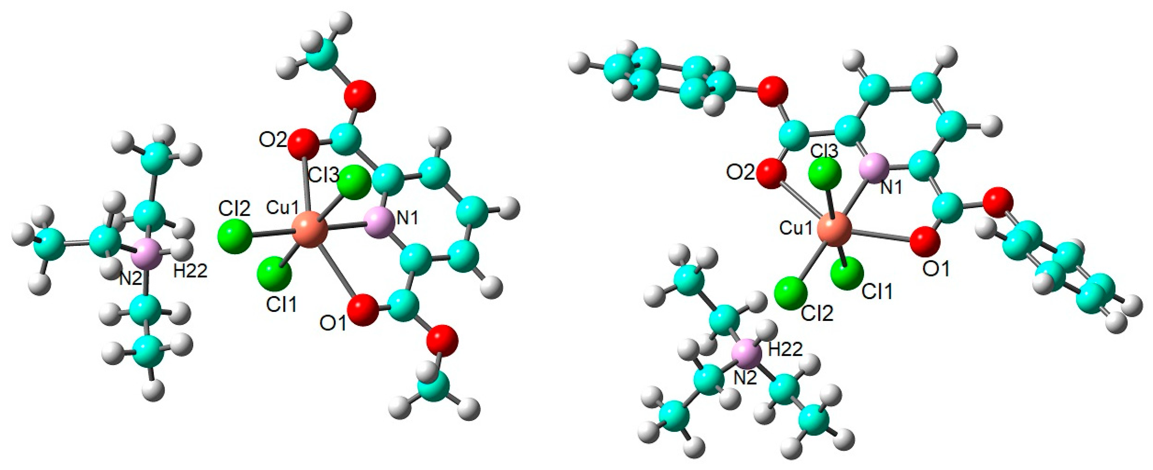
To achieve insight into the electronic structure of the compounds, density functional theory (DFT) calculations in the gas phase and various solvent environments are performed (B3LYP/6–31+G(d,p) using Gaussian). The Cu atom was treated using LANL2DZ relativistic pseudo potentials. The final optimized geometry of the compounds in the gas phase is depicted in Figure 4. The calculated C2v-symmetric structure of the ion pair (HNEt3)+···[Cu(pydicMe2)Cl3] in the gas phase is very similar to the crystal structure, e.g., with two strong hydrogen bonds N2–H22···Cl1 (2.98 Å) and N2–H22···Cl2 (2.01 Å).
In CH2Cl2, acetone, DMF, and MeCN solution, the geometry of the complexes remains rather intact. Only the Cu–Cl3 bond is markedly elongated with a slight tendency of longer bonds for more polar solvents along this series. Even more pronounced is the increasing distance between the ions due to the shielding of electrostatic interactions in these solutions (see Tables S3–S7 in the Supplementary Materials). The computed dipole moments of the complexes are significantly increased in solution compared with the gas phase (Table 1) and the electric permittivity of the solvents clearly has a very pronounced effect on the dipole moments, even in CH2Cl2. In order to investigate the solubility and thermodynamic stability of (HNEt3)[Cu(pydicR2)Cl3] in the surrounding medium, the solvation energies are calculated. The solvation energy of the system is determined by calculating the free energy differences in the solvent and the gas phase (Table 1). The solvation energies vary from about −110.88 to −135.12 kJ/mol and increase together with the dipole moment of the complex when increasing the electric permittivity of the solvents. Negative values of solvation energies denote that solvation of these complexes is spontaneous. To get an idea about the kinetic stability and chemical reactivity of these complexes, we also calculated the difference between the highest occupied molecular orbital (HOMO) and the lowest unoccupied molecular orbital (LUMO) (ELUMOEHOMO) for the complexes in each solvent (Table 1). The spatial distributions of HOMO, SOMO, and LUMO of the complexes are supplied in the Supplementary Materials (Figure S8). In general, molecules with a small energy gap are more polarizable [43] and will have higher chemical activity and lower kinetic stability [44]. Overall, the HOMO−LUMO gap decreases as the electric permittivity of the solvents declines along the series DMF (ε = 37.22) > MeCN (ε = 35.69) >> acetone (ε = 20.49) >> CH2Cl2 (ε = 8.93). However, the absolute values are quite similar for all four solvents, which means that a pronounced activation by the polar solvents DMF or MeCN compared with acetone or CH2Cl2 seems not to be the case.
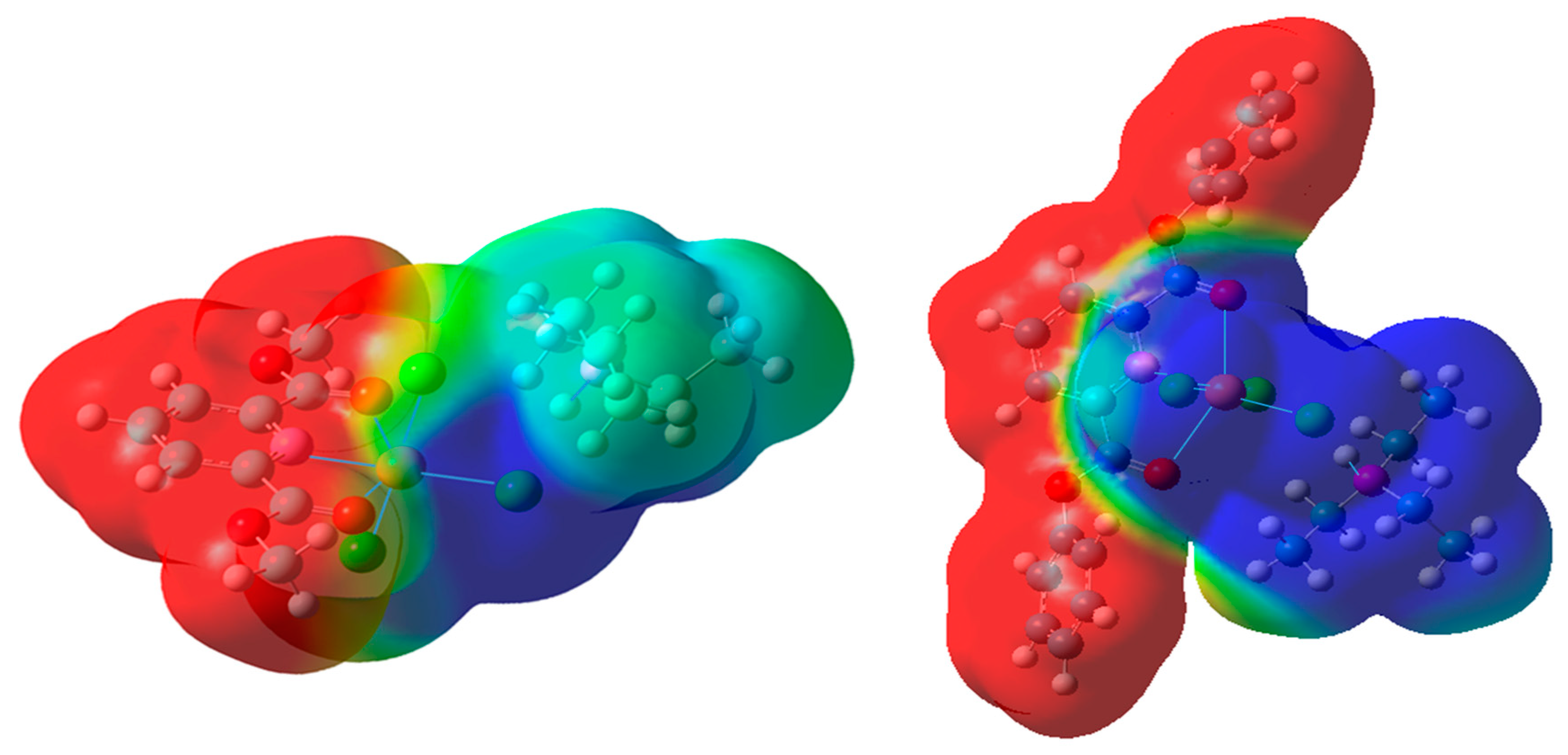
The electron density of (HNEt3)[Cu(pydicMe2)Cl3] and (HNEt3)[Cu(pydicPh2)Cl3] in the gas phase can be visualized from the three-dimensional electrostatic potential map (EPM) surfaces (Figure 5). The distribution of negative and positive charges within the EPM surfaces are depicted by red and blue colors, respectively. As a result of change in electron densities in EPM surfaces upon solvation, the charges of the anions and cations both decrease along the series. DMF > MeCN > acetone > CH2Cl2 >> gas phase (Table 1, Figures S9 and S10 in the Supplementary Materials).
To better understand the inclusion of the solvent effect on the DFT calculations and also the nature of interactions in the (HNEt3)[Cu(pydicR2)Cl3] compounds, natural bond orbital (NBO) analyses were carried out as implemented in the Gaussian program package. In the NBO analysis, the donor and acceptor interaction energies can be estimated through the second-order perturbation theory, which is described by the equation below.
E ( 2 ) = q i F ( i , j ) 2 ε i ε j
where qi is the donor orbital occupancy, εj and εi are diagonal elements, and F(i,j) is the off-diagonal NBO Fock matrix element. The most important donor-acceptor interactions with high second-order perturbation energies E(2) are provided in Table 2 (data for the pydicPh derivative in Table S8 in the Supplementary Materials).
As the interaction between (HNEt3)+ and [Cu(pydicR2)Cl3] as the electron acceptor and electron donor increases, the calculated E(2) value become larger, as expected. Furthermore, the increase in donating tendency from electron donors to electron acceptors is consistent with the enhancement of conjugation of the whole system from R = Me to R = Ph. The results suggest strong orbital interactions between the antibonding molecular orbitals of the Cu atom (LP*) and the antibonding orbitals of the N2–H22 bond (BD*) in the gas phase, which leads to a stabilization of 18.91 kJ/mol for (HNEt3)[Cu(pydicMe2)Cl3] and 21.30 kJ/mol for (HNEt3)[Cu(pydicPh2)Cl3], respectively. In solution with an effect of the solvents, this donor-acceptor behavior between (HNEt3)+ and [Cu(pydicMe2)Cl3] leads to a reduction of the intermolecular charge transfer with lower values of E(2) (1.66). The table also reveals that the E(2) in the LP*(8) Cu1 and the BD*(1) N2–H22 and also the LP(3) Cl2 and BD*(1) N2–H22 are decreasing with an increasing dielectric constant of the solvent. The NBO analysis results illustrate that, in the gas phase, LP(4) Cl2 and LP(4) Cl1 participate as donors and the LP*(7) Cu1 acts as an acceptor with the strongest intramolecular charge transfer interactions (Table 2). In solvent environments, the E(2) in the LP(4) Cl2 and LP*(7) Cu1 and also the E(2) in the LP(4) Cl1 and LP*(7) Cu1 are enlarged with increasing dielectric constants of the solvent. However, this trend for the E(2) in the LP(4) Cl3 and LP*(7) Cu1 are completely reversed. The trend of charge transfer from LP(1) O1, LP(1) O2, and LP(1) N1 as donors and LP* Cu1 as an acceptor are increased with a growing dielectric constant of the solvents.

2.3. Molecular Dynamics (MD) Simulations of Bulk Solutions

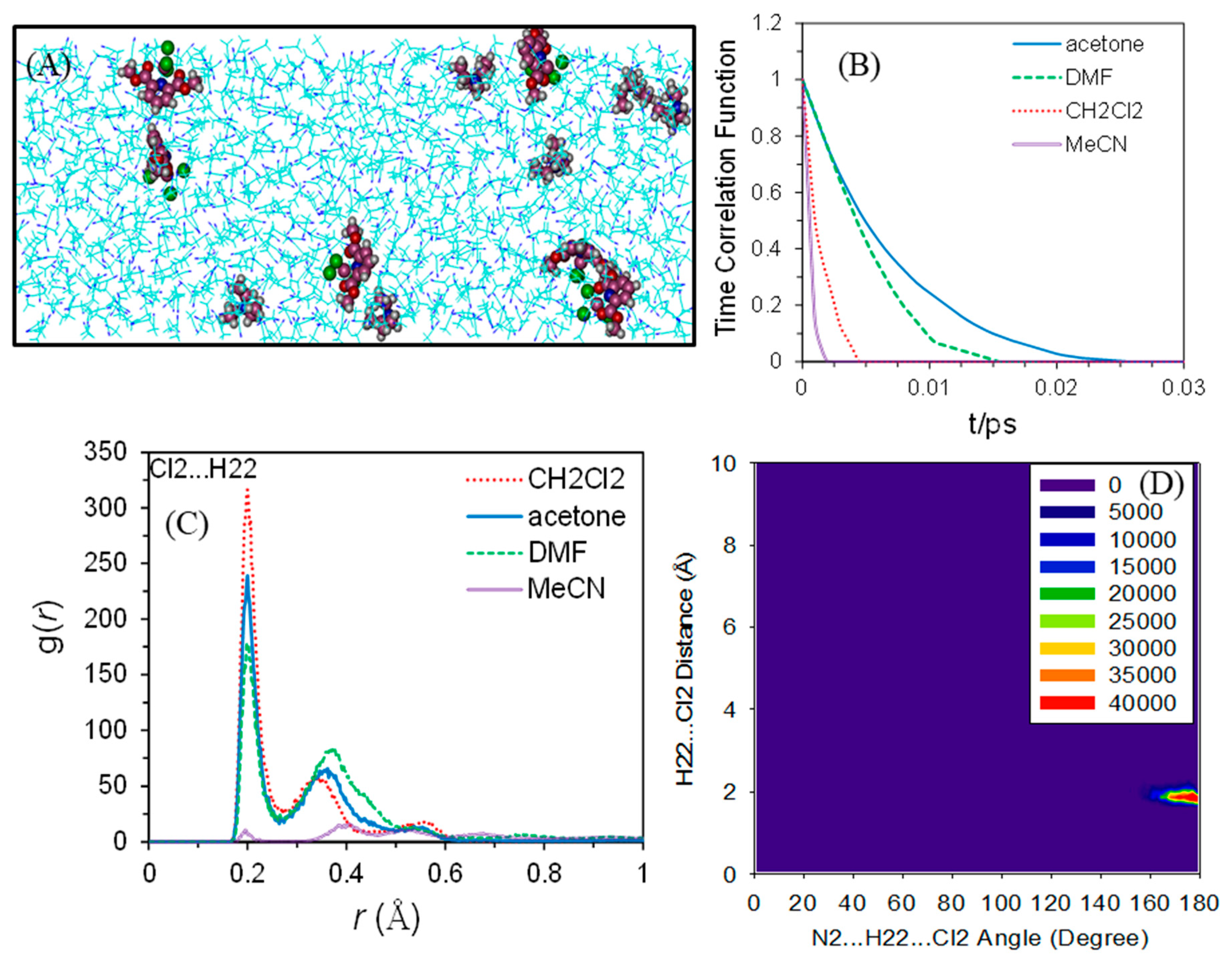
In addition, we also performed molecular dynamic simulations using the GROMACS 4.5.5 package and utilizing the GROMOS force field 53A6. Six ion pairs solvated in 1200 solvent molecules were simulated for each compound. As an example, Figure 6A shows the snapshot of the (HNEt3)+···[Cu(pydicMe2)Cl3] ion pairs solvated in MeCN after 10 ns of simulation. The anion-cation correlation lifetime in various solvents is depicted in Figure 6B. The lifetime of the hydrogen bond between the Cl2 atom of the anion and H22 of the cation for the (HNEt3)+···[Cu(pydicMe2)Cl3] system tends to diminish along the series acetone > DMF > CH2Cl2 > MeCN, which is not along the general polarity (permittivity) of these solvents. The radial distribution functions (g(r)) between the Cl2 atom of the anion and H22 of the cation are calculated and illustrated in Figure 6C for (HNEt3)+···[Cu(pydicMe2)Cl3] ion pairs in various solvents.
The first peak of the radial distribution function is located at around 1.84 Å, which is an indicator of a strong hydrogen bond. Combined radial/angular distribution function (CDF) is a powerful tool for defining hydrogen bond criteria, since it offers much more information than g(r) [45]. The combined radial/angular distribution function (see Figure 6D) indicates that this preferred interaction occurs for N2···H22···Cl2 angles (α) between 160° and 180°.
All of these calculated data points show a marked difference between the gas phase and solvent surrounding these ion pairs and emphasizing the importance of the proper inclusion of solvents in any DFT calculations. According to our calculations, the electronic structure of the studied solutes is influenced in a different manner by the non-coordinating CH2Cl2, the weakly coordinating acetone, and MeCN and DMF, which are considered suitable ligands for CuII. With increasing polarity along the series gas phase < CH2Cl2 < acetone < MeCN < DMF solutions, the Cl3–Cu bond is markedly elongated, the distance between the cations and anions are increased, and the dipole moments as well as the extent of charge localization of the cation and anion increases. However, the differences between CH2Cl2 and MeCN or DMF solutions are not very pronounced. Although more detailed kinetic calculations would be necessary to make a statement on the dissociation of the complexes in MeCN or DMF solution, we assume from the present calculations that the complexes remain stable in solution, which is important in view of the following spectroscopy and electrochemical measurements in the solution.

2.4. EPR Spectroscopy of Solids and Solutions

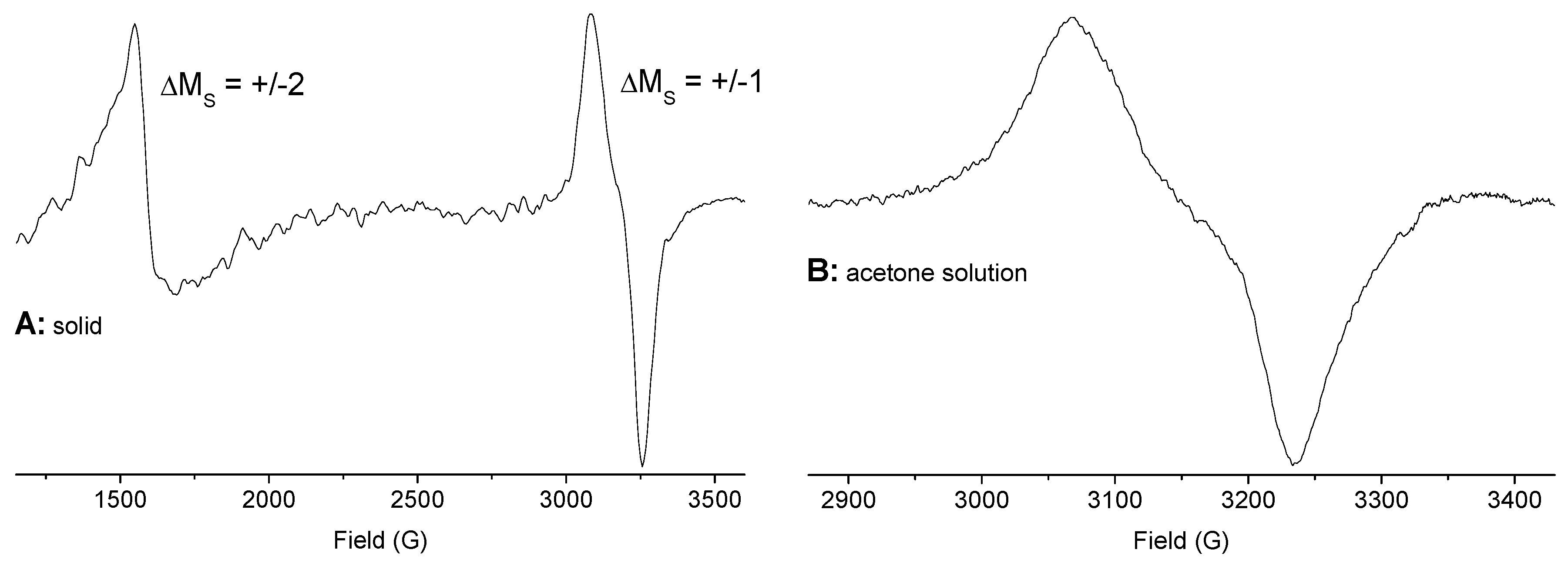
The X-band EPR spectrum of [{Cu(pydic(IPh)2)Cl}2(µ-Cl)2] in the solid shows a very intense signal for the forbidden ΔMS = ±2 transition and a rhombic ΔMS = ±1 signal (Figure 7), which is completely in line with the binuclear character of the complex and a triplet ground state. In acetone solution, the spectrum is essentially retained and, thus, the binuclear character remains unchanged. A similar spectrum with very similar g values has been reported for [{Cu(pydicEt2)Cl}2(µ-Cl)2] [23], while the derivative [{Cu(pydiciPr2)Cl}2(µ-Cl)2] exhibits a markedly different averaged g value (Table 3) and no ΔMS = ±2 transition is observed. The difference has been attributed to the different geometries for the central Cu(µ-Cl)2Cu unit, which is planar for the pydicEt2 complex but bent for the iso-propyl derivative [23]. Thus, our results are fully in line with the DFT-calculated structure of [{Cu(pydic(IPh)2)Cl}2(µ-Cl)2], which shows a planar Cu(µ-Cl)2Cu core (Figure 1). Although the averaged g value is quite similar for the related binuclear CuII complex[{Cu(pydotH2)Cl}2(µ-Cl)2] (pydotH2 = 2,6-bis(1-hydroxy-1-o-tolyl-ethyl-η2-O,O’)pyridine)[46], the g anisotropy Δg is markedly increased, while the ΔMS = ±2 is far less intense. This reflects the different “ligand setting” in this complex containing an ONO pincer ligand based on pyridine-2,6-dimethanol. As deduced from EXAFS data, the O, N, O atoms of the pydotH2 ligand and the bridging chlorido ligand form a square plane of the tightly bound ligands. From this bridging Cl, an elongated Cu–Cl bond connects to the second CuII, while the terminal chloride ligand has a short Cu–Cl bond [46]. Thus, while in the binuclear complexes [{Cu(pydicR2)Cl}2(µ-Cl)2] with R = Me, Et, iPr and IPh, the tight binding ligands are N, Cl, and 2× µ-Cl with the two square planes fused by the bridging µ-Cl atoms. In the pydotH2 complex, the two square planes O,N,O,µ-Cl and the central Cu(µ-Cl)2Cu core are perpendicular to each other. The EPR spectra fully reflect this difference.
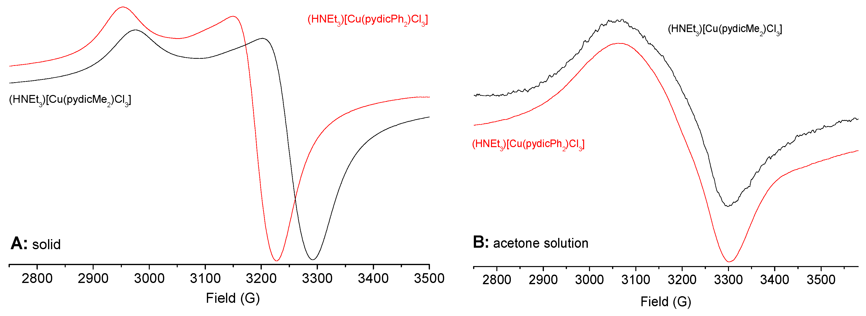
The mononuclearcompounds (HNEt3)[Cu(pydicMe2)Cl3] and (HNEt3)[Cu(pydicPh2)Cl3] reveal EPR spectra of axial symmetry with gǀǀ > g in the solid (Figure 8), which is typical for elongated octahedral or square pyramidal CuII complexes [23,47,48,49,50,51,52]. The Cu hyperfine structure (coupling to the 63Cu (69.17%) and 65Cu (30.83%) nuclei with I = 3/2 [53]) was not observed in line with observations for similar mononuclear complexes [46,52]. A rhombic EPR spectrum was reported for the complex [Cu(pydotH2)(DMF)Cl2] [46], which has an axially elongated distorted pseudo-octahedral geometry with the O,N,O ligand located in the square plane of tight Cu–ligand bonds. Yet, in (HNEt3)[Cu(pydicMe2)Cl3] and (HNEt3)[Cu(pydicPh2)Cl3], the N,Cl3 coordination represents the short = strong bonds and the Ocarbonyl functions are in the elongated positions. Nevertheless, the averaged g values and g anisotropy (Δg) are very similar. Far smaller averaged g values were reported for the square pyramid or trigonal bipyramidal complexes listed in Table 3. Thus, the observed spectra represent the overall geometry around CuII than the ligand strength of Cl, DMF, and O atoms in the O, N, O ligands.
Samples dissolved in acetone or MeCN (at ambient temperatures) exhibit axial signal symmetry even though the averaged g values and the g anisotropy (Δg) are markely decreased. We conclude that the ion pairs and the anionic complexes in (HNEt3)[Cu(pydicR2)Cl3] are basically retained in acetone or MeCN solution with minor alterations in complex geometry. This is completely in line with our DFT calculations showing changes in the geometry and charge transfer of the ion pairs upon solvation and likely shows no dissociation.

2.5. UV–Vis Absorption Spectroscopy

The binuclear complex [{Cu(pydic(IPh)2)Cl}2(µ-Cl)2] and the mononuclear compounds (HNEt3)[Cu(pydicMe2)Cl3] and (HNEt3)[Cu(pydicPh2)Cl3] dissolved in MeCN show intense UV bands around 290 or 260, respectively, which is assignable to ligand-centered π–π* transitions (Table 4, spectra in the Supplementary Materials). The three complexes all show a characteristic absorption band at around 460 nm, which is assignable to a ligand(Cl)-to-metal(Cu) charge transfer LMCT transition. In the visible range, broad bands assigned to d–d transitions (a2,b1,b2→a1 in C2v symmetry) were observed. Their exact energies are difficult to determine but lie around 820 nm for (HNEt3)[Cu(pydicMe2)Cl3], 890 nm for (HNEt3)[Cu(pydicPh2)Cl3], and 830 nm for [{Cu(pydic(IPh)2)Cl}2(µ-Cl)2]. These are in line with values recently reported for related CuII complexes with O,N,O ligands of the pyridine-2,6-dimethanol type [46,54]. In the spectrum of K2[CuCl4] in MeCN, the long wavelength d-d band is observed at 1074 nm, which is in line with a markedly stronger ligand field for the [Cu(pydicR2)Cl3] and [{Cu(pydic(IPh)2)Cl}2(µ-Cl)2] complexes.

2.6. Electrochemical Measurements

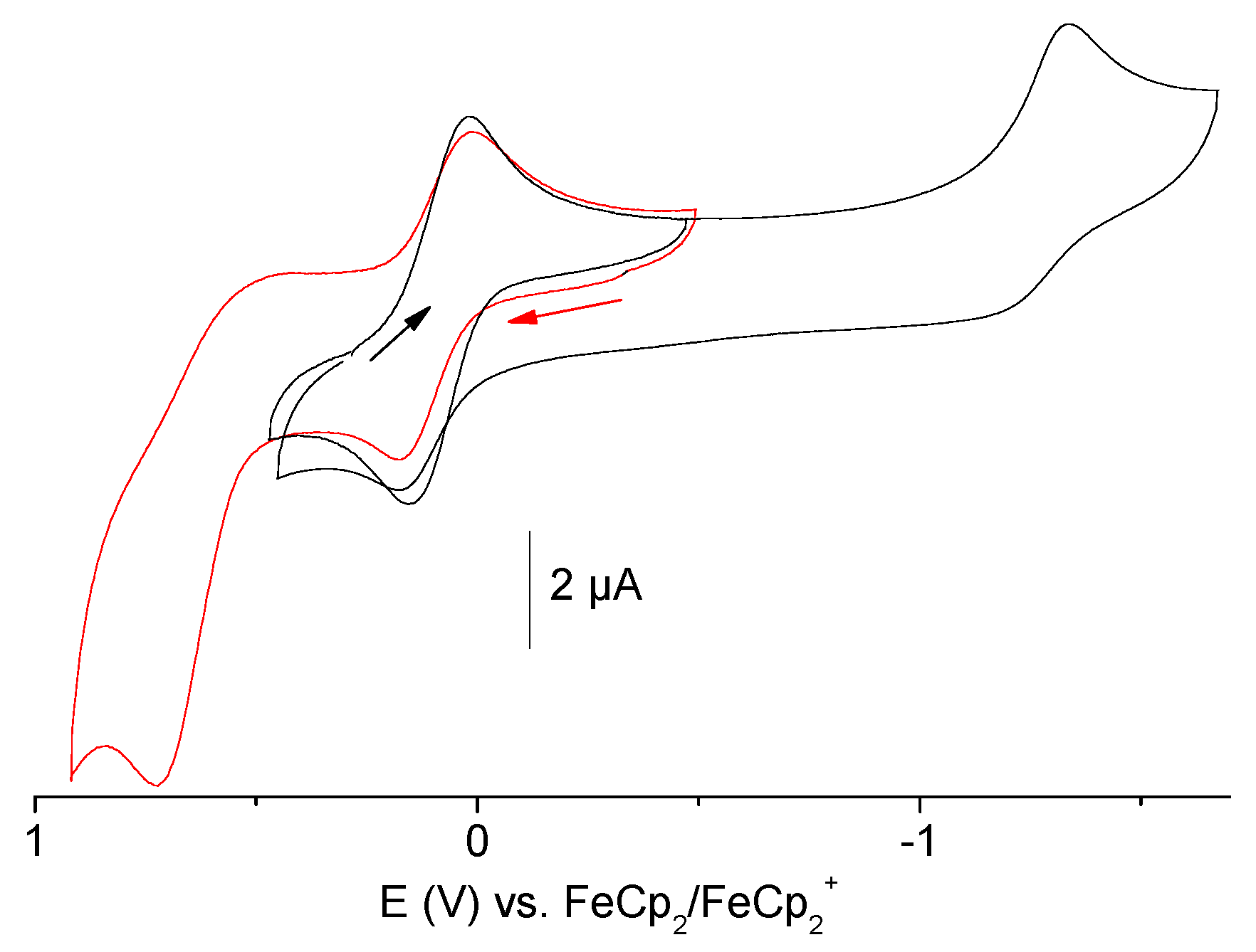
The electrochemical properties of the new complexes and the new ligand pydic(IPh)2 were studied by cyclic voltammetry in MeCN/nBu4NPF6 solution. The complex [{Cu(pydic(IPh)2)Cl}2(µ-Cl)2] shows a reversible redox wave around 0 V vs. ferrocene/ferrocenium, which can be assigned to the CuII/CuI couple (Figure 9). Additionally, an irreversible oxidation wave at 0.73 V and an irreversible reduction wave at −1.33 V were observed. They both occurred for the uncoordinated ligand at similar potentials (Table 5) and were, thus, assigned to ligand-centered processes. Assuming two electrons for the reversible CuII/CuI wave of the binuclear complex, the oxidation wave is markedly larger and more than two electrons might be involved in this process. The redox features of the mononuclear compounds (HNEt3)[Cu(pydicMe2)Cl3] and (HNEt3)[Cu(pydicPh2)Cl3] are very similar, with a reversible CuII/CuI wave at around 0.1 V, irreversible oxidations at around 1 V, and reductions at around −1.3 V (Figure S4 in the Supplementary Materials). For the phenyl derivative [Cu(pydicPh2)Cl3], the second reduction wave is partially reversible, while the methyl derivative shows a broad irreversible wave. The bulky phenyl groups seem to stabilize the reduced complex, which is in line with the idea that the reduction is ligand-centered.
Spectro-electrochemical (SEC), in situ UV–vis absorption spectra during cathodic or anodic electrolysis measurements of (HNEt3)[Cu(pydicPh2)Cl3] and [{Cu(pydic(IPh)2)Cl}2(µ-Cl)2] confirm the full reversibility of the CuII/CuI waves. However, no conclusive spectra were obtained for the reduced species at around −1.3 or the oxidized complexes at around +1 V (for figures, see Supplementary Materials). Importantly, the reduction wave at −1.3 V for (HNEt3)[Cu(pydicPh2)Cl3], which appears reversible in the relatively fast cyclic voltammetry experiment with a timescale of a few seconds turns out to be completely irreversible in the much slower SEC experiments, which are run on a minute timescale.

3. Experimental Section

3.1. Methods and Instrumentation

NMR spectra were recorded on a Bruker Avance II 300 MHz spectrometer (Bruker, Rheinhausen, Germany), using a triple resonance 1H, nBB inverse probe head. The unambiguous assignment of the 1H and 13C resonances was obtained from 1H NOESY, 1H COSY, gradient selected 1H, 13C HSQC, and HMBC experiments. All 2D NMR experiments were performed using standard pulse sequences from the Bruker pulse program library. Chemical shifts were relative to TMS. UV–vis absorption spectra were measured on a Shimadzu UV-3600 photo spectrometer (Shimadzu Europe, Duisburg, Germany). Elemental analyses were carried out using a HEKAtech CHNS EuroEA 3000 Analyzer (Hekatech, Wegberg, Germany). EPR spectra were recorded in the X-band on a Bruker System ELEXSYS 500E equipped with a Bruker Variable Temperature Unit ER 4131VT (500 to 100 K) (Bruker, Rheinhausen, Germany). The g values were calibrated using a dpph sample. Electrochemical experiments were carried out in 0.1 M nBu4NPF6 solutions using a three-electrode configuration (glassy carbon working electrode, Pt counter electrode, Ag/AgCl pseudo reference), an Autolab PGSTAT30 potentiostat (Metrohm, Filderstadt, Germany), and a function generator. Experiments were run at a scan rate of 100 mV/s at ambient temperature. The ferrocene/ferrocenium couple (FeCp2/FeCp2+) served as the internal reference. UV–vis spectro-electrochemical measurements were performed with an optical transparent thin-layer electrochemical (OTTLE) cell [55,56].

3.2. Crystal Structure Determination

The measurements were performed at 293(2) K using graphite mono-chromatized Mo Kα radiation (λ = 0.71073 Å) on IPDS II (STOE and Cie., Darmstadt, Germany). The structures were solved by direct methods using SHELX-97 and WinGX (SHELXS-97) [57,58,59] and refined by full-matrix least-squares techniques against F2 (SHELXL-2017/1) [60,61]. The numerical absorption corrections (X-RED V1.22; Stoe & Cie, 2001) were performed after optimizing the crystal shapes using the X-SHAPE V1.06 (Stoe & Cie, 1999) [62,63]. The non-hydrogen atoms were refined with anisotropic displacement parameters. H atoms were included by using appropriate riding models. CCDC 1878796 ([Cu(OH2)6][{Cu(pydic)}2(µ-Cl)2]), 1878795 ((HNEt3)[Cu(pydicMe2)Cl3]), and 1878792 (pydic(IPh)2) contain the full crystallographic data. These data can be obtained free of charge at www.ccdc.cam.ac.uk/conts/retrieving.html or from the Cambridge Crystallographic Data Center, 12 Union Road, Cambridge, CB2 1EZ UK. Fax: +44-1223-336-033; Email: [email protected].

3.3. Quantumchemical and Molecular Dynamics Calculations

DFT-calculations on the structures of mononuclear [Cu(pydic(IPh)2)Cl2] vs. binuclear [{Cu(pydic(IPh)2)Cl}2(µ-Cl)2] were carried out on a def-SV(P)[64]/B3LYP [65,66,67] level using the TURBOMOLE [68] program package and TmoleX user interface [69].
(HNEt3)[Cu(pydicR2)Cl3] (R = Me or Ph) structures were optimized using DFT calculations at the B3LYP/6–31+G(d,p) level in different environments (gas phase, various solvents) using the Gaussian 09 program [70]. The Cu atom was treated using LANL2DZ relativistic pseudo potentials. The solvent effect was introduced by applying the polarized continuum model (PCM) embedded in the Gaussian 09 package. In the natural bond, the orbital (NBO) analysis and the donor and acceptor interaction energies can be estimated through the second-order perturbation theory, described using the equation below.
E ( 2 ) = q i F ( i , j ) 2 ε i ε j
where qi is the donor orbital occupancy, εj and εi are diagonal elements, and F(i,j) is the off-diagonal NBO Fock matrix element.
Molecular dynamics (MD) simulations on (HNEt3)[Cu(pydicR2)Cl3] (R = Me or Ph) were carried out using the GROMACS package [71] and utilizing the GROMOS force field 53A6 [72]. Six-ion pairs solvated in 1200 solvent molecules were simulated for each compound. The equations of motion have been solved by the leapfrog integrator with a time step of 2 fs. After initial energy minimizations and preliminary simulations adjustments in NVT and NPT ensembles, MD simulations were extended for an additional time of 10 ns collecting statistical data under the NPT condition at T = 298 K with periodic boundary conditions in all directions. The temperature was maintained at 298 K using the Berendsen thermostat [73] and the pressure was kept at 1 bar by coupling semi-isotopically to a barostat with a coupling constant of 1 ps. Both the electrostatic and van der Waals interactions used a short-range cutoff of 1.2 nm and the long-range electrostatics were evaluated using the particle mesh Ewald (PME) method [74].

3.4. Syntheses

3.4.1. General

Water-free reactions were carried out under inert gas conditions and performed using Schlenk techniques. Solvents were dried using a MBRAUN MB SPS-800 solvent purification system. Pyridine-2,6-dicarbonyl dichloride [51] was synthesized according to a literature procedure.
Data pyridine-2,6-dicarbonyl dichloride: Elemental analysis calcd. for C7H3NO2Cl2 (204.01): C 41.21, H 1.48, N 6.87; found: C 41.13, H 1.30, N 6.83%. 1H NMR (300 MHz, acetone-d6): δ = 8.55–8.41 (m, 3H, 3,4,5Py). 13C NMR (75.47 MHz, acetone-d6): δ = 170 (2C, C=O), 150 (2C, 2,6Py), 142 (1C, 4Py), 131 (2C, 3,5Py) ppm.

3.4.2. Synthesis of bis(2-iodophenyl)pyridine-2,6-dicarboxylic ester pydic(IPh)2 and [{Cu(pydic(IPh)2)Cl}2(µ-Cl)2]

Synthesis of pydic(IPh)2. A total of 1.0 g (5 mmol) pyridine-2,6-dicarbonyl dichloride was dissolved in 20 mL dry diethyl ether. In addition, 2.3 g (10 mmol) of 2-iodo-phenol were dissolved in 30 mL dry diethyl ether. Furthermore, 10 mg 4-dimethylaminopyridine (5%mol) and 0.7 mL NEt3 were added to the alcohol mixture, which was then cooled using an ice bath. The acid chloride solution was dropped slowly into the alcohol mixture and precipitation of the product started immediately. After stirring for 1 h at 0 °C, 0.7 mL NEt3 were added at an ambient temperature and the mixture was stirred for 16 h. The formed colorless precipitate was filtered off and washed with 5 mL diethyl ether. The remaining powder was dried yielding 2.35 g (82%) of a colorless solid. Elemental analysis calcd. for C19H11NO4I2 (571.10): C 39.96, H 1.94, N 2.45; found: C 39.89, H 1.93, N 2.43%. 1H NMR (300 MHz, dmf-d7): δ = 8.64 (d, 2H, 3,4Py), 8.47 (t, 1H, 5Py), 7.94 (d, 2H, 3Ph), 7.51 (t, 2H, 5Ph), 7.47 (d, 2H, 6Ph), 7.11 (t, 2H, 4Ph) ppm.
Synthesis of [{Cu(pydic(IPh)2)Cl}2(µ-Cl)2]. 100 mg (0.18 mmol) pydic(IPh)2 were dissolved in 15 mL MeOH. 24 mg (0.18 mmol) anhydrous CuCl2 were dissolved in 10 mL MeOH and both solutions were mixed and stirred at an ambient temperature for 16 h. The solvent was evaporated to yield a brown powder. Recrystallization from MeOH gave 85 mg (67%) of brown microcrystalline material. Elemental analysis calcd. for C19H11CuCl2NO4I2 (705.56): C 32.34, H 1.57, N 1.99; found: C 32.33, H 1.62, N 2.02.

3.4.3. One Pot Reactions

Synthesis of (HNEt3)[Cu(pydicMe2)Cl3]. 100 mg (0.5 mmol) pyridine-2,6-dicarbonyl dichloride were dissolved in 15 mL dried MeOH while 67 mg (0.5 mmol) anhydrous CuCl2 were added, which was followed by 1 mL (excess) anhydrous NEt3. The reaction mixture was stirred at ambient temperature for 16 h. The resulting green solution was evaporated to dryness and the residue was recrystallized from MeOH solution to yield 175 mg (0.37 mmol, 75%) green-brown crystals. C15H25CuCl3N2O4 (467.27) calcd: C 38.56, H 5.39, N 6.00; found: C 38.63, H 5.39, N 6.02%.
Synthesis of (HNEt3)[Cu(pydicPh2)Cl3]. A total of 200 mg (1 mmol) pyridine-2,6-dicarbonyl dichloride were mixed with 15 g phenol and 132 mg (1 mmol) anhydrous CuCl2 followed by the addition of 1 mL NEt3. The mixture was warmed up to 50 °C to get a clear homogenous mixture (melting point of phenol = 40.5 °C) and stirred for 96 h. During that time, the excess phenol solidified in the upper part of the flask and the product complex remained as brown oil. The oil was transferred to a new flask and cooled down to 20 °C, upon which the oil solidified as a brownish powder. The crude product was suspended in acetone and carefully filtered. Slow evaporation yielded 350 mg (0.6 mmol, 60%) of a green-yellow microcrystalline powder. C25H29CuCl3N2O4 (591.41) calcd.: C 50.77, H 4.94, N 4.74; found: C 50.72, H 4.98, N 4.76%.

3.4.4. Complexes from pydicCl2 Hydrolysis

In a typical reaction, 100 mg (0.5 mmol) pyridine-2,6-dicarbonyl dichloride were dissolved in 15 mL moist MeOH. 85 mg (0.5 mmol) CuCl2·2H2O and 1 mL (excess) NEt3 were added and the whole mixture was stirred at an ambient temperature for 16-24 h. The resulting green solutions were slowly evaporated and individual crystals were collected. Thus, 33 mg (24%) turquoise crystals of [Cu(pydic)(OH2)2]n were obtained. Elemental analysis calcd. for C7H7CuNO6 (264.68): C 31.76, H 2.67, N 5.29; found: C 31.79, H 2.72, N 5.24%, see also the XRD Section. In the same way, 21 mg (12%) blue-green crystals of [Cu(OH2)6][{Cu(pydic)}2(µ-Cl)2] were obtained. Elemental analysis calcd. for C14H18Cu3Cl2N2O14 (699.84): C 21.74, H 1.82, N 3.62; found: C 21.80, H 1.79, N 3.58%, see also XRD section.

4. Conclusions

New CuII complexes of the pyridine-2,6-dicarboxylic ester ligands (pydicR2; with R = Me, Ph and IPh) were synthesized and characterized. The pyridine-2,6-carboxylic esters pydicR2 with R = Me or Ph form the unprecedented mononuclear CuII complexes [Cu(pydicR2)Cl3] in one-pot reactions starting from pyridine-2,6-carboxychloride pydicCl2, anhydrous CuII chloride, and NEt3 in MeOH or PhOH solution under non-aqueous conditions. The triethyl ammonium salts (HNEt3)[Cu(pydicR2)Cl3] were isolated. The methyl derivative was crystallized to allow an XRD structure determination. Synthesis attempts in the presence of water yielded the CuII complexes [Cu(pydic)(OH2)2]n and [Cu(OH2)6][{Cu(pydic)}2(µ-Cl)2] resulting from pydicCl2 hydrolysis. The new pydic(IPh)2 (IPh = 2-iodo-phenyl) ester ligand was synthesized and reacted with anhydrous CuCl2 yielding the new binuclear complex [{Cu(pydic(IPh)2)Cl}2(µ-Cl)2]. EPR, UV–vis absorption spectroscopic and electrochemical investigations confirm similarities between copper complexes of pydic ester ligands and corresponding Cu complexes of pyridine-2,6-dialcohols. HOMO–LUMO gap, dipole moments, and electrostatic potential maps for the ion pairs (HNEt3)[Cu(pydicR2)Cl3] are calculated using DFT methods. These quantities were decreasing along the series DMF > MeCN > acetone > CH2Cl2 >>> gas phase. The second order delocalization energies obtained from the NBO analysis confirm the occurrence of the intramolecular charge transfer between the (HNEt3)+ and [Cu(pydicR2)Cl3] ions. However, increasing the dielectric constant of the solvent environment leads to a reduction of charge transfers between cations and anions. Furthermore, time correlation functions as well as radial and combined distribution functions between cations and anions were calculated by MD simulations. All of these data show a marked difference between the gas phase and a different solvent media ranging from the non-coordinating CH2Cl2 and the weakly coordinating acetone to the MeCN and DMF, which are both frequently used as ligands in CuII complexes. We assume that the spectroscopy and electrochemistry in such solutions represent the complexes under study and not dissociated CuII species.

Supplementary Materials

The following are available online at https://www.mdpi.com/2304-6740/7/4/53/s1. (A) Figures S1–S10 showing crystal and molecular structures, CVs, UV–vis absorption spectra and electrostatic potential maps of (HNEt3)[Cu(pydicR2)Cl3] in solvents. (B) Tables S1–S8 presenting full crystal structure and refinement data alongside with calculated structure parameters and second-order perturbation energies. (C) Experimental details on structure solution and description of structural details of pydic(IPh)2 and [Cu(OH2)6][{Cu(pydic)}2(µ-Cl)2]. (D) Cif and Checkcif files of the ligand pydic(IPh)2 and the compounds [Cu(OH2)6][{Cu(pydic)}2(µ-Cl)2] and (HNEt3)[Cu(pydicMe2)Cl3].

Author Contributions

K.B. carried out synthesis, analysis, and spectroscopic and electrochemical analysis of the compounds. K.B. and A.S. collected data and solved and refined XRD datasets. A.S., M.H.D., and A.R.Z. performed the quantum chemical calculations. A.R.Z. coordinated these calculations. A.K. designed the project and wrote the manuscript. All authors have given approval to the final version of the manuscript.

Funding

This research was funded by Studienstiftung des Deutschen Volkes (K.B.). A.K. acknowledges the German Academic Exchange Service (DAAD)—KD_0001052598-2 for a short-time guest lectureship and the Shiraz University.

Acknowledgments

We thank Ingo Pantenburg (University of Cologne) for crystal data collection.

Conflicts of Interest

The authors declare no conflict of interest.

References

  1. Xiang, S.; Bao, D.-X.; Wang, J.; Li, Y.-C.; Zhao, X.-Q. Luminescent lanthanide coordination compounds with pyridine-2,6-dicarboxylic acid. J. Lumin. 2017, 186, 273–282. [Google Scholar] [CrossRef]
  2. Celestine, M.J.; Bullock, J.L.; Boodram, S.; Rambaran, V.H.; Holder, A.A. Interesting properties of p-, d-, and f-block elements when coordinated with dipicolinic acid and its derivatives as ligands: Their use as inorganic pharmaceuticals. Rev. Inorg. Chem. 2015, 35, 57–67. [Google Scholar] [CrossRef]
  3. Kirillov, A.M.; Shul’Pin, G.B. Pyrazinecarboxylic acid and analogs: Highly efficient co-catalysts in the metal-complex-catalyzed oxidation of organic compounds. Coord. Chem. Rev. 2013, 257, 732–754. [Google Scholar] [CrossRef]
  4. Teo, P.; Hor, T.A. Spacer directed metallo-supramolecular assemblies of pyridine carboxylates. Coord. Chem. Rev. 2011, 255, 273–289. [Google Scholar] [CrossRef]
  5. Abdelhameed, R.M.; Ananias, D.; Silva, A.M.S.; Rocha, J. Building Light-Emitting Metal–Organic Frameworks by Post-Synthetic Modification. ChemistrySelect 2017, 2, 136–139. [Google Scholar] [CrossRef]
  6. Yang, R.; Li, H.H.; Van Hecke, K.; Cui, G.H. Cobalt(II) and Copper(II) Complexes Constructed from Bis(benzimidazole) and 2,6-Pyridinedicarboxylate Co-ligands: Synthesis, Crystal Structures, and Catalytic Properties. Z. Anorg. Allgem. Chem. 2015, 641, 642–649. [Google Scholar] [CrossRef]
  7. Derikvand, Z.; Dorosti, N.; Hassanzadeh, F.; Shokrollahi, A.; Mohammadpour, Z.; Azadbakht, A. Three new supramolecular compounds of copper(II), cobalt(II) and zirconium(IV) with pyridine-2,6-dicarboxylate and 3,4-diaminopyridine: Solid and solution states studies. Polyhedron 2012, 43, 140–152. [Google Scholar] [CrossRef]
  8. Kim, M.K.; Bae, K.-L.; Ok, K.M. From Pincers to Steps: Synthesis, Structure, Characterization, and Transformation of a New Helical Calcium–Organic Framework, Ca[NC5H3(CO2)2](H2O)1.5. Cryst. Growth Des. 2011, 11, 930–932. [Google Scholar] [CrossRef]
  9. Stavila, V.; Bulimestru, I.; Gulea, A.; Colson, A.C.; Whitmire, K.H. Hexaaquacobalt(II) and hexaaquanickel(II) bis(μ-pyridine-2,6-dicarboxylato)bis[(pyridine-2,6-dicarboxylato)bismuthate(III)] dihydrate. Acta Crystallogr. Sect. C Cryst. Struct. Commun. 2011, 67, m65–m68. [Google Scholar] [CrossRef]
  10. Zhang, G.-C.; Chen, S.-P.; Yang, Q.; Gao, S.-L. Thermochemistry on K2[M(DPA)2]·7H2O(s) (M = Cu and Ni, H2DPA = pyridine-2,6-dicarboxylic acid). Thermochim. Acta 2011, 518, 66–71. [Google Scholar] [CrossRef]
  11. Das, B.; Baruah, J.B. Coordinated cations in dipicolinato complexes of divalent metal ions. Inorg. Chim. Acta 2010, 363, 1479–1487. [Google Scholar] [CrossRef]
  12. Felloni, M.; Blake, A.J.; Hubberstey, P.; Wilson, C.; Schröder, M. Solvent Control of Supramolecular Architectures Derived from 4,4′-Bipyridyl-Bridged Copper(II) Dipicolinate Complexes. Cryst. Growth Des. 2009, 9, 4685–4699. [Google Scholar] [CrossRef]
  13. Furet, E.; Costuas, K.; Rabiller, P.; Maury, O. On the Sensitivity of f Electrons to Their Chemical Environment. J. Am. Chem. Soc. 2008, 130, 2180–2183. [Google Scholar] [CrossRef]
  14. Katada, H.; Seino, H.; Mizobe, Y.; Sumaoka, J.; Komiyama, M. Crystal structure of Ce(IV)/dipicolinate complex as catalyst for DNA hydrolysis. J. Biol. Inorg. Chem. 2008, 13, 249–255. [Google Scholar] [CrossRef] [PubMed]
  15. Hakimi, M.; Mardani, Z.; Moeini, K.; Mohr, F. Coordination behavior of dimethyl pyridine-2,6-dicarboxylate towards mercury(II), cadmium(II) and chromium(III) in the solid- and gaseous state supported by CSD studies. Polyhedron 2015, 102, 569–577. [Google Scholar] [CrossRef]
  16. Hakimi, M.; Moeini, K.; Mardani, Z.; Schuh, E.; Mohr, F. Silver(I) coordination polymer and nine-coordinated cadmium(II) complex with dimethyl pyridine-2,6-dicarboxylate supported by solid state and electrochemical studies. J. Coord. Chem. 2013, 66, 1129–1141. [Google Scholar] [CrossRef]
  17. Kapoor, P.; Pathak, A.; Kaur, P.; Venugopalan, P.; Kapoor, R. Steric control of coordination: Unusual coordination mode of dimethylpyridine-2,6-dicarboxylate in a new dinuclear copper(II) complex [(dmpc)(Cl)(µ-Cl)Cu2(µ-Cl)(Cl)(dmpc)] and reversal of the coordination mode in [Cu(dmpc)(H2O)3](ClO4)2. Trans. Met. Chem. 2004, 29, 251–258. [Google Scholar] [CrossRef]
  18. Steinkamp, T.; Karst, U. Detection scheme for bioassays based on 2,6-pyridinedicarboxylic acid derivatives and enzyme-amplified lanthanide luminescence. Anal. Chim. Acta 2004, 526, 27–34. [Google Scholar] [CrossRef]
  19. Pointillart, F.; Bourdolle, A.; Cauchy, T.; Maury, O.; le Gal, Y.; Golhen, S.; Cador, O.; Ouahab, L. In Solution Sensitization of Er(III) Luminescence by the 4-Tetrathiafulvalene-2,6-pyridinedicarboxylic Acid Dimethyl Antenna Ligand. Inorg. Chem. 2012, 51, 978–984. [Google Scholar] [CrossRef]
  20. Creber, M.L.; Orrell, K.G.; Osborne, A.G.; Šik, V.; Hursthouse, M.B.; Light, M.E. Solution fluxionality of some pyridine-2,6-dialdehydes, -diketones and -diesters when acting as bidentate ligands in rhenium(I) and platinum(IV) complexes. Crystal structure of [ReBr(CO)3L] (L = methylethyldipicolinate). Polyhedron 2001, 20, 1973–1982. [Google Scholar] [CrossRef]
  21. Goher, M.A.; Mautner, F.; Hafez, A.K.; Youssef, A. Synthesis and characterization of copper(I) complexes of dimethyldipicolinate (dmdp) and X-ray structure of the mononuclear six-coordinate complex [Cu(dmdp)2]ClO4. Polyhedron 2003, 22, 515–520. [Google Scholar] [CrossRef]
  22. Kapoor, P.; Pathak, A.; Kapoor, R.; Venugopalan, P.; Corbella, M.; Rodríguez, M.; Robles, J.; Llobet, A. Structural, Electronic, and Magnetic Consequences of O-Carbonyl vs O-Alkoxy Ester Coordination in New Dicopper Complexes Containing the Cu2(μ-Cl)2 Core. Inorg. Chem. 2002, 41, 6153–6160. [Google Scholar] [CrossRef]
  23. Platas-Iglesias, C.; Piguet, C.; André, N.; Bünzli, J.-C.G. Lanthanide triple-stranded helical complexes with a substituted 2,6-pyridinedicarboxylate. J. Chem. Soc. Dalton Trans. 2001, 3084–3091. [Google Scholar] [CrossRef]
  24. Muller, G.; Schmidt, B.; Jiricek, J.; Hopfgartner, G.; Riehl, J.P.; Bünzli, J.-C.G.; Piguet, C. Lanthanide triple helical complexes with a chiral ligand derived from 2,6-pyridinedicarboxylic acid. J. Chem. Soc. Dalton Trans. 2001, 2655–2662. [Google Scholar] [CrossRef]
  25. Renaud, F.; Piguet, C.; Bernardinelli, G.; Bünzli, J.-C.G.; Hopfgartner, G. In Search for Mononuclear Helical Lanthanide Building Blocks with Predetermined Properties: Lanthanide Complexes with Diethyl Pyridine-2, 6-Dicarboxylate. Chem. Eur. J. 1997, 3, 1660–1667. [Google Scholar] [CrossRef]
  26. Espinet, P.; García-Orodea, E.; Miguel, J.A. Mesogenic Palladium Complexes with Pincer Ligands Derived from Dipicolinic Acid. Inorg. Chem. 2000, 39, 3645–3651. [Google Scholar] [CrossRef] [PubMed]
  27. Rasshoffer, W.; Müller, V.M.; Vögtle, F. Noncyclic Crown Ether Type Esters and Their Complexes. Chem. Ber. 1979, 112, 2095–2119. [Google Scholar]
  28. Kirillov, A.M.; Haukka, M.; Da Silva, M.F.C.G.; Pombeiro, A.J.L. Synthesis, Characterization and Redox Behaviour of Mono- and Dicarbonyl Phosphane Rhenium(I) Complexes Bearing N-, N,N- and N,O-Type Ligands. Eur. J. Inorg. Chem. 2007, 2007, 1556–1565. [Google Scholar] [CrossRef]
  29. Senegas, J.-M.; Bernardinelli, G.; Imbert, D.; Bünzli, J.-C.G.; Morgantini, P.-Y.; Weber, J.; Piguet, C. Connecting Terminal Carboxylate Groups in Nine-Coordinate Lanthanide Podates: Consequences on the Thermodynamic, Structural, Electronic, and Photophysical Properties. Inorg. Chem. 2003, 42, 4680–4695. [Google Scholar] [CrossRef] [Green Version]
  30. Bogdan, N.; Grosu, I.; Benoît, G.; Toupet, L.; Ramondenc, Y.; Condamine, E.; Silaghi-Dumitrescu, I.; Plé, G. Molecular Rotors: Design, Synthesis, Structural Analysis, and Silver Complex of New [7.7]Cyclophanes. Org. Lett. 2006, 12, 2619–2622. [Google Scholar] [CrossRef]
  31. Weng, Z.; Teo, S.; Hor, T.S.A. Metal Unsaturation and Ligand Hemilability in Suzuki Coupling. Acc. Chem. Res. 2007, 40, 676–684. [Google Scholar] [CrossRef] [PubMed]
  32. Demerseman, B.; Toupet, L. Enolate-Phosphane Ligands Providing a Tool for the Selective Substitution of Triphenylphosphane by Carbon Monoxide or Trimethylphosphane in Complex {Ru(Cp)[η2-P,O-Ph2PCH2C(tBu)=O](PPh3)}[PF6], and Subsequent Reactivity Towards Terminal Alkynes. Eur. J. Inorg. Chem. 2006, 2006, 1573–1581. [Google Scholar] [CrossRef]
  33. Saes, B.W.H.; Verhoeven, D.G.A.; Lutz, M.; Gebbink, R.J.M.K.; Moret, M.-E. Coordination of a Diphosphine−Ketone Ligand to Ni(0), Ni(I), and Ni(II): Reduction-Induced Coordination. Organometallics 2015, 34, 2710–2713. [Google Scholar] [CrossRef]
  34. Weber, B.; Werner, H. Osmium(II) and Osmium(IV) Complexes with Phosphane–Ethers, –Esters, and –Amines as Mono- and Bidentate Ligands. Eur. J. Inorg. Chem. 2007, 2007, 2072–2082. [Google Scholar] [CrossRef]
  35. Mecking, S.; Keim, W. Cationic Palladium η3-Allyl Complexes with Hemilabile P,O-Ligands: Synthesis and Reactivity. Insertion of Ethylene into the Pd−Allyl Function. Organometallics 1996, 15, 2650–2656. [Google Scholar] [CrossRef]
  36. Sileo, E.E.; Quinteros-Rivero, M.F.; Rivero, B.E.; Rigotti, G.; Blesa, M.A. Structure and reactivity of copper(II) carboxylates I. Copper(II) dipicolinates. Solid State Ionics 1994, 73, 153–159. [Google Scholar] [CrossRef]
  37. Cingi, M.B.; Villa, A.C.; Gastini, C.; Nardelli, M. Crystal and molecular structures of divalent metal-complexes with pyridinecarboxylic acids. II. (Pyridine-2,6-dicarboxylato)diaquocopper(II), [Cu(C7H3NO4)(OH2)2]. Gazz. Chim. Ital. 1971, 101, 825–832. [Google Scholar]
  38. Glowiak, T.; Podgórska, I. X-ray, Spectroscopic and Magnetic Studies of Hexaaquacopper(II) Di(diphenylphosphate) Diglycine. Inorg. Chim. Acta 1986, 125, 83–88. [Google Scholar] [CrossRef]
  39. Kirillova, M.V.; da Silva, M.F.C.G.; Kirillov, A.M.; da Silva, J.J.R.F.; Pombeiro, A.J.L. 3D hydrogen bonded heteronuclear CoII, NiII, CuII and ZnII aqua complexes derived from dipicolinic acid. Inorg. Chim. Acta 2007, 360, 506–512. [Google Scholar] [CrossRef]
  40. Yue, C.; Lin, Z.; Chen, L.; Jiang, F.; Hong, M. The 2D–3D networks with infinite channels or difform chains of copper(II) complexes via weak non-covalent interactions. J. Mol. Struct. 2005, 779, 16–22. [Google Scholar] [CrossRef]
  41. Wang, W.-Z.; Liu, X.; Liao, D.-Z.; Jiang, Z.-H.; Yan, S.-P.; Wang, G.-L. A novel one-dimensional, ferromagnetic copper(II) complex with chloro-bridges and hydrogen bonds: [(PDA)2Cu2(µ-Cl)2][CuPz2(H2O)4] (H2PDA = pyridine-2,6-dicarboxylic acid, Pz = pyrazole). Inorg. Chem. Commun. 2001, 4, 327–331. [Google Scholar] [CrossRef]
  42. Steiner, T. The Hydrogen Bond in the Solid State. Angew. Chem. Int. Ed. 2002, 41, 48–76. [Google Scholar] [CrossRef]
  43. Powell, B.J.; Baruah, T.; Bernstein, N.; Brake, K.; McKenzie, R.H.; Meredith, P.; Pederson, M.R. A first-principles density-functional calculation of the electronic and vibrational structure of the key melanin monomers. J. Chem. Phys. 2004, 120, 8608–8615. [Google Scholar] [CrossRef] [Green Version]
  44. Gussoni, M.; Castiglioni, C. Infrared intensities. Use of the CH-stretching band intensity as a tool for evaluating the acidity of hydrogen atoms in hydrocarbons. J. Mol. Struct. 2000, 521, 1–18. [Google Scholar] [CrossRef]
  45. Brehm, M.; Kirchner, B. TRAVIS—A Free Analyzer and Visualizer for Monte Carlo and Molecular Dynamics Trajectories. J. Chem. Inf. Model. 2011, 51, 2007–2023. [Google Scholar] [CrossRef] [PubMed]
  46. Klein, A.; Butsch, K.; Elmas, S.; Biewer, C.; Heift, D.; Nitsche, S.; Schlipf, I.; Bertagnolli, H. Oxido-pincer complexes of copper(II)—An EXAFS and EPR study of mono- and binuclear [(pydotH2)CuCl2]n (n = 1 or 2). Polyhedron 2012, 31, 649–656. [Google Scholar] [CrossRef]
  47. Halcrow, M.A. Jahn-Teller Distortions in Transition Metal Compounds, and Their Importance in Functional Molecular and Inorganic Materials. Chem. Soc. Rev. 2013, 42, 1784–1795. [Google Scholar] [CrossRef]
  48. Hathaway, B.; Billing, D. The electronic properties and stereochemistry of mono-nuclear complexes of the copper(II) ion. Coord. Chem. Rev. 1970, 5, 143–207. [Google Scholar] [CrossRef]
  49. Koval, I.A.; Sgobba, M.; Huisman, M.; Lüken, M.; Saint-Aman, E.; Gamez, P.; Krebs, B.; Reedijk, J. A remarkable anion effect on the crystal packing of two analogous copper complexes from a thiophene-containing phenol-based ligand. Inorg. Chim. Acta 2006, 359, 4071–4078. [Google Scholar] [CrossRef]
  50. Thakurta, S.; Roy, P.; Rosair, G.; Gómez-García, C.J.; Garribba, E.; Mitra, S. Ferromagnetic exchange coupling in a new bis(µ-chloro)-bridged copper(II) Schiff base complex: Synthesis, structure, magnetic properties and catalytic oxidation of cycloalkanes. Polyhedron 2009, 28, 695–702. [Google Scholar] [CrossRef]
  51. Yraola, F.; Albericio, F.; Corbella, M.; Royo, M. [{Cu(pzPh)(Opo)}2(μ-Cl)2]: A new dinuclear copper(II) complex with a chloride bridge and mixed blocking ligands. Inorg. Chim. Acta 2008, 361, 2455–2461. [Google Scholar] [CrossRef]
  52. Pérez, A.L.; Neuman, N.I.; Baggio, R.; Ramos, C.A.; Dalosto, S.D.; Rizzi, A.C.; Brondino, C.D. Exchange interaction between S = 1/2 centers bridged by multiple noncovalent interactions: Contribution of the individual chemical pathways to the magnetic coupling. Polyhedron 2017, 123, 404–410. [Google Scholar] [CrossRef]
  53. Rieger, P.H. Electron Spin Resonance—Analysis and Interpretation; RCS Publishing: Cambridge, UK, 2007. [Google Scholar]
  54. Klein, A.; Elmas, S.; Butsch, K. Oxido Pincer Ligands—Exploring the Coordination Chemistry of Bis(hydroxymethyl)pyridine Ligands for the Late Transition Metals. Eur. J. Inorg. Chem. 2009, 2009, 2271–2281. [Google Scholar] [CrossRef]
  55. Kaim, W.; Fiedler, J. Spectroelectrochemistry: The best of two worlds. Chem. Soc. Rev. 2009, 38, 3373. [Google Scholar] [CrossRef]
  56. Kaim, W.; Klein, A. Spectroelectrochemistry; RSC Publishing: Cambridge, UK, 2008. [Google Scholar]
  57. Sheldrick, G.M. A short history of SHELX. Acta Crystallogr. Sect. A Found. Crystallogr. 2008, 64, 112–122. [Google Scholar] [CrossRef] [PubMed]
  58. Sheldrick, G.M. SHELX-97, Programs for Crystal Structure Analysis; University of Göttingen: Göttingen, Germany, 1997. [Google Scholar]
  59. Farrugia, L.J. WinGX and ORTEP for Windows: An update. J. Appl. Crystallogr. 2012, 45, 849–854. [Google Scholar] [CrossRef]
  60. Sheldrick, G.M. SHELXL-2017/1, Program for the Solution of Crystal Structures; University of Göttingen: Göttingen, Germany, 2017. [Google Scholar]
  61. Sheldrick, G.M. Crystal structure refinement with SHELXL. Acta Crystallogr. Sect. C Struct. Chem. 2015, 71, 3–8. [Google Scholar] [CrossRef]
  62. STOE X-RED. Data Reduction Program, Version 1.22/Windows; STOE & Cie: Darmstadt, Germany, 2001. [Google Scholar]
  63. STOE X-SHAPE. Crystal Optimisation for Numerical Absorption Correction, Version 1.06/Windows; STOE & Cie: Darmstadt, Germany, 1999. [Google Scholar]
  64. Horn, H.; Schäfer, A.; Ahlrichs, R. Fully optimized contracted Gaussian basis sets for atoms Li to Kr. J. Chem. Phys. 1992, 97, 2571–2577. [Google Scholar]
  65. Lee, C.; Yang, W.; Parr, R.G. Development of the Colle-Salvetti correlation-energy formula into a functional of the electron density. Phys. Rev. B 1988, 37, 785–789. [Google Scholar] [CrossRef]
  66. Becke, A.D. A new mixing of Hartree–Fock and local density-functional theories. J. Chem. Phys. 1993, 98, 1372–1377. [Google Scholar] [CrossRef]
  67. Becke, A.D. Density-functional thermochemistry. III. The role of exact exchange. J. Chem. Phys. 1993, 98, 5648–5652. [Google Scholar] [CrossRef]
  68. TURBOMOLE 7.0; TURBOMOLE GmbH: Karlsruhe, Germany, 2015.
  69. Steffen, C.; Thomas, K.; Huniar, U.; Hellweg, A.; Rubner, O.; Schroer, A. TmoleX-A graphical user interface for TURBOMOLE. J. Comput. Chem. 2010, 31, 2967–2970. [Google Scholar] [CrossRef]
  70. Frisch, M.J.; Trucks, G.W.; Schlegel, H.B.; Scuseria, G.E.; Robb, M.A.; Cheeseman, J.R.; Scalmani, G.; Barone, V.; Mennucci, B.; Petersson, G.A.; et al. Gaussian 09; Gaussian Inc.: Wallingford, CT, USA, 2009. [Google Scholar]
  71. Hess, B.; Kutzner, C.; Van Der Spoel, D.; Lindahl, E. GROMACS 4: Algorithms for Highly Efficient, Load-Balanced, and Scalable Molecular Simulation. J. Chem. Theory Comput. 2008, 4, 435–447. [Google Scholar] [CrossRef] [PubMed]
  72. Lemkul, J.A.; Allen, W.J.; Bevan, D.R. Practical Considerations for Building GROMOS-Compatible Small-Molecule Topologies. J. Chem. Inf. Model. 2010, 50, 2221–2235. [Google Scholar] [CrossRef] [PubMed]
  73. Berendsen, H.J.; Postma, J.V.; van Gunsteren, W.F.; DiNola, A.R.H.J.; Haak, J.R. Molecular Dynamics with Coupling to an External Bath. J. Chem. Phys. 1984, 81, 3684–3690. [Google Scholar] [CrossRef]
  74. Essmann, U.; Perera, L.; Berkowitz, M.L.; Darden, T.; Lee, H.; Pedersen, L.G. A Smooth Particle Mesh Ewald Method. J. Chem. Phys. 1995, 103, 8577–8593. [Google Scholar] [CrossRef]
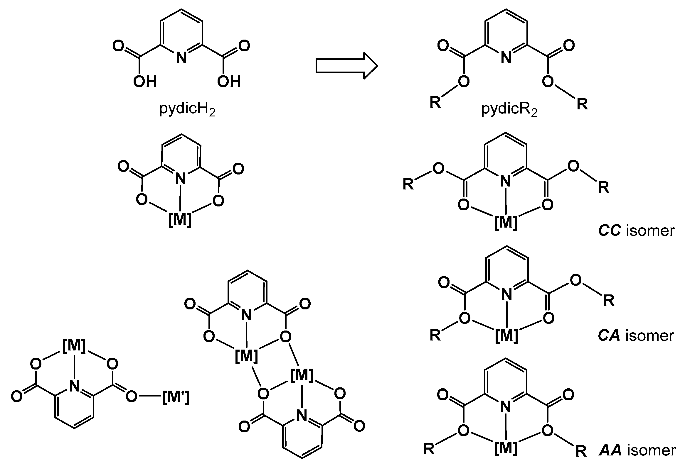
Scheme 1. Binding modes of pydic2− compared to pyridine-2,6-dicarboxylic ester ligands (R = alkyl or aryl; [M] = metal ions or metal complex fragments).
Inorganics 07 00053 sch001
Scheme 2. Two reaction strategies to synthesize CuII complexes of pydicR2 ligands (DMAP = dimethyl 4-aminopyridine).
Inorganics 07 00053 sch002
Figure 1. DFT-calculated optimized geometry of mononuclear [Cu(pydic(IPh)2)Cl2] (left) and binuclear [{Cu(pydic(IPh)2)Cl}2(µ-Cl)2] (right). Calculated at the def-SV(P)/B3LYP level.
Inorganics 07 00053 g001
Figure 2. Left: ORTEP representation (50% probability level) of the molecular structure of [Cu(OH2)6][{Cu(pydic)}2(µ-Cl)2]. H atoms were omitted for clarity. Right: Crystal structure of [Cu(OH2)6][{Cu(pydic)}2(µ-Cl)2] along the crystallographic c-axis.
Inorganics 07 00053 g002
Figure 3. ORTEP representation (50% probability level) of the molecular structure of (HNEt3)[Cu(pydicMe2)Cl3]. H atoms are omitted for clarity. Right: Crystal structure of (HNEt3)[Cu(pydicMe2)Cl3] along the crystallographic a-axis.
Inorganics 07 00053 g003
Figure 4. DFT-calculated geometry-optimized (B3LYP/6–31+G(d,p), Gaussian) structures of (HNEt3)[Cu(pydicMe2)Cl3] and (HNEt3)[Cu(pydicPh2)Cl3] in the gas phase with numbering.
Inorganics 07 00053 g004
Figure 5. Electrostatic potential maps of (HNEt3)[Cu(pydicMe2)Cl3] (left) and (HNEt3)[Cu(pydicPh2)Cl3] (right) in the gas phase. Contours are color-coded from red (negative) to blue (positive).
Inorganics 07 00053 g005
Figure 6. (A) Snapshots of the DFT calculated dynamic behavior of six ion pairs (HNEt3)[Cu(pydicMe2)Cl3] in MeCN. (B) Time correlation functions, (C) the radial distribution function, and (D) the combined radial/angular distribution function between the Cl2 atom of the anion and H22 of the cation in (HNEt3)+···[Cu(pydicMe2)Cl3].
Inorganics 07 00053 g006
Figure 7. X-band EPR spectra of [{Cu(pydic(IPh)2)Cl}2(µ-Cl)2] at 298 K. (A) in the solid. (B) in acetone solution.
Inorganics 07 00053 g007
Figure 8. X-band EPR spectra of (HNEt3)[Cu(pydicMe2)Cl3] and (HNEt3)[Cu(pydicPh2)Cl3]. (A) in the solid, (B) in acetone solution measured at 298 K.
Inorganics 07 00053 g008
Figure 9. Cyclic voltammogramms of [{Cu(pydic(IPh)2)Cl}2(µ-Cl)2] in MeCN/nBu4NPF6.
Inorganics 07 00053 g009
Table 1. DFT-calculated dipole moments and solvation energies of (HNEt3)[Cu(pydicMe2)Cl3] and (HNEt3)[Cu(pydicPh2)Cl3] in the gas phase and solvent media.
Compound Gas PhaseCH2Cl2AcetoneMeCNDMF
(HNEt3)[Cu(pydicMe2)Cl3]dipole moment (Debye)8.2712.4913.2213.4913.51
solvation energy (kJ/mol) −110.88−128.30−134.74−135.12
ELUMOEHOMO (kJ/mol)323.15382.64390.20392.91393.06
charge [Cu(pydicMe2)Cl3]−0.84006 −0.90691−0.91645−0.91969−0.92005
(HNEt3)[Cu(pydicPh2)Cl3]dipole moment (Debye)7.9712.2913.1313.4413.46
solvation energy (kJ/mol) −124.86−139.25−146.15−146.55
ELUMOEHOMO (kJ/mol)314.22369.83376.68379.41379.57
charge
[Cu(pydicPh2)Cl3]
−0.84929 −0.90889 −0.92116 −0.92426−0.92447
Table 2. DFT-calculated second-order perturbation energies E(2) (kJ/mol) of selected orbital interactions in (HNEt3)[Cu(pydicMe2)Cl3] in the gas phase and solvents a.
DonorAcceptorGas PhaseCH2Cl2AcetoneMeCNDMF
LP*(8) Cu1BD*(1) N2–H2218.918.247.286.956.90
LP(1) Cl1LP*(7) Cu148.5350.5451.2151.4251.46
LP(4) Cl1LP*(6) Cu199.6298.7899.2999.5499.54
LP(4) Cl1LP*(7) Cu1167.40179.53181.21181.84181.88
LP(1) Cl2LP*(8) Cu161.3057.9157.7457.6157.70
LP(4) Cl2LP*(6) Cu1107.15126.40128.37128.99129.04
LP(4) Cl2LP*(8) Cu1153.55157.32156.65156.23156.27
LP(3) Cl2BD*(1) N2–H2266.1947.4942.3040.5440.30
LP(1) Cl3LP*(7) Cu172.7264.1063.2663.0163.01
LP(4) Cl3LP*(6) Cu1123.85105.31102.84102.00101.96
LP(4) Cl3LP*(7) Cu1206.65192.67191.04190.50190.50
LP(1) O1LP*(9) Cu131.5934.6435.1535.4035.40
LP(1) O2LP*(9) Cu136.3637.2437.2437.2037.20
LP(1) N1LP*(6) Cu163.3969.8370.6370.8870.88
LP(1) N1LP*(8) Cu164.5271.5572.7273.1873.22
a LP = lone pairs, LP* = antibonding molecular orbitals of Cu. BD* = antibonding orbitals.
Table 3. X-band EPR data of the reported and comparable CuII complexes a.
Compoundsgavg||
or g1
g
or g2
g3ΔgState
reported compounds
[{Cu(pydic(IPh)2)Cl}2(µ-Cl)2]2.1572.2602.1152.0950.165solid
[{Cu(pydic(IPh)2)Cl}2(µ-Cl)2]2.1302.1972.1022.0910.106acetone
(HNEt3)[Cu(pydicMe2)Cl3]2.1822.3512.098 0.253solid
(HNEt3)[Cu(pydicMe2)Cl3]2.1312.2112.091 0.120acetone
(HNEt3)[Cu(pydicPh2)Cl3]2.1962.3622.113 0.249solid
(HNEt3)[Cu(pydicPh2)Cl3]2.1262.2012.089 0.112acetone
related mononuclear complexes
[Cu(pydic)(H2O)2] (sqp) b2.1562.3242.1022.0430.281solid
[Cu(pydimH2)Cl2] (sqp) c2.1582.3112.0942.0680.243solid
[Cu(pydipH2)Cl2] (tbp) c2.1382.2202.2201.973 e0.223solid
[Cu(pydotH2)(DMF)Cl2] (aeo) c2.1942.3532.1202.1080.245solid
related binuclear complexes
[{Cu(pydicEt2)Cl}2(µ-Cl)2] d2.1472.212.122.110.100solid
[{Cu(pydiciPr2)Cl}2(µ-Cl)2] d2.1302.172.162.060.110solid
[{Cu(pydotH2)Cl}2(µ-Cl)2] c2.1482.4182.0811.9460.472solid
a measured at 298 K; gav = averaged g value = (g + 2 g)/3 or (g1 + g2 + g3)/3; Δg = gg or g1 − g3. b From Reference [52].c From Reference [46]. sqp = square pyramidal, tbp = trigonal bipyramidal, aeo = axially elongated pseudo-octahedral. d From Reference [23].
Table 4. Absorption maxima for CuII complexes a.
Compoundsλ/nm (ε/L·mol–1·cm–1) c
(HNEt3)[Cu(pydicMe2)Cl3]261 (48400), 462 (3290), 822 (160)
(HNEt3)[Cu(pydicPh2)Cl3]261 (44400), 307 (14400) b, 462 (4580), 890 (310)
[{Cu(pydic(IPh)2)Cl}2(µ-Cl)2]289 (27100), 464 (3740), 830 (200)
K2[CuCl4]312, 346sh, 462, 1074
a Measured in MeCN. b Absorption assigned to π(Ph)–π(Py) transitions. c extinction coefficient.
Table 5. Selected electrochemical data a.
CompoundsEpa oxE½ CuI/CuIIEpc red
pydic(IPh)20.72-−1.52
pydicPh20.91-−1.63
pydicMe20.86-−1.60
[{Cu(pydic(IPh)2)Cl}2(µ-Cl)2]0.730.08−1.33
(HNEt3)[Cu(pydicPh2)Cl3]1.110.09−1.32
(HNEt3)[Cu(pydicMe2)Cl3]0.840.14−1.33
K2[CuCl4]1.120.08−1.48
a From cyclic voltammetry in MeCN/nBu4NPF6 solutions. Half-wave potentials E½ for reversible waves, cathodic peak potentials Epc for irreversible reduction, and anodic peak potentials Epa for irreversible oxidation. All potentials in V vs. FeCp2/FeCp2+.