Article

Development of Immunohistochemical Methods for Casein Detection in Meat Products

by
Ludmila Kalčáková
1,
Matej Pospiech
1,
Bohuslava Tremlová
1,
Zdeňka Javůrková
1,* and
Irina Chernukha
2
1
Department of Plant Origin Foodstuffs Hygiene and Technology, Faculty of Veterinary Hygiene and Ecology, University of Veterinary and Pharmaceutical Sciences Brno, 612 24 Brno, Czech Republic
2
The V.M. Gorbatov Federal Research Center for Food Systems of Russian Academy of Sciences, Talalikhina ul. 26, 109316 Moscow, Russia
*
Author to whom correspondence should be addressed.
Foods 2021, 10(1), 28; https://doi.org/10.3390/foods10010028
Submission received: 23 November 2020 / Revised: 19 December 2020 / Accepted: 22 December 2020 / Published: 24 December 2020
(This article belongs to the Special Issue Advances in the Approach Food Allergy)

Abstract

:
To increase production efficiency of meat products, milk protein additives are often used. Despite a number of advantages, use of dairy ingredients involves a certain risk, namely the allergenic potential of milk proteins. A number of methods have been developed to detect milk-origin raw materials in foodstuffs, including immunological reference methods. This study presents newly developed immunohistochemical (IHC) methods for casein detection in meat products. Casein was successfully detected directly in meat products where sensitivity was determined at 1.21 and specificity at 0.28. The results obtained from the IHC were compared with the Enzyme-Linked Immuno Sorbent Assay (ELISA) and there was no statistically significant difference between the IHC and ELISA methods (p > 0.05). The correspondence between the methods was 72% in total. The highest correspondence was reached in frankfurters (90%), the lowest in canned pâté (44%).

1. Introduction

Nowadays, meat products represent foodstuffs with a wide range of non-meat ingredients. They are used for various reasons, including the economic, technological, or marketing side of production as well as to enhance taste and flavor. One of the non-meat ingredients used in meat products is milk protein. Milk proteins are obtained from milk and dairy products [1]. Within the meat industry, a significant position among the various ground meat products is occupied by sausages, where milk protein additives are utilized as emulsifiers or production cost reducers. Another effect, however, is to improve the yield, slicing quality, flavor, and nutritional value of the final product [2]. Caseins are made up of several proteins (αs1-, αs2-, β-, and κ-) capable of forming micelles (130–160 nm circular structures). The emulsifying properties are explained by forming structures with high surface activity. In the meat industry, caseins are used in variously modified forms. The most widespread form is sodium caseinate that is characterized by its high solubility and ability to increase volume of the meat dough with a higher capacity to bind water. It also has a significant effect as a fat emulsifier during the treatment [3]. Sodium caseinate is often used as an additive for dry-fermented sausages due to its emulsifying properties [4]. Because of its excellent functional properties, addition of milk protein ingredients into ground meat products has a great potential. Examples of successful use in such meat products include caseinate in chicken nuggets, whey protein concentrate in sausages, sodium caseinate with whey powder in Turkish meat rolls, or milk co-precipitates in chicken loaf [5]. Whey proteins are also composed of a number of proteins. Mainly β-lactoglobulin, α-lactalbumin, serum protein, immunoglobulins, and polypeptides. β-lactoglobulin is a globular dimeric protein that represents 50% of total whey proteins. After partial denaturation, it is able to form dimers with other milk proteins. Other functional whey proteins include immunoglobulins, which only account for 2% in whey but have significant emulsifying properties. The most common forms used in meat production are whey concentrates and isolates. An option is likewise the textured whey protein, which is used as a partial replacement of muscle in meat products up to 40% w/w [3]. Milk-origin raw materials are also used to maintain the juiciness of low-fat meat products. For example, milk powder, sodium caseinate, milk co-precipitate, and skim milk protein are considered to be water-binding substitutes for fat [6].
From the legislative point of view, dairy proteins are considered to be a component of foodstuffs and can therefore replace some additives. Thus, they are included among the essential ingredients in the ingredients list and restrictions on additives do not apply to them. However, according to E.U. legislation, milk is also classified as an allergen. Therefore, its presence must be declared according to the legislation [7].
The incidence of food allergies is estimated to range from 0.5% to 3% within Europe [8]. Nowadays, 0.6–2.5% of preschool children, 0.3% of older children and adolescents, and less than 0.5% of adults suffer from cow’s milk allergy globally. Unfortunately, the prevalence of allergy to cow’s milk is steadily increasing [9].
Cow’s milk contains approximately 30 potentially allergenic proteins. Out of this, about 30 to 35 g are contained in 1 dm3 of milk [10]. The primary cow’s milk allergens include caseins, β-lactoglobulin, and α-lactalbumin. The casein fractions are represented in the form of an isoform of different proportions. These are isoforms such as α-S1-casein (32%), α-S2-casein (10%), β-casein (28%), and κ-casein (10%). Whey proteins include α-lactalbumin, β-lactoglobulin, immunoglobulins, bovine serum albumin, and trace amounts of lactoferrin [8]. It is estimated that 66% of milk allergies are caused by β-lactoglobulin, 57% by casein, and significantly less by α-lactalbumin and bovine serum albumin (18%) [10]. D’Auria et al. [8] report that allergenic activity was observed for β-casein in 35% of patients, for α-S1-casein in 26%, as well as for κ-casein in 26%, whereas for β-lactoglobulin in 19%, for α-lactalbumin in 12%, for lactoferrin in 3%, and for bovine serum in 1%. As with all food allergens, there is currently no suitable treatment for cow’s milk allergy. The only therapy available is to avoid consuming milk and dairy products. However, allergenic proteins are not the only factor responsible for allergic reactivity as well as test immunoreactivity. The food processing, food matrix effect, and different degree of digestion can change the immunoreactivity. Several recent studies of immunoreactivity changes are described by Villa et al. [11]. None of them, nevertheless, describes immunoreactivity changes in meat products where strong matrix effect is expected. Likewise, they do not report effects of thermal processing on a milk-containing food matrix where detection of milk additives can be reduced [12].
The aim of this work was to develop an immunohistochemical method for direct detection of casein in meat products’ matrix and to validate it. Secondary objectives involved verification of immunoreactivity of common types of milk additives used in meat industry and demonstrating the masking effect of different meat matrices on the immunoreactivity of immunoassay applied.

2. Materials and Methods

Common milk additives available in the market were used. Specifically, calcium caseinate, whey protein with high lactose content, thermostable whey concentrate, and whey concentrate containing 85% of casein (MEGGLE Wasserburg GmbH & CO.KG, Wasserburg, Germany), as well as milk powder, whey powder, and buttermilk powder (LAKTOS, a.s., Prague, Czech Republic).
Two groups of model samples were prepared. The meat bases of the model samples were prepared from minced pork and beef meat in a 1:1 ratio. The first group was prepared with the addition of different types of milk additives in the concentration of 2.5%, which is a common addition into meat products to reach the required effect [3]. The additives used were calcium caseinate (C), whey powder (W1), whey protein with high lactose content (W2), thermostable whey concentrate (W3), buttermilk powder (W4), whey concentrate containing 85% of casein (W5), and milk powder (M1). The second group (samples C1–C7) was prepared with increasing concentrations of calcium caseinate (0.0001%; 0.001%; 0.01%; 0.1%; 1%; 2.5%; and 10%), and it was used to determine the limit of detection according to previous study [13]. The raw materials were ground and mixed in Vorwerk Thermomix 31 (Vorwerk, Wuppertal, Germany) at 7000 rpm for 2 min. Subsequently, each meat matrix was quantitatively transferred to a sample container and cooked in water bath until the core matrix was cooked at 70 °C for 10 min (Rommelsbacher, Dinkelsbühl, Germany).
For immunohistochemical methods, the samples were prepared according to the following procedure: four partial samples were frozen at −35 °C for each sample. After freezing, the samples were cut into 10 μm sections on a rotation microtome HM 550 (Microm, Walldorf, Germany). The partial samples were cut into two serial sections, while 50 µm were overcut. Totally eight sections for each sample were prepared. The sections were mounted on SuperFrost plus slides (Menzel-Glasser, Menzel GmbH & Co KG, Braunschweig, Germany). Before the immunohistochemical procedure, the slides were dried in a fridge. The immunohistochemical procedure followed in no later than seven days.
The immunohistochemical procedure was based on the IHC method described by Pospiech, Tremlová, Renčová, and Randulová [13] and Řezáčová-Lukášková et al. [14] with the following changes. The immunohistochemical VECTASTAIN Elite ABC KIT was used (VectorLaboratories, PK 6101, Burlingame, CA, USA). Rabbit anti-beta casein polyclonal antibody (Bioss Antibodies, Woburn, MA, USA) was used as the primary antibody, and antibody diluent (DakoCytomation Ref.S0809, Glostrup, Denmark) was used for dilution. In negative control, the primary antibody was substituted by antibody diluent (DakoCytomation ref. S0809). For antigen retrieval, Citrate-EDTA was used (Sigma-Aldrich, St. Louis, MO, USA) according to Bednářová, Pospiech, Tremlová, Řezáčová-Lukášková, and Bednář [15]. Avidin-biotin complex was used for multiplication of signal, the second phase included biotin-conjugated secondary antibody (VectorLaboratories, PK 6101, Burlingame, CA, USA), and the third phase applied avidin-biotin complex with horseradish peroxidase (VectorLaboratories, PK 6101, Burlingame, CA, USA). For immunological visualization, peroxidase substrate HistoGreen (Linaris GmbH, Mannheim, Denmark) and 3,3′-diaminobenzidine (DAB) (DakoCytomation, Glostrup, Denmark) were used. For background staining, Calleja, Toluidine Blue, and Nuclei Fast Red (MERCI s.r.o., Brno, Czech Republic) were utilized.
To analyze repeatability and reproducibility, two model samples were examined. The first guaranteed a positive (1% addition of 94% calcium caseinate) (R1) and the second guaranteed a negative pattern (without any milk content additive (R2). To verify repeatability, both analyzed samples (samples R1 and R2) were repeatedly examined five times on different days. To verify reproducibility, samples of R1 and R2 were examined also by another trained examiner. To analyze specificity, 25 meat products from the Czech and Russian market with and without declared content of milk addition were analyzed twice by two trained examiners. The meat products were chosen randomly in the retail market and comprised frankfurters (10), canned pâté (9), cooked salami (2), meat loaf (2), and ham mousse (2). Frankfurters M2–M7 were prepared by Russian normative technology according to COST P 52196-2011. These products contained 3% of dry cow defatted milk. The Czech producer confirmed milk addition in the analyzed products without quantitative declaration. The products quantity represents the frequency of milk or milk protein usage in the product groups. All validation criteria were evaluated after sample anonymization. According to laboratory practice, a sample is evaluated as positive in case of chromogen precipitation in minimally three histological sections where each IHC examination evaluated eight histological sections.
IHC results were confirmed by sandwich ELISA kit of Veratox® For Casein Allergen Test 8460 (Neogen, Lansing, MI, USA). The samples from the market were examined according to the kit procedure. Before weighting a 5 g portion, 100 g of the samples were homogenized.
Statistical analyses were performed in Unistat 6.1. (Unistat Ltd., London, UK) and MS Excel (Microsoft Corporation, Washington, DC, USA) software. To determine the significance differences of the IHC and ELISA, McNemar’s test [16] utilizing test criterion calculation of χ2 (chi quadrate) was used.

3. Results and Discussion

3.1. Method Development

In immunohistochemical methods, there are several critical steps that can negatively affect the outcome of the immunohistochemical reaction. When developing a new method, it is therefore necessary to verify these critical steps before validating and verifying the method itself. The key step is not just the selection of a suitable primary antibody but also its concentration. A polyclonal primary antibody was chosen because this type is commonly used in most commercial kits and it is readily available. The recommended dilution for IHC application was determined by the primary antibody manufacturer at 1:200 to 1:400, for ELISA and western blotting at 1:300 to 1:1000 dilution. For immunohistochemical methods, higher concentrations are used due to lower sensitivity compared with ELISA or western blotting. Within the method development, dilution of the primary antibody was verified at 1:10; 1:100; 1:500; 1:1000; and 1:1500. The result is presented in Table 1.
The results were evaluated qualitatively, the optimal dilution proved to be the 1:500 dilution. With regard to economic considerations, dilutions of 1:1000 and 1:1500 may also be used, but only in the case of expected high milk protein content. If milk proteins are used as functional additives in the meat product, concentrations of up to 2% of caseinates and up to 3.5% of whey proteins can be expected [17], thus a dilution of 1:1000 would be sufficient. However, in the case of cross-contamination monitoring, a 1:500 dilution is strongly recommended with regards to the low concentrations.
For proper interpretation of the signal in IHC of immunohistochemical methods, a sufficient contrast between the observed signal (antigen) and other structures (background) is also critical. Caseinate forms a gel-like structure overlapping the muscle proteins [18]. Thus, it is important to find sufficiently contrasting targeted background stainings over the selected chromogen with regard to the fact of color bonding where caseinate overlaps the muscle protein. In order to obtain the strongest signal possible, two chromogens and three different stainings were compared in the C6 sample. The results are presented in Table 2.
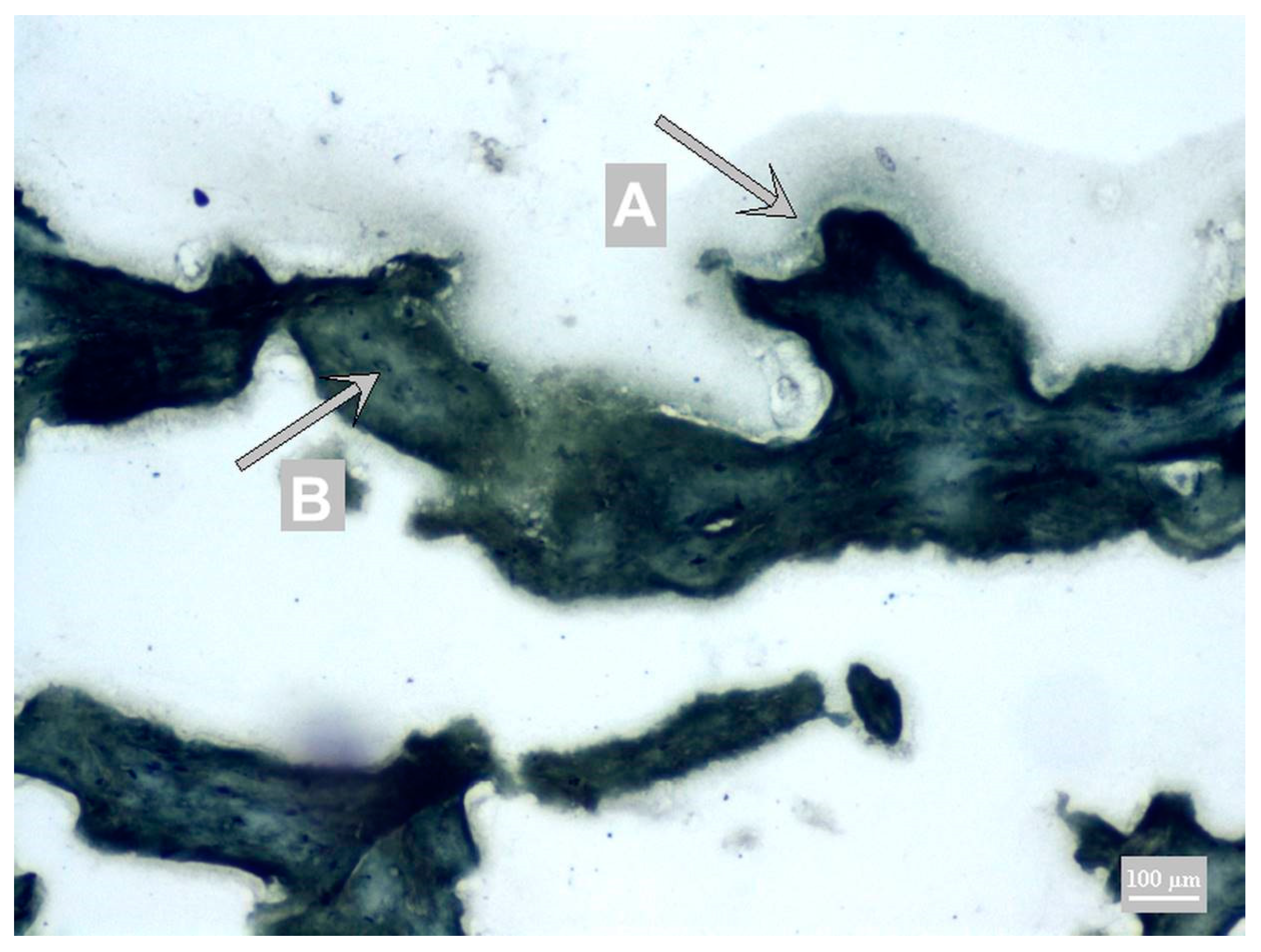
As described by Thomas and Lemmer [19], DAB and HistoGreen chromogens have the same sensitivity. However, when comparing these two with peroxidase substrates, differences in contrast are evident. DAB provides a clear and smooth but sharply bounded and consistent form of immunoreactive structures, and it is typically characterized by regular incision rates in terms of intensity and density. HistoGreen creates coarse grain structures and tends to change the color quickly. The choice of DAB chromogen with background Toluidine Blue staining was based on the work by Řezáčová-Lukášková et al. [14], where high contrast was achieved for wheat protein. Likewise, high contrast was shown using the DAB chromogen with Calleja staining [13] for soy protein. These assumptions were not confirmed as part of the dairy protein detection. DAB chromogen highlighted the presence of milk proteins in brown. However, in combination with Calleja staining, they did not achieve sufficient contrast (Figure 1), as the other structures of the sample (especially the muscle) are colored yellow-green, from green to brown.
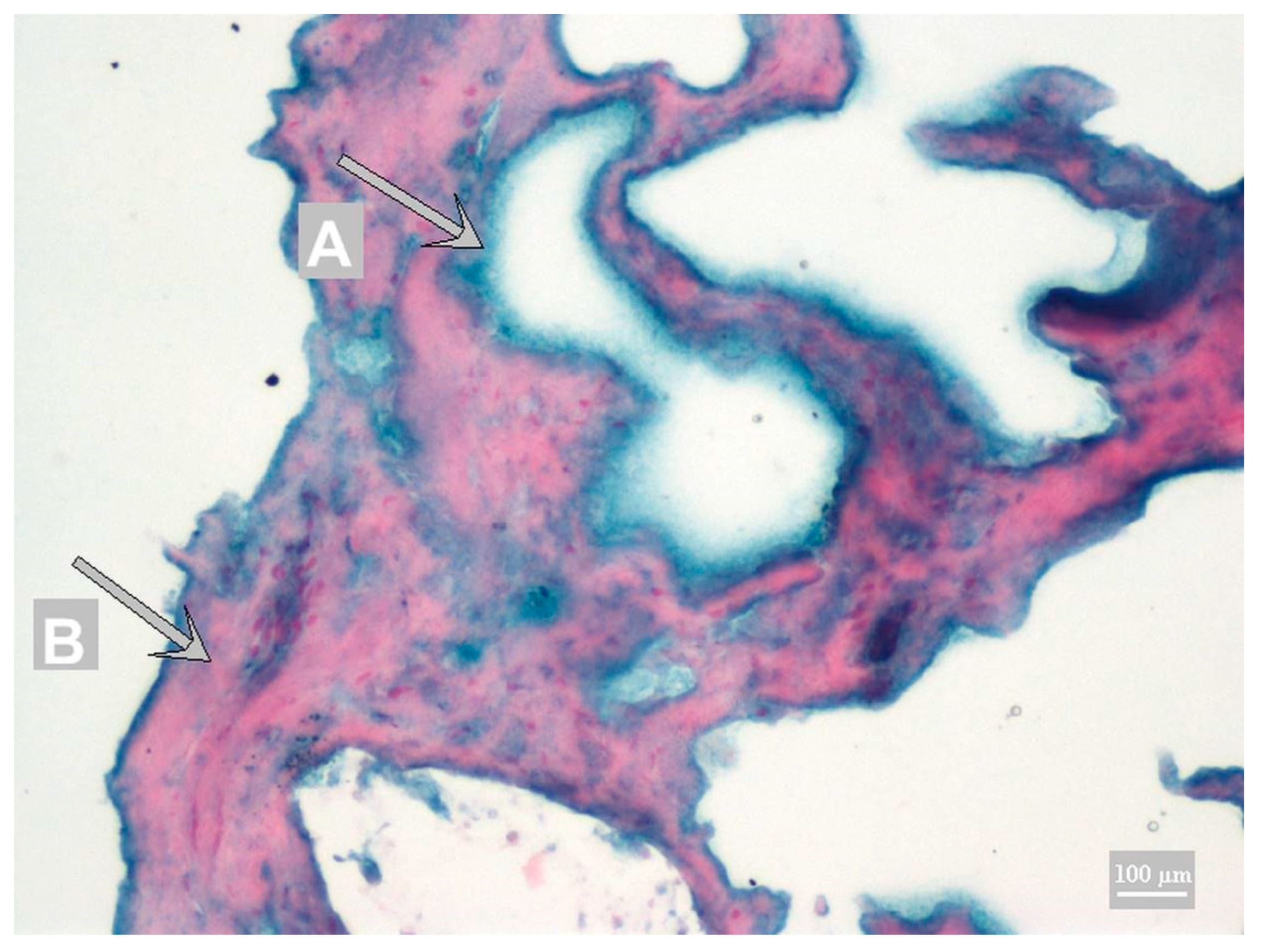
The combination with Toluidine Blue achieved a different color spectrum of the background (Figure 2).
The Toluidine Blue stains other structures blue, blue-violet to blue-gray. Because none of the stainings achieved sufficient contrast, Fast Nuclei Red staining was tested as a new staining for IHC. Using Fast Nuclei Red in combination with DAB chromogen did not achieve sufficient contrast. Sufficient contrast was achieved only with the Fast Nuclei Red in combination with HistoGreen chromogen (Figure 3).
An alternative to immunochemical methods includes immunofluorescence methods, which do not need background staining, as a specific wavelength emission is measured [20]. However, thereby we lose accompanying information about the protein formation and, in particular, localization of the monitored protein in the product structure.

3.2. Method Validation

The qualitative methods (i.e., methods determining the presence or absence of the analyte/allergenic compound) are common for laboratory examination, especially as screening methods prior to a more expensive examination by a quantitative method. The advantages of their use are the costs-reduction and time-saving [21]. Similarly, in an official document by the European Commission [22] and Magnusson [23], the following parameters were provided: limit of detection, repeatability and reproducibility, sensitivity, and selectivity/specificity.

3.3. Limit of Detection (LOD)

Determination of the milk proteins added to meat products is not studied very much. Studies based on immunological methods most often deal with the detection of adulteration of different types of milk or cheeses with cow’s milk. The detection limit of these methods ranges from 0.001% (v/v) to 15% (v/v) [24,25] and depends on the method used and the antigen chosen using different milk proteins or immunoglobulins. Commercial immunoassay kits, where the detection limit ranges from 0.1–5 mg kg−1 are currently available. Likewise, the limit of detection (LOD) is dependent on the method used, the antigen selected, and also on the analyzed matrix [26]. LOD in our developed IHC was determined at 0.0001% (w/w) of caseinate that represents 9.4 mg kg−1 of casein (Table 3).
According to Trullols, Ruisanchez, and Rius [21], LOD was determined as positively evaluated samples in all eight replicates. The detection limit of the applied ELISA method was determined at 2.5 mg kg−1 of casein according to its manufacturer. Analytical methods, such as ELISA, are used routinely to obtain semiquantitative or quantitative results. For the determination of milk, sandwich ELISA kits with sensitivity of up to 1 mg kg−1 are commercially available. There are also kits that are formed only to detect a specific milk protein (bovine serum albumin, casein, beta-lactoglobulin) and others to detect whole milk or whey protein with the achievement of LOD values within the range of 0.5 to 5 mg kg−1 [27]. To quantify casein in food products (e.g., cured sausages), Spizzirri and Crillo [28] used the S-ELISA (sandwich ELISA) method using rabbit and goat anti-casein antibodies where LOD was 0.5 mg kg−1. According to Gern, Yang, Evrard, and Sampson [29], the presence of 136 μg mL−1 of casein and 5 μg mL−1 of whey was enough to induce allergic reactions. It means that the developed and commonly used methods are adequate for milk and whey protein detection in case of consumer protection.

3.4. Repeatability and Reproducibility

Repeatability and reproducibility are optional parameters for qualitative methods. In case of IHC assays, where the protocols are performed on different days, the impact of preanalytical factors were described in many studies [30,31]. The repeatability and reproducibility were provided for the evaluation of the environmental influence. The repeatability was established to be 100% for positive as well as negative samples (Table 4). Based on personal factors during examination, the reproducibility was performed. The reproducibility was also established as 100% (Table 5).

3.5. Sensitivity and Specificity

Fuchs [32] reports that allergy to cow’s milk proteins is probably the most common food allergy affecting the European population. Milk allergy is, however, caused by several proteins that differ not only in their allergenic potential but also in post-processing stability. Whey proteins of β-lactoglobulin as well as serum albumin and immunoglobulin lose their allergenic potential during heat treatment and enzymatic cleavage. In contrast, caseins and α-lactalbumin are considered thermostable. This applies mainly to casein with a loose tertiary, highly hydrated structure. Caseins are not significantly affected by severe heat treatment but are very susceptible to all proteinases and exopeptidases [33]. This fact is also confirmed by Spizzirri and Crillo [28] claiming that casein is a thermostable protein; in contrast, whey β-lactoglobulin is thermally labile and irreversibly denatured or aggregated with casein micelles and α-lactalbumin after heat treatment. However, the research by Bu, Luo, Zheng, and Zheng [34] disproves the claim that thermosetting proteins are thermolabile. In their study, they found that α-lactalbumin and β-lactoglobulin have even higher IgG binding after pasteurization and sterilization than before heat treatment. β-casein is able to maintain its immunoreactivity even after warming up to 55 °C [9]. For meat products, cooking at 70 °C for 10 min is mostly used. Nevertheless, based on a number of studies, we can assume that even such a warm-up will not cause a sufficient reduction in the allergenicity of caseins. Different immunoreactivity was also confirmed for the analytical methods. In ELISA, extraction effectivity is important [35] in comparison with IHC using direct detection of antigen in food matrix where extraction effectivity is negligible for solid antigen. The solubility as well as immunoreactivity can be changed by protein aggregation. The aggregate can be detected by the IHC method easily, as it is known from histological diagnostic as well as in cases of decreasing immunoreactivity. The important issues for immunoreactivity of IHC methods are pre- and postanalytical factors [31]. For confirmation of immunoreactivity, various dairy additives were tested. Immunoreactivity was evaluated on model samples with 2.5% content of additives: calcium caseinate (C1–7), whey protein (W1–W5), and milk addition (M1). For all additions, the casein immunoreactivity was confirmed (Table 6).
This result points to the suitability of polyclonal antibodies for the IHC and generally supports immunological based methods. The content of casein in dry whey protein concentrate was confirmed also in another study [36], and it is in conformity with our results. Heat treatment, however, is not the only way that can affect the immunoreactivity of raw materials used. In the production of dried sausages, there is a number of reactions that make sausages obtain their characteristic texture, flavor, and smell. They include in particular hydrolysis and autooxidation of lipids, proteolysis, and transformation of amino acids to aromatic compounds. In addition, the fermentation, such as lactic acid bacteria, has a major influence on the formation of small peptides and free amino acids. Dried sausages often include sodium caseinate, which undergoes transformations similar to meat protein (i.e., the formation of small peptides and free amino acids). These small peptides contribute to the development of the characteristic taste of dry fermented products and also fulfill important bioactive functions, such as antioxidant and antihypertensive activity in dried meat products. Within analysis of casein degradation, it was found that most of the peptide residues in the food remain from β-casein [4]. Many studies also focus on the values of the minimum observed dose inducing allergic reactions to milk and its products. Several of these studies have demonstrated severe reactions to very small amounts of these allergens. An example is a severe anaphylactic reaction that was caused by consumption of a sausage containing cow’s milk (corresponding to casein content of 60 mg) [27]. In that case, casein was selected as the most suitable protein for IHC detection. Considering casein sensitivity to proteinases, an enzymatic method cannot be used as one of the antigen retrieval techniques [15]. It is not easy to verify the various effects that can occur in foodstuffs. Demonstration of truly positive and negative samples is commonly used for this purpose. For the purpose of validation in a diverse matrix of meat products, products from the real retail market were evaluated according to Trullols, Ruisanchez, and Rius [21]. The results are presented in Table 7. Correspondence level of the IHC method with ELISA for all the analyzed meat groups reached 72% in total.
The IHC sensitivity was set at 1.21 and the specificity at 0.28 according to Trullols, Ruisanchez, and Rius [21]. Sensitivity is the ability of a method to detect truly positive samples as positive. It can be calculated as test positives/total number of known positives. Specificity is defined as the ability of a method to detect truly negative samples as negative. It can be expressed as test negatives/total number of known negatives [Trullols et al., 2004]. Model meat products with various additive concentrations, different dairy additive types, and products from the retail network were used for the calculation. The method is therefore highly sensitive for the detection of milk additives. The concentration does not show a strong impact on the developed methods. In contrast to the IHC method, the ELISA kit reaches sensitivity of 1.15 and specificity of 0.4. The specificity in both methods should be considered as indicative because only two negative samples were evaluated. Thus, in contrast to the ELISA method, the IHC method is less sensitive.
McNemar’s Test (Chi-square statistic) was used to compare the qualitative results of both methods applied [13,21] where model samples and meat products were evaluated. The comparison shows that even with respect to certain differences, especially in the number of negatively evaluated results by IHC (n = 7) and ELISA (n = 5), including dubious samples, both methods are comparable (p > 0.05) (Table 7 and Table 8).
The reason for choosing ELISA for the comparison was that it is commonly used for the determination of milk proteins in foodstuffs and it is also the official screening method in many countries [37]. For meat products, determination of defatted milk powder was also validated already at 10 mg kg−1 [38]. These authors also reported a relatively high level of reproducibility of interlaboratory accuracy (Allergeneye ELISA) for milk with the relative standard deviation for reproducibility (RSDR) amounting to 6.8–10.5% for all processed foodstuffs. Similar results in interlaboratory reproducibility (RSDR 7–14% and 12–17%) were achieved by Matsuda et al. [37], who tested the presence of milk in sausages using ELISA methods: FASTKIT and ELISA FASPEK. However, compared with the above, the ELISA method is still not considered to be sufficiently perfect. A perfect method is expected to incorporate features such as high accuracy and sensitivity, low costs, and rapid analysis of various types of technologically processed food products. There is a variety of commercially available ELISA kits; nevertheless, none of them meets the above requirements. The biggest problem is false-negative results in heat-treated products [39]. The false-negative results were demonstrated also in this work where ELISA reached 15.3% and IHC 19.2% in total. In frankfurters, ELISA reached 10% and IHC 0%. In canned pâté, ELISA reached 33.3% and IHC 55.5% of false-negative results.
Lactose was declared in a chorizo product (L1) where it is added as part of the starter to improve the fermentation process using lactic acid bacteria [40]. Our results obtained by ELISA and IHC were positive, contrary to the original assumption. The positivity of the sample with declared lactose content can be caused by contamination of this additive during the production. Contamination of the lactose additive by milk proteins might be due to its production from cow’s milk or whey. Lactose is used not only in the food industry but also in the pharmaceutical industry where it is used as an ineffective ingredient, a filler in many pharmaceutical preparations. The problem of lactose contamination by milk proteins has already been recognized in the past in pharmaceutical products [41]. Therefore, five different whey products commonly used in the food industry were tested—Table 5 (W1–W5). Both methods, the IHC as well as ELISA, confirmed the presence of casein in these products. Our study confirmed also the presence of casein in lactose additive. A small content of casein in lactose was also confirmed not only among various manufacturers but also among individual batch preparations from the same manufacturer. From this point of view, we can assume that the sample from the market network examined by us was impacted by a casein contamination of lactose or unsatisfactory purification during lactose producing.
Canned pâté products also differed from the assumptions, where the IHC method did not confirm presence of milk additive. This included a doubtful sample of M12 and false-negative samples of M11 and M16–M18, which represents 44%. In this group of products, milk additives are primarily used as emulsifiers [3,4]. This property and high degree of homogenization of the matrix are attributed to the negative result of the IHC assay, when milk proteins are surrounded by the fat particles and subsequently form an emulsion matrix with water and fat [42]. This phenomenon leads to the spreading of proteins on a larger area, and the resulting precipitate is of weaker intensity. With respect to the non-specific binding always present, the signal intensity is then lost. A similar result was also shown by the ELISA examination where M13, M15, and M17 were false-negative (33%). Our results are in agreement with Zaffran and Sathe [43], who refer that immunoreactivity can be changed by food processing. Weakening of signal intensity could also be due to denaturation of proteins that might be caused by higher temperatures and pressure used in the production of canned meat products. The food processing with protein spreading also can explain false negative results of IHC. This hypothesis, however, is in contradiction to many authors who have demonstrated the allergenic potential of caseins in heat-treated milk even after temperatures above 170 °C were applied for up to 60 min [44]. As reported by D’Auria et al. [8], caseins are resistant to high temperatures, and it can be said that they represent most of the proteins contained in heat-treated foodstuffs. Heat resistance can be affected by thiol group binding κ-casein [45]. However, low immunoreactivity of canned pâté remains to be further explored. Dupont et al. [44] describe the possibility of reducing allergenicity and allergic reactions of children consuming heat-treated milk. When evaluating the allergenicity, the team of authors came to the finding that caseins appeared to be heavily allergenic even at temperatures of 170 °C for up to 60 min of heating. Although α-casein is thermally stable, it is influenced by the presence of phosphate, which is essential for the proteins’ function of binding calcium. Because caseins are resistant to high temperatures, they can be said to be the major allergenic proteins contained in heat-treated foodstuffs [8]. This statement was also confirmed in other heat-treated meat products (frankfurter type) analyzed in our study. Comparison of both methods shows that the IHC method is comparable with ELISA for this group of meat products. Of the 10 analyzed samples of frankfurter-type products, correspondence was achieved in 9 samples (90%) because ELISA showed a false-negative response in one case. On the other hand, canned pâté correspondence reached 40%. The correspondence for ham mousse was 100%, for salami 50%, and for meat loaf 100% (Table 7). However, in these three product groups, only a small number of samples was evaluated, and data are only approximated. Further studies should focus on immunoreactivity changes in pâté and other processed meat products.

4. Conclusions

Milk proteins are a frequent additive especially to emulsified meat products. The immunohistochemical method for the detection of casein was developed and validated in this work. The immunohistochemical methods using the HistoGreen chromogen and Nuclear Fast Red background staining were evaluated as the most appropriate. The detection limit of the method was determined at 9.4 mg kg−1, repeatability and reproducibility were established at 100%, sensitivity was 1.21, and specificity was 0.28.
The casein, as one of the stable milk proteins, was confirmed as an appropriate antigen for immunologically based tests. IHC as well as ELISA detected casein in calcium caseinate, whey powder, whey protein with high lactose content, thermostable whey concentrate, buttermilk powder, whey concentrate containing 85% of casein, as well as milk.
Nevertheless, the impact of food processing to casein immunoreactivity was observed. The canned pâté demonstrated the highest masking effect of its matrix for booth methods. ELISA reached 33.3% and IHC 55.5% of false-negative results. The lowest masking effect was confirmed for whole meat products following meat loaf, salami, and frankfurters. This study is a pilot study for evaluation of food processing impact on immunoreactivity using the direct antigen detection in food matrix.

Author Contributions

Conceptualization: M.P.; methodology, validation, and form analysis: M.P. and L.K.; writing—original draft preparation: M.P. and L.K.; writing—review and editing: Z.J.; supervision: B.T. and I.C.; project administration: M.P.; funding acquisition: M.P. All authors have read and agreed to the published version of the manuscript.

Funding

This work was supported by the Internal Grant Agency of FVHE UVPS Brno under Grant IGA 27/2014/FVHE and the institutional research funds of UVPS Brno.

Conflicts of Interest

No potential conflict of interest was reported by the authors.

References

  1. O’Regan, J.; Ennis, M.P.; Mulvihill, D.M. Milk proteins. In Handbook of Hydrocolloids; Woodhead Publishing: Cambridge, UK, 2009; pp. 298–358. [Google Scholar] [CrossRef]
  2. Kumar, S.; Zanzad, P.N.; Ambadkar, R.K.; Rindhe, S.N.; Kumar, P.; Karle, S.D. Storage stability of chicken sausage incorporated with selected levels of sodium caseinate. J. Vet. Public Health 2011, 9, 33–37. [Google Scholar]
  3. Tarté, R. Ingredients in Meat Products: Properties, Functionality and Applications; Springer Science & Business Media: Berlin/Heidelberg, Germany, 2009; ISBN 978-0-387-71327-4. [Google Scholar]
  4. Mora, L.; Escudero, E.; Aristoy, M.C.; Toldrá, F. A peptidomic approach to study the contribution of added casein proteins to the peptide profile in Spanish dry-fermented sausages. Int. J. Food Microbiol. 2015, 212, 41–48. [Google Scholar] [CrossRef] [PubMed] [Green Version]
  5. Mallika, E.N.; Prabhakar, K.; Reddy, P.M. Low Fat Meat Products-An Overview. Vet. World 2009, 2, 364–366. [Google Scholar]
  6. Jalal, H.; Mir, S.; Wani, S.A.; Sofi, A.H.; Pal, M.A.; Rather, F. Development of low fat meat products. Int. J. Food Nutr. Saf. 2013, 4, 98–107. [Google Scholar]
  7. European Commission. Regulation (EU) No 1169/2011 of the European Parliament and of the Council of 25 October 2011 on the provision of food information to consumers, amending Regulations (EC) No 1924/2006 and (EC) No 1925/2006 of the European Parliament and of the Council, and repealing Commission Directive 87/250/EEC, Council Directive 90/496/EEC, Commission Directive 1999/10/EC, Directive 2000/13/EC of the European Parliament and of the Council, Commission Directives 2002/67/EC and 2008/5/EC and Commission Regulation (EC). Off. J. Eur. Union L 2011, 304, 18–63. [Google Scholar]
  8. D’Auria, E.; Mameli, C.; Piras, C.; Cococcioni, L.; Urbani, A.; Zuccotti, G.V.; Roncada, P. Precision medicine in cow’s milk allergy: Proteomics perspectives from allergens to patients. J. Proteom. 2018, 188, 173–180. [Google Scholar] [CrossRef]
  9. Hochwallner, H.; Schulmeister, U.; Swoboda, I.; Spitzauer, S.; Valenta, R. Cow’s milk allergy: From allergens to new forms of diagnosis, therapy and prevention. Methods 2014, 66, 22–33. [Google Scholar] [CrossRef]
  10. Miciński, J.; Kowalski, I.M.; Zwierzchowski, G.; Szarek, J.; Pierożyński, B.; Zabłocka, E. Characteristics of cow’s milk proteins including allergenic properties and methods for its reduction. Pol. Ann. Med. 2013, 20, 69–76. [Google Scholar] [CrossRef]
  11. Villa, C.; Costa, J.; Oliveira, M.B.P.; Mafra, I. Bovine milk allergens: A comprehensive review. Compr. Rev. Food Sci. Food Saf. 2018, 17, 137–164. [Google Scholar] [CrossRef] [Green Version]
  12. Downs, M.L.; Taylor, S.L. Effects of thermal processing on the enzyme-linked immunosorbent assay (ELISA) detection of milk residues in a model food matrix. J. Agric. Food Chem. 2010, 58, 10085–10091. [Google Scholar] [CrossRef]
  13. Pospiech, M.; Tremlova, B.; Renčová, E.; Randulová, Z. Immunohistochemical detection of soya protein–optimisation and verification of the method. Czech J. Food Sci. 2009, 27, 11–19. [Google Scholar] [CrossRef] [Green Version]
  14. Řezáčová-Lukášková, Z.; Tremlová, B.; Pospiech, M.; Renčová, E.; Randulová, Z.; Steinhauser, L.; Reichová, A.; Bednář, J. Comparison of immunohistochemical, histochemical and immunochemical methods for the detection of wheat protein allergens in meat samples and cooked, dry, raw and fermented sausage samples. Food Addit. Contam. Part A 2011, 28, 817–825. [Google Scholar] [CrossRef] [PubMed]
  15. Bednářová, M.; Pospiech, M.; Tremlová, B.; Řezáčová-Lukášková, Z.; Bednář, J. Antigen retrieval and fixation of sections on slides for immunohistochemical detection of soya protein in meat products. J. Food Nutr. Res. 2015, 54, 1–8. [Google Scholar]
  16. Hendl, J. Review of Statistical Methods of Data Processing; Portál: Prague, Czech Republic, 2004; ISBN 80-7178-820-1. [Google Scholar]
  17. Petracci, M.; Bianchi, M.; Mudalal, S.; Cavani, C. Functional ingredients for poultry meat products. Trends Food Sci. Technol. 2013, 33, 27–39. [Google Scholar] [CrossRef]
  18. Barbut, S. Effects of caseinate, whey and milk powders on the texture and microstructure of emulsified chicken meat batters. Lwt-Food Sci. Technol. 2006, 39, 660–664. [Google Scholar] [CrossRef]
  19. Thomas, M.A.; Lemmer, B. HistoGreen: A new alternative to 3,3′-diaminobenzidine-tetrahydrochloride-dihydrate (DAB) as a peroxidase substrate in immunohistochemistry? Brain Res. Protoc. 2005, 14, 107–118. [Google Scholar] [CrossRef]
  20. Petrášová, M.; Pospiech, M.; Tremlová, B.; Javůrková, Z. Immunofluorescence detection of pea protein in meat products. Food Addit. Contam. Part A 2016, 33, 1283–1289. [Google Scholar] [CrossRef]
  21. Trullols, E.; Ruisanchez, I.; Rius, F.X. Validation of qualitative analytical methods. Trac. Trends Anal. Chem. 2004, 23, 137–145. [Google Scholar] [CrossRef] [Green Version]
  22. European Commission. Commission Decision 2002/657/EC of 12 August 2002 implementing Council Directive 96/23/EC concerning the performance of analytical methods and the interpretation of results. Off. J. Eur. Union 2002, 221, 8–36. [Google Scholar]
  23. Magnusson, B. The Fitness for Purpose of Analytical Methods: A Laboratory Guide to Method Validation and Related Topics; Eurachem: Teddington, UK, 2014; ISBN 978-91-87461-59-0. Available online: www.eurachem.org (accessed on 18 November 2020).
  24. Borková, M.; Snášelova, J. Possibilities of different animal milk detection in milk and dairy products–a review. Czech J. Food Sci. 2005, 23, 41–50. [Google Scholar] [CrossRef] [Green Version]
  25. Hurley, I.P.; Coleman, R.C.; Ireland, H.E.; Williams, J.H. Use of sandwich IgG ELISA for the detection and quantification of adulteration of milk and soft cheese. Int. Dairy J. 2006, 16, 805–812. [Google Scholar] [CrossRef]
  26. Schubert-Ullrich, P.; Rudolf, J.; Ansari, P.; Galler, B.; Führer, M.; Molinelli, A.; Baumgartner, S. Commercialized rapid immunoanalytical tests for determination of allergenic food proteins: An overview. Anal. Bioanal. Chem. 2009, 395, 69–81. [Google Scholar] [CrossRef] [PubMed]
  27. EFSA Panel on Dietetic Products, Nutrition and Allergies (NDA). Scientific Opinion on the evaluation of allergenic foods and food ingredients for labelling purposes. EFSA J. 2014, 12, 3894.
  28. Spizzirri, U.G.; Cirillo, G. (Eds.) Food Safety: Innovative Analytical Tools for Safety Assessment; Scrivener Publishing: Salem, MA, USA, 2017. [Google Scholar] [CrossRef]
  29. Gern, J.E.; Yang, E.; Evrard, H.M.; Sampson, H.A. Allergic reactions to milk-contaminated nondairy products. N. Engl. J. Med. 1991, 324, 976–979. [Google Scholar] [CrossRef]
  30. Taylor, C.R. Quality assurance and standardization in immunohistochemistry. A proposal for the annual meeting of the Biological Stain Commission, June, 1991. Biotech. Histochem. 1992, 67, 110–117. [Google Scholar] [CrossRef]
  31. Taylor, C.; Shi, S.R.; Cote, R. Antigen retrieval for immunohistochemistry. Appl. Immunohistochem. 1996, 4, 144–166. [Google Scholar]
  32. Fuchs, M. Potravinové Alergie; Food Allergies; Maxdorf: Praha, Czech Republic, 2013; p. 43. ISBN 978-80-7345-335-0. [Google Scholar]
  33. Wal, J.M. Cow’s milk allergens. Allergy 1998, 53, 1013–1022. [Google Scholar] [CrossRef]
  34. Bu, G.; Luo, Y.; Zheng, Z.; Zheng, H. Effect of heat treatment on the antigenicity of bovine α-lactalbumin and β-lactoglobulin in whey protein isolate. Food Agric. Immunol. 2009, 20, 195–206. [Google Scholar] [CrossRef]
  35. Diaz-Amigo, C. Towards a comprehensive validation of ELISA kits for food allergens. Case 2-milk. Food Anal. Methods 2010, 3, 351–356. [Google Scholar] [CrossRef]
  36. Nishanthi, M.; Chandrapala, J.; Vasiljevic, T. Properties of whey protein concentrate powders obtained by spray drying of sweet, salty and acid whey under varying storage conditions. J. Food Eng. 2017, 214, 137–146. [Google Scholar] [CrossRef]
  37. Matsuda, R.; Yoshioka, Y.; Akiyama, H.; Aburatani, K.; Watanabe, Y.; Matsumoto, T.; Morishita, N.; Sato, H.; Mishima, T.; Gamo, R.; et al. Interlaboratory evaluation of two enzyme-linked immunosorbent assay kits for the detection of egg, milk, wheat, buckwheat, and peanut in foods. J. AOAC Int. 2006, 89, 1600–1608. [Google Scholar] [CrossRef] [PubMed]
  38. Kato, S.; Yagi, T.; Kato, A.; Yamamoto, S.; Akimoto, M.; Arihara, K. Interlaboratory study of ELISA kits for the detection of egg and milk protein in processed foods. J. AOAC Int. 2015, 98, 810–816. [Google Scholar] [CrossRef] [PubMed]
  39. Spychaj, A.; Pospiech, E.; Iwańska, E.; Montowska, M. Detection of allergenic additives in processed meat products. J. Sci. Food Agric. 2018, 98, 4807–4815. [Google Scholar] [CrossRef] [PubMed]
  40. González-Fernández, C.; Santos, E.M.; Jaime, I.; Rovira, J. Influence of starter cultures and sugar concentrations on biogenic amine contents in chorizo dry sausage. Food Microbiol. 2003, 20, 275–284. [Google Scholar] [CrossRef]
  41. Nowak-Wegrzyn, A.; Shapiro, G.G.; Beyer, K.; Bardina, L.; Sampson, H.A. Contamination of dry powder inhalers for asthma with milk proteins containing lactose. J. Allergy Clin. Immunol. 2004, 113, 558–560. [Google Scholar] [CrossRef]
  42. Youssef, M.K.; Barbut, S. Physicochemical effects of the lipid phase and protein level on meat emulsion stability, texture, and microstructure. J. Food Sci. 2010, 75, S108–S114. [Google Scholar] [CrossRef]
  43. Zaffran, V.D.; Sathe, S.K. Immunoreactivity of Biochemically Purified Amandin from Thermally Processed Almonds (Prunus dulcis L.). J. Food Sci. 2018, 83, 1805–1809. [Google Scholar] [CrossRef]
  44. Dupont, C.; Chouraqui, J.P.; Linglart, A.; Bocquet, A.; Darmaun, D.; Feillet, F.; Frelut, M.L.; Girardet, J.P.; Hankard, R.; Rozé, J.C.; et al. Nutritional management of cow’s milk allergy in children: An update. Arch. Pédiatrie 2018, 25, 236–243. [Google Scholar] [CrossRef]
  45. Sletten, G.B.; Holden, L.; Egaas, E.; Faeste, C.K. Differential influence of the degree of processing on immunogenicity following proteolysis of casein and β-lactoglobulin. Food Agric. Immunol. 2008, 19, 213–228. [Google Scholar] [CrossRef]
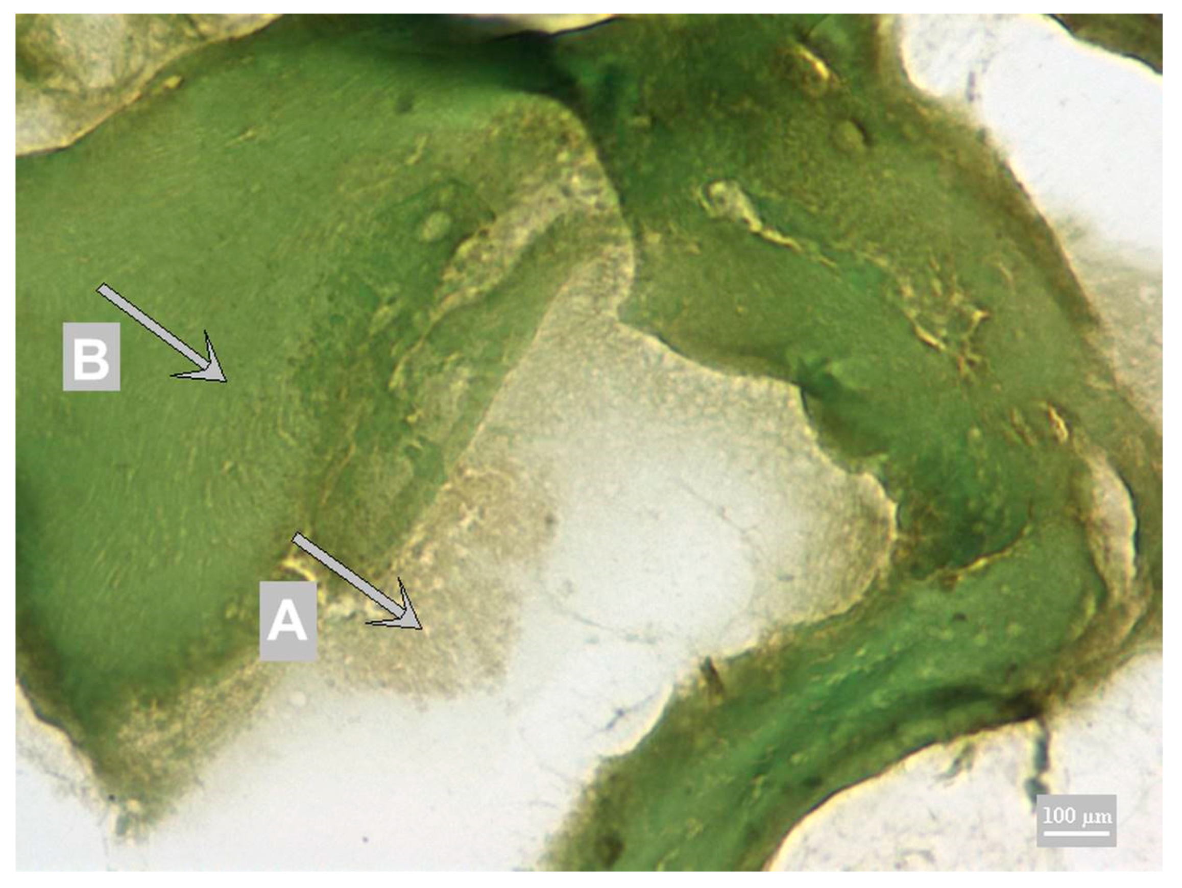
Figure 1. Calleja staining with DAB chromogen; A chromogen precipitate—calcium caseinate (gray-brown); B muscle fiber (green).
Foods 10 00028 g001
Figure 2. Toluidine Blue with DAB chromogen; A chromogen precipitate—calcium caseinate (blue-brown); B muscle fiber (blue).
Foods 10 00028 g002
Figure 3. Fast Nuclei Red with HistoGreen chromogen; A chromogen precipitate—calcium caseinate (green); B muscle fiber (red).
Foods 10 00028 g003
Table 1. Effect of primary antibody dilution on IHC detection of beta-casein in selected model samples.
Model SampleCaseinate AdditionPrimary Antibody Dilution
1:101:1001:5001:10001:1500
C51%+++++++++++++++
C62.5%++++++++++++
+: weak signal; ++: strong signal; +++: very strong signal; C5 and C6: calcium caseinate.
Table 2. Contrast between chromogens and background staining in calcium caseinate 2.5% model sample.
ChromogensBackground Staining
CallejaToluidine BlueFast Nuclei Red
DAB++-
HistoGreen--+++
+: weak signal; +++: very strong signal.
Table 3. Limit of detection.
Caseinate Addition (%)Quantity of Casein (mg kg−1)No. of RepetitionsPositive/Negative
0080/8
0.00019.488/0
0.0019488/0
0.0194088/0
0.1940088/0
194,00088/0
10940,00088/0
Table 4. Repeatability of IHC examination.
RepetitionR1R2
1YesNo
2YesNo
3YesNo
4YesNo
5YesNo
Correspondence100%100%
R1: positive control; R2: negative control.
Table 5. Reproducibility of IHC examination by different examiners.
ExaminerR1R2
AYesNo
AYesNo
AYesNo
AYesNo
AYesNo
BYesNo
BYesNo
BYesNo
BYesNo
BYesNo
Correspondence100%100%
A: examiner 1; B: examiner 2; R1: positive control; R2: negative control.
Table 6. IHC and ELISA immunoreactivity to different additive types in model meat products.
SampleC1C2C3C4C5C6C7W1W2W3W4W5M1
IHC+++++++++++++
ELISA+++++++++++++
Note: C1–7: calcium caseinate; W1: whey powder; W2: whey protein with high lactose content; W3: thermostable whey concentrate; W4: buttermilk powder; W5: whey concentrate containing 85% of casein; M1: milk; +: positive reaction.
Table 7. IHC and ELISA immunoreactivity to casein in commercial meat products.
Meat Product
Type of Additives and IdentificationM2M3M4M5M6M7M8M9M10M11M12M13M14M15M16M17M18M19M20Co1Co2Ch1Ch2Ch3L1
IHC+++++++++− *+/− *+++− *− *++++++
ELISA++++− *++++++− *+− *+++++ **++++
Meat ProductFRCPHMCPFRSAMLFRMLSA
Correspondence of IHC and ELISA methods
Meat ProductTotal (%)
FR905/6
(83%)
3/3
(100%)
1/1
(100%)
CP44 1/1
(100%)
3/8
(37.5%)
HM100 2/2
(100%)
SA50 0/1
(0%)
1/1
(100%)
ML100 1/1
(100%)
1/1
(100%)
All Types72 18/25 (72)
M2–20: milk; Co1–2: control sample without any milk protein; Ch1–3: cheese; L1: lactose; +: positive; −: negative, +/−: dubious reaction; *: false negative; **: false positive; FR: frankfurters; CP: canned pâté; HM: ham mousse; SA: salami; ML: meat loaf; IHC/ELISA (% correspondence).
Table 8. IHC and ELISA comparison by Chi-square statistic.
IHC+IHC−Sum
ELISA+29534
ELISA−325
Sum33740
Chst0.5
p = 0.4795
Dubious samples were calculated as positive and also negative ones.
Publisher’s Note: MDPI stays neutral with regard to jurisdictional claims in published maps and institutional affiliations.