Article

Lactiplantibacillus plantarum LOC1 Isolated from Fresh Tea Leaves Modulates Macrophage Response to TLR4 Activation

1
Central Research Institute, ITO EN Ltd., Shizuoka 421-0516, Japan
2
Laboratory of Animal Food Function, Graduate School of Agricultural Science, Tohoku University, Sendai 980-8576, Japan
3
Laboratory of Immunobiotechnology, Reference Centre for Lactobacilli (CERELA-CONICET), Tucuman CP4000, Argentina
4
International Education and Research Center for Food and Agricultural Immunology (CFAI), Graduate School of Agricultural Science, Tohoku University, Sendai 980-8576, Japan
*
Authors to whom correspondence should be addressed.
Foods 2022, 11(20), 3257; https://doi.org/10.3390/foods11203257
Submission received: 26 September 2022 / Revised: 14 October 2022 / Accepted: 17 October 2022 / Published: 18 October 2022

Abstract

:
Previously, we demonstrated that Lactiplantibacillus plantarum LOC1, originally isolated from fresh tea leaves, was able to improve epithelial barrier integrity in in vitro models, suggesting that this strain is an interesting probiotic candidate. In this work, we aimed to continue characterizing the potential probiotic properties of the LOC1 strain, focusing on its immunomodulatory properties in the context of innate immunity triggered by Toll-like receptor 4 (TLR4) activation. These studies were complemented by comparative and functional genomics analysis to characterize the bacterial genes involved in the immunomodulatory capacity. We carried out a transcriptomic study to evaluate the effect of L. plantarum LOC1 on the response of murine macrophages (RAW264.7 cells) to the activation of TLR4. We demonstrated that L. plantarum LOC1 exerts a modulatory effect on lipopolysaccharide (LPS)-induced inflammation, resulting in a differential regulation of immune factor expression in macrophages. The LOC1 strain markedly reduced the LPS-induced expression of some inflammatory cytokines (IL-1β, IL-12, and CSF2) and chemokines (CCL17, CCL28, CXCL3, CXCL13, CXCL1, and CX3CL1), while it significantly increased the expression of other cytokines (TNF-α, IL-6, IL-18, IFN-β, IFN-γ, and CSF3), chemokines (IL-15 and CXCL9), and activation markers (H2-k1, H2-M3, CD80, and CD86) in RAW macrophages. Our results show that L. plantarum LOC1 would enhance the intrinsic functions of macrophages, promoting their protective effects mediated by the stimulation of the Th1 response without affecting the regulatory mechanisms that help control inflammation. In addition, we sequenced the LOC1 genome and performed a genomic characterization. Genomic comparative analysis with the well-known immunomodulatory strains WCSF1 and CRL1506 demonstrated that L. plantarum LOC1 possess a set of adhesion factors and genes involved in the biosynthesis of teichoic acids and lipoproteins that could be involved in its immunomodulatory capacity. The results of this work can contribute to the development of immune-related functional foods containing L. plantarum LOC1.

1. Introduction

Lactiplantibacillus plantarum are nomadic lactic acid bacteria (LAB) [1] and are known for their significant intraspecific versatility. This species of LAB is frequently isolated from dairy foods as well as fresh and fermented plant and meat. L. plantarum are also inhabitants of the gastrointestinal and urogenital tracts of humans and animals [2]. Consistent with its environmental range, L. plantarum strains have larger genomes compared with other species of LAB [3]. L. plantarum is widely used in the industry to produce numerous fermented foods, and certain strains are used as probiotics for the improvement of human and animal health.
Some L. plantarum strains have been shown to exert immunomodulatory effects on the host. Studies have demonstrated that strains such as L. plantarum CRL1506, MPL16 and LRCC5310 are able to regulate the Toll-like receptor 3 (TLR3)-mediated immune response in the intestinal mucosa and to increase the production of interferon (IFN)-γ and IFN-β as well as antiviral factors increasing the protection against viral infections [4,5,6,7]. L. plantarum YU was shown to strongly augment IL-12 production by intestinal antigen-presenting cells [8], which was associated with TLR2 stimulation. On the other hand, it was reported that L. plantarum strains are also able to regulate TLR4-mediated immunity. It was shown that the exopolysaccharide (EPS) of L. plantarum L-14 is able to regulate the inflammatory response triggered by lipopolysaccharide (LPS) administration to mouse RAW 264.7 macrophages [9]. The EPS from the L-14 strain reduced the expression of the inducible nitric oxide synthase and diminished the proinflammatory mediators interleukin (IL)-6, IL-1β and tumor necrosis factor (TNF)-α. L. plantarum L15 was shown to reduce the expression of TLR4, MyD88 and genes related to the NF-κB signaling pathway, inducing increased protection against ulcerative colitis [10], while mice treated with a mixture of three L. plantarum strains (KLDS 1.0318, KLDS 1.0344, and KLDS 1.0386) had significantly lower production of intestinal TNF-α, IL-6 and IL-12 and reduced gut tissue injury after the challenge with LPS [11]. Thus, the immunomodulatory probiotic or immunobiotic L. plantarum strains, particularly those with the ability to regulate TLR-mediated immune responses, are interesting alternatives to develop functional foods with the capacity to improve immune health [4,5,12].
Members of the Lactobacillus group have been isolated from fermented tea leaves including the species Lactiplantibacillus pentosus [13], Limosilactobacillus fermentum [14] and L. plantarum [15]. Meanwhile, the isolation of Lactobacillus strains from fresh tea leaves (Camellia sinensis, Theaceae) has been less explored [16]. In this regard, we recently isolated LAB strains from fresh tea leaves and evaluated the impact of selected strains on the intestinal barrier integrity using co-culture in vitro models of the small and large intestine [17]. Among the studied strains, L. plantarum LOC1 was able to improve epithelial barrier integrity and the expression of occludin and mucin genes in Caco-2/HT29-MTX mono- or co-cultures challenged with dextran sodium sulfate (DSS). These results showed that L. plantarum LOC1 is an interesting probiotic candidate with beneficial effects on the intestinal barrier.
To the best of our knowledge, few studies have evaluated the potential probiotic and immunobiotic properties of L. plantarum strains from fresh tea leaves. Carrying out these studies would be of great importance to find microorganisms with beneficial properties for health, which also have biotechnological properties not found in bacteria of dairy origin. Therefore, the objective of this work was to continue characterizing the potential probiotic properties of the LOC1 strain, focusing on its immunomodulatory properties in the context of innate immunity triggered by TLR4 activation. These studies were complemented by comparative and functional genomics analysis to characterize the bacterial genes involved in the immunomodulatory capacity.

2. Materials and Methods

2.1. Microorganisms

Lactiplantibacillus plantarum strains LOC1 and LOC3 were isolated from fresh tea leaves from a Kagoshima tea plantation in Japan [17]. Lacticaseibacillus rhamnosus CRL1505 and L. plantarum CRL1506 belong to CERELA-CONICET Culture Collection (Tucuman, Argentina) and were originally isolated from goat milk [6]. Stock cultures were stored at −80 °C in De Man, Rogosa and Sharpe (MRS) broth (Oxoid, Basingstoke, UK) broth containing 30% glycerol. Lactobacilli were propagated twice in MRS broth prior to use. Bacteria were grown overnight anaerobically at 37 °C in MRS broth. Lactobacilli from the stationary phase were harvested by centrifugation at 8000× g, followed by two washes with PBS.

2.2. Cell Cultures

RAW264.7, a mouse macrophage cell line (RIKEN Cell Bank, Tsukuba, Japan), was cultured in high-glucose Dulbecco’s modified Eagle medium (DMEM) (Thermo Fisher Scientific, Tokyo, Japan) containing 10% fetal bovine serum (FBS), 100 U/mL penicillin, and 100 μg/mL streptomycin (Thermo Fisher Scientific, Tokyo, Japan) at 37 °C in a humidified incubator containing 5% CO2.
Mouse intestinal epithelial (MIE) cells were maintained in Dulbecco’s modified Eagle’s medium (DMEM) (Thermo Fisher Scientific, Tokyo, Japan) supplemented with 10% fetal calf serum, 100 U/mL streptomycin, and 100 mg/mL penicillin at 37 °C in an atmosphere of 5% CO2. MIE cells grow rapidly and have a cobblestone morphology, which is a typical feature of intestinal epithelial cells [18].

2.3. Cell Stimulation with Lactobacilli and LPS

RAW264.7 cells were plated at a density of 5 × 10⁵ cells/well in 12-well plates and cultured for overnight. After changing medium, macrophages were cultivated for 3 h in the absence or presence of lactobacilli (5 × 10⁷ cells/mL). For LPS challenge experiments, RAW264.7 cells were cultivated overnight as described above, and after changing medium, lactobacilli (1 × 10⁸ cells/mL) were added to stimulate the cells for 24 h. Then, wells were washed with medium 3 times to eliminate lactobacilli and stimulated with LPS (50 ng/mL) for 3 h.
For the MIE–macrophage co-culture system, the Transwell culture system was used. MIE cells were seeded in the apical surface at a concentration of 2.5 × 10⁵ cells/well in 12-well tissue culture plates (Corning, pore size 0.4 µm) and cultured for three days. RAW264.7 cells were plated at a density of 5 × 10⁵ cells/well in 12-well plates and cultured overnight. After incubation, both cells were overlaid. For the evaluation of the immunomodulatory activity of lactobacilli in the MIE–immune cell co-culture system, the apical surface containing MIE cells was stimulated with lactobacilli strains (1 × 10⁸ cells/mL) for 48 h. Then, the basolateral surface containing RAW264.7 cells was washed with PBS, and macrophages were stimulated with LPS (50 ng/mL) for 3 h.

2.4. Microarray Analysis

The isolation of total RNA from L. plantarum-treated and non-lactobacilli-treated control macrophages was performed with the RNeasy Mini Kit (Qiagen, Tokyo, Japan). Samples were treated with DNAse and the integrity of RNA molecules was assessed with the RNA 6000 Nano Kit and the Agilent 2100 Bioanalyzer (Agilent Technologies, Santa Clara, CA, USA). Then, the biosynthesis of complementary DNA was performed with 200 ng of RNA. The SurePrint G3 Mouse GE 8 × 60K Ver.2.0 Microarray (Agilent Technologies) was used for hybridization at Hokkaido System Science Co. Microarray scanning was performed with the Microarray Scanner from Agilent Technologies, while digitization was performed with Agilent Feature Extraction 10.7.3.1.
The normalization of data and the analysis of gene expression were performed with GeneSpring software version 13.1 (Agilent Technologies). Genes up- and down-regulated significantly in samples (stimulated with LPS or L. plantarum plus LPS) with respect to control samples (without LPS stimulation) were considered. Two criteria were used for the selection of genes with significant changes in transcript abundance: a cutoff in transcript abundance of at least 2-fold and a t-test p value of less than 0.05. The Limma package from Bioconductor in R software (version 3.2.5) was used for the statistical studies. The log2 ratio was used to express the results. Genes whose expressions were log2 > 1 and p < 0.05 were annotated using PANTHER 11.1 (pantherdb.org, accessed on 4 April 2022) and analyzed according to the Gene Ontology (GO) classification. Microarray data were submitted to NCBI-GEO under the accession number GSE213389.

2.5. Two-Step Real-Time Quantitative PCR

Two-step real-time quantitative PCR (qPCR) was performed to characterize the expression of selected genes in macrophages. Total RNA isolation was performed with TRIzol reagent (Invitrogen). The Quantitect reverse transcription (RT) kit (Qiagen, Tokyo, Japan) was used to obtain the complementary DNAs following the manufacturer’s recommendations. The Platinum SYBR green qPCR SuperMix UDG with ROX (Invitrogen) and the 7300 real-time PCR system (Applied Biosystems, Warrington, UK) were used for qPCR. Table S1 show the sequence of primers used in this work. The PCR cycling conditions were described previously [4]. The reaction mixtures contained 5 μL of sample cDNA and 15 μL of master mix, including primers. β-actin expression was used to normalize cDNA levels in the samples.

2.6. L. plantarum LOC1 Genome Analysis

The identification of L. plantarum LOC1 at genus and species levels was performed with PCR and pheS sequence analysis. Whole-genome DNA from L. plantarum LOC1 was prepared following the procedure described previously [17].
The Prokaryotic Genome Annotation Pipeline (PGAP) v4.8 was used to predict bacterial genes. For this purpose, the stand-alone configuration was used. The functional characterization of individual genes in the LOC1 genome was performed with the BlastKOALA tool [19]. The analysis of glycosylhydrolases and glycosyltransferases was performed with the dbCAN2 server [20]. The amino acid sequences of extracellular proteins and adhesion factors as well as the proteins involved in the biosynthesis of EPSs, lipoproteins, and teichoic acids were obtained from the GenBank database. BLAST in the stand-alone mode [21], was used to search those genes in the genomes of L. plantarum. Figures showing the presence/absence of genes were performed with Python 3.7 using the Pandas and Seaborn libraries.

2.7. Statistical Analysis

Statistical analyses were performed using GLM and REG procedures available in the SAS computer program (SAS, 1994). Comparisons between mean values were carried out using one-way ANOVA and Fisher’s least significant difference (LSD) test. For these analyses, p values < 0.05 were considered significant.

3. Results

3.1. L. plantarum Isolated from Fresh Tea Leaves Modulate the Immunotranscriptomic Response of Macrophages Triggered by TLR4 Activation

We first aimed to evaluate the response of RAW macrophages to the activation of TLR4 with different doses of LPS. For this purpose, macrophages were stimulated with 50, 100, 250, 500 and 1000 ng/mL of LPS and 3 h later, the expressions of TNF-α, IL-1β and IL-10 were evaluated by qPCR (Figure 1A–C). The stimulation of macrophages with 50 ng/mL of LPS significantly augmented the expression of TNF-α, IL-1β and IL-10. The increase of 2 folds in LPS concentration (100 ng/mL) duplicated the expression levels of the three cytokines evaluated when compared to the lower dose of LPS (Figure 1A–C). Of note, further increases in LPS concentrations did not modify the expression levels of TNF-α, IL-1β or IL-10 compared to the observed in macrophages stimulated with 100 ng/mL. Then, the dose of 100 ng/mL of LPS was selected for further experiments.
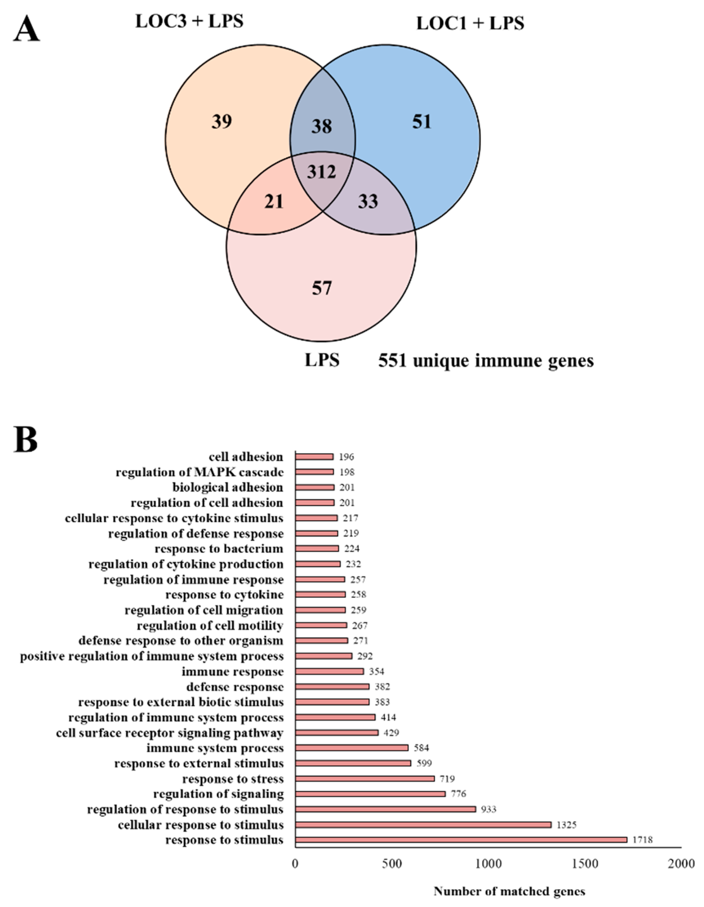
The transcriptomic response of RAW macrophages to the challenge with LPS and the influence of L. plantarum LOC1 and LOC3 strains on this response was then investigated. Microarray analysis was performed in macrophages 3 h after the stimulation with the TLR4 agonist. When LPS-treated macrophages were compared with unchallenged RAW cells it was found that there were 4789 unique genes (Figure 1D) and 4687 unique genes (Figure 1E) up-regulated and down-regulated, respectively. Out of these differentially regulated genes, 551 were assigned to immune-related functions according to the GO database (Figure 2A). Changes in the immunotranscriptome response in macrophages after LPS challenge included transcripts in the following GO Biological Process pathways: “response to stimulus”, “response to stress”, “immune system process”, “cell surface receptor signaling pathway”, “response to external stimulus”, “regulation of the immune system process”, and “defense response” (Figure 2B).
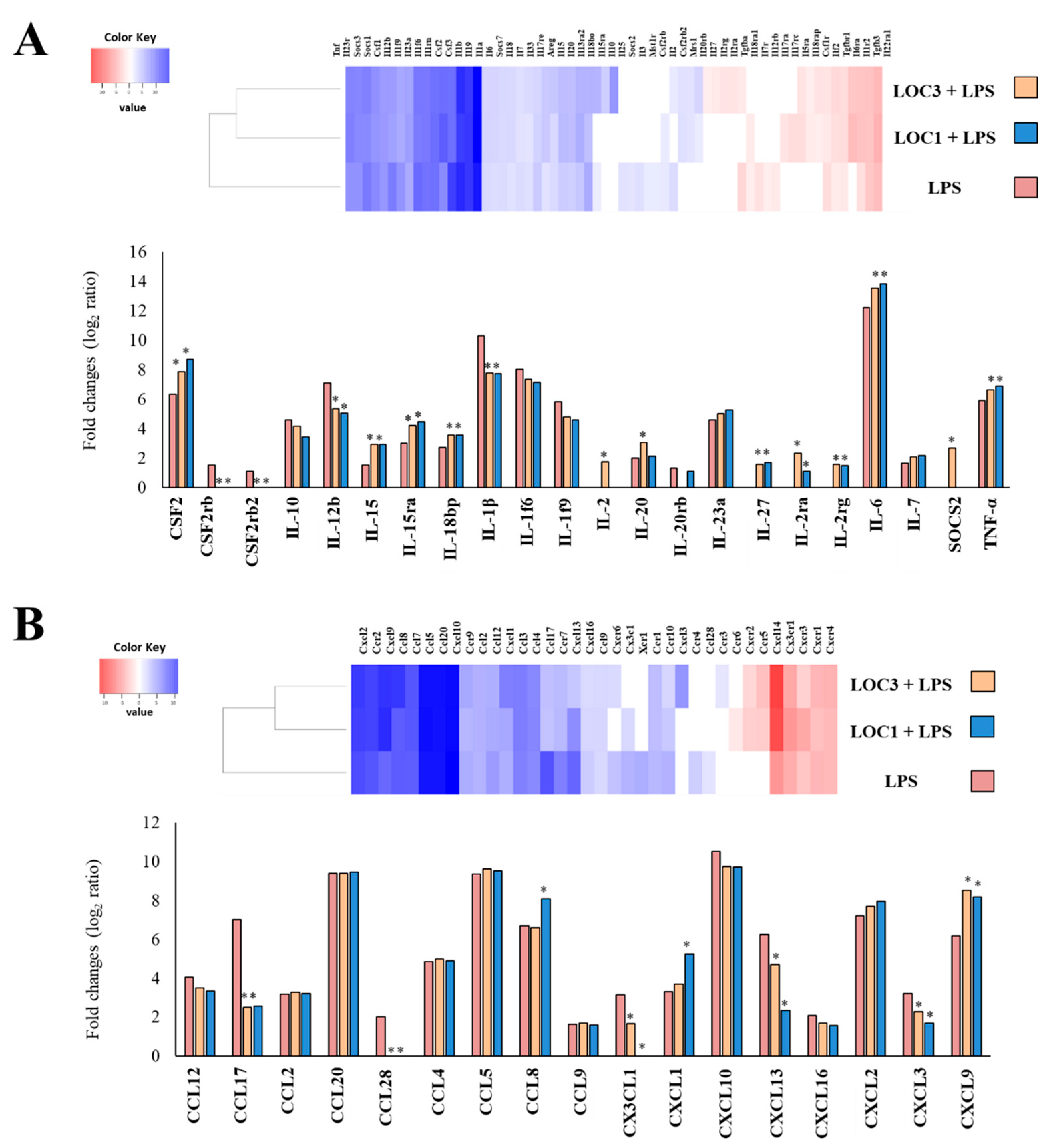
The most remarkable changes in macrophages after stimulation with LPS were found in the expression of cytokines, chemokines and surface markers genes. LPS challenge augmented the expression of the inflammatory cytokines TNF-α (log2 ratio 5.9), IL-1β (10.3), IL-6 (12.2), IL-12b (7.1), IL-15 (1.5), IL-18bp (2.7), and IL-23a (4.6) (Figure 3A). In addition, the microarray analysis revealed significantly increases in the expression levels of the factors CSF2 (6.3), CSF2rb (1.5) and CSF2rb2 (1.5) (Figure 3A) as well as in the chemokines CCL2 (3.1), CCL4 (4.8), CCL5 (9.3), CCL8 (6.7), CCL9 (1.3), CCL12 (4.1), CCL17 (7.1), CCL20 (9.3), CCL28 (2.1), CX3CL1 (3.1), CXCL1 (3.3), CXCL2 (7.2), CXCL3 (3.2), CXCL9 (6.1), CXCL10 (10.5), CXCL12 (7.2), CXCL13 (6.2), and CXCL16 (2.1) (Figure 3B). Of note, augmented expression of the regulatory cytokine IL-10 (4.6) was also detected in LPS-challenged macrophages (Figure 3A). Significantly increased expression of CD40 (5.7), CD69 (5.6), CD80 (3.2), CD83 (6.8), CD164 (2.6), CD200r1 (2.5), Fcgr1 (2.1), Fcgr4 (2.1), Siglec1 (2.6) and SiglecF (3.3) were observed in RAW macrophages after the activation of TLR4 (Figure S1). In addition, the stimulation of macrophages with LPS increased the expression of IFNA7 (1.2) and IFNB1 (8.6) as well as several interferon-induced genes including OAS1a (2.5), OAS2 (4.6), OAS3 (3.5), DXH58 (2.7), IFIT3 (9.7), IFI209 (7.5), IFIT1b11 (6.5), and IFIT3b (9.5) (Figure S1).
Next, we analyzed microarray data to evaluate the effect of the strains L. plantarum LOC1 and LOC3 on the immunotranscriptomic response of macrophages stimulated with LPS. The comparative analysis of microarray profiles indicated that both LOC1 and LOC3 strains differentially modulated the expression of several genes related to the innate immune response triggered by TLR4 activation in macrophages. The Venn diagram analysis was used to find genes that were uniquely and commonly modulated between L. plantarum-treated and control macrophages (Figure 2A). Of the 551 differentially expressed genes in the Venn diagram analysis, 51 were unique to the cells stimulated with the LOC1 strain before the LPS challenge, while 39 were unique for the LOC3 group. In addition, 38 genes were common to LOC1 treatment plus LPS and LOC3 plus LPS groups. It was also observed that 312 genes were common to all the three treatments (Figure 2A).
The heat-map cluster analysis focused on the expression of cytokines, chemokines (Figure 3), surface markers and IFN-related genes (Figure S1) depicts the transcriptomic patterns of differentially modulated genes between L. plantarum-treated and control macrophages. The treatment with LOC1 plus LPS clustered closer to the treatment with LOC3 plus LPS and, both clustered separated from the control. Closer examination of gene expression revealed differences in cytokines and chemokines genes sheared by L. plantarum-treated macrophages and controls (Figure 3). Most remarkable differences were found in the genes for CSF2, IL-6, TNF-α, IL-15 and IL-18bp that were significantly higher in lactobacilli-treated macrophages than in controls. In addition, the expression levels of IL-1β and IL-12b were lower in macrophages treated with the LOC strains than in controls (Figure 3A). Interestingly, although no differences were found between L. plantarum-treated macrophages and controls when the expression of IL-10 was analyzed, it was detected that macrophages stimulated with the LOC1 strain significantly up-regulated the expression of the regulatory factors IL-27 and SOCS2. L. plantarum LOC3 was able to increase the expression of IL-27 but not SOCS2 (Figure 3A). The LOC strains were also able to significantly reduce the expressions of CCL17, CCL28, CX3CL1, CXCL13 and CXCL3, being the LOC1 strain more efficient than the LOC3 to reduce the expression levels of CXCCL1 and CXCL13 (Figure 3B). Both L. plantarum strains increased the expression of CXCL9, while only the LOC3 strain augmented the expression of CCL8 and CXCL1 when compared to controls (Figure 3B). The LOC strains reduced the expression of CD38, CD83, CD164, CD200r2, CD209a, and SiglecF in macrophages challenged with LPS, while they augmented the expressions of CD200r4, CD80, CD86, H2-K1 and H2-M3 (Figure S1). Only L. plantarum LOC1 increased the expression of CD200b, while only LOC3 augmented Siglec1 (Figure S1). When the IFN-related genes were analyzed, it was observed that both L. plantarum LOC1 and LOC3 significantly increased the expression of IFNB1, IFNA7, IFIT3, IFIT1b11, and IFIT3b (Figure S1). Furthermore, significantly up-regulated expression of IFNA1, IFNA2, IFNA5, IFNA13, IFNA14, IFITM7, and IFNAB was detected in macrophages treated with the LOC strains when compared to controls (Figure S1).

3.2. L. plantarum LOC1 Modulate the TLR4-Mediated Immune Response in Macrophages Similarly to Other Immunobiotic Strains

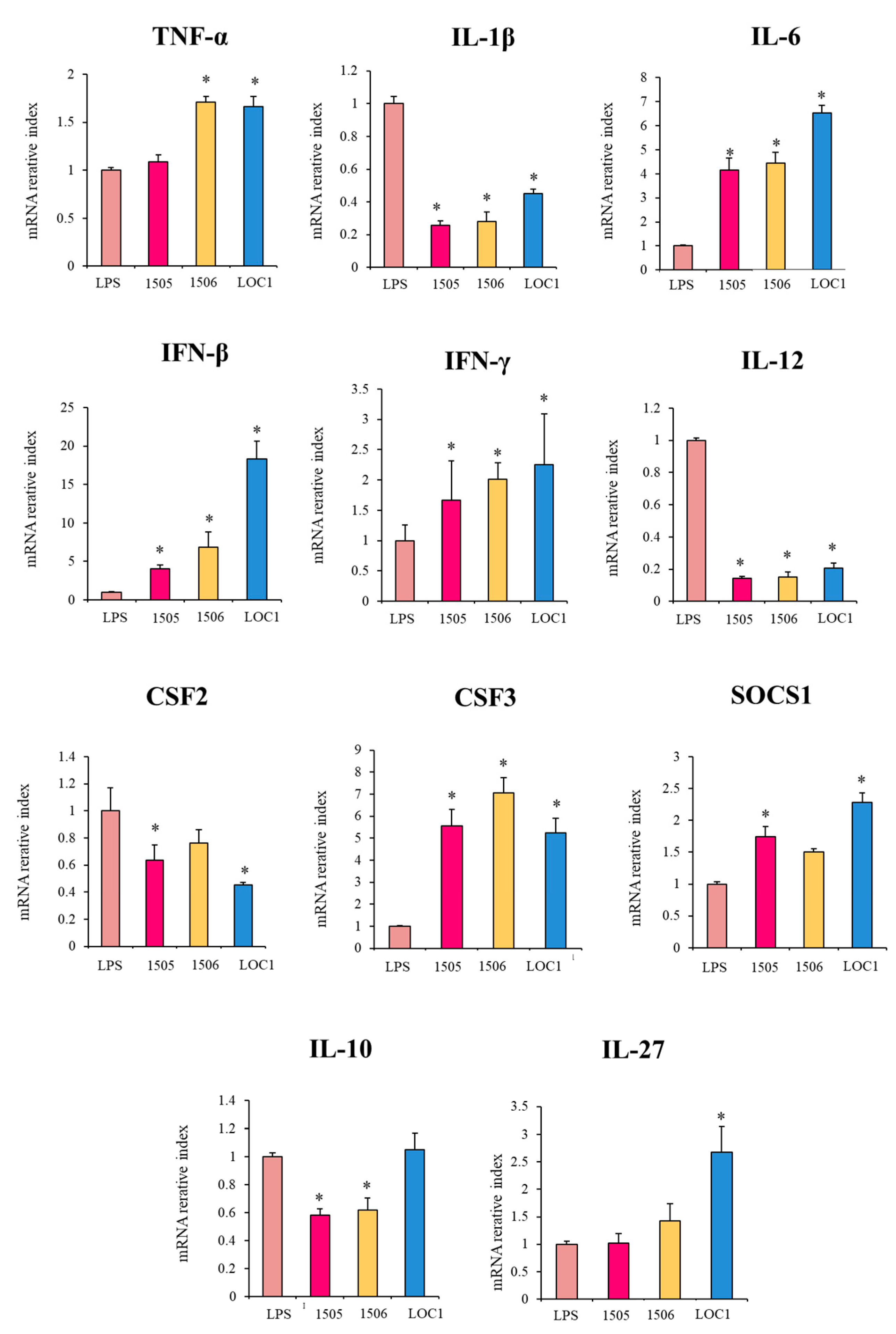
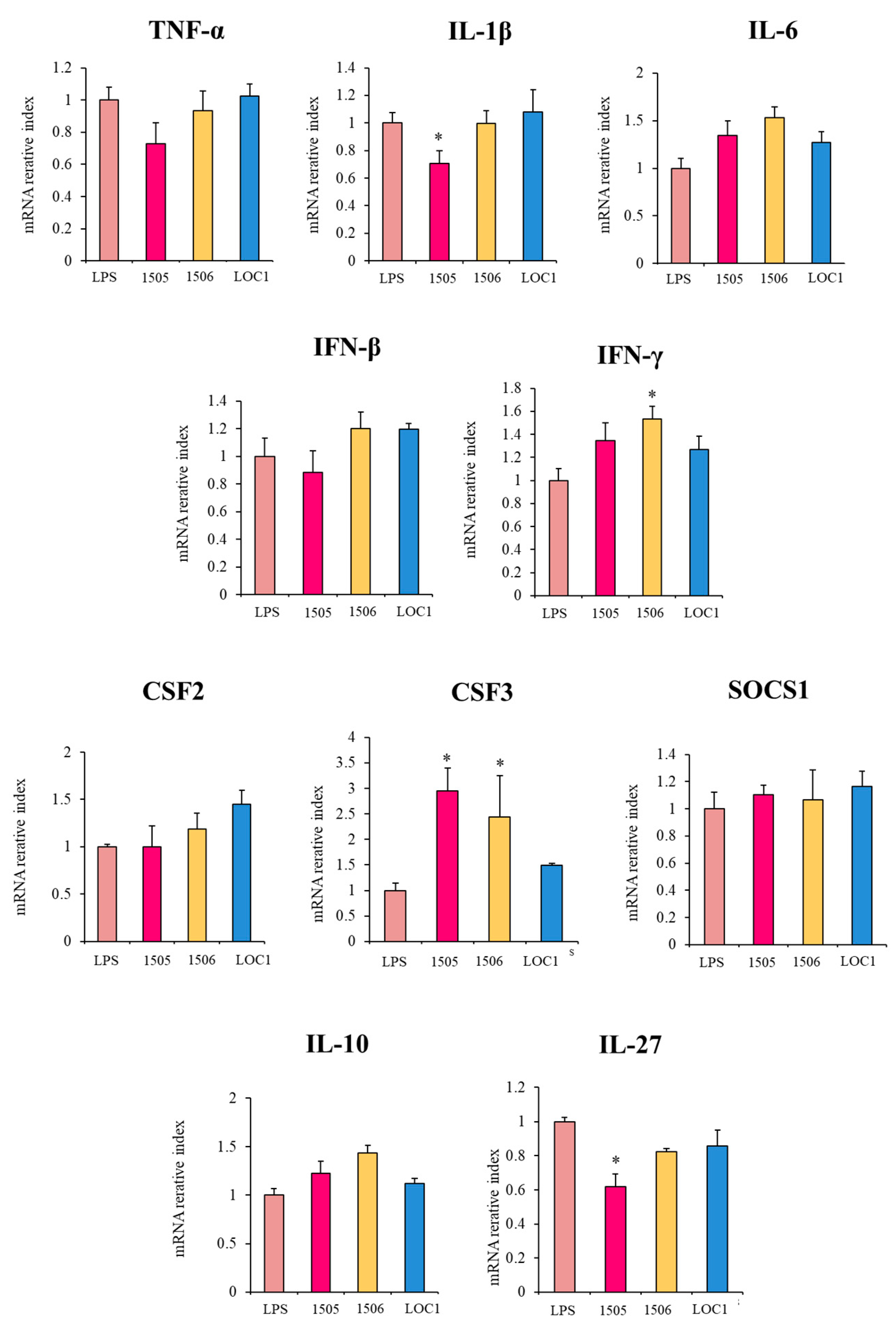
In order to confirm the changes induced by L. plantarum LOC1 in the immunotranscriptome response of LPS-challenged macrophages, qPCR was performed on selected genes. Genes with significant differences between LOC1-treated and non-treated macrophages were chosen. The expression of IL-10 was also evaluated. In addition, the influence of the well-characterized immunobiotic strains L. rhamnosus CRL1505 [22] and L. plantarum CRL1506 [23] on the response of macrophages to TLR4 activation was also studied for comparisons. As shown in Figure 4, the transcriptional changes evaluated by qPCR indicated a similar overall trend in the transcription of microarray. The treatment of macrophages with L. plantarum LOC1 significantly increased their expression levels of TNF-α, IL-6, IFN-β, IFN-γ, and CSF3 in response to LPS challenge. The LOC1 strain also reduced the expression of IL-1β, IL-12, and CSF2 in macrophages stimulated with the TLR4 agonist. Similarly, the treatment of macrophages with the CRL1505 and CRL1506 strains significantly augmented the expressions of IL-6, IFN-β, IFN-γ, and CSF3 and reduced IL-1β and IL-12 (Figure 4). No significant reductions were observed for TNF-α and CSF2 in macrophages treated with L. rhamnosus CRL1505 and L. plantarum CRL1506, respectively. Of note, L. plantarum LOC1 was more efficient than CRL1505 and CRL1506 strains to increase the expression of IFN-β (Figure 4). As expected, the LOC1 strain did not induce changes in IL-10 expression, while both CRL1505 and CRL1506 significantly reduced the expression levels of the regulatory cytokine. In addition, L. rhamnosus CRL1505 and L. plantarum LOC1 enhanced the expression of SOCS1, while only the LOC1 strain up-regulated IL-27 when compared to LPS-challenged macrophages (Figure 4).
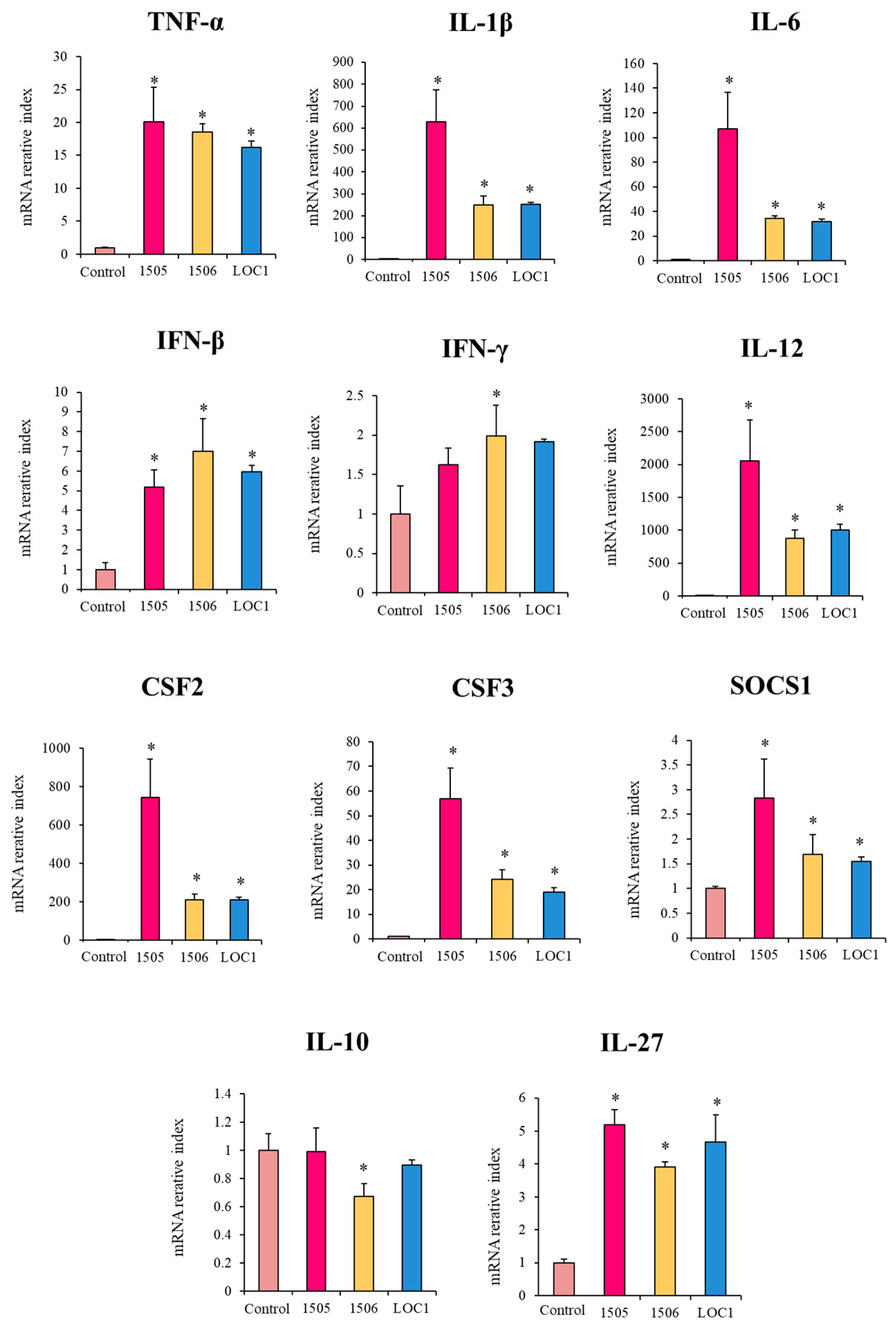
We also evaluated the effect of lactobacilli in the expression of selected immune factors in macrophages in the absence of LPS challenge. As shown in Figure 5, the three strains increased the expression of TNF-α, IL-1β, IL-6, IFN-β, IL-12, CSF2 and CSF3 in macrophages when compared to untreated controls. The three strains were equally effective in up-regulating TNF-α and IFN-β expressions, while the levels of IL-1β, IL-6, IL-12, CSF2 and CSF3 were significantly higher in macrophages treated with L. rhamnosus CRL1505 than in cells stimulated with the L. plantarum strains. In addition, the CRL1506 strain was the only one able to increase the expression of IFN-γ (Figure 5). The three strains augmented the expression of SOCS1 and IL-27. The lactobacilli treatment did not induce modifications in the expression levels of IL-10 except for L. plantarum CRL1506 that reduced this regulatory cytokine (Figure 5).

3.3. L. plantarum LOC1 Does Not Modulate the TLR4-Mediated Immune Response in Macrophages Indirectly Througth Intestinal Epithelial Cells

It was demonstrated that intestinal epithelial cells play a crucial role in the interaction of the intestinal immune system with microbes. Epithelial cells influence immune responses by orchestrating communication between intestinal microbes and mucosal innate immune cells such as macrophages [24]. Thus, we aimed to evaluate whether L. plantarum LOC1, L. rhamnosus CRL1505 or L. plantarum CRL1506 were able to influence the response of macrophages to LPS challenge indirectly through intestinal epithelial cells. For this purpose, murine intestinal epitheliocytes (MIE cells) co-cultures with RAW macrophages were prepared. MIE cells were stimulated with LOC1, CRL1505 or CRL1506 strains and then macrophages were challenged with the TLR4 agonist, and the expressions of immune factors were evaluated by qPCR (Figure 6). No significant differences were observed between control and lactobacilli-treated cells when the expressions of TNF-α, IL-6, IFN-β, CSF2, SOCS1 and IL-10 were evaluated. The treatment with L. rhamnosus CRL1505 reduced the expression of IL-1β and IL-27, while the CRL1506 improved IFN-γ (Figure 6). Both, L. rhamnosus CRL1505 and L. plantarum CRL1506 significantly increased the expression levels of CSF3. Of note, the treatment with L. plantarum LOC1 did not induce changes in TNF-α, IL-6, IFN-β, CSF2, CSF3, SOCS1 or IL-10 (Figure 6).

3.4. General Genomic Characteristics of L. plantarum LOC1

We next aimed to carry out comparative and functional genomics studies in order to identify the bacterial gene(s) responsible for the immunomodulatory properties of L. plantarum LOC1. For this purpose, the complete genome of the LOC1 strain was sequenced with the Illumina MiSeq platform and uploaded to the NCBI repository (accession number BOUN00000000).
The whole genome size and GC content of L. plantarum LOC1 was 3.13 Mb and 44.7%, respectively (Table 1). This is in agreement with the genomic analysis of L. plantarum strains that showed a mean size and GC content of 3.32 Mb and 44.5%, respectively [25].
The KEGG database and the BlastKOALA tool were used for the functional characterization of the LOC1 strain genome (Figure 7). L. plantarum CRL1506 and the well-characterized probiotic strain WCFS1 were used for comparisons. No differences were observed in the number of metabolism pathway genes associated with carbohydrates (213 ± 2), glycan biosynthesis and metabolism (59 ± 1), amino acids (135 ± 2), lipids (41 ± 2), nucleotides (65 ± 2) or energy metabolism (65 ± 2) among the strains. In addition, no significant differences were found in cofactors and vitamins (69 ± 3), polyketides (18 ± 3) or biosynthesis of other secondary metabolites (27 ± 3) when the genomes of LOC1, CRL1506 and WCFS1 were compared (Figure 7). There were also no differences in the numbers of genes associated with “genetic information processing”, “environmental information processing” or “cellular processes” between the three lactobacilli strains (Figure S2).
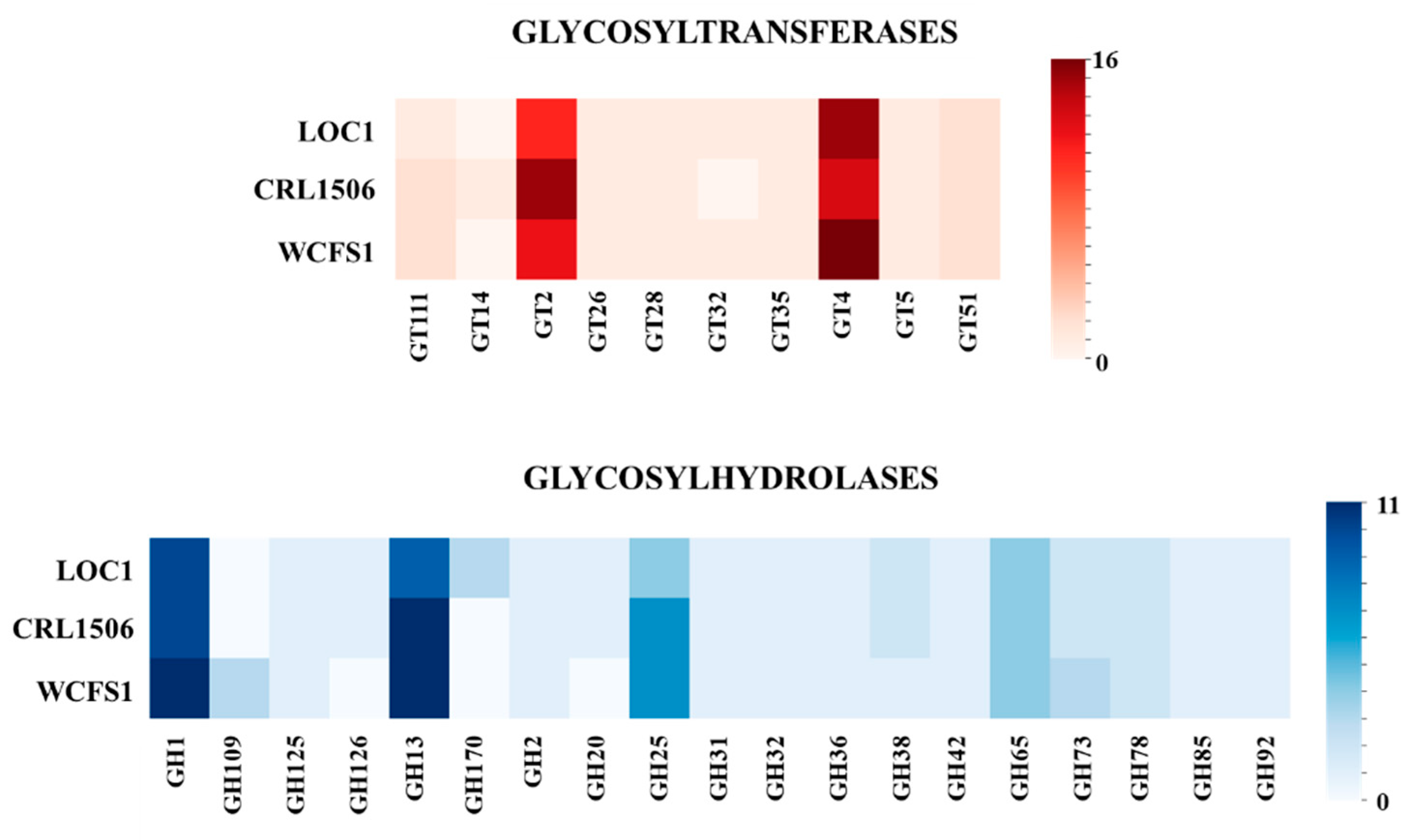
Previous genomic analyzes found that in the species L. plantarum the most abundant sugar metabolism genes belonged to the glycosylhydrolases (GH) and glycosyltransferases (GT) families [26]. Thus, the numbers of genes for GT and GH were also analyzed in the LOC1 strain (Figure 8). Genes for several GT and GH families were detected in the genomes of L. plantarum LOC1. Similar to L. plantarum WCSF1 the LOC1 strain had numbers of GT2 and GT4 that were lower and higher than the CRL1506 strain, respectively. In addition, lower numbers of GH13 and GH25 were found in the LOC1 strain when compared to L. plantarum WCSF1 and CRL1506 (Figure 8). L. plantarum LOC1 had also higher numeber of GH170 than the other two strains. Similar to L. plantarum CRL1506, the LOC1 strain had numbers of GH1, GH109 and GH73 that were lower than the WCSF1 strain.

3.5. Study of the Genes Associated with the Expression of Surface Molecules in L. plantarum LOC1

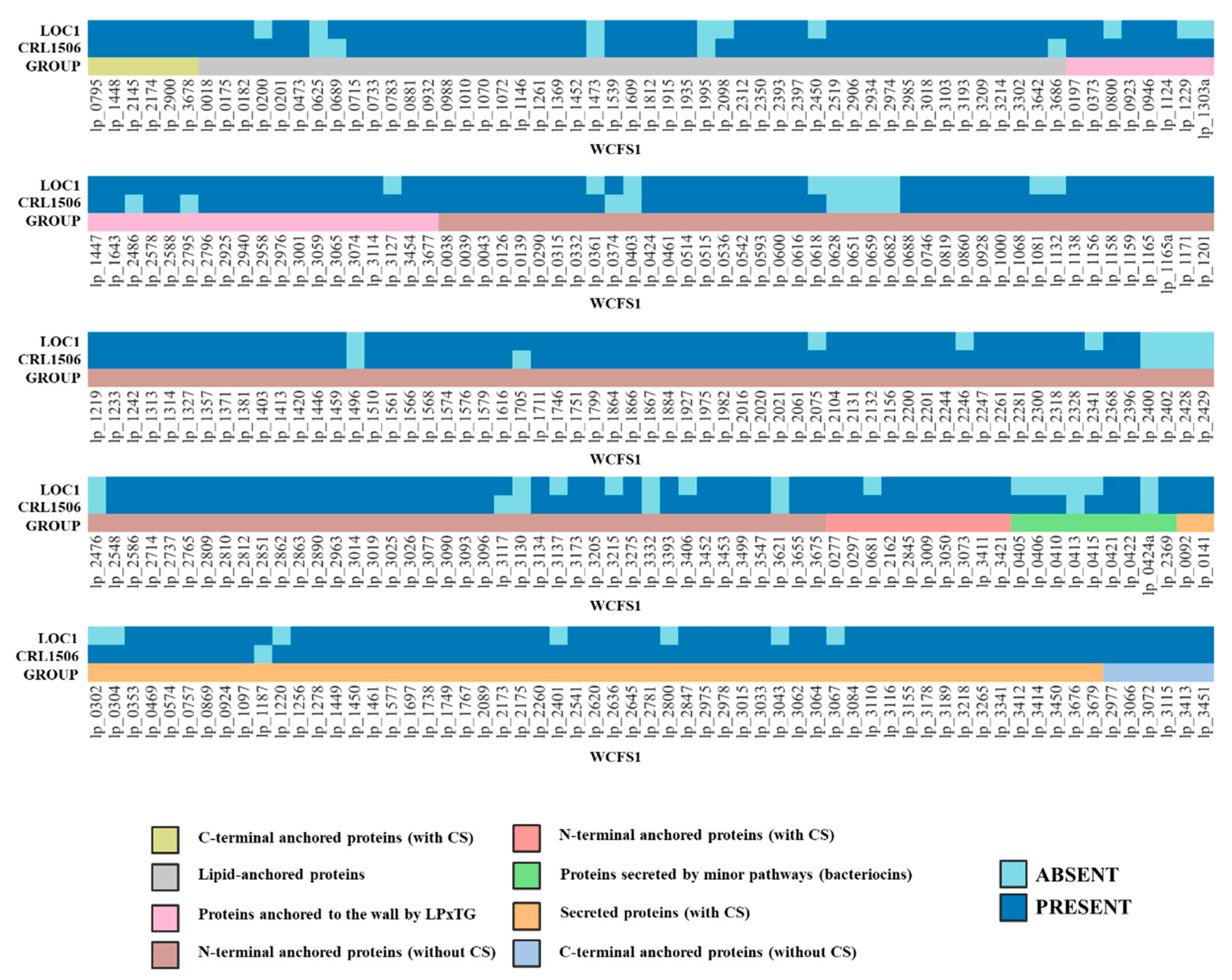
It was reported that surface-exposed proteins play key roles in the interactions of lactobacilli with their environments [27,28]. Therefore, we first evaluated the genes coding surface-exposed and secreted proteins in L. plantarum LOC1 and compared them with CRL1506 and WCSF1 strains (Figure 9). The extracellular proteins in the LOC1 strain were studied by in silico analysis considering the subcellular location (surface-expressed or secreted), the secretory mechanism and the anchorage type, following the guidelines recently described by our group [28]. Notable differences were detected between L. plantarum LOC1, CRL1506 and WCSF1 when analyzing the 304 genes for extracellular proteins. Of note, the proteins ABC transporter-substrate binding protein (lp_0200), prophage P2a protein 7 (lp_2450), cell surface protein precursor with LPXTG-motif (lp_0800), mucus-binding protein with LPXTG-motif cell wall anchor (lp_3127), preprotein translocase SecE (lp_0616), extracellular protein (lp_1132), transcriptional attenuator LytR family (lp_2075), bacterial type II secretion/trafficking system extracellular protein (lp_2246), class A beta-lactamase (lp_2341), PTS system EIIB component (lp_3137), zinc ribbon domain-containing protein (lp_3215), membrane-bound cell surface hydrolase (lp_3393), prophage P1 lysin (lp_0681), bacteriocin precursor peptide PlnK (lp_0405), bacteriocin precursor peptide PlnJ (lp_0406), bacteriocin precursor peptide PlnN (lp_0410), plantaricin A precursor induction factor (lp_0415), extracellular transglycosylase (lp_0302), extracellular transglycosylase (lp_0304), polysaccharide biosynthesis protein (lp_1220), prophage P2a lysin (lp_2401), transcription regulator MarR (lp_2800), extracellular zinc metalloproteinase (lp_3043), and the cell surface protein CscB (lp_3067) that are present in the genomes of the CRL1506 and WCSF1 strains were absent in L. plantarum LOC1 (Figure 9). In addition, the proteins cell surface lipoprotein precursor (lp_0689), ABC transporter-substrate binding protein (lp_3686), mucus-binding protein with LPXTG-motif cell wall anchor (lp_2486), cell surface protein precursor (lp_2795), lysine-rich extracellular protein (lp_0374), cell surface protein CscC (lp_3117) and glycosylhydrolase (lp_1187) were detected in the genome of the LOC1 strain but not in L. plantarum CRL1506 (Figure 9).
We also investigated the presence of genes involved in the adhesion to the gastrointestinal tract in the genome of L. plantarum LOC1. For this purpose, a bioinformatic analysis was carried out considering different types of molecules with mucus/mucin-binding domains, which were recently characterized and described in a comparative genomic study using strains of L. plantarum possessing differential immunomodulatory activities [28]. Then, the proteins MucBP1, MucBP2, MucBP3, MucBP4, MucBP5, MucBP-DUF1, MucBP-DUF2, Muc-MubB2-MBG, Muc-MubB2-YGX, Muc-MubB2, YceG and Ig-like were searched in the genome of the LOC1 strain (Figure 10). Similar to the WCSF1 strain, L. plantarum LOC1 does not possess the proteins YceG, Ig-like, and Muc-MubB2. The proteins MucBP3 and Muc-MubB2-YGX were also absent in the genome of the LOC1 strain.
Proteins containing domains for chitin, collagen, and fibronectin adhesion have been described in the genome of L. plantarum WCFS1 [29,30,31,32]. Then, the presence of the genes coding for collagen binding proteins CBP1 and CBP2, fibronectin binding proteins FBP1 and FBP2, as well as Msa (mannose-specific binding), GAPDH (glyceraldehyde-3-phosphate dehydrogenase), LuxS (autoinducer-2), AAD (alpha-acetolactate decarboxylase) and CnaB (Cna protein type B) proteins were searched in the genome of the LOC1 strain and compared with L. plantarum CRL1506 and WCFS1 (Figure 9). All these adhesion factors were detected in the genome of the LOC1 strain.
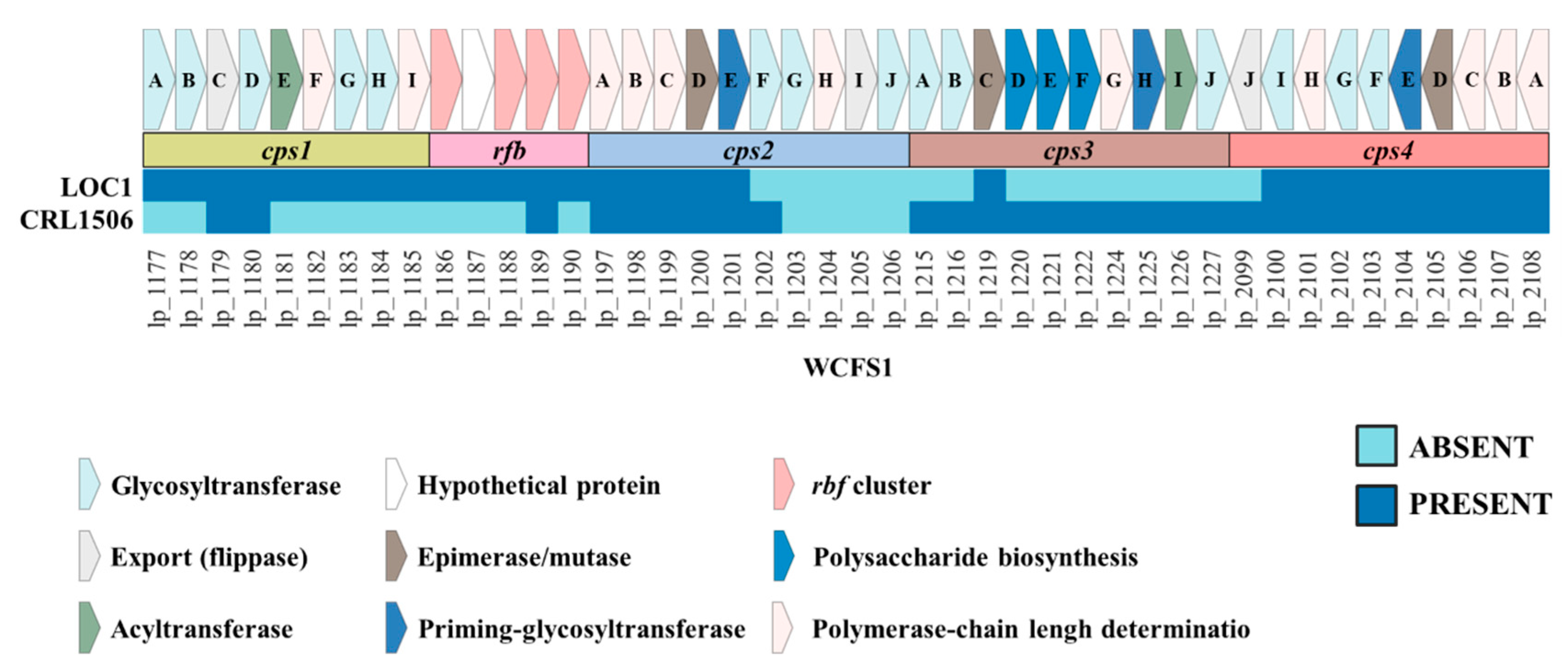
EPSs are also involved in the interaction of LAB with the host. It was shown that L. plantarum have four clusters for the biosynthesis of EPS [33] and recently we demonstrated that strains may differ markedly in the presence of those clusters in their genomes [28]. Thus, we performed a sequence comparison using L. plantarum WCFS1 as a reference to characterize the EPS clusters in the genome of the LOC1 strain (Figure 11). The genes corresponding to the eps1 cluster (or cps1) and eps4 cluster (or cps4) were found in the L. plantarum LOC1 except for the eps4J gene of cluster eps4. The genes corresponding to the eps3 cluster (or cps3) were not found in the genomes of any of the LOC1 strain, except for the eps3C gene (Figure 11). For the eps2 (or cps2) cluster, it was observed that L. plantarum LOC1 had the first five genes (eps2A, eps2B, eps2C, eps2D and eps2E) that have been reported to be highly conserved among L. plantarum strains [33,34]. The other genes of the eps2 cluster of the WCSF1 strain used as reference were not found in L. plantarum LOC1 and this is probably related to the fact that those genes possess very limited sequence homology between the corresponding regions from different strains [28,34]. It was shown that some strains including L. plantarum WCFS1 [34] possess a group of conserved genes called rfb (rmlACBD genes), which are involved the biosynthesis of rhamnose precursors during the synthesis of EPS molecules [35]. The rfb genes were detected in the genome of L. plantarum LOC1 (Figure 11).
Some studies have demonstrated that lipoproteins expressed on the surface of LAB may be involved in their immunomodulatory effect. In this regard, it was shown that the prolipoprotein diacylglyceryl transferase lgt gene (lp_0755), is of important for the immunomodulatory ability of L. plantarum WCFS1 [36]. Furthermore, it was shown that the WCFS1 possess three acyltransferases (lp_0856, lp_0925 and lp_1181) that are involved in lipoprotein triacylation. On the other hand, it was reported that teichoic (TA) and lipoteichoic (LTA) acids, and particularly the dltD gene involved in the incorporation of D-alanine into LTA, play important roles in the immunomodulatory capacities of probiotic L. plantarum strains [12,37,38,39]. Then, we investigated the presence of lgt, lp_0856, lp_0925, lp_1181 and dltD genes as well as other genes involved in TA and LTA biosynthesis in the genome of the LOC1 strain (Figure S3). All the genes were present in L. plantarum LOC1 and showed no differences with WCSF1 and CRL1506 strains, except for tagF1 (lp_0268) and tagF2 (lp_0269).

4. Discussion

Macrophages represent the first line of host immune defense in the intestine [40,41] and their proper activity requires a tight regulation of gene expression allowing a fine-tuned immune response. It was reported that probiotic microorganisms are able to beneficially modulate macrophage functioning by enhancing their ability to protect against pathogens and avoiding the generation of detrimental inflammation (reviewed in Wang et al. [42]). Of note, the ability of probiotic bacteria to modulate macrophages’ responses was shown to be strain specific and thus, each probiotic candidate must be tested to identify specific biological activities. The murine RAW 264.7 macrophage cell line has been shown to be a useful in vitro tool to select and characterize immunomodulatory probiotics [42]. Using RAW macrophages, we demonstrated in this work that L. plantarum LOC1 isolated from fresh tea modulates cytokine expressions. The LOC1 strain increased the expression of TNF-α, IL-1β, IL-6, IFN-β, IL-12, CSF2 and CSF3. In addition, L. plantarum LOC1 did not induce modifications in IL-10 but it augmented the expression of SOCS1 and IL-27. The changes induced by the LOC1 strain were characteristic and differed from those induced by other probiotic strains such as L. rhamnosus CRL1505 and L. plantarum CRL1506, highlighting that this property is strain dependent. In line with our results, it was found that RAW macrophages treated with L. acidophilus MTCC-10307 significantly up-regulate the expression of IL-10, IL-6, IL-12 and IFN-α, while the expression of TNF-α was down-regulated with low doses of lactobacilli but up-regulated at higher concentrations [43]. The treatment of RAW macrophages with L. rhamnosus ATCC 7469 increased the synthesis of TNF-α, IL-6, and IL-10, but was not able to enhance the production of IL-1β, IL-4 or IL-12 [44].
Our results suggest that L. plantarum LOC1 is a potent stimulator of macrophages. The improvement of TNF-α, IL-1β, IL-6, and IL-12 have been shown to be associated with the enhanced capacity of macrophages to protect against infections. Studies found that lactobacilli with the ability to reduce the severity of intestinal infections can improve the phagocytic activity of macrophages and their capacity to produce inflammatory factors such as TNF-α and IFN-γ [45,46]. It was also reported that immunostimulatory L. plantarum strains activate mouse macrophages enhancing IL-12 production and phagocytosis [47]. Furthermore, studies demonstrated that among the cytokines induced by immunomodulatory lactobacilli, the most remarkable effect was the increase in TNF-α, IFN-γ, IL-1β, IL-6, and IL-12 for most of the probiotic strains assayed [45,46,48]. Of note, the LOC1 strain also increased SOCS1 and IL-27 that are known to be involved in the control of inflammation [49,50]. Thus, the LOC1 strain is capable of modulating both the inflammatory and regulatory responses in macrophages. This property was put into further evidence when L. plantarum LOC1-treated macrophages were challenged with LPS.
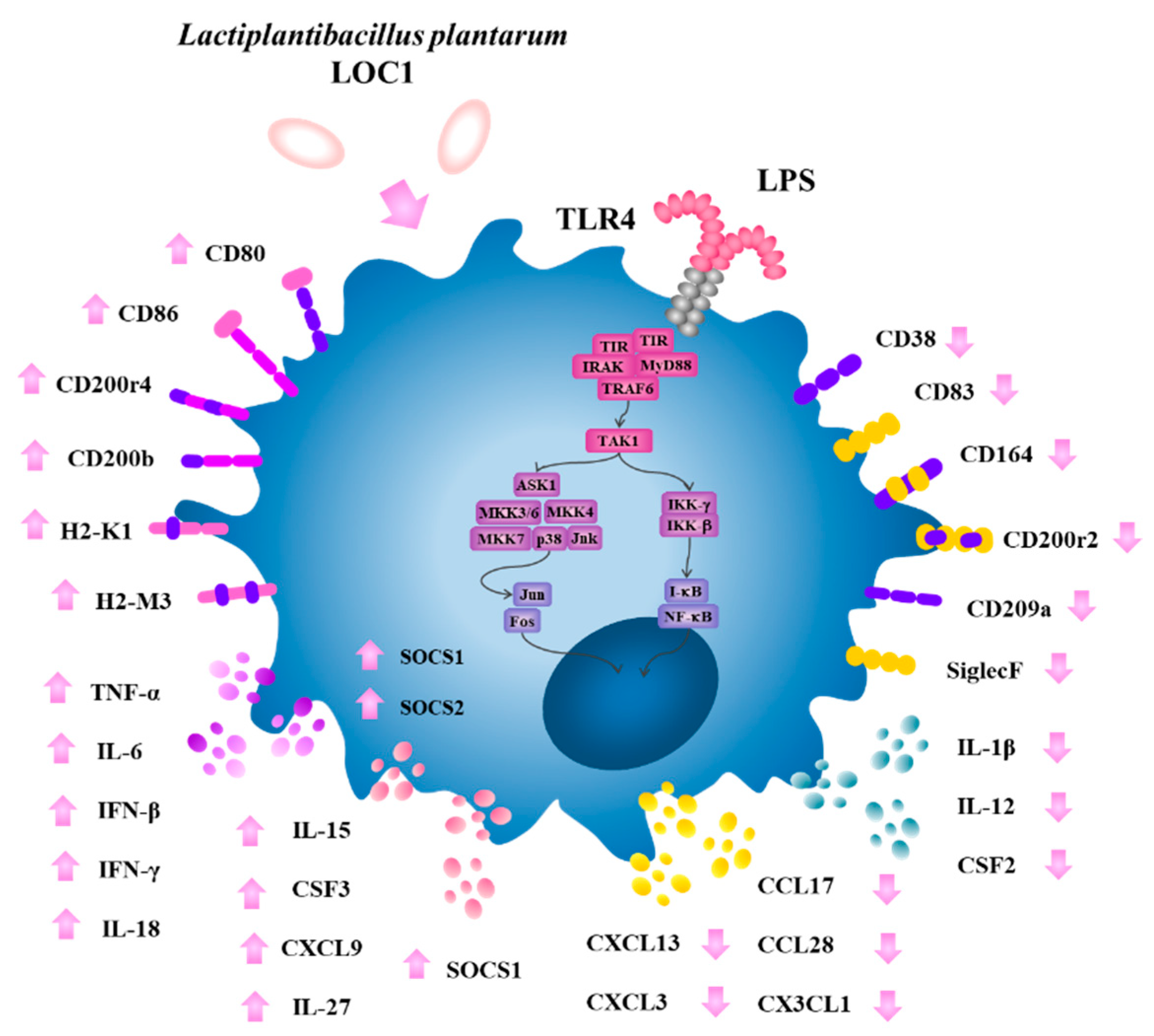
Several large-scale transcriptomic and proteomic studies have been performed with macrophages challenged with LPS with the aim of exploring different aspects of the TLR4 signaling and its impact on mucosal protection [40,41]. In this work, we carried out a transcriptomic study to evaluate the response of murine macrophages to the activation of TLR4 and in line with several other studies [51,52], we found that RAW macrophages mount a complex transcriptomic response characterized by changes in the expression of hundreds of genes with diverse immunological functions. Of note, the most remarkable changes in RAW macrophages after the stimulation with LPS were found in expression of cytokines, chemokines and surface markers genes. Using this transcriptomic approach, we also demonstrated that L. plantarum LOC1 exerts a modulatory effect on LPS-induced inflammation, resulting in a differential regulation of immune factor expression in macrophages. Specifically, we found that the LOC1 strain markedly reduced the LPS-induced expression of some inflammatory cytokines (IL-1β, IL-12, and CSF2) and chemokines (CCL17, CCL28, CXCL3, CXCL13, CXCL1, and CX3CL1), while it significantly increased the expression of other cytokines (TNF-α, IL-6, IL-18, IFN-β, IFN-γ, and CSF3), chemokines (IL-15 and CXCL9), and activation markers (H2-k1, H2-M3, CD80, and CD86) in RAW macrophages (Figure 12).
The cytokines TNF-α, IL-6, IL-18, IFN-β, IFN-γ, and CSF3 produced by macrophages play important roles in the protection again pathogens by promoting phagocytosis and mediating the recruitment and activation of effector immune cells into the site of infection. These functions are complemented by chemokines such as IL-15 and CXCL9 that induce the recruitment of γδ T cells [53] and T cells [54], respectively, which are essential for the protection against Gram negative pathogens. In addition to cytokine and chemokine up-regulation, macrophages increase several activation markers after their stimulation with LPS. In this regard, although MHC class II molecules are constitutively expressed on macrophages, its expression is significantly augmented by LPS [55]. We observed a significant increase in several histocompatibility class II antigens in RAW macrophages challenged with LPS. Of note, L. plantarum LOC1 significantly up-regulated the expression of histocompatibility 2, class II antigen K1 and histocompatibility 2, class II antigen M3 as well as the co-stimulatory molecules CD80 and CD86 (Figure 12). MHC class II and co-stimulatory proteins expressed in macrophages are of importance in the development and maintenance of immune responses mediated by T cells, particularly CD4+ T lymphocytes [55]. Thus, our results show that the LOC1 strain is able to stimulate macrophages to support T-cell-mediated immunity.
Of note, we also detected the reduction in inflammatory factor expressions in LPS-treated macrophages previously stimulated with L. plantarum LOC1 (Figure 12). The reduction in inflammatory cytokines expression in LPS-challenged RAW macrophages have been described for other LAB strains. It was shown that L. plantarum CKDB008 reduce the production of NO, TNF-α, IL-6, and IL-1β in LPS-stimulated RAW macrophages [56]. Lactobacilli strains isolated from different sources reduced the levels of TNF-α, IL-1β and IL-6 by inhibiting p38, ERK1/2 and SAPK/JNK pathways in RAW cells challenged with LPS [57]. Similarly, cellular fractions derived from the probiotic strain L. rhamnosus GG were shown to inhibit the activation of MAPK and NF-κB signaling pathways in LPS-stimulated RAW cells, leading to reduced TNF-α and IL-6 production [58]. Here, the treatment of macrophages with the LOC1 strain significantly reduced the expression of IL-1β, IL-12, and CSF2 as well as CCL17, CXCL3, CXCL13, CX3CL1 and CXCL1, which are chemokines for neutrophils, monocytes, macrophages, and B cells [55]. We also detected the up-regulation of SOCS-1 that is a negative regulator able to control cytokine signaling [49] and IL-27 that is an important regulator of inflammation that limit development of IFNγ-producing Th1 cells by stimulating IL-10 production by CD4+ T cells [50]. Furthermore, our transcriptomic analysis revealed a differential expression of CD200/CD200R molecules, which reduce the synthesis of proinflammatory mediators and induces the synthesis of anti-inflammatory factors in macrophages [59]. A reduction in the pro-inflammatory marker CD38 [60] was also observed in macrophages treated with the LOC1 strain.
In the presence of inflammatory stimuli such as LPS, macrophages change their gene expression, up-regulating proinflammatory cytokines and chemokines, oxidative metabolites, and proteases that play roles in the protection against pathogens. However, if the macrophage-mediated inflammatory response is not timely and effectively regulated, tissue-destructive pathology is induced [41]. Macrophages show considerable phenotype diversity and plasticity in response to environmental stimuli and can behave as both inflammatory and regulatory cells. Our results allow us to speculate that L. plantarum LOC1 would enhance the intrinsic functions of macrophages, promoting their protective effects mediated by the stimulation of the Th1 response without affecting the regulatory mechanisms that help control inflammation. Whether this effect is maintained in vivo, helping to improve protection against pathogens and protecting against inflammation simultaneously, is a topic that should be studied in the immediate future to position this strain as a probiotic to improve resistance to infections. In addition, detailed mechanistic studies are necessary to evaluate the immune receptor(s) used by L. plantarum LOC1 to exert its immunomodulatory effects.
We performed here for the first time a genomic characterization of L. plantarum LOC1. Genomic comparative analysis was performed with the well-known immunomodulatory strains WCSF1 and CRL1506. We focused our analysis on the molecules of the bacterial surface that could be responsible for interacting with the host’s immunological receptors including surface proteins, adhesion factors, EPS and LTA. The bioinformatic study revealed that L. plantarum LOC1 possesses several factors that have been proposed to be important for the interaction of this species of bacteria with the host (Figure 13).
Some studies have shown that EPS produced by L. plantarum strains are involved in their immunomodulatory properties. The EPS from L. plantarum NCU116 was shown to reduce intestinal inflammation through the STAT3 signaling pathway [61], while the EPS from L. plantarum L-14 reduced the expression of cyclooxygenase-2, inducible nitric oxide synthase as well as IL-6, TNF-α, and IL-1β in LPS-challenged RAW macrophages [9]. Thus, we analyzed the EPS genes cluster in the LOC1 strain. There is high diversity among EPS clusters identified in L. plantarum strains. Five different EPS clusters were described in this species of lactobacilli [62]. In the model strain WSCF1, four major EPS-synthetic gene clusters were identified and designated as cps1, cps2, cps3, and cps4 [34]. Two separated regions of the L. plantarum WCFS1 genome contain those clusters: while cps1, cps2, and cps3 are located in tandem in one region, the cps4 cluster is located separated [34]. Of note, the cps1 gene cluster controlled the molecular mass and the rhamnose composition of EPS [62], while cps4A-J genes in the cps4 cluster regulate the overall EPS yield. In fact, the deletion of these genes resulted in a reduction of less than half fold EPS production compared to wild-type bacteria [34]. Of note, both clusters of EPS were almost complete in the genome of L. plantarum LOC1, while cps3 was not observed and cps2 partially detected. Similarly, it has been shown that not all L. plantarum strains had the four clusters of EPS genes in their genomes [28,63]. Strains ATCC14197 and ST-III have only the csp3 and csp4 clusters, JMD1 only possess the cps4 cluster [63], while the immunomodulatory strain CRL1506 carries cps2, cps3 and cps4 clusters [28]. This variability between the strains could impact in their interaction with the host and particularly with the cells of the immune system such as macrophages. Considering that the LOC1 strain presented differences in EPS clusters in relation to the immunomodulatory strain CRL1506 but modulated in a similar way the response of RAW macrophages, it is possible to speculate that EPS would not be involved in the immunomodulatory activity of L. plantarum LOC1.
We also investigated genes coding for proteins involved in the adhesion of L. plantarum, which are thought to be important for the strains to fulfill their probiotic functions. In the probiotic WCFS1 strain, several genes with adhesion function were observed, including mucin-, collagen-, chitin-, and fibronectin-binding proteins [29], as well as agglutination factors [64]. In a previous work, when adhesion factors were evaluated in different L. plantarum strains possessing differential immunomodulatory properties, we found a great variability between strains [28]. Similarly, we described here that L. plantarum LOC1 has its own characteristic set of adhesion factors (Figure 13). Assessing whether any of these factors are essential for its immunomodulatory activity is an interesting topic for future research.
In addition, genes involved in the biosynthesis of TA, LTA and lipoproteins were also analyzed in the genome of L. plantarum LOC1. Those molecules have been associated with the immunomodulatory properties of L. plantarum strains such as J26 and WCSF1 [36,65]. Thus, the lipoproteins-related genes (lgt, lp_0856, lp_0925 and lp_1181), the TA-related genes (gtcA1, gtcA2, gtcA3, dltX, tagD1, tarI, tarJ, tarK and tarL) and the LTA-related gene dltD of L. plantarum LOC1 were searched by in silico analysis. No differences were found between the LOC1 strain and L. plantarum WSCF1 and CRL1506, indicating that these molecules would be similar in the three strains and, therefore, are molecules that could be involved in the immunomodulatory capacity of L. plantarum LOC1.
When the genes belonging to metabolic pathways of the three strains (LOC1, WSCF1 and CRL1506) were compared, no significant differences were detected except for genes involved in carbohydrate metabolism, particularly in the presence/absence of GH and GT. These findings are in line with previous studies that reported differences in genes involved in sugar transport and catabolism among L. plantarum strains [66]. These different metabolic capacities condition the fermentation and growth characteristics of each strain [67] and collaborate in determining the types of habitats in which the bacteria will be able to subsist as well as their potential biotechnological applications. It is likely that these differences are noticeable in this work since the genomic studies concentrated on L. plantarum strains that come from different origins: fresh tea leaves, goat milk and human saliva. An interesting point for future research would be to carry out comparative genomic studies of L. plantarum LOC1 with strains isolated from plant sources and from fermented tea or tea fresh leaves. The sequencing of the LOC1 strain genome carried out in this work makes it possible to perform those studies as well as others that are necessary to advance in the potential biotechnological applications of this strain.

5. Conclusions

In the current study, we demonstrated that L. plantarum LOC1 isolated from fresh tea leaves is an interesting probiotic strain candidate with immunomodulatory activity. In vitro transcriptomic studies demonstrated that the LOC1 strain differentially modulates the expression of immune factors in LPS-challenged macrophages suggesting that this candidate probiotic has the capacity to improve the beneficial functions of these immune cells. In addition, we sequenced the complete genome of L. plantarum LOC1 and genomic studies showed that the strain possess several factors described for other probiotic strains that may be involved in its interaction with macrophages. The results of this work can contribute to the development of immune-related functional foods containing L. plantarum LOC1. Further investigation into the in vivo beneficial immunomodulatory effects of the LOC1 strain are ongoing.

Supplementary Materials

The following supporting information can be downloaded at: https://www.mdpi.com/article/10.3390/foods11203257/s1, Figure S1: Effect of Lactiplantibacillus plantarum strains isolated from fresh tea leaves in murine RAW macrophages stimulated with the Toll-like receptor 4 (TLR4) agonist lipopolysaccharide (LPS). Macrophages were treated with L. plantarum LOC1 or LOC3 strains and then challenged with LPS. The expression of genes was determined by microarray analysis 3 h after LPS stimulation. Non-lactobacilli-treated macrophages stimulated with LPS were used as control. Heat-map analysis and fold expression changes of surface markers (A) and interferons (IFNs) and IFNs-related genes (B). Asterisks indicate significant differences between the indicated groups and LPS-challenged control macrophages, (*) p < 0.05. Figure S2: Study of the number of genes in the different functional categories associated with “genetic information processing”, “environmental information processing” and “cellular process” pathways of the genome of Lactiplantibacillus plantarum LOC1. The genome of the LOC1 strain was studied and compared with the probiotic strains WCFS1 and CRL1506 as reference. Functional characterization of genes was performed with the BlastKOALA tool according to the KEGG database. Figure S3: Analysis of the presence/absence of genes involved in the biosynthesis of lipoproteins, teichoic acids and lipoteichoic acids in Lactiplantibacillus plantarum LOC1. The genome of the LOC1 strain was studied and compared with the probiotic strains WCFS1 and CRL1506 as reference. Table S1: Nucleotide sequences of the specific primers used for real-time PCR.

Author Contributions

Conceptualization, J.V. and H.K.; methodology, M.S., Y.T., K.F. and I.S.; software, L.A.; validation, M.S. and J.V.; formal analysis, M.S., L.A. and J.V.; investigation, M.S., Y.T. and I.S.; resources, J.V. and H.K.; data curation, L.A.; writing—original draft preparation, J.V.; writing—review and editing, M.S. and H.K.; visualization, M.S., L.A. and J.V.; supervision, J.V. and H.K.; project administration, H.K.; funding acquisition, H.K. All authors have read and agreed to the published version of the manuscript.

Funding

This study was supported by Grant-in-Aid for Scientific Research (A) (19H00965) from the Japan Society for the Promotion of Science (JSPS) to Haruki Kitazawa, and by JSPS Core-to-Core Program, A. Advanced Research Networks entitled Establishment of international agricultural immunology research-core for a quantum improvement in food safety, and by AMED Grant Number JP21zf0127001. Kohtaro Fukuyama was supported by JST, the establishment of university fellowships towards the creation of science technology innovation, Grant Number JPMJFS2102.

Data Availability Statement

Data are contained within this article.

Conflicts of Interest

The authors declare no conflict of interest.

References

  1. Duar, R.M.; Lin, X.B.; Zheng, J.; Martino, M.E.; Grenier, T.; Pérez-Muñoz, M.E.; Leulier, F.; Gänzle, M.; Walter, J. Lifestyles in transition: Evolution and natural history of the genus Lactobacillus. FEMS Microbiol. Rev. 2017, 41, S27–S48. [Google Scholar] [CrossRef] [Green Version]
  2. Yu, A.O.; Goldman, E.A.; Brooks, J.T.; Golomb, B.L.; Yim, I.S.; Gotcheva, V.; Angelov, A.; Kim, E.B.; Marco, M.L. Strain diversity of plant-associated Lactiplantibacillus plantarum. Microb. Biotechnol. 2021, 14, 1990–2008. [Google Scholar] [CrossRef]
  3. Salvetti, E.; Harris, H.M.B.; Felis, G.E.; O’Toole, P.W. Comparative Genomics of the Genus Lactobacillus Reveals Robust Phylogroups that Provide the Basis for Reclassification. Appl. Environ. Microbiol. 2018, 84, 00993-18. [Google Scholar] [CrossRef] [Green Version]
  4. Albarracin, L.; Kobayashi, H.; Iida, H.; Sato, N.; Nochi, T.; Aso, H.; Salva, S.; Alvarez, S.; Kitazawa, H.; Villena, J. Transcriptomic analysis of the innate antiviral immune response in porcine intestinal epithelial cells: Influence of immunobiotic lactobacilli. Front. Immunol. 2017, 8, 57. [Google Scholar] [CrossRef] [Green Version]
  5. Albarracin, L.; Garcia-Castillo, V.; Masumizu, Y.; Indo, Y.; Islam, M.A.M.A.M.A.; Suda, Y.; Garcia-Cancino, A.; Aso, H.; Takahashi, H.; Kitazawa, H.; et al. Efficient Selection of New Immunobiotic Strains with Antiviral Effects in Local and Distal Mucosal Sites by Using Porcine Intestinal Epitheliocytes. Front. Immunol. 2020, 11, 543. [Google Scholar] [CrossRef]
  6. Villena, J.; Chiba, E.; Vizoso-Pinto, M.G.; Tomosada, Y.; Takahashi, T.; Ishizuka, T.; Aso, H.; Salva, S.; Alvarez, S.; Kitazawa, H. Immunobiotic Lactobacillus rhamnosus strains differentially modulate antiviral immune response in porcine intestinal epithelial and antigen presenting cells. BMC Microbiol. 2014, 14, 126. [Google Scholar] [CrossRef] [Green Version]
  7. Kim, K.; Lee, G.; Thanh, H.D.; Kim, J.H.; Konkit, M.; Yoon, S.; Park, M.; Yang, S.; Park, E.; Kim, W. Exopolysaccharide from Lactobacillus plantarum LRCC5310 offers protection against rotavirus-induced diarrhea and regulates inflammatory response. J. Dairy Sci. 2018, 101, 5702–5712. [Google Scholar] [CrossRef]
  8. Kawashima, T.; Hayashi, K.; Kosaka, A.; Kawashima, M.; Igarashi, T.; Tsutsui, H.; Tsuji, N.M.; Nishimura, I.; Hayashi, T.; Obata, A. Lactobacillus plantarum strain YU from fermented foods activates Th1 and protective immune responses. Int. Immunopharmacol. 2011, 11, 2017–2024. [Google Scholar] [CrossRef]
  9. Kwon, M.; Lee, J.; Park, S.; Kwon, O.H.; Seo, J.; Roh, S. Exopolysaccharide Isolated from Lactobacillus plantarum L-14 Has Anti-Inflammatory Effects via the Toll-Like Receptor 4 Pathway in LPS-Induced RAW 264.7 Cells. Int. J. Mol. Sci. 2020, 21, 9283. [Google Scholar] [CrossRef]
  10. Yu, P.; Ke, C.; Guo, J.; Zhang, X.; Li, B. Lactobacillus plantarum L15 Alleviates Colitis by Inhibiting LPS-Mediated NF-κB Activation and Ameliorates DSS-Induced Gut Microbiota Dysbiosis. Front. Immunol. 2020, 11, 575173. [Google Scholar] [CrossRef]
  11. Dong, J.; Ping, L.; Zhang, K.; Tang, H.; Liu, J.; Liu, D.; Zhao, L.; Evivie, S.E.; Li, B.; Huo, G. Immunomodulatory effects of mixed Lactobacillus plantarum on lipopolysaccharide-induced intestinal injury in mice. Food Funct. 2022, 13, 4914–4929. [Google Scholar] [CrossRef] [PubMed]
  12. Mizuno, H.; Arce, L.; Tomotsune, K.; Albarracin, L.; Funabashi, R.; Vera, D.; Islam, M.A.M.A.; Vizoso-Pinto, M.G.M.G.; Takahashi, H.; Sasaki, Y.; et al. Lipoteichoic Acid Is Involved in the Ability of the Immunobiotic Strain Lactobacillus plantarum CRL1506 to Modulate the Intestinal Antiviral Innate Immunity Triggered by TLR3 Activation. Front. Immunol. 2020, 11, 571. [Google Scholar] [CrossRef] [PubMed]
  13. Tanasupawat, S.; Pakdeeto, A.; Thawai, C.; Yukphan, P.; Okada, S. Identification of lactic acid bacteria from fermented tea leaves (miang) in Thailand and proposals of Lactobacillus thailandensis sp. nov., Lactobacillus camelliae sp. nov., and Pediococcus siamensis sp. nov. J. Gen. Appl. Microbiol. 2007, 53, 7–15. [Google Scholar] [CrossRef] [Green Version]
  14. Klayraung, S.; Viernstein, H.; Sirithunyalug, J.; Okonogi, S. Probiotic Properties of Lactobacilli Isolated from Thai Traditional Food. Sci. Pharm. 2008, 76, 485–504. [Google Scholar] [CrossRef] [Green Version]
  15. Horie, M.; Sato, H.; Tada, A.; Nakamura, S.; Sugino, S.; Tabei, Y.; Katoh, M.; Toyotome, T. Regional characteristics of Lactobacillus plantarum group strains isolated from two kinds of Japanese post-fermented teas, Ishizuchi-kurocha andAwa-bancha. Biosci. Microbiota Food Health 2019, 38, 11. [Google Scholar] [CrossRef] [PubMed] [Green Version]
  16. Gharaei-Fathabad, E.; Eslamifar, M. Isolation and applications of one strain of Lactobacillus paraplantarum from tea leaves (Camellia sinensis). Am. J. Food Technol. 2011, 6, 429–434. [Google Scholar] [CrossRef]
  17. Tsujikawa, Y.; Suzuki, M.; Sakane, I. Isolation, identification, and impact on intestinal barrier integrity of Lactiplantibacillus plantarum from fresh tea leaves (Camellia sinensis). Biosci. Microbiota Food Health 2021, 40, 186. [Google Scholar] [CrossRef]
  18. Kanaya, T.; Miyazawa, K.; Takakura, I.; Itani, W.; Watanabe, K.; Ohwada, S.; Kitazawa, H.; Rose, M.T.; McConochie, H.R.; Okano, H.; et al. Differentiation of a murine intestinal epithelial cell line (MIE) toward the M cell lineage. Am. J. Physiol. Gastrointest. Liver Physiol. 2008, 295, G273–G284. [Google Scholar] [CrossRef] [Green Version]
  19. Kanehisa, M.; Sato, Y.; Morishima, K. BlastKOALA and GhostKOALA: KEGG Tools for Functional Characterization of Genome and Metagenome Sequences. J. Mol. Biol. 2016, 428, 726–731. [Google Scholar] [CrossRef] [Green Version]
  20. Zhang, H.; Yohe, T.; Huang, L.; Entwistle, S.; Wu, P.; Yang, Z.; Busk, P.K.; Xu, Y.; Yin, Y. dbCAN2: A meta server for automated carbohydrate-active enzyme annotation. Nucleic Acids Res. 2018, 46, W95–W101. [Google Scholar] [CrossRef]
  21. Johnson, M.; Zaretskaya, I.; Raytselis, Y.; Merezhuk, Y.; McGinnis, S.; Madden, T.L. NCBI BLAST: A better web interface. Nucleic Acids Res. 2008, 36, 5–9. [Google Scholar] [CrossRef] [PubMed]
  22. Villena, J.; Kitazawa, H. The Modulation of Mucosal Antiviral Immunity by Immunobiotics: Could They Offer Any Benefit in the SARS-CoV-2 Pandemic? Front. Physiol. 2020, 11, 699. [Google Scholar] [CrossRef] [PubMed]
  23. Villena, J.; Li, C.; Vizoso-Pinto, M.G.; Sacur, J.; Ren, L.; Kitazawa, H. Lactiplantibacillus plantarum as a Potential Adjuvant and Delivery System for the Development of SARS-CoV-2 Oral Vaccines. Microorganisms 2021, 9, 683. [Google Scholar] [CrossRef] [PubMed]
  24. Kayama, H.; Okumura, R.; Takeda, K. Interaction Between the Microbiota, Epithelia, and Immune Cells in the Intestine. Annu. Rev. Immunol. 2020, 38, 23–48. [Google Scholar] [CrossRef]
  25. Carpi, F.M.; Coman, M.M.; Silvi, S.; Picciolini, M.; Verdenelli, M.C.; Napolioni, V. Comprehensive pan-genome analysis of Lactiplantibacillus plantarum complete genomes. J. Appl. Microbiol. 2021, 132, 592–604. [Google Scholar] [CrossRef]
  26. Mao, B.; Yin, R.; Li, X.; Cui, S.; Zhang, H.; Zhao, J.; Chen, W. Comparative Genomic Analysis of Lactiplantibacillus plantarum Isolated from Different Niches. Genes 2021, 12, 241. [Google Scholar] [CrossRef]
  27. Fernández Ramírez, M.D.; Nierop Groot, M.N.; Smid, E.J.; Hols, P.; Kleerebezem, M.; Abee, T. Role of cell surface composition and lysis in static biofilm formation by Lactobacillus plantarum WCFS1. Int. J. Food Microbiol. 2018, 271, 15–23. [Google Scholar] [CrossRef]
  28. Albarracin, L.; Tonetti, F.R.; Fukuyama, K.; Suda, Y.; Zhou, B.; Baillo, A.A.; Fadda, S.; Saavedra, L.; Kurata, S.; Hebert, E.M.; et al. Genomic Characterization of Lactiplantibacillus plantarum Strains Possessing Differential Antiviral Immunomodulatory Activities. Bacteria 2022, 1, 136–160. [Google Scholar] [CrossRef]
  29. Boekhorst, J.; Wels, M.; Kleerebezem, M.; Siezen, R.J. The predicted secretome of Lactobacillus plantarum WCFS1 sheds light on interactions with its environment. Microbiology 2006, 152, 3175–3183. [Google Scholar] [CrossRef] [Green Version]
  30. Jia, F.-F.; Zheng, H.-Q.; Sun, S.-R.; Pang, X.-H.; Liang, Y.; Shang, J.-C.; Zhu, Z.-T.; Meng, X.-C. Role of luxS in Stress Tolerance and Adhesion Ability in Lactobacillus plantarum KLDS1.0391. Biomed Res. Int. 2018, 2018, 4506829. [Google Scholar] [CrossRef]
  31. Wang, G.; Zhang, M.; Zhao, J.; Xia, Y.; Lai, P.F.H.; Ai, L. A surface protein from Lactobacillus plantarumincreases the adhesion of lactobacillus strains to human epithelial cells. Front. Microbiol. 2018, 9, 2858. [Google Scholar] [CrossRef] [PubMed] [Green Version]
  32. Holst, B.; Glenting, J.; Holmstrøm, K.; Israelsen, H.; Vrang, A.; Antonsson, M.; Ahrné, S.; Madsen, S.M. Molecular Switch Controlling Expression of the Mannose-Specific Adhesin, Msa, in Lactobacillus plantarum. Appl. Environ. Microbiol. 2019, 85, e02954-18. [Google Scholar] [CrossRef] [PubMed] [Green Version]
  33. Huang, T.; Peng, Z.; Hu, M.; Xiao, Y.; Liu, Z.-g.; Guan, Q.-q.; Xie, M.-y.; Xiong, T. Interactions between Lactobacillus plantarum NCU116 and its environments based on extracellular proteins and polysaccharides prediction by comparative analysis. Genomics 2020, 112, 3579–3587. [Google Scholar] [CrossRef] [PubMed]
  34. Remus, D.M.; van Kranenburg, R.; van Swam, I.I.; Taverne, N.; Bongers, R.S.; Wels, M.; Wells, J.M.; Bron, P.A.; Kleerebezem, M. Impact of 4 Lactobacillus plantarum capsular polysaccharide clusters on surface glycan composition and host cell signaling. Microb. Cell Fact. 2012, 11, 149. [Google Scholar] [CrossRef] [Green Version]
  35. Péant, B.; LaPointe, G.; Gilbert, C.; Atlan, D.; Ward, P.; Roy, D. Comparative analysis of the exopolysaccharide biosynthesis gene clusters from four strains of Lactobacillus rhamnosus. Microbiology 2005, 151, 1839–1851. [Google Scholar] [CrossRef] [Green Version]
  36. Lee, I.C.; van Swam, I.I.; Boeren, S.; Vervoort, J.; Meijerink, M.; Taverne, N.; Starrenburg, M.; Bron, P.A.; Kleerebezem, M. Lipoproteins Contribute to the Anti-inflammatory Capacity of Lactobacillus plantarum WCFS1. Front. Microbiol. 2020, 11, 1822. [Google Scholar] [CrossRef]
  37. Grangette, C.; Nutten, S.; Palumbo, E.; Morath, S.; Hermann, C.; Dewulf, J.; Pot, B.; Hartung, T.; Hols, P.; Mercenier, A. Enhanced antiinflammatory capacity of a Lactobacillus plantarum mutant synthesizing modified teichoic acids. Proc. Natl. Acad. Sci. USA 2005, 102, 10321–10326. [Google Scholar] [CrossRef] [Green Version]
  38. Hirose, Y.; Murosaki, S.; Fujiki, T.; Yamamoto, Y.; Yoshikai, Y.; Yamashita, M. Lipoteichoic acids on Lactobacillus plantarum cell surfaces correlate with induction of interleukin-12p40 production. Microbiol. Immunol. 2010, 54, 143–151. [Google Scholar] [CrossRef]
  39. Bron, P.A.; Tomita, S.; van Swam, I.I.; Remus, D.M.; Meijerink, M.; Wels, M.; Okada, S.; Wells, J.M.; Kleerebezem, M. Lactobacillus plantarum possesses the capability for wall teichoic acid backbone alditol switching. Microb. Cell Fact. 2012, 11, 123. [Google Scholar] [CrossRef] [Green Version]
  40. Kota, R.S.; Rutledge, J.C.; Gohil, K.; Kumar, A.; Enelow, R.I.; Ramana, C.V. Regulation of gene expression in RAW 264.7 macrophage cell line by interferon-gamma. Biochem. Biophys. Res. Commun. 2006, 342, 1137–1146. [Google Scholar] [CrossRef]
  41. Muller, P.A.; Matheis, F.; Mucida, D. Gut macrophages: Key players in intestinal immunity and tissue physiology. Curr. Opin. Immunol. 2020, 62, 54–61. [Google Scholar] [CrossRef] [PubMed]
  42. Wang, Y.; Liu, H.; Zhao, J. Macrophage Polarization Induced by Probiotic Bacteria: A Concise Review. Probiotics Antimicrob. Proteins 2020, 12, 798–808. [Google Scholar] [CrossRef] [PubMed]
  43. Pradhan, B.; Guha, D.; Ray, P.; Das, D.; Aich, P. Comparative Analysis of the Effects of Two Probiotic Bacterial Strains on Metabolism and Innate Immunity in the RAW 264.7 Murine Macrophage Cell Line. Probiotics Antimicrob. Proteins 2016, 8, 73–84. [Google Scholar] [CrossRef] [PubMed]
  44. Jorjão, A.L.; De Oliveira, F.E.; Leão, M.V.P.; Carvalho, C.A.T.; Jorge, A.O.C.; De Oliveira, L.D. Live and Heat-Killed Lactobacillus rhamnosus ATCC 7469 May Induce Modulatory Cytokines Profiles on Macrophages RAW 264.7. Sci. World J. 2015, 2015, 716749. [Google Scholar] [CrossRef] [Green Version]
  45. Maldonado Galdeano, C.; De Moreno De Leblanc, A.; Vinderola, G.; Bibas Bonet, M.E.; Perdigón, G. Proposed model: Mechanisms of immunomodulation induced by probiotic bacteria. Clin. Vaccine Immunol. 2007, 14, 485–492. [Google Scholar] [CrossRef] [Green Version]
  46. Salva, S.; Villena, J.; Alvarez, S. Immunomodulatory activity of Lactobacillus rhamnosus strains isolated from goat milk: Impact on intestinal and respiratory infections. Int. J. Food Microbiol. 2010, 141, 82–89. [Google Scholar] [CrossRef]
  47. Kojima, N.; Kojima, S.; Hosokawa, S.; Oda, Y.; Zenke, D.; Toura, Y.; Onohara, E.; Yokota, S.I.; Nagaoka, M.; Kuroda, Y. Wall teichoic acid-dependent phagocytosis of intact cell walls of Lactiplantibacillus plantarum elicits IL-12 secretion from macrophages. Front. Microbiol. 2022, 13, 986396. [Google Scholar] [CrossRef]
  48. Villena, J.; Salva, S.; Agüero, G.; Alvarez, S. Immunomodulatory and protective effect of probiotic Lactobacillus casei against Candida albicans infection in malnourished mice. Microbiol. Immunol. 2011, 55, 434–445. [Google Scholar] [CrossRef]
  49. Wormald, S.; Hilton, D.J. Inhibitors of cytokine signal transduction. J. Biol. Chem. 2004, 279, 821–824. [Google Scholar] [CrossRef] [Green Version]
  50. De Oca, M.M.; De Labastida Rivera, F.; Winterford, C.; Frame, T.C.M.; Ng, S.S.; Amante, F.H.; Edwards, C.L.; Bukali, L.; Wang, Y.; Uzonna, J.E.; et al. IL-27 signalling regulates glycolysis in Th1 cells to limit immunopathology during infection. PLoS Pathog. 2020, 16, e1008994. [Google Scholar] [CrossRef]
  51. de Bus, I.; van Krimpen, S.; Hooiveld, G.J.; Boekschoten, M.V.; Poland, M.; Witkamp, R.F.; Albada, B.; Balvers, M.G.J. Immunomodulating effects of 13- and 16-hydroxylated docosahexaenoyl ethanolamide in LPS stimulated RAW264.7 macrophages. Biochim. Biophys. Acta Mol. Cell Biol. Lipids 2021, 1866, 158908. [Google Scholar] [CrossRef] [PubMed]
  52. Kopczynski, M.; Rumienczyk, I.; Kulecka, M.; Statkiewicz, M.; Pysniak, K.; Sandowska-Markiewicz, Z.; Wojcik-Trechcinska, U.; Goryca, K.; Pyziak, K.; Majewska, E.; et al. Selective Extracellular Signal-Regulated Kinase 1/2 (ERK1/2) Inhibition by the SCH772984 Compound Attenuates In Vitro and In Vivo Inflammatory Responses and Prolongs Survival in Murine Sepsis Models. Int. J. Mol. Sci. 2021, 22, 10204. [Google Scholar] [CrossRef] [PubMed]
  53. Takano, M.; Nishimura, H.; Kimura, Y.; Mokuno, Y.; Washizu, J.; Itohara, S.; Nimura, Y.; Yoshikai, Y. Protective roles of gamma delta T cells and interleukin-15 in Escherichia coli infection in mice. Infect. Immun. 1998, 66, 3270–3278. [Google Scholar] [CrossRef] [Green Version]
  54. Elia, G.; Guglielmi, G. CXCL9 chemokine in ulcerative colitis. Clin. Ter. 2018, 169, E235–E241. [Google Scholar] [CrossRef]
  55. Tarique, A.A.; Logan, J.; Thomas, E.; Holt, P.G.; Sly, P.D.; Fantino, E. Phenotypic, functional, and plasticity features of classical and alternatively activated human macrophages. Am. J. Respir. Cell Mol. Biol. 2015, 53, 676–688. [Google Scholar] [CrossRef] [PubMed]
  56. Noh, H.J.; Park, J.M.; Kwon, Y.J.; Kim, K.; Park, S.Y.; Kim, I.; Lim, J.H.; Kim, B.K.; Kim, B.Y. Immunostimulatory Effect of Heat-Killed Probiotics on RAW264.7 Macrophages. J. Microbiol. Biotechnol. 2022, 32, 638–644. [Google Scholar] [CrossRef] [PubMed]
  57. Khanna, S.; Bishnoi, M.; Kondepudi, K.K.; Shukla, G. Isolation, characterization and anti-inflammatory mechanism of probiotics in lipopolysaccharide-stimulated RAW 264.7 macrophages. World J. Microbiol. Biotechnol. 2020, 36, 1323–1327. [Google Scholar] [CrossRef]
  58. Qi, S.R.; Cui, Y.J.; Liu, J.X.; Luo, X.; Wang, H.F. Lactobacillus rhamnosus GG components, SLP, gDNA and CpG, exert protective effects on mouse macrophages upon lipopolysaccharide challenge. Lett. Appl. Microbiol. 2020, 70, 118–127. [Google Scholar] [CrossRef]
  59. Kern, K.; Pierre, S.; Schreiber, Y.; Angioni, C.; Thomas, D.; Ferreirós, N.; Geisslinger, G.; Scholich, K. CD200 selectively upregulates prostaglandin E 2 and D 2 synthesis in LPS-treated bone marrow-derived macrophages. Prostaglandins Other Lipid Mediat. 2017, 133, 53–59. [Google Scholar] [CrossRef]
  60. Amici, S.A.; Young, N.A.; Narvaez-Miranda, J.; Jablonski, K.A.; Arcos, J.; Rosas, L.; Papenfuss, T.L.; Torrelles, J.B.; Jarjour, W.N.; Guerau-de-Arellano, M. CD38 is robustly induced in human macrophages and monocytes in inflammatory conditions. Front. Immunol. 2018, 9, 1593. [Google Scholar] [CrossRef]
  61. Zhou, X.; Hong, T.; Yu, Q.; Nie, S.; Gong, D.; Xiong, T.; Xie, M. Exopolysaccharides from Lactobacillus plantarum NCU116 induce c-Jun dependent Fas/Fasl-mediated apoptosis via TLR2 in mouse intestinal epithelial cancer cells. Sci. Rep. 2017, 7, 14247. [Google Scholar] [CrossRef] [PubMed] [Green Version]
  62. Deo, D.; Davray, D.; Kulkarni, R. A Diverse Repertoire of Exopolysaccharide Biosynthesis Gene Clusters in Lactobacillus Revealed by Comparative Analysis in 106 Sequenced Genomes. Microorganisms 2019, 7, 444. [Google Scholar] [CrossRef] [PubMed] [Green Version]
  63. Garcia-Gonzalez, N.; Bottacini, F.; van Sinderen, D.; Gahan, C.G.M.; Corsetti, A. Comparative Genomics of Lactiplantibacillus plantarum: Insights into Probiotic Markers in Strains Isolated from the Human Gastrointestinal Tract and Fermented Foods. Front. Microbiol. 2022, 13, 1353. [Google Scholar] [CrossRef] [PubMed]
  64. Pretzer, G.; Snel, J.; Molenaar, D.; Wiersma, A.; Bron, P.A.; Lambert, J.; De Vos, W.M.; Van Der Meer, R.; Smits, M.A.; Kleerebezem, M. Biodiversity-based identification and functional characterization of the mannose-specific adhesin of Lactobacillus plantarum. J. Bacteoriology 2005, 187, 6128–6136. [Google Scholar] [CrossRef] [PubMed] [Green Version]
  65. Smelt, M.J.; de Haan, B.J.; Bron, P.A.; van Swam, I.; Meijerink, M.; Wells, J.M.; Kleerebezem, M.; Faas, M.M.; de Vos, P. The Impact of Lactobacillus plantarum WCFS1 Teichoic Acid D-Alanylation on the Generation of Effector and Regulatory T-cells in Healthy Mice. PLoS ONE 2013, 8, e63099. [Google Scholar] [CrossRef] [Green Version]
  66. Molenaar, D.; Bringel, F.; Schuren, F.H.; De Vos, W.M.; Siezen, R.J.; Kleerebezem, M. Exploring Lactobacillus plantarum Genome Diversity by Using Microarrays. J. Bacteriol. 2005, 187, 6119. [Google Scholar] [CrossRef] [Green Version]
  67. Siezen, R.J.; Tzeneva, V.A.; Castioni, A.; Wels, M.; Phan, H.T.K.; Rademaker, J.L.W.; Starrenburg, M.J.C.; Kleerebezem, M.; van Hylckama Vlieg, J.E.T. Phenotypic and genomic diversity of Lactobacillus plantarum strains isolated from various environmental niches. Environ. Microbiol. 2010, 12, 758–773. [Google Scholar] [CrossRef]
Figure 1. Effect of Lactiplantibacillus plantarum strains isolated from fresh tea leaves in murine RAW macrophages stimulated with the Toll-like receptor 4 (TLR4) agonist lipopolysaccharide (LPS). The expression of the immune factors TNF-α (A), IL-β (B) and IL-10 (C) was determined 3 h after different doses of LPS stimulation. Asterisks indicate significant differences between the indicated groups and control macrophages, (*) p < 0.05, (**) p < 0.01. Macrophages were treated with L. plantarum LOC1 or LOC3 strains and then challenged with LPS. The expression of genes was determined by microarray analysis 3 h after LPS stimulation. Non-lactobacilli-treated macrophages stimulated with LPS were used as control. Venn diagrams showing the number of differentially up-regulated (D) and down-regulated (E) genes for each experimental group.
Foods 11 03257 g001
Figure 2. Effect of Lactiplantibacillus plantarum strains isolated from fresh tea leaves in murine RAW macrophages stimulated with the Toll-like receptor 4 (TLR4) agonist lipopolysaccharide (LPS). Macrophages were treated with L. plantarum LOC1 or LOC3 strains and then challenged with LPS. The expression of genes was determined by microarray analysis 3 h after LPS stimulation. Non-lactobacilli-treated macrophages stimulated with LPS were used as control. Venn diagrams showing the number of differentially regulated immune (A) genes for each experimental group. Number of matched genes categorized according to the Gene Ontology (GO) database (B).
Foods 11 03257 g002
Figure 3. Effect of Lactiplantibacillus plantarum strains isolated from fresh tea leaves in murine RAW macrophages stimulated with the Toll-like receptor 4 (TLR4) agonist lipopolysaccharide (LPS). Macrophages were treated with L. plantarum LOC1 or LOC3 strains and then challenged with LPS. The expression of genes was determined by microarray analysis 3 h after LPS stimulation. Non-lactobacilli-treated macrophages stimulated with LPS were used as control. Heat-map analysis and fold expression changes of cytokines (A) and chemokines (B). Asterisks indicate significant differences between the indicated groups and LPS-challenged control macrophages, (*) p < 0.05.
Foods 11 03257 g003
Figure 4. Effect of Lactiplantibacillus plantarum LOC1 in murine RAW macrophages stimulated with the Toll-like receptor 4 (TLR4) agonist lipopolysaccharide (LPS). Macrophages were treated with L. plantarum LOC1 and then challenged with LPS. The expression of immune factors was determined 3 h after LPS stimulation. Macrophages treated with the probiotic strains Lacticaseibacillus rhamnosus CRL1505 or L. plantarum CRL1506 and then challenged with LPS were used for comparisons. Results represent data from three independents. Asterisks indicate significant differences between the indicated groups and LPS-challenged control macrophages, (*) p < 0.05.
Foods 11 03257 g004
Figure 5. Effect of Lactiplantibacillus plantarum LOC1 in murine RAW macrophages. Cells were treated with L. plantarum LOC1 and the expression of immune factors was determined 12 h after lactobacilli stimulation. Macrophages treated with the probiotic strains Lacticaseibacillus rhamnosus CRL1505 or L. plantarum CRL1506 were used for comparisons. Results represent data from three independents. Asterisks indicate significant differences between the indicated groups and untreated control macrophages, (*) p < 0.05.
Foods 11 03257 g005
Figure 6. Effect of Lactiplantibacillus plantarum LOC1 in murine intestinal epithelial (MIE) cells and RAW macrophage co-cultures stimulated with the Toll-like receptor 4 (TLR4) agonist lipopolysaccharide (LPS). MIE cells in co-culture with macrophages were treated with L. plantarum LOC1 and then challenged with LPS. The expression of immune factors was determined in macrophages 3 h after LPS stimulation. Co-cultures treated with the probiotic strains Lacticaseibacillus rhamnosus CRL1505 or L. plantarum CRL1506 and then challenged with LPS were used for comparisons. Results represent data from three independents. Asterisks indicate significant differences between the indicated groups and LPS-challenged control co-cultures, (*) p < 0.05.
Foods 11 03257 g006
Figure 7. Study of the number of genes in the different functional categories associated with metabolic pathways of the genome of Lactiplantibacillus plantarum LOC1. The genome of the LOC1 strain was studied and compared with the probiotic strains WCFS1 and CRL1506 as reference. Functional characterization of genes was performed with the BlastKOALA tool according to the KEGG database.
Foods 11 03257 g007
Figure 8. Analysis of the number of genes of the different families of glycosyltransferases (GT) and glycosylhydrolases (GH) in Lactiplantibacillus plantarum LOC1. The genome of the LOC1 strain was studied and compared with the probiotic strains WCFS1 and CRL1506 as reference.
Foods 11 03257 g008
Figure 9. Analysis of the presence/absence of extracellular proteins in Lactiplantibacillus plantarum LOC1. The genomes of LOC1 and CRL1506 strains were studied and compared with the probiotic strain WCFS1 as reference (lp_ genes). Proteins were grouped in C-terminal anchored proteins with and without cleavage sites (CS), N-terminal anchored proteins with and without CS, lipid-anchored proteins, proteins anchored to the wall by the LPxTG domain, secreted proteins with CS and proteins secreted by minor pathways.
Foods 11 03257 g009
Figure 10. Analysis of the presence/absence of genes involved in adhesion in Lactiplantibacillus plantarum LOC1. The genome of the LOC1 strain was studied and compared with the probiotic strains WCFS1 and CRL1506 as reference.
Foods 11 03257 g010
Figure 11. Analysis of the presence/absence of genes involved in the biosynthesis of exopolysaccharides (EPS) in Lactiplantibacillus plantarum LOC1. The genomes of LOC1 and CRL1506 strains were studied and compared with the probiotic strain WCFS1 as reference (lp_ genes). Four clusters for EPS biosynthesis (cps1, cps2, cps3 and cps4) and the rfb cluster involved in the incorporation of rhamnose to EPS are shown.
Foods 11 03257 g011
Figure 12. Schematic representation of the immune factor expressions differentially by Lactiplantibacillus plantarum LOC1 in murine RAW macrophages stimulated with the Toll-like receptor 4 (TLR4) agonist lipopolysaccharide (LPS). Up- and down-regulation of immune genes with respect to LPS-treated control macrophages are shown with arrows.
Foods 11 03257 g012
Figure 13. Schematic representation of glycosylhydrolases (GH), glycosyltransferases (GT), adhesion factors and exopolysaccharide (EPS) detected in the Lactiplantibacillus plantarum LOC1 genome. The genome of the LOC1 strain was studied and compared with the probiotic strains WCFS1 and CRL1506 as reference.
Foods 11 03257 g013
Table 1. General genomic characteristics of the L. plantarum LOC1. The genome of the LOC1 strain was studied and compared with the probiotic strains WCFS1 and CRL1506 as reference.
Lactoplantibacillus plantarumLOC1CRL1506WCFS1
HostCamellia sinensisCapra aegagrus hircusHomo sapiens
OrigenFresh tea leavesMilkSaliva
Genome size (pb)3,138,5053,228,0963,348,624
G+C content (%)44.744.544.4
Genes307830513154
Coding sequences (total)290729673062
Protein coding sequences283429183015
ARNr
(5s, 16s, 23s)
3
(1, 1, 1)
13
(6, 4, 3)
16
(6, 5, 5)
ARNt576772
Access numberBOUN00000000LNCP00000000AL935263.2
Publisher’s Note: MDPI stays neutral with regard to jurisdictional claims in published maps and institutional affiliations.