Article

High Prevalence of Enterobacterales in the Smear of Surface-Ripened Cheese with Contribution to Organoleptic Properties

1
Laboratory of Food Microbiology, Institute of Food, Nutrition and Health, ETH Zurich, Schmelzbergstrasse 7, 8092 Zurich, Switzerland
2
Laboratory of Food Biotechnology, Institute of Food, Nutrition and Health, ETH Zurich, Schmelzbergstrasse 7, 8092 Zurich, Switzerland
*
Author to whom correspondence should be addressed.
Foods 2022, 11(3), 361; https://doi.org/10.3390/foods11030361
Submission received: 30 December 2021 / Revised: 21 January 2022 / Accepted: 22 January 2022 / Published: 26 January 2022
(This article belongs to the Special Issue Current Advances in Cheese Microbiology)

Abstract

:
The smear of surface-ripened cheese harbors complex microbiota mainly composed of typical Gram-positive aerobic bacteria and yeast. Gram-negative bacteria are usually classified as un-wanted contaminants. In order to investigate the abundance and impact of Gram-negative bacte-ria naturally occurring in the smear of surface-ripened cheese, we performed a culture-based analysis of smear samples from 15 semi-hard surface-ripened cheese varieties. The quantity, di-versity and species distribution of Proteobacteria in the surface smear of the analyzed cheese vari-eties were unexpectedly high, and comprised a total of 22 different species. Proteus and Morganella predominated most of the analyzed cheese varieties, while Enterobacter, Citrobacter, Hafnia and Serratia were also found frequently. Further physiological characterization of Proteus isolates re-vealed strong proteolytic activity, and the analysis of volatiles in the smear cheese surface head-space suggested that Enterobacterales produce volatile organic flavor compounds that contribute to the organoleptic properties of surface-ripened cheese. Autochthonous members of Enterobac-terales were found in 12 of the 15 smear samples from surface-ripened cheeses, suggesting that they are part of the typical house microbiota that shape the organoleptic properties of the cheese rather than represent unwanted contaminants. However, further investigation on safety issues of the individual species should be performed in order to manage the health risk for consumers.

1. Introduction

The production of cheese has a long tradition in Europe that has given rise to a majority of commercially important cheese varieties [1]. About one third of the yearly cheese production of Switzerland comprises semi-hard cheeses, while the majority are surface-ripened varieties with washed rind such as Appenzeller or Raclette cheese [2].
The development of a complex microbial biocoenosis in the surface smear during the ripening process is characteristic of these semi-hard surface-ripened cheese varieties. This surface smear microbiota is not only responsible for the red–orange coloration of the cheese surface, it also greatly determines the organoleptic properties of this type of cheese—including its intense sulfuric smell [3]. When the equilibrium of surface smear microbiota is unbalanced, undesirable contaminants such as spoilage microorganisms or pathogens may grow on the cheese surface, which may cause rind defects or pose a potential threat to consumers’ health [4].
The surface smear develops naturally on cheese surface that is regularly washed with brine and ripened at suitable conditions such as 13–15 °C and high relative humidity (>95%). At the end of the ripening process, the surface smear microbiota consists mainly of salt-tolerant yeast and aerobic Gram-positive bacteria [3,5]. While bacterial count in the surface smear can exceed 109 CFU cm−2, yeast cells are less abundant with counts of 105–107 CFU cm−2 [3,6,7]. Typical surface smear bacteria like Arthrobacter, Brevibacterium, Corynebacterium and Microbacterium may account for 50–90% of the total smear bacteria, whereas for the coagulase-negative Staphylococcus species, it is reported that they represent only about 1–5% on the surface of mature Tilsit-type cheese [5]. Micrococcus and halo-tolerant bacteria known from marine environments, such as Halomonas or Marinilactibacillus, occur usually in lower numbers in the smear of surface-ripened cheese [8,9]. However, the cheese surface smear also provides a convenient habitat for enterococci, enterobacteria and molds that are usually not expected to belong to the characteristic surface smear microbiota [10,11,12,13]. In particular, Gram-negative bacteria are frequently detected on the cheese surface as well as in the cheese matrix, but in differing abundances [14,15,16,17,18,19,20,21]. Sometimes the presence of Proteobacteria on surface-ripened cheese can result in deterioration of quality, such as purple rind defects caused by indigo- and indirubin-producing strains of Proteus and Psychrobacter [22]. In general, the outermost edible part of the cheese and the surface seem to offer more favorable conditions for the survival of Gram-negative bacteria and, in addition, are more readily accessible to environmental microorganisms [23]. Thus, the typical ‘house microbiota’ assembled by adventitious microorganisms on production equipment, such as wooden boards and shelves, milk vats, production surfaces or cheese brines, may serve as a source for Gram-negative bacteria on cheese [7,24,25]. Furthermore, raw milk cheeses might contain Gram-negative bacteria ab initio inside and upon the cheese matrix due to prior colonization of the milk during processing, whereas Gram-negative bacteria in cheese from pasteurized milk are rather derived from secondary contaminations during cheese manufacture [26,27].
In the literature, the abundance and function of Gram-negative bacteria in the surface smear of cheese are usually documented for individual cheese varieties [28]. Therefore, this study is intended to provide a broad overview on the topic by analyzing the diversity, abundance and function of Gram-negative bacteria in the surface smear of several varieties of semi-hard surface-ripened cheeses produced from raw and pasteurized milk. For this purpose, we monitored the proportional distribution of Gram-negative species isolated from the cheese surface smear by 16S rRNA gene amplicon sequencing. Furthermore, representative bacterial strains were tested for their physiological characteristics, such as proteolytic and lipolytic activities, in regard to cheese flavor compound production. Volatile cheese flavor compounds of the surface smear were analyzed by quantitative instrumental gas chromatography analysis in order to link the potential contribution of Gram-negative bacteria in the surface smear to flavor generation, and to discuss their role in the area of conflict between beneficial contribution to organoleptic properties and food safety aspects.

2. Materials and Methods

2.1. Cheese Samples

In total, 15 samples (V1–V15) of Swiss semi-hard surface-ripened cheeses were obtained from retail trade in Switzerland and subjected to microbial analysis of the cheese surface smear composition (Table 1). Cheese samples were acquired either as vacuum film-prepacked from self-service shelves or wrapped in cheese wrapper from the cheese counter. As indicated in Table 1, the cheese varieties included in this study were produced by different production systems and could be assigned thus: Mountain-like, Trappist-like, Raclette-like and Tilsit-like cheese. All the cheese varieties were produced from cow’s milk, either raw, after thermization (light heat treatment step, usually between 57–68 °C for 15 s) or pasteurization (72–75 °C, 30 s), and varied in fat content in dry matter from 25–55%.

2.2. Sampling Procedure and Microbial Analysis by Culturing

From all investigated cheeses, a rectangle of 8 cm2 in size and 2–3 mm thickness (corresponding to 2.3 ± 0.2 g weight) was collected from the cheese surface and further processed as described in a previous study [13]. Colony-forming units (CFU) were determined by surface plating of serial dilutions of the homogenized cheese surface smear on Tryptic Glucose Yeast Agar (TGYA; Biolife, Milano, Italy), supplemented with 1% (w/v) peptone from casein (Merck, Darmstadt, Germany), Violet Red Bile Glucose Agar (VRBG; Biolife, Milano, Italy), Endo Agar (Endo; Biolife, Milano, Italy) and Plate Count Skim Milk Agar (PCSMA; Merck, Darmstadt, Germany), supplemented with 0.0005% (w/v) crystal violet (Roth, Karlsruhe, Germany) and 0.0005% (w/v) vancomycin hydrochloride (Roth, Karlsruhe, Germany) for inhibition of Gram-positive microbiota (PCAI) as described by Delbès et al., 2007 [10]. The incubation properties were as follows: total mesophilic aerobic bacterial counts on TGYA (3 days 30 °C aerobic incubation, followed by 7 days 22 °C room temperature incubation under daylight), Gram-negative colony counts on PCAI (2 days 30 °C aerobic for PCAI or anaerobic incubation for PCAIan), Enterobacteriaceae colony counts on VRBG Agar (1 day 37 °C aerobic incubation) and coliform bacteria on Endo Agar (1 day 37 °C aerobic incubation) for isolation purposes. Colony counts on TGYA, PCAI and VRBG were determined as the weighted average.

2.3. Isolation of Gram-Negative Bacteria and Genotypic Identification by Partial 16S rRNA and gyrB Sequencing

Three representative colonies from each colony type identified on VRBG, PCAI, PCAIan or Endo plates were picked and purified by the streak plate method on the same culture media. Two additional subcultures were performed on half-concentrated Brain Heart Infusion Agar (BHI½) containing 1.85% (w/v) BHI broth (Biolife, Milano, Italy), 0.25% (w/v) sodium chloride (Merck, Darmstadt, Germany) and 1.5% (w/v) agar (Roth, Karlsruhe, Germany).
For identification of bacterial isolates and comparative analysis by partial 16S rDNA or gyrB sequencing, DNA extraction was performed from 2 mL of cells grown overnight at 37 °C in BHI½ containing 0.25% (w/v) sodium chloride. Initially, cells were washed twice in 1 mL phosphate-buffered saline (PBS) pH 7.4, containing 10 mM potassium chloride (Merck, Darmstadt, Germany), 137 mM sodium chloride (Merck, Darmstadt, Germany), 10 mM disodium hydrogen phosphate (Roth, Karlsruhe, Germany) and 2 mM potassium dihydrogen phosphate (Fluka Chemie GmbH, Buchs, Switzerland) [29]. DNA was extracted by the use of the GenEluteTM Bacterial Genomic DNA Kit (Sigma-Aldrich Chemie GmbH, Steinheim, Germany) according to the manufacturer’s instructions, with the exception of doubling heated incubation times and DNA elution performed with 50 µL preheated (55 °C) Milli-Q Water (Millipore AG, Zug, Switzerland). DNA concentration was determined using a ND-1000 Spectrophotometer (NanoDrop® Technologies, Wilmington, USA).
Amplification of 16S rRNA genes was performed using universal broad-range primers Forward (5′-AGA GTT TGA TCM TGG CTC AG-3′; M = A/C) and Reverse (5′-TAC CAG GGT ATC TAA TCC TGT T-3′) corresponding to positions EC8-27 and EC781-802 of the Escherichia coli 16S rRNA gene according to Brosius et al., 1978 [30]. Amplification of gyrB genes from bacterial isolates resulting in Polymerase Chain Reaction (PCR) amplicons of 1260 bp length was performed using universal primers UP1 and UP2r, as described by Yin et al., 2008 [31]. A 20 µL PCR mixture containing either 1 µL each of forward primer (10 pmol µL−1) and reverse primer (10 pmol µL−1) for 16S rRNA gene amplification, respectively 2 µL each of forward primer UP2r (10 pmol µL−1) and reverse primer UP1 (10 pmol µL−1) for gyrB gene amplification, 10 µL PCR Master Mix (2×) (Thermo Fisher Scientific Inc., Waltham, USA), 7 µL, respectively 5 µL, nuclease free water (Thermo Fisher Scientific Inc., Waltham, USA) and 1 µL DNA (1.5–7 ng/µL). For colony PCR, the DNA was substituted with a tiny amount of cells grown on solid media, which were transferred by a tooth pick to the PCR mixture and complemented by 1 µL of nuclease-free water. The thermocycler (Primus 25 advanced; Clemens GmbH, Waldbüttelbrunn, Germany) was programmed to perform an initial denaturation step at 95 °C for 10 min, followed by 25 cycles, each of 1 min at 95 °C, 1 min at 55 °C and 2 min at 72 °C, and a final extension at 72 °C for 10 min for 16S rRNA gene amplification, whereas PCR cycling parameters and cycle amounts for gyrB gene amplification were applied as proposed to be optimal by Yin et al., 2008 [32]. PCR amplicons were purified prior to sequencing through the use of a PCR clean-up kit (Sigma-Aldrich, Steinheim, Germany) in accordance with the manufacturer’s instructions. Amplicon sequencing with sequencing primer RTU3 (5′-GWA TTA CCG CGG CKG CTG-3′; W = A/T; K = G/T) (5 pmol µL−1) corresponding to position EC519-536, respectively with sequencing primer UP1 (10 pmol µL−1), was performed by GATC Biotech AG (Konstanz, Germany) [30].
Electropherograms of the resulting 16S rRNA or gyrB DNA sequences were carefully analyzed and manually edited when necessary. The sequences were compared to GenBank database sequences using the Basic Local Alignment Search Tool (BLAST) algorithm provided by the National Center for Biotechnology Information (NCBI; Bethesda, MD, USA) and SepsiTestTM BLAST online tool (Molzym GmbH & Co. KG, Bremen, Germany) [32]. Best BLAST hits of database sequence comparison were recorded. Percentage values of ≥99% identity of the query sequence to the closest relative database sequence were considered as species identification, whereas percentage values between <99% and ≥97% were considered as identification on a genus level. If the two different BLAST approaches from a single sequence resulted in different species assignments, or if the BLAST results from gyrB and 16S rDNA sequences revealed different species assignments, the valid genus designation was recorded.

2.4. Determination of Proteolytic and Lipolytic Activity of Gram-Negative Isolates

Selected Gram-negative bacteria were analyzed for their proteolytic and lipolytic properties by testing hydrolysis of proteins and fats embedded in solid media. Bacteria were grown overnight in 5 mL BHI½ broth at 37 °C under aerobic conditions to a cell density of about 108 cells mL−1, as verified by OD measurement on a spectrophotometer Libra S22 (Biochrom Ltd., Cambridge, England). For testing the proteolytic activity of isolates, 5 µL of overnight culture was spotted in triplicates on Caso Bouillon Skim Milk Agar (CBSMA) containing 4% (w/v) Caso Bouillon Broth (Roth, Karlsruhe, Germany), 5% (w/v) skim milk powder (Oxoid LTD., Basingstroke, Hampshire, England) and 1.5% (w/v) agar (Roth, Karlsruhe, Germany). Lipolytic activity was determined using 5 µL of an overnight culture spotted in triplicates on Tributyrin Agar (TBA) containing 0.5% (w/v) peptone from casein (Merck, Darmstadt, Germany), 0.3% (w/v) yeast extract (Merck, Darmstadt, Germany), 0.4% (w/v) tween 80 (Merck, Darmstadt, Germany), 1.5% (w/v) agar (Roth, Karlsruhe, Germany) and 1% (w/v) glycerintributyrate (Merck, Darmstadt, Germany). Incubation of each strain was performed under aerobic and anaerobic conditions at 4 °C, 22°C (room temperature) and 30 °C for 12 days. Proteolytic and lipolytic bacterial activity was recorded as an agar clearing zone (halo) surrounding the bacterial growth after 2, 5 and 12 days of incubation. The extent of the proteolytic or lipolytic active zone was quantified by measuring the diameter of the halo in mm and subtracting the diameter in mm of the colony formed by the tested strain. The average for three tested colonies of the same bacterial strain and the according standard deviation were calculated. Clearing zones or overgrowing colonies with a diameter of more than 40 mm were recorded as a proteolytic or lipolytic activity of >40 mm.

2.5. Determination of Extended Spectrum β–Lactamase (ESBL)-Mediated Resistance of Enterobacterales Isolated from the Cheese Surface Smear

Using the disk diffusion method, antimicrobial susceptibility was tested for 27 of the previously isolated surface smear bacteria of the genera Proteus, Morganella, Enterobacter, Citrobacter, Hafnia and Serratia. Cells grown on BHI½ agar containing 0.25% (w/v) sodium chloride were diluted in 0.85% (w/v) sodium chloride solution (Biomérieux, Geneva, Switzerland) until a McFarland standard of 0.5 was reached (Vitek Densicheck; Biomérieux, Geneva, Switzerland). For confluent growth, cells were distributed equally on Mueller–Hinton Agar (Biomérieux, Geneva, Switzerland) through the use of a cotton swab (Huberlab, Aesch, Switzerland) soaked in diluted bacterial suspension. The antibiotics applied were cefoxitin (30 µg), ceftazidime (10 µg), amoxicillin with clavulanic acid (20 µg with 10 µg), cefotaxime (30 µg), cefpodoxime (10 µg), ceftriaxone (30 µg), piperacillin with tazobactam (30 µg with 6 µg) and cefepime (30 µg) (all BD BBLTM Sensi-DiscTM; Becton Dickinson AG, Allschwil, Switzerland). Zones of antibiotic resistances were evaluated 24 h after incubation at 30 °C in accordance with current European Committee on Antimicrobial Susceptibility Testing (EUCAST) guidelines [33].

2.6. Volatile Organic Flavor Compound Analysis of Cheese Surface Smear

A cheese portion of Tilsit-like variety V2, obtained directly from the manufacturer at the end of the ripening period of 70–110 days, was analyzed for volatile organic flavor compounds (VOC) released from the cheese surface smear by the Institute for Food Sciences (Agroscope, Berne, Switzerland). For this purpose, volatiles deriving from the cheese surface smear were identified by headspace solid-phase microextraction coupled with gas chromatography mass spectrometry (HS-SPME-GC-MS) or gas chromatography pulsed flame-photometric detection (HS-SPME-GC/PFPD). Individual chemical compounds detected in significant amounts in the VOC profiles were compared to volatile flavor compound patterns in bibliographic data to identify chemical compounds that presumably originate from the metabolic activity of members of Enterobacterales.

3. Results

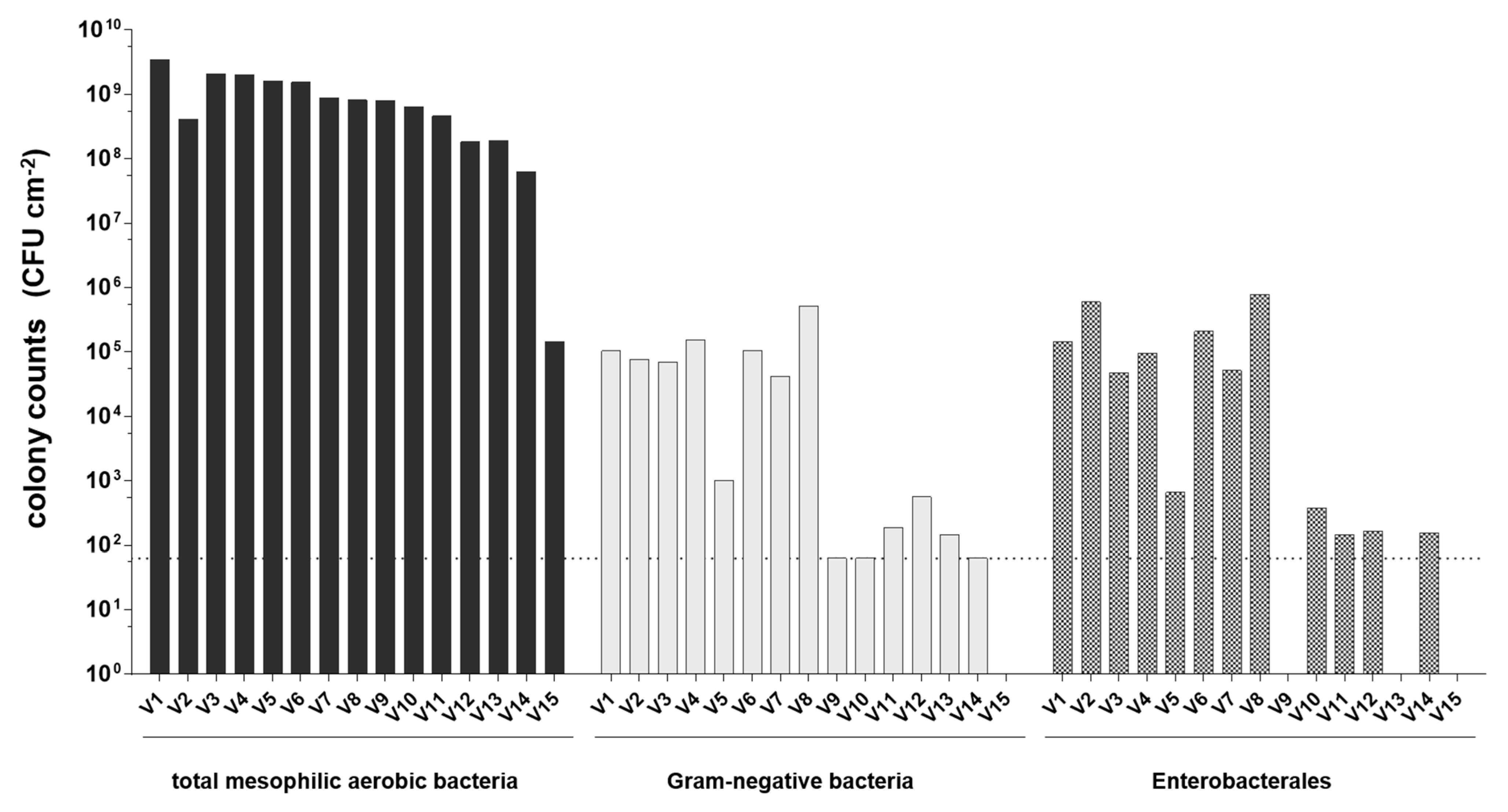
3.1. Abundance of Total Mesophilic Aerobic Bacteria, Gram-Negative Bacteria and Enterobacterales in Cheese Surface Smear

The weighted average colony counts in CFU cm−2 determined by culturing of the surface smear microbiota of 15 different semi-hard surface-ripened cheese varieties are illustrated in Figure 1. Counts for total mesophilic aerobic bacteria were in the range of 6.4 × 10⁷ to 3.5 × 10⁹ CFU cm−2 with the exception of one sample (V15) revealing a much lower abundance of mesophilic aerobic bacteria and a lack of Gram-negative bacteria. All other smear samples contained Gram-negative bacteria in a broad range from the detection limit of 6.3 × 101 CFU cm−2 up to 5.1 × 10⁵ CFU cm−2. With the exception of surface smear samples V9 and V13, all samples that contained Gram-negative bacteria harbored members of Enterobacterales in the range of 1.6 × 10² CFU cm−2 to 7.8 × 10⁵ CFU cm−2. Thus, in contrast to counts for total mesophilic aerobic bacteria, the abundance of Enterobacterales and Gram-negative bacteria varied strongly in the samples of different cheese varieties.

3.2. Genotypic Identification of Gram-Negative Isolates by Partial Sequencing of 16S rRNA and gyrB Genes

A total of 85 Gram-negative isolates from the surface smear of the 15 surface-ripened cheese varieties were further processed for molecular identification based on partial 16S rDNA and gyrB sequencing (Table 2). Overall, the results revealed a high diversity comprising 22 different species that belonged to 14 different genera of Proteobacteria. Most isolates could be assigned to the γ-Proteobacteria class and 84.7% of all isolates represented Enterobacterales, whereas Xanthomonadaceae were represented by single isolates. Representatives of β-Proteobacteria not belonging to the order Enterobacterales comprised mainly Alcaligenaceae with 9.4% of total isolates and a single isolate of Uruburuella suis belonging to Neisseriaceae. Further single isolates represented members of Caulobacteraceae and Phyllobacteriaceae, which belong to α-Proteobacteria. Eight of the fifteen cheese varieties exclusively harbored members of Enterobacterales, whereas four cheese varieties additionally featured Alcaligenaceae or Neisseriaceae. The most abundant Gram-negative genera have been Proteus and Morganella with 37.7% and 23.5%, respectively, represented by the species Proteus vulgaris, Proteus hauseri and Morganella morganii. Proteus spp. were isolated from 11 of the 15 cheese varieties investigated in this study, and Morganella spp. were detected in 9 of the 15 cheese varieties (Table 2). Further frequent isolates corresponded to Citrobacter sp. (9.4%), Enterobacter sp. (5.9%), and Serratia sp. (3.5%). Providencia sp., Providencia heimbachae, Providencia rettgeri and Serratia proteamaculans were represented by single isolates only. Escherichia coli, as a typical fecal indicator, was not present. The family Alcaligenaceae was represented by Pusillimonas sp. (4.7%), Advenella sp. (2.4%) and a single isolate of Kerstersia gyiorum.
Only two cheese varieties (V9 and V13) that harbored Gram-negative bacteria did not reveal a predomination of Enterobacterales. Cheese variety V9 harbored Stenotrophomonas maltophilia as the sole representative of Proteobacteria, whereas the surface microbiota of cheese variety V13 revealed a wide variety of different Proteobacteria, including Advenella sp., Advenella kashmirensis, Brevundimonas diminuta, Defluvibacter, Pusillimonas and Stenotrophomonas. In general, the numbers for different genera of Proteobacteria determined in the surface smear samples varied between 101 CFU cm−2 for the genera Brevundimonas and Defluvibacter and 104 CFU cm−2 for Advenella, Citrobacter, Morganella, Proteus, Providencia and Pusillimonas. Enterobacter, Hafnia, Kerstersia, Serratia, Stenotrophomonas and Uruburuella were represented with 102 to 103 CFU cm−2.

3.3. Proteolytic and Lipolytic Activities of Isolated Proteobacteria

Many representatives of Enterobacterales are known to contribute to the formation of key aroma compounds in cheese by their enzymatic activity [34]. Consequently, 14 selected surface smear isolates from cheese varieties V2, V3 and V5 were tested for their proteolytic and lipolytic potential by applying a spot-on-the-lawn test. Only Proteus isolates revealed proteolytic activity in situ on Caso Bouillon Skim Milk Agar (Table 3) with isolates V2.4, V2.8 and V5.3 showing highest proteolytic activity, as the whole CBSMA Agar plate was cleared at the latest after 5 days of incubation. Three Proteus isolates (V2.5, V2.8 and V5.3) revealed proteolytic activity also under anaerobic conditions, but only after 12 d of incubation (data not shown).
Lipolytic activity of isolates was tested on Tributyrin Agar (TBA). In general, lipolytic activity was much less frequently observed than proteolytic activity. Halomonas and Proteus isolates from cheese sample V2 revealed lipolytic activity after 12 days’ aerobic incubation at room temperature and 30 °C, whereas a Providencia heimbachae isolate from cheese sample V5 showed lipolytic activity in vitro after 12 days at 30 °C. No lipolytic activity could be observed under anaerobic growth conditions.

3.4. Volatile Carbonic Flavor Compounds Produced by Gram-Negative Isolates of the Cheese Surface Smear

To investigate the potential contribution of Proteobacteria to the typical flavor of semi-hard surface-ripened cheese, a headspace atmosphere analysis was performed on cheese variety V2, which was known to harbor different Proteobacteria such as Advenella, Morganella, Proteus, Providencia and Pusillimonas. Application of HS-SPME-GC-MS and HS-SPME-GC/PFPD revealed a variety of different volatile carbonic flavor compounds such as alcohols, aldehydes, carboxylic acids, ketones and sulfur compounds (data not shown). The majority of aroma compounds detected by the analysis were sulfur compounds (dimethyl sulfide, dimethyl disulfide, dimethyl trisulfide, hydrogen sulfide, methanethiol and methylthioacetate), followed by carboxylic acids (acetic acid, butyric acid, formic acid and 3-methylbutyric acid) and ketones (acetoin, 2-butanone, 2-nonanone and 1-phenylethanone). Alcohol compounds determined in the headspace comprised 3-methylbutanol and phenylethanol. Interestingly, many of the volatile organic compounds detected in significant amounts by headspace atmosphere analysis of cheese variety V2 are known to be produced by representatives of Enterobacterales [14,16,19,35,36,37,38,39,40].

3.5. Extended Spectrum β–Lactamase (ESBL)-Mediated Resistances of Enterobacterales Isolated from the Cheese Surface Smear

To investigate potential health risk factors of individual smear bacteria, selected isolates that come into consideration as ESBL producers were tested for their antibiotic susceptibility and the ability to produce extended spectrum β–lactamase. However, none of the Morganella, Enterobacter, Citrobacter, Proteus, Hafnia and Serratia isolates featured ESBL production, whereas all the Morganella isolates as well as one isolate each of the genera Enterobacter, Citrobacter and Hafnia expressed AmpC β-lactamase, which is intrinsically present in these species.

4. Discussion

The present study revealed an unexpectedly high abundance and diversity of Proteobacteria species on the surface of different semi-hard surface-ripened cheese varieties at the end of the ripening stage. The composition of Gram-negative bacterial population of the surface microbiota varied considerably between different cheese varieties. High counts of up to 7.8 × 10⁵ CFU cm−2 Gram-negative bacteria were determined for the majority of cheese varieties analyzed in the present study. These values were comparable to counts of approx. 105 CFU cm−2 Gram-negative bacteria determined for Tilsit-type cheeses as reported in the literature for [5,13]. Differences in the quantity of Gram-negative bacteria might be influenced by differing production technologies, seasonal variation or the milk type applied for cheese production. With up to 109 CFU cm−2, the quantity of total aerobic mesophilic bacteria in cheese smear was very similar for all cheese varieties analyzed in this study, and corresponded well to values reported for various surface-ripened cheese varieties in the literature [3,5,6,8].
At least 22 different species of Proteobacteria were isolated, belonging to 14 different genera. With 84.7% of the 85 identified isolates, Enterobacterales represented the most frequent Gram-negative bacteria. About half of the analyzed cheese varieties featured exclusively members of Enterobacterales, whereas only two varieties lacked its representatives. The observed predomination of Enterobacterales as major representatives of Gram-negative bacteria isolated from cheese surface microbiota is consistent with data from previous studies on surface-ripened cheese [12,41]. In this study, Enterobacterales were mainly represented by isolates of the genera Proteus and Morganella, whereas Citrobacter, Enterobacter, Serratia, Providencia and Hafnia were isolated to a much lesser extent. Besides Enterobacterales, Gram-negative bacteria regularly detected on the surface of surface-ripened cheeses belonged mainly to Proteobacteria families such as Alcaligenaceae, Caulobacteraceae, Halomonadaceae, Moraxellaceae, Oceanospirillaceae, Pseudoalteromonadaceae, Pseudomonadaceae, Vibrionaceae and Xanthomonadaceae [12,13,15,20,24,42,43,44]. On Livarot, a French surface-ripened soft cheese, Gram-negative bacteria accounted for 32% of the overall bacteria isolated from the cheese surface [15]. A recent study on the temporal differences in the microbial composition of Époisses cheese rinds during ripening and storage also revealed a dominance of Gram-negative species. At the end of ripening, about 70% of all 16S rRNA gene sequences were derived from Gram-negative bacteria, with the most abundant genera being Psychrobacter, Halomonas, Mesonia and Vibrio [28].
Proteobacteria other than Enterobacterales isolated from the surface of cheese varieties investigated in this study were mainly represented by single isolates, and comprised members of Alcaligenaceae (Advenella, Kerstersia and Pusillimonas) as well as Caulobacteraceae (Brevundimonas), Phyllobacteriaceae (Defluvibacter), Neisseriaceae (Uruburuella) and Xanthomonadaceae (Stenotrophomonas). Brevundimonas diminuta and Stenotrophomonas sp. were already reported to be part of the surface smear microbiota of different surface-ripened French cheeses [12]. Advenella kashmirensis as well as Pusillimonas sp. have been described for Austrian mountain cheese [45]. To our best knowledge, for the genera Defluvibacter, Kerstersia and Uruburuella this is the first description of an association with cheese. Defluvibacter sp. represents typical environmental bacteria isolated from aqueous habitats such as the activated sludge of waste water plants [46]. In contrast, Kerstersia gyiorum has been isolated from various human clinical samples such as wounds, feces or chronic infection sites [47]. Although the definite pathogenic potential has not been reported for this species, its contribution to chronic infections is discussed [47]. Likewise, Uruburuella suis, isolated first from clinical specimens of animals, seems to be related to respiratory diseases in pigs [48].
Our study revealed no correlation between the abundance of Gram-negative bacteria or species diversity and the type of milk applied for cheese production, whether raw, thermized or pasteurized. This is consistent with the finding that on an average, similar percentages of Proteobacteria were determined in raw milk and pasteurized milk cheese using high-throughput sequencing analysis of 62 different cheese samples [20]. Furthermore, the type of production, whether artisanal manufacturing, traditional or mass production, did not influence the quantity and diversity of Proteobacteria. It is reported that members of Proteobacteria populate functional habitats such as milk handling surfaces or cheese maturation surfaces in artisanal cheese manufacturing plants [24]. In particular, coliform bacteria and Pseudomonadaceae are known to be part of biofilms on wooden vats and shelves from which inoculation might take place during cheese production or ripening [25]. Halotolerant Proteobacteria such as Psychrobacter, Pseudoalteromonas and Vibrio are supposed to establish a ‘house microbiota’, as they are similarly abundant in aging rooms and cheese surfaces [24]. While cheeses produced from pasteurized milk might exclusively be colonized by a secondary contamination with Proteobacteria, those produced from raw or thermized milk might contain indigenous Proteobacteria originating from the milk. Raw milk can be populated by significant proportions of mainly psychrotrophic Proteobacteria (Enterobacteriaceae, Pseudomonas, Acinetobacter) that might flourish during cold storage, and even dominate dairy tank milk [27,42]. As Gram-negative bacteria are known to be adherent to stainless steel and therefore exhibit a high biofilm-forming potential, they are able to colonize dairy pipelines leading to potential recontamination of pasteurized milk [26]. However, for San Simón cheese it was demonstrated that counts for Enterobacteriaceae in the surface smear constantly decreased during a 6-week ripening period, which was related to the influence of the combined effects of physico-chemical parameters [19].
It is assumed that members of Enterobacterales contribute to typical cheese flavor development by the lipolytic breakdown of milk fat and the generation of semi-volatile fatty acids and the according ethyl esters, while peptidase and deaminase activity on proteins contributes to the production of volatile aroma compounds [35,40]. In this study, lipolytic activity was observed for isolates of Halomonas sp., Proteus sp., and Providencia heimbachae, which seems to be the first description of lipolytic activity for Halomonas and Providencia. According to the literature, Citrobacter spp. and Enterobacter spp. may also contribute to aroma compound production in cheese although their proteolytic and/or lipolytic activity in vitro seems to be less pronounced [49].
Proteus spp., in particular Proteus vulgaris strains, were frequently isolated from the surface of surface-ripened cheeses, and their potential role in cheese ripening has already been intensively discussed [12,13,14,15,36,40,50]. Proteus vulgaris was able to successfully colonize and dominate the surface of pilot-scale cheese while significantly contributing to its organoleptic properties through the production of aldehydes [14]. Therefore, the presence of Proteus vulgaris might be desirable for the development of typical aroma compounds [50]. In agreement with the high caseinolytic activity reported for crude culture supernatants of Proteus vulgaris, all Proteus strains investigated in this study featured strong proteolytic activity [40]. In this study, Enterobacterales isolates other than Proteus spp. showed no casein degradation, although this was described for cheese smear isolates affiliated to the genera Enterobacter or Citrobacter [16,34,49]. Accordingly, this enzymatic activity seems to be a strain-dependent feature [34]. In experimental cheese produced from milk artificially inoculated with Enterobacteriaceae, the degradation of major casein fractions could be assigned to their specific proteolytic activity [34].
Enzyme-driven catabolic activities such as proteolysis and the subsequent catabolism of amino acids or lipolysis, besides lactose and citrate metabolism implying glycolysis, impact to a great extent the organoleptic properties of cheese [51,52]. In the literature, various Proteobacteria species are described to produce a wide variety of volatile compounds in large quantities when growing in milk, cheese model medium or cheese surface smear, thereby impacting cheese flavor during ripening [14,16,36,37,40]. A variety of the aroma compounds typical for cheese flavor were detected in significant amounts in the volatile compound pattern of the headspace atmosphere of cheese variety V2 analyzed in this study. Mainly volatile compounds such as carboxylic acids, ketones and sulfur compounds could be related to Enterobacterales isolates after comparing the results to bibliographic data. As Morganella and Proteus spp. predominated the Gram-negative fraction of isolates from the surface smear of cheese variety V2, we concluded that they may contribute to the respective volatile aroma compounds detected in the headspace atmosphere of cheese variety V2. In particular, Proteus vulgaris is known to produce a wide variety of volatile compounds with low perception threshold in large amounts (dimethyl disulfide, 3-methylbutanol) in cheese model medium, and has a prevalence on aroma compound production during the ripening of a model cheese [14,40]. A Proteus vulgaris isolate from the surface of surface-ripened French cheese was able to produce inter alia dimethyl-trisulfide, dimethyl-sulfide, 2-butanone, 1-phenylethanone and phenylethanol in liquid medium [36]. These volatile compounds were also detected in the headspace of cheese variety V2, providing further evidence that Proteus sp. may contribute to the formation of the volatile sulfur compounds that represent garlic odor notes known to be increasingly produced at the end of the ripening process [40]. This assumption is supported by a study which indicates that using human senses (odor and taste), it is possible to predict high levels of Enterobacteriaceae in soft cheeses made from raw milk [53].
As Proteus vulgaris and Hafnia alvei produce a wide variety and large quantity of volatile compounds, non-pathogenic and non-biogenic amine-producing strains thereof were proposed as industrially applicable microorganisms for the production of natural cheese flavor compounds [49]. Hafnia alvei inoculated in pilot-scale cheese was able to establish successfully at the beginning of the ripening process in the cheese community and clearly intensified the production of volatile compounds, mainly by production of volatile sulfur compounds [37]. Hence, non-pathogenic strains of Hafnia alvei are commercially available for the purpose of conferring a flavor comparable to raw milk cheese to that produced from pasteurized milk [37]. Accordingly, as one of few Enterobacterales, Hafnia alvei was included in an authoritative list of microbial food cultures with practical use by the International Dairy Federation (IDF) [54]. However, the potential for production of biogenic amines in vitro or the presence of antibiotic resistance genes might question the use of such bacteria in cheese production [12,16,17].
Concerning the presence of Proteobacteria in food products such as cheese, the positive as well as negative aspects considering the risks and benefits of Gram-negative bacteria in cheese surface smear are up for debate. Many traditional cheeses harbor Proteobacteria, which are considered to belong to natural autochthonous microbiota and are thought to have a beneficial impact on cheese sensory characteristics [14,15,16,17,41]. However, a previous study on the development of a defective smear after prepackaging of surface-ripened cheese in plastic foil revealed that the presence of certain members of Proteobacteria may take a turn for the worse. The vacuum foil prepackaging of surface-ripened cheese can result in a shift in the microbial composition of the smear microbiota towards Proteobacteria, which would then contribute to the development of negative organoleptic properties in the surface smear of the cheese [55]. Furthermore, certain members of Enterobacterales found in cheese are regarded as markers for unsatisfactory hygiene practice during food processing due to their relation to bacterial strains of fecal origin [56]. Threshold and tolerance values for the number of indicator microorganisms revealing bacteriological safety exist in the food legislation of almost all European countries. In the case of Swiss cheese, Swiss food hygiene legislation prescribes limit values for Escherichia coli in cheese produced from heat-treated milk but not for raw milk cheeses. However, limit values are defined for Salmonella spp. as food safety criteria in cheeses produced from raw milk or milk with a heat treatment below pasteurization temperature (thermized milk). Accordingly, as no Escherichia coli or Salmonella spp. were isolated from the cheese surface, the occurrence of Citrobacter, Enterobacter, Hafnia, Morganella, Proteus, Providencia and Serratia in cheese smear is in agreement with current regulations.
Beside hygiene-related aspects, food safety issues challenge the occurrence of Gram-negative bacteria in cheese. According to the European Food Safety Authority (EFSA), none of the Gram-negative bacterial species identified in this study qualified for presumption of safety (QPS) status when intended as biological culture added to foods [57]. Food-borne outbreaks related to Gram-negative bacteria in cheese are rare, and cover mainly Salmonella, enteropathogenic or shiga toxin-producing E. coli or Brucella spp. [58]. Intense hygienic efforts and improved technology applied in the dairy process, such as pasteurization and proper acidification of milk during cheese production as well as the preservation of semi-hard cheese by salting, minimize the risk for transfer of food-borne pathogens. Furthermore, certain strains of Gram-negative species exhibited an antilisterial effect in vitro and in situ in previous studies [59]. Though several Proteobacteria species isolated in this study (Brevundimonas diminuta, Citrobacter sp., Enterobacter sp., Hafnia alvei, Morganella morganii, Proteus vulgaris, Stenotrophomonas maltophilia, Serratia proteamaculans) represent opportunistic pathogens (classified to biosafety level 2), they are associated with diseases that are rarely serious.
Antibiotic resistances such as extended spectrum β-lactamase (ESBL)-mediated resistance of Enterobacteriaceae concern a further safety issue. Several isolates of Enterobacterales from this study have been tested for extended spectrum β-lactamase (ESBL) production, but none of the isolates exhibited such a resistance. The detected AmpC β-lactamases are intrinsically present in many species. They are chromosomally encoded and usually expressed at low levels [60,61]. Considering the literature about ESBL producing Enterobacteriaceae related to cheese, potential β-lactam resistance was detected mainly in Escherichia coli strains isolated from cheese produced in different countries [60]. As the prevalence of ESBL in Europe is high, the emergence of antibiotic resistance in food-related bacteria needs to be closely monitored [62]. Although the consumption of antibiotic-resistant Enterobacteriaceae may not present an immediate risk to human health, antibiotic-resistant genes might be transferred from commensal bacteria to obligate pathogenic bacteria on the cheese surface or bacteria of the human intestinal microbiota [12].

5. Conclusions

This study provides valuable insights into the occurrence of Enterobacterales in cheese surface microbiota and their presumable contribution to flavor development in cheese. An unexpectedly high diversity of Gram-negative bacteria was detected in the smears of different semi-hard surface-ripened cheese varieties. Proteus spp. and Morganella spp. were most prominent, independent of the milk type applied for cheese production or the type of manufacturing process. The quantity of Gram-negative bacteria in the surface smear differed between different cheese varieties, and could have been influenced by dairy ‘house microbiota’. Proteus spp. seemed to contribute in particular to the organoleptic properties of surface-ripened cheese by their strong proteolytic activity. Furthermore, a variety of volatile flavor compounds with low perception threshold was detected in the headspace atmosphere of the surface of cheese variety V2, which most likely resulted from the enzymatic activity of Enterobacterales. On the other hand, several isolated Enterobacterales species represented opportunistic pathogens. Therefore, safety issues related to these species need to be investigated in order to manage the risk to consumer health.
In conclusion, cheese manufacturers are encouraged to produce as tasteful a cheese as possible, with an appealing appearance and of high quality, stability and safety, independent of the seasonal variability induced in basic raw material. Autochthonous-occurring Enterobacterales on the cheese surface appear to be valuable for the organoleptic properties of various cheese varieties, and must not entirely be considered as undesired contaminants. Naturally occurring surface smear microbiota comprising Enterobacterales may protect the surface against unwanted mold spoilage or pathogenic contaminants. Consequently, abandonment of Enterobacterales from the surface smear due to food safety reasons might result in cheese lacking the typical flavor that is important for the authenticity of its variety.

Author Contributions

Conceptualization, L.M. and M.S.; Methodology, J.S.R., L.A., L.M. and M.S.; Formal analysis, J.S.R., H.V.L. and L.A.; Investigation, J.S.R., H.V.L. and L.A.; Resources, L.M. and M.S.; Data curation, J.S.R., L.A., L.M. and M.S.; Validation, J.S.R.; Writing—original draft preparation, J.S.R.; Writing—review and editing, M.S. and J.S.R.; Visualization, J.S.R. and M.S.; Supervision, J.S.R., L.M. and M.S.; Project administration, L.M. and M.S.; Funding acquisition, L.M. and M.S. All authors have read and agreed to the published version of the manuscript.

Funding

This research was funded by Innosuisse—Swiss Innovation Agency, grant number 10273-3 and the APC was funded by ETH Zurich.

Institutional Review Board Statement

Not applicable.

Informed Consent Statement

Not applicable.

Data Availability Statement

Data supporting the reported results can be found on the ETH Zurich Research Collection: https://doi.org/10.3929/ethz-a-010476499.

Acknowledgments

This work was supported by the Swiss Innovation Promotion Agency CTI, project number 10273-3. The authors gratefully acknowledge the contribution of Emmanuelle Arias-Roth and Katharina Breme from Agroscope, Institute for Food Sciences, Berne, Switzerland for analysis of volatile flavor compounds. Katja Lucke is kindly acknowledged for the technical expertise in antibiotic resistance testing. For assisting in executing microbiological experimental work, the authors like to thank most sincerely Alex Baumann, Selina Jost, Samuel Schneider and Martin Knobel.

Conflicts of Interest

The authors declare no conflict of interest.

References

  1. McSweeney, P.L.H.; Ottogalli, G.; Fox, P.F. Diversity of cheese varieties: An overview. In Cheese: Chemistry, Physics and Microbiology; Fox, P.F., McSweeney, P.L.H., Cogan, T.M., Guinee, T.P., Eds.; Elsevier: Amsterdam, The Netherlands, 2004; Volume 2, pp. 1–23. [Google Scholar]
  2. Switzerlands Cheese Market in Figures 2020. Available online: https://www.cheesesfromswitzerland.com/fileadmin/content/SCM_Folder_2020_Taschenstatistik_300x234mm_E_RZ_lowres.pdf (accessed on 17 December 2021).
  3. Brennan, N.M.; Cogan, T.M.; Loessner, M.J.; Scherer, S. Bacterial surface-ripened cheeses. In Cheese: Chemistry, Physics and Microbiology; Fox, P.F., McSweeney, P.L.H., Cogan, T.M., Guinee, T.P., Eds.; Elsevier: Amsterdam, The Netherlands, 2004; Volume 2, pp. 199–225. [Google Scholar]
  4. Rademaker, J.; Peinhopf, M.; Rijnen, L.; Bockelmann, W.; Noordman, W. The surface microflora dynamics of bacterial smear-ripened Tilsit cheese determined by T-RFLP DNA population fingerprint analysis. Int. Dairy J. 2005, 15, 785–794. [Google Scholar] [CrossRef]
  5. Bockelmann, W.; Willems, K.; Neve, H.; Heller, K. Cultures for the ripening of smear cheeses. Int. Dairy J. 2005, 15, 719–732. [Google Scholar] [CrossRef]
  6. Brennan, N.M.; Ward, A.C.; Beresford, T.P.; Fox, P.F.; Goodfellow, M.; Cogan, T.M. Biodiversity of the bacterial flora on the surface of a smear cheese. Appl. Environ. Microbiol. 2002, 68, 820–830. [Google Scholar] [CrossRef] [PubMed] [Green Version]
  7. Mounier, J.; Goerges, S.; Gelsomino, R.; Vancanneyt, M.; Vandemeulebroecke, K.; Hoste, B.; Brennan, N.; Scherer, S.; Swings, J.; Fitzgerald, G. Sources of the adventitious microflora of a smear ripened cheese. J. Appl. Microbiol. 2006, 101, 668–681. [Google Scholar] [CrossRef]
  8. Mounier, J.; Gelsomino, R.; Goerges, S.; Vancanneyt, M.; Vandemeulebroecke, K.; Hoste, B.; Scherer, S.; Swings, J.; Fitzgerald, G.F.; Cogan, T.M. Surface microflora of four smear-ripened cheeses. Appl. Environ. Microbiol. 2005, 71, 6489–6500. [Google Scholar] [CrossRef] [Green Version]
  9. Roth, E.; Schwenninger, S.M.; Eugster-Meier, E.; Lacroix, C. Facultative anaerobic halophilic and alkaliphilic bacteria isolated from a natural smear ecosystem inhibit Listeria growth in early ripening stages. Int. J. Food Microbiol. 2011, 147, 26–32. [Google Scholar] [CrossRef]
  10. Delbès, C.; Ali-Mandjee, L.; Montel, M.C. Monitoring bacterial communities in raw milk and cheese by culture-dependent and-independent 16S rRNA gene-based analyses. Appl. Environ. Microbiol. 2007, 73, 1882–1891. [Google Scholar] [CrossRef] [Green Version]
  11. Ogier, J.-C.; Serror, P. Safety assessment of dairy microorganisms: The Enterococcus genus. Int. J. Food Microbiol. 2008, 126, 291–301. [Google Scholar] [CrossRef]
  12. Coton, M.; Delbès-Paus, C.; Irlinger, F.; Desmasures, N.; Le Fleche, A.; Stahl, V.; Montel, M.C.; Coton, E. Diversity and assessment of potential risk factors of Gram-negative isolates associated with French cheeses. Food Microbiol. 2011, 29, 88–98. [Google Scholar] [CrossRef]
  13. Amato, L.; Ritschard, J.S.; Kurtz, O.; Arias-Roth, E.; Lacroix, C.; Schuppler, M.; Meile, L. Microbial composition of defect smear—A problem evolving during foil-prepacked storage of red-smear cheeses. Int. Dairy J. 2012, 27, 77–85. [Google Scholar] [CrossRef]
  14. Deetae, P.; Mounier, J.; Bonnarme, P.; Spinnler, H.; Irlinger, F.; Helinck, S. Effects of Proteus vulgaris growth on the establishment of a cheese microbial community and on the production of volatile aroma compounds in a model cheese. J. Appl. Microbiol. 2009, 107, 1404–1413. [Google Scholar] [CrossRef] [PubMed]
  15. Larpin-Laborde, S.; Imran, M.; Bonaïti, C.; Bora, N.; Gelsomino, R.; Goerges, S.; Irlinger, F.; Goodfellow, M.; Ward, A.C.; Vancanneyt, M.; et al. Surface microbial consortia from Livarot, a French smear-ripened cheese. Can. J. Microbiol. 2011, 57, 651–660. [Google Scholar] [CrossRef] [PubMed]
  16. Chaves-López, C.; De Angelis, M.; Martuscelli, M.; Serio, A.; Paparella, A.; Suzzi, G. Characterization of the Enterobacteriaceae isolated from an artisanal Italian ewe’s cheese (Pecorino Abruzzese). J. Appl. Microbiol. 2006, 101, 353–360. [Google Scholar] [CrossRef] [PubMed]
  17. Maifreni, M.; Frigo, F.; Bartolomeoli, I.; Innocente, N.; Biasutti, M.; Marino, M. Identification of the Enterobacteriaceae in Montasio cheese and assessment of their amino acid decarboxylase activity. J. Dairy Res. 2013, 80, 122–127. [Google Scholar] [CrossRef] [PubMed]
  18. Irlinger, F.; Layec, S.; Hélinck, S.; Dugat-Bony, E. Cheese rind microbial communities: Diversity, composition and origin. FEMS Microbiol. Let. 2014, 362, 1–11. [Google Scholar] [CrossRef] [PubMed] [Green Version]
  19. Tornadijo, M.; García, M.; Fresno, J.; Carballo, J. Study of Enterobacteriaceae during the manufacture and ripening of San Simón cheese. Food Microbiol. 2001, 18, 499–509. [Google Scholar] [CrossRef]
  20. Quigley, L.; O’Sullivan, O.; Beresford, T.P.; Ross, R.P.; Fitzgerald, G.F.; Cotter, P.D. High-Throughput Sequencing for detection of subpopulations of bacteria not previously associated with artisanal cheeses. Appl. Environ. Microbiol. 2012, 78, 5717–5723. [Google Scholar] [CrossRef] [Green Version]
  21. Unno, R.; Suzuki, T.; Matsutani, M.; Ishikawa, M. Evaluation of the relationships between microbiota and metabolites in soft-type ripened cheese using an integrated omics approach. Front. Microbiol. 2021, 12, 681185. [Google Scholar] [CrossRef]
  22. Kamelamela, N.; Zalesne, M.; Morimoto, J.; Robbat, A.; Wolfe, B.E. Indigo- and indirubin-producing strains of Proteus and Psychrobacter are associated with purple rind defect in a surface-ripened cheese. Food Microbiol. 2018, 76, 543–552. [Google Scholar] [CrossRef]
  23. Peng, S.; Schafroth, K.; Jakob, E.; Stephan, R.; Hummerjohann, J. Behaviour of Escherichia coli strains during semi-hard and hard raw milk cheese production. Int. Dairy J. 2013, 31, 117–120. [Google Scholar] [CrossRef] [Green Version]
  24. Bokulich, N.A.; Mills, D.A. Facility-specific “house” microbiome drives microbial landscapes of artisan cheesemaking plants. Appl. Environ. Microbiol. 2013, 79, 5214–5223. [Google Scholar] [CrossRef] [PubMed] [Green Version]
  25. Didienne, R.; Defargues, C.; Callon, C.; Meylheuc, T.; Hulin, S.; Montel, M.-C. Characteristics of microbial biofilm on wooden vats (‘gerles’) in PDO Salers cheese. Int. J. Food Microbiol. 2012, 156, 91–101. [Google Scholar] [PubMed]
  26. Ksontini, H.; Kachouri, F.; Hamdi, M. Dairy biofilm: Impact of microbial community on raw milk quality. J. Food Qual. 2013, 36, 282–290. [Google Scholar] [CrossRef]
  27. Quigley, L.; O’Sullivan, O.; Stanton, C.; Beresford, T.P.; Ross, R.P.; Fitzgerald, G.F.; Cotter, P.D. The complex microbiota of raw milk. FEMS Microbiol. Rev. 2013, 37, 664–698. [Google Scholar] [CrossRef] [PubMed] [Green Version]
  28. Irlinger, F.; Monnet, C. Temporal differences in microbial composition of Époisses cheese rinds during ripening and storage. J. Dairy Sci. 2021, 104, 7500–7508. [Google Scholar] [CrossRef]
  29. Sambrook, J.; Russell, D.W. Molecular Cloning; Cold Spring Harbor Laboratory Press: Woodbury, NY, USA, 2001; Volume 1–3. [Google Scholar]
  30. Brosius, J.; Palmer, M.L.; Kennedy, P.J.; Noller, H.F. Complete nucleotide sequence of a 16S ribosomal RNA gene from Escherichia coli. Proc. Natl. Acad. Sci. USA 1978, 75, 4801–4805. [Google Scholar] [CrossRef] [Green Version]
  31. Yin, H.; Cao, L.; Xie, M.; Chen, Q.; Qiu, G.; Zhou, J.; Wu, L.; Wang, D.; Liu, X. Bacterial diversity based on 16S rRNA and gyrB genes at Yinshan mine, China. Syst. Appl. Microbiol. 2008, 31, 302–311. [Google Scholar] [CrossRef]
  32. Altschul, S.F.; Madden, T.L.; Schäffer, A.A.; Zhang, J.; Zhang, Z.; Miller, W.; Lipman, D.J. Gapped BLAST and PSI-BLAST: A new generation of protein database search programs. Nucl. Acids Res. 1997, 25, 3389–3402. [Google Scholar] [CrossRef] [Green Version]
  33. European Committee on Antimicrobial Susceptibility Testing. EUCAST Guidelines for Detection of Resistance Mechanisms and Specific Resistances of Clinical and/or Epidemiological Importance; EUCAST: Basel, Switzerland, 2013. [Google Scholar]
  34. Morales, P.; Fernández-García, E.; Nuñez, M. Caseinolysis in cheese by Enterobacteriaceae strains of dairy origin. Lett. Appl. Microbiol. 2003, 37, 410–414. [Google Scholar] [CrossRef]
  35. Dahl, S.; Tavaria, F.K.; Xavier Malcata, F. Relationships between flavour and microbiological profiles in Serra da Estrela cheese throughout ripening. Int. Dairy J. 2000, 10, 255–262. [Google Scholar] [CrossRef]
  36. Deetae, P.; Bonnarme, P.; Spinnler, H.; Helinck, S. Production of volatile aroma compounds by bacterial strains isolated from different surface-ripened French cheeses. Appl. Microbiol. Biotechnol. 2007, 76, 1161–1171. [Google Scholar] [CrossRef] [PubMed]
  37. Irlinger, F.; In Yung, S.A.Y.; Sarthou, A.-S.; Delbès-Paus, C.; Montel, M.-C.; Coton, E.; Coton, M.; Helinck, S. Ecological and aromatic impact of two Gram-negative bacteria (Psychrobacter celer and Hafnia alvei) inoculated as part of the whole microbial community of an experimental smear soft cheese. Int. J. Food Microbiol. 2012, 153, 332–338. [Google Scholar] [CrossRef] [PubMed]
  38. Morales, P.; Feliu, I.; Fernández-García, E.; Nun, M. Volatile compounds produced in cheese by Enterobacteriaceae strains of dairy origin. J. Food Protect. 2004, 67, 567–573. [Google Scholar] [CrossRef]
  39. Tornadijo, E.; Fresno, J.M.; Carballo, J.; Martín-Sarmiento, R. Study of Enterobacteriaceae throughout the manufacturing and ripening of hard goats’ cheese. J. Appl. Bacteriol. 1993, 75, 240–246. [Google Scholar] [CrossRef] [PubMed]
  40. Deetae, P.; Spinnler, H.-E.; Bonnarme, P.; Helinck, S. Growth and aroma contribution of Microbacterium foliorum, Proteus vulgaris and Psychrobacter sp. during ripening in a cheese model medium. Appl. Microbiol. Biotechnol. 2009, 82, 169–177. [Google Scholar] [CrossRef] [PubMed]
  41. Montel, M.-C.; Buchin, S.; Mallet, A.; Delbès–Paus, C.; Vuitton, D.A.; Desmasures, N.; Berthier, F. Traditional cheeses: Rich and diverse microbiota with associated benefits. Int. J. Food Microbiol. 2014, 177, 136–154. [Google Scholar] [CrossRef]
  42. Mounier, J.; Monnet, C.; Jacques, N.; Antoinette, A.; Irlinger, F. Assessment of the microbial diversity at the surface of Livarot cheese using culture-dependent and independent approaches. Int. J. Food Microbiol. 2009, 133, 31–37. [Google Scholar] [CrossRef] [Green Version]
  43. Ogier, J.-C.; Lafarge, V.; Girard, V.; Rault, A.; Maladen, V.; Gruss, A.; Leveau, J.-Y.; Delacroix-Buchet, A. Molecular fingerprinting of dairy microbial ecosystems by use of Temporal Temperature and Denaturing Gradient Gel Electrophoresis. Appl. Env. Microbiol. 2004, 70, 5628–5643. [Google Scholar] [CrossRef] [Green Version]
  44. Leriche, F.; Bordessoules, A.; Fayolle, K.; Karoui, R.; Laval, K.; Leblanc, L.; Dufour, E. Alteration of raw-milk cheese by Pseudomonas spp.: Monitoring the sources of contamination using fluorescence spectroscopy and metabolic profiling. J. Microbiol. Meth. 2004, 59, 33–41. [Google Scholar] [CrossRef]
  45. Schornsteiner, E.; Mann, E.; Bereuter, O.; Wagner, M.; Schmitz-Esser, S. Cultivation-independent analysis of microbial communities on Austrian raw milk hard cheese rinds. Int. J. Food Microbiol. 2014, 180, 88–97. [Google Scholar] [CrossRef]
  46. Fritsche, K.; Auling, G.; Andreesen, J.R.; Lechner, U. Defluvibacter lusatiae gen. nov., sp. nov., a new chlorophenol-degrading member of the α-2 subgroup of Proteobacteria. Syst. Appl. Microbiol. 1999, 22, 197–204. [Google Scholar] [CrossRef]
  47. Pence, M.A.; Sharon, J.; McElvania Tekippe, E.; Pakalniskis, B.L.; Ford, B.A.; Burnham, C.-A.D. Two cases of Kerstersia gyiorum isolated from sites of chronic infection. J. Clin. Microbiol. 2013, 51, 2001–2004. [Google Scholar] [CrossRef] [PubMed] [Green Version]
  48. Silipo, A.; Sturiale, L.; De Castro, C.; Lanzetta, R.; Parrilli, M.; Garozzo, D.; Molinaro, A. Structure of the lipopolysaccharide isolated from the novel species Uruburuella suis. Carbohydr. Res. 2012, 357, 75–82. [Google Scholar] [CrossRef]
  49. Wessels, D.; Jooste, P.J.; Mostert, J.F. Psychrotrophic, proteolytic and lipolytic properties of Enterobacteriaceae isolated from milk and dairy products. Int. J. Food Microbiol. 1989, 9, 79–83. [Google Scholar] [CrossRef]
  50. Deetae, P.; Saint-Eve, A.; Spinnler, H.E.; Helinck, S. Critical effect of oxygen on aroma compound production by Proteus vulgaris. Food Chem. 2011, 126, 134–139. [Google Scholar] [CrossRef]
  51. Duthoit, F.; Callon, C.; Tessier, L.; Montel, M.-C. Relationships between sensorial characteristics and microbial dynamics in “Registered Designation of Origin” Salers cheese. Int. J. Food Microbiol. 2005, 103, 259–270. [Google Scholar] [CrossRef] [PubMed]
  52. Marilley, L.; Casey, M. Flavours of cheese products: Metabolic pathways, analytical tools and identification of producing strains. Int. J. Food Microbiol. 2004, 90, 139–159. [Google Scholar] [CrossRef]
  53. Westlinga, M.; Danielsson-Thama, M.-L.; Jassb, J.; Nilsena, A.; Öströma, A.; Thama, W. Contribution of Enterobacteriaceae to sensory characteristics in soft cheeses made from raw milk. Proc. Food Sci. 2016, 7, 17–20. [Google Scholar] [CrossRef] [Green Version]
  54. Bourdichon, F.; Casaregola, S.; Farrokh, C.; Frisvad, J.C.; Gerds, M.L.; Hammes, W.P.; Harnett, J.; Huys, G.; Laulund, S.; Ouwehand, A.; et al. Food fermentations: Microorganisms with technological beneficial use. Int. J. Food Microbiol. 2012, 154, 87–97. [Google Scholar] [CrossRef]
  55. Ritschard, J.S.; Amato, L.; Kumar, Y.; Müller, B.; Meile, L.; Schuppler, M. The role of the surface smear microbiome in the development of defective smear on surface ripened red smear cheese. AIMS Microbiol. 2018, 4, 622–641. [Google Scholar] [CrossRef]
  56. Martín, M.C.; Martínez, N.; del Rio, B.; Ladero, V.; Fernández, M.; Alvarez, M.A. A novel real-time polymerase chain reaction-based method for the detection and quantification of lactose-fermenting Enterobacteriaceae in the dairy and other food industries. J. Dairy Sci. 2010, 93, 860–867. [Google Scholar] [CrossRef] [PubMed] [Green Version]
  57. EFSA Panel on Biological Hazards (BIOHAZ). Scientific Opinion on the update of the list of QPS-recommended biological agents intentionally added to food and feed as notified to EFSA (2017–2019). EFSA J. 2020, 18, 5966. [Google Scholar]
  58. De Buyser, M.-L.; Dufour, B.; Maire, M.; Lafarge, V. Implication of milk and milk products in food-borne diseases in France and in different industrialised countries. Int. J. Food Microbiol. 2001, 67, 1–17. [Google Scholar] [CrossRef]
  59. Imran, M.; Bré, J.-M.; Guéguen, M.; Vernoux, J.-P.; Desmasures, N. Reduced growth of Listeria monocytogenes in two model cheese microcosms is not associated with individual microbial strains. Food Microbiol. 2013, 33, 30–39. [Google Scholar] [CrossRef]
  60. Amador, P.; Fernandes, R.; Prudêncio, C.; Brito, L. Resistance to β-lactams in bacteria isolated from different types of Portuguese cheese. Int. J. Mol. Sci. 2009, 10, 1538–1551. [Google Scholar] [CrossRef] [PubMed] [Green Version]
  61. Hanson, N.D. AmpC β-lactamases: What do we need to know for the future? J. Antimicrob. Chemother. 2003, 52, 2–4. [Google Scholar] [CrossRef] [Green Version]
  62. Cantón, R.; Novais, A.; Valverde, A.; Machado, E.; Peixe, L.; Baquero, F.; Coque, T.M. Prevalence and spread of extended-spectrum β-lactamase-producing Enterobacteriaceae in Europe. Clin. Microbiol. Infect. 2008, 14, 144–153. [Google Scholar] [CrossRef] [Green Version]
Figure 1. Weighted average in CFU cm−2 for total mesophilic aerobic bacteria, Gram-negative bacteria and Enterobacterales in the surface smear of semi-hard surface-ripened cheese varieties (V1 to V15). The detection limit is indicated by the horizontal dotted line. Lack of bars indicates samples with numbers below the detection limit.
Foods 11 00361 g001
Table 1. Specifications of semi-hard surface-ripened cheese samples analyzed in this study.
SampleCheese TypeWeight aMilk Type bFat Content cProduction Type
V1Trappist-like0.4 kgpasteurized45%artisanal
V2Tilsit-like4 kgthermized45%traditional
V3Tilsit-like4 kgpasteurized45%traditional
V4Tilsit-like (mild/creamy)4 kgpasteurized45%traditional
V5Trappist-like0.8 kgthermized51%traditional
V6Raclette-like4 kgthermized25%artisanal
V7Raclette-like5 kgpasteurized30%artisanal
V8Tilsit-like (mild/creamy)3 kgthermized53%artisanal
V9Mountain cheese-like5 kgthermized45%artisanal
V10Tilsit-like (mild/creamy)6 kgpasteurized31%traditional
V11Tilsit-like7 kgraw milk48%traditional
V12Tilsit-like (mild/creamy)4 kgpasteurized55%industrial
V13Tilsit-like7 kgraw milk45%traditional
V14Raclette-like8 kgraw milk51–54%traditional
V15Tilsit-like (mild/creamy)2 kgpasteurized45%industrial
a weight of a whole round cheese wheel according to cheese manufacturer; b according to cheese manufacturer; c in dry matter according to cheese manufacturer.
Table 2. Overview of Gram-negative bacteria identified in the smear of semi-hard surface-ripened cheese samples V1 to V15.
Occurrence in Surface Smear of Cheese Sample
V1V2V3V4V5V6V7V8V9V10V11V12V13V14V15
Bacterial SpeciesIsolation Media
Advenella sp.++VRBG/PCAI
Advenella kashmirensis+Endo
Brevundimonas diminuta+PCAI
Citrobacter sp.+++VRBG/PCAI/PCAIan/Endo
Defluvibacter sp.+VRBG
Enterobacter spp.+++VRBG/PCAI/PCAIan/Endo
Hafnia alvei+PCAIan
Kerstersia gyiorum+Endo
Morganella spp.+++VRBG/PCAI/Endo
Morganella morganii++++++++VRBG/PCAI/PCAIan/Endo
Proteus spp.++++++++++VRBG/PCAI/Endo
Proteus hauseri++VRBG/PCAI
Proteus vulgaris+++VRBG/PCAI
Providencia sp.+VRBG
Providencia heimbachae+VRBG
Providencia rettgeri+VRBG
Pusillimonas spp.+++VRBG/Endo
Serratia spp.++PCAI/PCAIan
Serratia proteamaculans+PCAIan
Stenotrophomonas sp.+PCAI
S. maltophilia+PCAI
Uruburuella suis+PCAI
Endo, Endo Agar; PCAI, Plate Count Skim Milk Agar supplemented with crystal violet and vancomycin-hydrochloride (PCAIan, PCAI anaerobic incubation); VRBG, Violet Red Bile Glucose Agar.
Table 3. Proteolytic activity of selected Proteobacteria isolates under different environmental conditions.
Proteolysis Zone (mm) a
Aerobic Conditions
RT30 °C
Isolate No.Species2 d5 d12 d2 d5 d12 d
V2.4Proteus sp. 29 ± 2>40>4027 ± 5>40>40
V2.5Proteus sp. 4 ± 23 ± 1>407 ± 12>40
V2.6Proteus sp. -5 ± 112 ± 4-2 ± 111 ±6
V2.7Proteus sp. 2 ± 119 ± 2>403 ± 1>40>40
V2.8Proteus sp. 38 ± 4>40>40->40>40
V2.9Proteus sp. -6 ± 220 ± 52 ± 17 ± 225 ± 2
V5.3Proteus sp. >40>40>40>40>40>40
a The proteolysis zone in mm was defined as the diameter of the spot showing clearance due to proteolysis after subtraction of the diameter of the grown colony. RT, room temperature (22 °C); d, days of incubation.
Publisher’s Note: MDPI stays neutral with regard to jurisdictional claims in published maps and institutional affiliations.

