
Antimicrobial Resistance and Virulence of Non-Typhoidal Salmonella from Retail Foods Marketed in Bangkok, Thailand

 , and
, and
Abstract
:1. Introduction
2. Materials and Methods
2.1. Sample Collection and Bacterial Isolation and Identification
2.2. Serotyping of the Salmonella Isolates
2.3. Determination of Intestinal Cell Invasion by Salmonella Isolates
2.4. Antimicrobial Resistance Profiles
2.5. Polymerase Chain Reaction for Determination of Drug Resistance and Virulence Genes of the Salmonella Isolates
2.6. Statistical Analysis
3. Results
3.1. Prevalence and Serotypes of Salmonella in Retail Food Samples
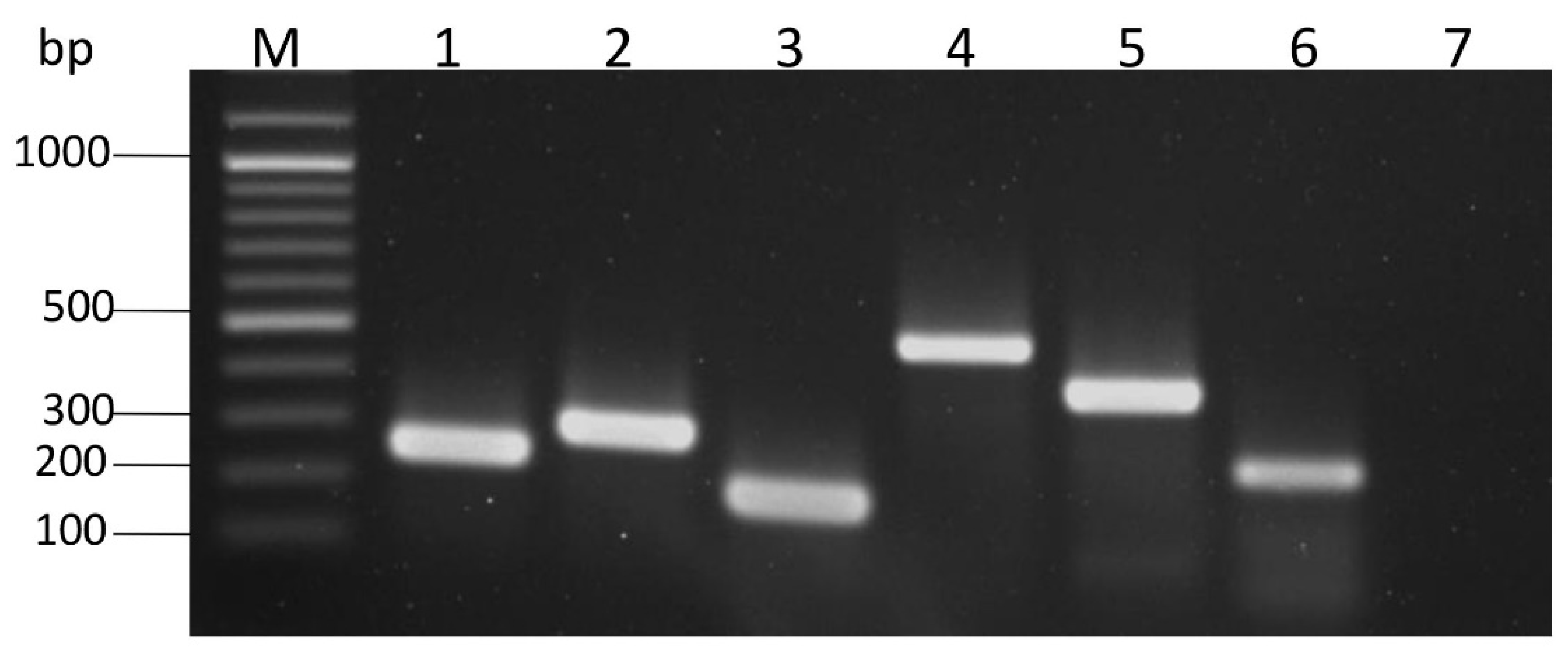
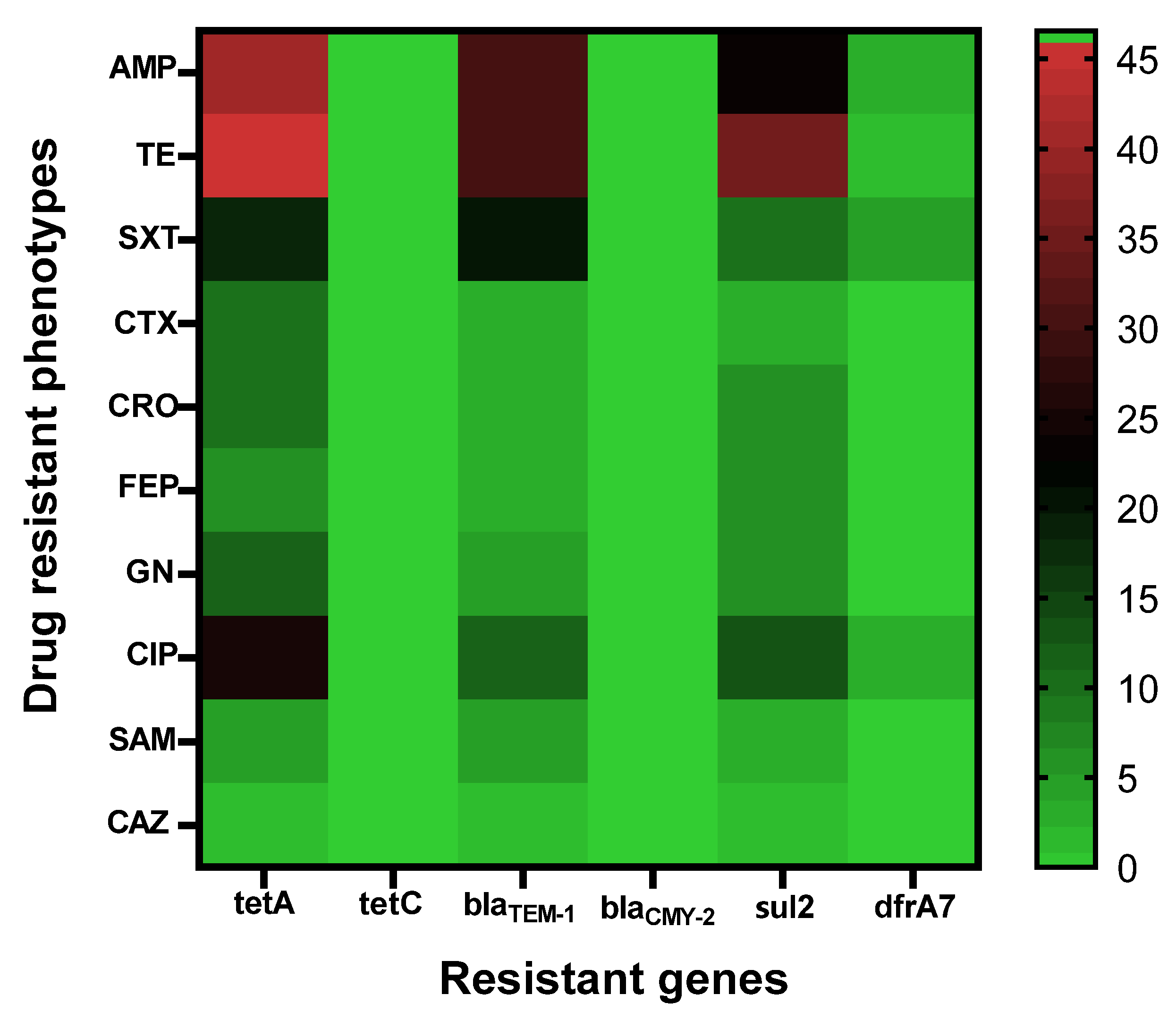
3.2. Antimicrobial and Virulence Genotypes of the Salmonella Isolates
3.3. Antimicrobial Phenotypes of the Salmonella Isolates
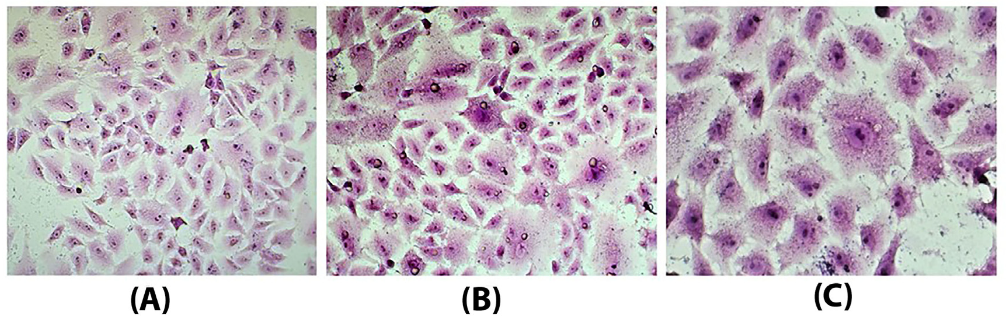
3.4. Caco-2 Invasion Assay on Isolates
4. Discussion
5. Conclusions
Author Contributions
Funding
Data Availability Statement
Conflicts of Interest
References
- Majowicz, S.E.; Musto, J.; Scallan, E.; Angulo, F.J.; Kirk, M.; O’brien, S.J.; Jones, T.F.; Fazil, A.; Hoekstra, R.M. International Collaboration on Enteric Disease “Burden of Illness” Studies. The global burden of nontyphoidal Salmonella gastroenteritis. Clin. Infect. Dis. 2010, 50, 882–889. [Google Scholar]
- Morgado, M.E.; Jiang, C.; Zambrana, J.; Upperman, C.R.; Mitchell, C.; Boyle, M.; Sapkota, A.R.; Sapkota, A. Climate change, extreme events, and increased risk of salmonellosis: Foodborne diseases active surveillance network (FoodNet), 2004–2014. Environ. Health 2021, 20, 1–10. [Google Scholar]
- Popa, G.L.; Papa, M.I. Salmonella spp. infection-A continuous threat worldwide. Germs 2021, 11, 88. [Google Scholar]
- Pouokam, G.B.; Foudjo, B.U.; Samuel, C.; Yamgai, P.F.; Silapeux, A.K.; Sando, J.T.; Atonde, G.F.; Frazzoli, C. Contaminants in foods of animal origin in cameroon: A one health vision for risk management “from Farm to Fork”. Front. Public Health 2017, 5, 197. [Google Scholar]
- Golden, C.E.; Rothrock, M.J., Jr.; Mishra, A. Mapping foodborne pathogen contamination throughout the conventional and alternative poultry supply chains. Poult. Sci. 2021, 100, 101157. [Google Scholar]
- Fàbrega, A.; Vila, J. Salmonella enterica serovar Typhimurium skills to succeed in the host: Virulence and regulation. Clin. Microbiol. Rev. 2013, 26, 308–341. [Google Scholar]
- Foley, S.L.; Johnson, T.J.; Ricke, S.C.; Nayak, R.; Danzeisen, J. Salmonella pathogenicity and host adaptation in chicken-associated serovars. Microbiol. Mol. Biol. Rev. 2013, 77, 582–607. [Google Scholar]
- CDC. Antibiotic Resistance Threats in the United States; U.S. Department of Health and Human Services, CDC: Atlanta, GA, USA, 2019.
- Chen, H.M.; Wang, Y.; Su, L.H.; Chiu, C.H. Nontyphoid Salmonella infection: Microbiology, clinical features, and antimicrobial therapy. Pediatr. Neonatol. 2013, 54, 147–152. [Google Scholar]
- World Health Organization (WHO). Salmonella (Non-Typhoidal). 2018. Available online: https://www.who.int/news-room/fact-sheets/detail/salmonella-(non-typhoidal) (accessed on 19 September 2020).
- Collazo, C.M.; Galán, J.E. The invasion-associated type-III protein secretion system in Salmonella–A review. Gene 1997, 192, 51–59. [Google Scholar]
- Hensel, M. Salmonella pathogenicity island 2. Mol. Microbiol. 2000, 36, 1015–1023. [Google Scholar]
- Murray, R.A.; Lee, C.A. Invasion genes are not required for Salmonella enterica serovar Typhimurium to breach the intestinal epithelium: Evidence that Salmonella pathogenicity island 1 has alternative functions during infection. Infect. Immun. 2000, 68, 5050–5055. [Google Scholar]
- Boddicker, J.D.; Knosp, B.M.; Jones, B.D. Transcription of the Salmonella invasion gene activator, hilA, requires HilD activation in the absence of negative regulators. J. Bacteriol. 2003, 185, 525–533. [Google Scholar]
- Golubeva, Y.A.; Sadik, A.Y.; Ellermeier, J.R.; Slauch, J.M. Integrating global regulatory input into the Salmonella pathogenicity island 1 type III secretion system. Genetics 2012, 190, 79–90. [Google Scholar]
- Swamy, S.C.; Barnhart, H.M.; Lee, M.D.; Dreesen, D.W. Virulence determinants invA and spvC in salmonellae isolated from poultry products, wastewater, and human sources. Appl. Environ. Microbiol. 1996, 62, 3768–3771. [Google Scholar]
- Guerra, B.; Soto, S.M.; Argüelles, J.M.; Mendoza, M.C. Multidrug resistance is mediated by large plasmids carrying a class 1 integron in the emergent Salmonella enterica serotype [4, 5, 12: I:−]. Antimicrob. Agent. Chemother. 2001, 45, 1305–1308. [Google Scholar]
- Cardona-Castro, N.; Restrepo-Pineda, E.; Correa-Ochoa, M. Detection of hilA gene sequences in serovars of Salmonella enterica subspecies enterica. Memórias Inst. Oswaldo Cruz. 2002, 97, 1153–1156. [Google Scholar]
- Martin, L.C.; Weir, E.K.; Poppe, C.; Reid-Smith, R.J.; Boerlin, P. Characterization of blaCMY-2 plasmids in Salmonella and Escherichia coli isolates from food animals in Canada. Appl. Environ. Microbiol. 2012, 78, 1285–1287. [Google Scholar]
- Glenn, L.M.; Lindsey, R.L.; Folster, J.P.; Pecic, G.; Boerlin, P.; Gilmour, M.W.; Harbottle, H.; Zhao, S.; McDermott, P.F.; Fedorka-Cray, P.J.; et al. Antimicrobial resistance genes in multidrug-resistant Salmonella enterica isolated from animals, retail meats, and humans in the United States and Canada. Microb. Drug Resist. 2013, 19, 175–184. [Google Scholar]
- Sabry, M.A.; Abdel-Moein, K.A.; Abdel-Kader, F.; Hamza, E. Extended-spectrum β-lactamase-producing Salmonella serovars among healthy and diseased chickens and their public health implication. J. Glob. Antimicrob. Resist. 2020, 22, 742–748. [Google Scholar]
- Pavelquesi, S.L.S.; de Oliveira Ferreira, A.C.A.; Rodrigues, A.R.M.; de Souza Silva, C.M.; Orsi, D.C.; da Silva, I.C.R. Presence of Tetracycline and Sulfonamide Resistance Genes in Salmonella spp.: Literature Review. Antibiotics 2021, 10, 1314. [Google Scholar]
- ISO 6579:2002; Microbiology of Food and Animal Feeding Stuffs–Horizontal Method for the Detection of Salmonella spp. International Organization for Standardization (ISO): Geneva, Switzerland, 2002.
- Assaf, A.; Cordella, C.B.; Thouand, G. Raman spectroscopy applied to the horizontal methods ISO 6579: 2002 to identify Salmonella spp. in the food industry. Anal. Bioanal. Chem. 2014, 406, 4899–4910. [Google Scholar]
- Aslanzadeh, J. Biochemical profile-based microbial identification systems. In Advanced Techniques in Diagnostic Microbiology; Springer: Boston, MA, USA, 2006; pp. 84–116. [Google Scholar]
- Grimont, P.A.; Weill, F.X. Antigenic formulae of the Salmonella serovars. WHO Collab. Cent. Ref. Res. Salmonella 2007, 9, 1–166. [Google Scholar]
- Gal-Mor, O.; Suez, J.; Elhadad, D.; Porwollik, S.; Leshem, E.; Valinsky, L.; McClelland, M.; Schwartz, E.; Rahav, G. Molecular and cellular characterization of a Salmonella enterica serovar Paratyphi A outbreak strain and the human immune response to infection. Clin. Vaccine Immunol. 2012, 19, 146–156. [Google Scholar]
- Alam, S.B.; Mahmud, M.; Akter, R.; Hasan, M.; Sobur, A.; Nazir, K.H.; Noreddin, A.; Rahman, T.; El Zowalaty, M.E.; Rahman, M. Molecular detection of multidrug resistant Salmonella species isolated from broiler farm in Bangladesh. Pathogens 2020, 9, 201. [Google Scholar]
- Zou, M.; Keelara, S.; Thakur, S. Molecular characterization of Salmonella enterica serotype Enteritidis isolates from humans by antimicrobial resistance, virulence genes, and pulsed-field gel electrophoresis. Foodborne Pathog. Dis. 2012, 9, 232–238. [Google Scholar]
- Thung, T.Y.; Mahyudin, N.A.; Basri, D.F.; Radzi, C.W.M.; Nakaguchi, Y.; Nishibuchi, M.; Radu, S. Prevalence and antibiotic resistance of Salmonella Enteritidis and Salmonella Typhimurium in raw chicken meat at retail markets in Malaysia. Poult. Sci. J. 2016, 95, 1888–1893. [Google Scholar]
- Zambrana-Torrelio, C.; Murray, K.A.; Daszak, P. One health and hotspots of food-borne EIDs. In Improving Food Safety through a One Health Approach: Workshop Summary; National Academies Press: Washington, DC, USA, 2012. [Google Scholar]
- Poppe, C.; Martin, L.C.; Gyles, C.L.; Reid-Smith, R.; Boerlin, P.; McEwen, S.A.; Prescott, J.F.; Forward, K.R. Acquisition of resistance to extended-spectrum cephalosporins by Salmonella enterica subsp. enterica serovar Newport and Escherichia coli in the turkey poult intestinal tract. Appl. Environ. Microbiol. 2005, 71, 1184–1192. [Google Scholar]
- Fonseca, E.L.; Mykytczuk, O.L.; Asensi, M.D.; Reis, E.M.; Ferraz, L.R.; Paula, F.L.; Ng, L.K.; Rodrigues, D.P. Clonality and antimicrobial resistance gene profiles of multidrug-resistant Salmonella enterica serovar Infantis isolates from four public hospitals in Rio de Janeiro, Brazil. J. Clin. Microbiol. 2006, 44, 2767–2772. [Google Scholar]
- Chai, L.C.; Robin, T.; Ragavan, U.M.; Gunsalam, J.W.; Bakar, F.A.; Ghazali, F.M.; Radu, S.; Kumar, M.P. Thermophilic Campylobacter spp. in salad vegetables in Malaysia. Int. J. Food Microbiol. 2007, 117, 106–111. [Google Scholar]
- Dominguez, M.; Jourdan-Da Silva, N.; Vaillant, V.; Pihier, N.; Kermin, C.; Weill, F.X.; Delmas, G.; Kerouanton, A.; Brisabois, A.; de Valk, H. Outbreak of Salmonella enterica serotype Montevideo infections in France linked to consumption of cheese made from raw milk. Foodborne Pathog. Dis. 2009, 6, 121–128. [Google Scholar]
- Chotinan, S.; Tadee, P. Epidemiological Survey of S. Enteritidis Pulsotypes from Salmonellosis Outbreak in Chiang Mai and Samut Songkhram Provinces, Thailand. Vet. Integr. Sci. 2015, 13, 73–80. [Google Scholar]
- Afshari, A.; Baratpour, A.; Khanzade, S.; Jamshidi, A. Salmonella Enteritidis and Salmonella Typhimorium identification in poultry carcasses. Iran. J. Microbiol. 2018, 10, 45. [Google Scholar]
- Altaf Hussain, M.; Wang, W.; Sun, C.; Gu, L.; Liu, Z.; Yu, T.; Ahmad, Y.; Jiang, Z.; Hou, J. Molecular Characterization of Pathogenic Salmonella Spp. From Raw Beef in Karachi, Pakistan. Antibiotics 2020, 9, 73. [Google Scholar]
- Cardoso, M.O.; Ribeiro, A.R.; Santos, L.R.; Pilotto, F.; de Moraes, H.L.; Salle, C.T.; Rocha, S.L.; Nascimento, V.P. Antibiotic resistance in Salmonella Enteritidis isolated from broiler carcasses. Braz. J. Microbiol. 2006, 37, 368–371. [Google Scholar]
- Ghazaey, S.; Mirmomeni, M.H. Microbial-resistant Salmonella Enteritidis isolated from poultry samples. Rep. Biochem. Mol. Biol. 2012, 1, 9. [Google Scholar]
- Lin, D.; Chen, K.; Chan, E.W.C.; Chen, S. Increasing prevalence of ciprofloxacin-resistant food-borne Salmonella strains harboring multiple PMQR elements but not target gene mutations. Sci. Rep. 2015, 5, 14754. [Google Scholar]
- Darwin, K.H.; Miller, V.L. Molecular basis of the interaction of Salmonella with the intestinal mucosa. Clin. Microbiol. Rev. 1999, 12, 405–428. [Google Scholar]
- Dibb-Fuller, M.P.; Allen-Vercoe, E.; Thorns, C.J.; Woodward, M.J. Fimbriae-and flagella-mediated association with and invasion of cultured epithelial cells by Salmonella Enteritidis. Microbiology 1999, 145, 1023–1031. [Google Scholar]
- Van Asten, F.J.; Hendriks, H.G.; Koninkx, J.F.; Van der Zeijst, B.A.; Gaastra, W. Inactivation of the flagellin gene of Salmonella enterica serotype Enteritidis strongly reduces into differentiated Caco-2 cells. FEMS Microbiol. Lett. 2000, 185, 175–179. [Google Scholar]
- van Asten, F.J.; Hendriks, H.G.; Koninkx, J.F.; van Dijk, J.E. Flagella-mediated bacterial motility accelerates but is not required for Salmonella serotype Enteritidis invasion of differentiated Caco-2 cells. Int. J. Med. Microbiol. 2004, 294, 395–399. [Google Scholar]
- Sharma, I.; Das, K. Detection of invA gene in isolated Salmonella from marketed poultry meat by PCR assay. J. Food Process. Technol. 2016, 7, 2. [Google Scholar]
- Solano, C.; García, B.; Valle, J.; Berasain, C.; Ghigo, J.M.; Gamazo, C.; Lasa, I. Genetic analysis of Salmonella Enteritidis biofilm formation: Critical role of cellulose. Mol. Microbiol. 2002, 43, 793–808. [Google Scholar]
- Pang, J.C.; Lin, J.S.; Tsai, C.C.; Tsen, H.Y. The presence of major world-wide clones for phage type 4 and 8 Salmonella enterica serovar Enteritidis and the evaluation of their virulence levels by invasiveness assays in vitro and in vivo. FEMS Microbiol. Lett. 2006, 263, 148–154. [Google Scholar]
- Pan, Z.; Carter, B.; Núñez-García, J.; AbuOun, M.; Fookes, M.; Ivens, A.; Woodward, M.J.; Anjum, M.F. Identification of genetic and phenotypic differences associated with prevalent and non-prevalent Salmonella Enteritidis phage types: Analysis of variation in amino acid transport. Microbiology 2009, 155, 3200–3213. [Google Scholar]
- Borges, K.A.; Furian, T.Q.; Borsoi, A.; Moraes, H.L.; Salle, C.T.; Nascimento, V.P. Detection of virulence-associated genes in Salmonella Enteritidis isolates from chicken in South of Brazil. Pesqui. Vet. Bras. 2013, 33, 1416–1422. [Google Scholar]
- Thung, T.Y.; Radu, S.; Mahyudin, N.A.; Rukayadi, Y.; Zakaria, Z.; Mazlan, N.; Tan, B.H.; Lee, E.; Yeoh, S.L.; Chin, Y.Z.; et al. Prevalence, virulence genes and antimicrobial resistance profiles of Salmonella serovars from retail beef in Selangor, Malaysia. Front. Microbiol. 2018, 8, 2697. [Google Scholar]
- Mthembu, T.P.; Zishiri, O.T.; El Zowalaty, M.E. Molecular detection of multidrug-resistant Salmonella isolated from livestock production systems in South Africa. Infect. Drug Resist. 2019, 12, 3537. [Google Scholar]

| Gene Name | Oligonucleotide Sequence (5′-3′) | Product Size (bp) | Annealing Temperature (°C) | Reference |
|---|---|---|---|---|
| invA | Forward: ACAGTGCTCGTTTACGACCTGAAT Reverse: AGACGACTGGTACTGATCGATAAT | 244 | 60 | [28] |
| hilA | Forward: CGTGAAGGGATTATCGCAGT Reverse: GTCCGGGAATACATCTGAGC | 296 | 56 | [29] |
| blaTEM-1 | Forward: TTGGGTGCACGAGTGGGT Reverse: TAATTGTTGCCGGGAAGC | 504 | 56 | [30] |
| blaCMY-2 | Forward: ATAACCACCCAGTCACGC Reverse: CAGTAGCGAGACTGCGCA | 631 | 52 | [31] |
| sul2 | Forward: CGGCATCGTCAACATAACC Reverse: GTGTGCGGATGAAGTCAG | 405 | 60 | [31] |
| tetA | Forward: GCTACATCCTGCTTGCCTTC Reverse: CATAGATCGCCGTGAAGAGG | 210 | 52 | [32] |
| tetC | Forward: CTTGAGAGCCTTCAACCCAG Reverse: ATGGTCGTCATCTACCTGCC | 418 | 52 | [32] |
| dfrA7 | Forward: GGTAATGGCCCTGATATCCC Reverse: TGTAGATTTGACCGCCACC | 265 | 50 | [33] |
| Salmonella Isolates | Source | Antibiotic-Resistant Profile | Salmonella Serotype | Virulence Gene | Drug Resistance Associated Gene | ||||||
|---|---|---|---|---|---|---|---|---|---|---|---|
| invA | hilA | tetA | tetC | blaTEM-1 | blaCMY-2 | sul2 | dfrA7 | ||||
| Sal1 | pork | AMP, TE, and SXT | B | + | + | + | − | + | − | + | − |
| Sal2 | pork | AMP, TE, and SXT | B | + | + | + | − | + | − | + | − |
| Sal3 | pork | AMP and SXT | E | + | + | + | − | + | − | + | + |
| Sal4 | pork | AMP, CTX, CRO, FEP, GN, and TE | E | + | + | + | − | − | − | + | − |
| Sal5 | pork | AMP, CTX, CRO, FEP, GN, and TE | E | + | + | + | − | − | − | + | − |
| Sal6 | pork | AMP, TE, CIP, and SXT | E | + | + | + | − | + | − | + | + |
| Sal7 | pork | AMP, CTX, CRO, FEP, GN, and TE | E | + | + | − | − | − | − | + | − |
| Sal8 | pork | AMP and TE | C | + | + | + | − | + | − | + | − |
| Sal9 | pork | − | E | + | + | + | − | − | − | − | − |
| Sal10 | pork | AMP, CTX, CRO, FEP, GN, and TE | E | + | + | + | − | − | − | − | − |
| Sal11 | pork | − | E | + | + | + | − | − | − | − | − |
| Sal12 | pork | AMP and TE | B | + | + | + | − | + | − | + | − |
| Sal13 | pork | AMP | C | + | + | + | − | − | − | − | − |
| Sal14 | pork | AMP, TE, CIP, and SXT | B | + | + | − | − | + | − | − | − |
| Sal15 | pork | AMP, CTX, CRO, FEP, GN, and TE | E | + | + | + | − | − | − | − | − |
| Sal16 | pork | AMP, SAM, CAZ, CTX, CRO, FEP, GN, and TE | B | + | + | + | − | + | − | + | − |
| Sal17 | chicken | AMP, SAM, TE, and SXT | B | + | + | + | − | + | − | − | − |
| Sal18 | chicken | − | I | + | + | + | − | − | − | − | − |
| Sal20 | chicken | − | I | + | + | + | − | − | − | − | − |
| Sal21 | chicken | − | C | + | + | + | − | − | − | − | − |
| Sal22 | chicken | − | C | + | + | − | − | − | − | − | − |
| Sal23 | chicken | CIP | C | + | + | + | − | − | − | − | − |
| Sal24 | chicken | CIP | C | + | + | + | − | − | − | − | − |
| Sal25 | chicken | − | E | + | + | + | − | − | − | − | − |
| Sal26 | chicken | TE and CIP | B | + | + | + | − | − | − | − | − |
| Sal27 | chicken | CIP | C | + | + | + | − | − | − | − | − |
| Sal28 | chicken | − | C | + | + | + | − | − | − | − | − |
| Sal29 | chicken | − | Non A-I | + | + | + | − | − | − | − | − |
| Sal30 | chicken | AMP, TE, CIP, and SXT | B | + | + | + | − | + | − | − | − |
| Sal31 | chicken | AMP, TE, CIP, and SXT | B | + | + | + | − | + | − | − | − |
| Sal32 | chicken | TE | C | + | + | + | − | − | − | + | − |
| Sal33 | chicken | CIP | C | + | + | + | − | − | − | − | − |
| Sal34 | chicken | TE and CIP | C | + | + | + | − | − | − | + | − |
| Sal35 | chicken | TE and CIP | C | + | + | − | − | − | − | + | − |
| Sal36 | chicken | AMP, TE, and SXT | B | + | + | + | − | + | − | − | − |
| Sal37 | chicken | TE | C | + | + | + | − | − | − | + | − |
| Sal38 | chicken | − | C | + | + | + | − | − | − | − | |
| Sal39 | chicken | AMP, TE, and SXT | B | + | + | + | − | + | − | + | − |
| Sal40 | chicken | AMP, SAM, TE, and CIP | C | + | + | + | − | + | − | + | − |
| Sal42 | chicken | − | C | + | + | + | − | − | − | − | − |
| Sal43 | chicken | TE | B | + | + | + | − | − | − | + | − |
| Sal44 | chicken | GN, TE, CIP, and SXT | B | + | + | + | − | + | − | + | − |
| Sal45 | chicken | CIP and SXT | E | + | + | + | − | − | − | − | + |
| Sal46 | chicken | AMP, TE, and SXT | B | + | + | + | − | + | − | − | − |
| Sal47 | chicken | AMP and CIP | C | + | + | + | − | − | − | − | − |
| Sal48 | chicken | − | G | + | + | + | − | − | − | − | − |
| Sal50 | chicken | AMP, TE, and CIP | E | + | + | − | − | + | − | + | − |
| Sal52 | chicken | TE | C | + | + | + | − | − | − | + | − |
| Sal53 | chicken | TE and CIP | C | + | + | + | − | − | − | + | − |
| Sal54 | chicken | CIP | C | + | + | + | − | − | − | + | − |
| Sal55 | chicken | AMP and TE | C | + | + | + | − | + | − | + | − |
| Sal56 | chicken | AMP, CTX, CRO, FEP, GN, TE, and CIP | B | + | + | + | − | + | − | − | − |
| Sal57 | beef | − | B | + | + | − | − | − | − | − | − |
| Sal58 | beef | − | B | + | + | − | − | − | − | − | − |
| Sal59 | beef | − | E | + | + | − | − | − | − | − | − |
| Sal60 | beef | − | E | + | + | − | − | − | − | − | − |
| Sal62 | beef | − | E | + | + | − | − | − | − | − | − |
| Sal63 | beef | − | C | + | + | − | − | − | − | − | − |
| Number of isolates (%) | 58 (100) | 58 (100) | 0 (0) | 19 (32.76) | 0 (0) | 23 (39.66) | 3 (5.17) | ||||
| Antimicrobial Agent | Number of Isolates Tested | Anti-Biogram Phenotypes of Salmonella Isolates Number of Isolates (%) | ||
|---|---|---|---|---|
| Sensitive | Intermediate | Resistant | ||
| Group Penicillin | ||||
| ampicillin (AMP) | 58 | 32 (55.17) | 0 (0) | 26 (44.83) |
| Group Combined β-lactam agents | ||||
| ampicillin/sulbactam (SAM) | 58 | 49 (84.49) | 6 (10.34) | 3 (5.17) |
| piperacillin/tazobactam (TZP) | 58 | 56 (96.55) | 2 (3.45) | 0 (0) |
| Group Cephalosporin | ||||
| cefepime (FEP) | 58 | 51 (87.93) | 0 (0) | 7 (12.07) |
| cefotaxime (CTX) | 58 | 47 (81.03) | 4 (6.90) | 7 (12.07) |
| ceftazidime (CAZ) | 58 | 52 (89.66) | 5 (8.62) | 1 (1.72) |
| ceftriaxone (CRO) | 58 | 51 (87.93) | 0 (0) | 7 (12.07) |
| Group Aminoglycoside | ||||
| gentamicin (GN) | 58 | 51 (87.93) | 0 (0) | 7 (12.07) |
| amikacin (AK) | 58 | 58 (100) | 0 (0) | 0 (0) |
| Group Carbapenem | ||||
| ertapenem (ERT) | 58 | 58 (100) | 0 (0) | 0 (0) |
| meropenem (MEM) | 58 | 46 (79.11) | 12 (20.89) | 0 (0) |
| imipenem (IPM) | 58 | 54 (93.10) | 4 (6.90) | 0 (0) |
| Group Tetracycline | ||||
| tetracycline (TE) | 58 | 26 (44.83) | 0 (0) | 32 (55.17) |
| Group Fluoroquinolone | ||||
| ciprofloxacin (CIP) | 58 | 4 (6.90) | 34 (58.62) | 20 (34.48) |
| Group Folate pathway antagonist | ||||
| trimethoprime/sulfamethoxazole (SXT) | 58 | 46 (79.31) | 0 (0) | 12 (20.69) |
| ESBL | Number of isolates tested | Number of positive isolates (%) | Number of negative isolates (%) | |
| ceftazidime | 58 | 7 (12.07) | 51 (87.93) | |
| cefotaxime | 58 | 7 (12.07) | 51 (87.93) | |
Publisher’s Note: MDPI stays neutral with regard to jurisdictional claims in published maps and institutional affiliations. |
© 2022 by the authors. Licensee MDPI, Basel, Switzerland. This article is an open access article distributed under the terms and conditions of the Creative Commons Attribution (CC BY) license (https://creativecommons.org/licenses/by/4.0/).
Share and Cite
Kong-Ngoen, T.; Santajit, S.; Tunyong, W.; Pumirat, P.; Sookrung, N.; Chaicumpa, W.; Indrawattana, N. Antimicrobial Resistance and Virulence of Non-Typhoidal Salmonella from Retail Foods Marketed in Bangkok, Thailand. Foods 2022, 11, 661. https://doi.org/10.3390/foods11050661
Kong-Ngoen T, Santajit S, Tunyong W, Pumirat P, Sookrung N, Chaicumpa W, Indrawattana N. Antimicrobial Resistance and Virulence of Non-Typhoidal Salmonella from Retail Foods Marketed in Bangkok, Thailand. Foods. 2022; 11(5):661. https://doi.org/10.3390/foods11050661
Chicago/Turabian StyleKong-Ngoen, Thida, Sirijan Santajit, Witawat Tunyong, Pornpan Pumirat, Nitat Sookrung, Wanpen Chaicumpa, and Nitaya Indrawattana. 2022. "Antimicrobial Resistance and Virulence of Non-Typhoidal Salmonella from Retail Foods Marketed in Bangkok, Thailand" Foods 11, no. 5: 661. https://doi.org/10.3390/foods11050661
APA StyleKong-Ngoen, T., Santajit, S., Tunyong, W., Pumirat, P., Sookrung, N., Chaicumpa, W., & Indrawattana, N. (2022). Antimicrobial Resistance and Virulence of Non-Typhoidal Salmonella from Retail Foods Marketed in Bangkok, Thailand. Foods, 11(5), 661. https://doi.org/10.3390/foods11050661