Article

Metagenomic and Functional Characterization of Two Chilean Kefir Beverages Reveals a Dairy Beverage Containing Active Enzymes, Short-Chain Fatty Acids, Microbial β-Amyloids, and Bio-Film Inhibitors

by
Claudia Ibacache-Quiroga
1,2,*,
Karoll González-Pizarro
2,
Mariam Charifeh
2,
Christian Canales
3,
Rodrigo Díaz-Viciedo
2,4,
Oliver Schmachtenberg
5,6 and
M. Alejandro Dinamarca
1,2,*
1
Escuela de Nutrición y Dietética, Facultad de Farmacia, Universidad de Valparaíso, Valparaíso 2360102, Chile
2
Centro de Micro-Bioinnovación, Universidad de Valparaíso, Valparaíso 2360102, Chile
3
Facultad de Ingeniería y Tecnología, Universidad San Sebastián, Concepción 4080871, Chile
4
Escuela de Química y Farmacia, Facultad de Farmacia, Universidad de Valparaíso, Valparaíso 2360102, Chile
5
Instituto de Biología, Facultad de Ciencias, Universidad de Valparaíso, Valparaíso 2360102, Chile
6
Centro Interdisciplinario de Neurociencias (CINV), Universidad de Valparaíso, Valparaíso 2381850, Chile
*
Authors to whom correspondence should be addressed.
Foods 2022, 11(7), 900; https://doi.org/10.3390/foods11070900
Submission received: 18 February 2022 / Revised: 16 March 2022 / Accepted: 17 March 2022 / Published: 22 March 2022
(This article belongs to the Section Nutraceuticals, Functional Foods, and Novel Foods)

Abstract

:
Kefir beverage is a probiotic food associated with health benefits, containing probiotic microorganisms and biomolecules produced during fermentation. The microbial composition of these beverages varies among countries, geographical regions, and the substrates, therefore, the characterization of kefir beverages is of great relevance in understanding their potential health-promoting and biotechnological applications. Therefore, this study presents the metagenomic and functional characterization of two Chilean kefir beverages, K02 and K03, through shotgun and amplicon-based metagenomic, microbiological, chemical, and biochemical studies. Results show that both beverages’ microbiota were mainly formed by Bacteria (>98%), while Eukarya represented less than 2%. Regarding Bacteria, the most abundant genera were Acetobacter (93.43% in K02 and 80.99% in K03) and Lactobacillus (5.72% in K02 and 16.75% in K03), while Kazachstania was the most abundant genus from Eukarya (42.55% and 36.08% in K02 and K03). Metagenomic analyses revealed metabolic pathways for lactose and casein assimilation, biosynthesis of health-promoting biomolecules, and clusters for antibiotic resistance, quorum sensing communication, and biofilm formation. Enzymatic activities, microbial β-amyloids, and short-chain fatty acids (acetic acid and propionic acid) were also detected in these beverages. Likewise, both kefir beverages inhibited biofilm formation of the opportunistic pathogen Pseudomonas aeruginosa.

1. Introduction

Kefir is an ancestral dairy beverage originating from the Russian Caucasus and the Tibetan regions, produced by a microbial consortium through fermentation. This microbial consortium is composed of bacteria and yeast, naturally packed into a macrostructure known as “kefir grains” [1] that is used as the starter for the fermentation process. The microbial composition of kefir varies among countries, geographical regions, and due to the substrates used for fermentation [2,3,4]; therefore, different bacterial and yeast genera have been identified in kefir beverages and kefir grains [1,2,3]. Among commonly isolated bacterial genera are Lactobacillus, Lactococcus, Luconostoc, Acetobacter, Enterococcus, Pediococcus, and Gluconobacter [4,5,6,7,8], while the yeast component includes Saccharomyces, Candida, Kluyveromyces, Torulaspora, Kazachstania, Rhodosporidium, Pichia, and Yarrowia [9,10,11].
In the kefir grains, the microorganisms are embedded within an extracellular matrix composed of proteins and polysaccharides [12], forming a microbial ecosystem where microbe-microbe, microbe-environment, and microbe-substrate interactions occur [13,14]. The microorganisms of this ecosystem metabolize fat, proteins, and carbohydrates present in milk, generating different high-value biomolecules, such as vitamins and organic compounds, with known health benefits [15,16].
The potentially beneficial properties of kefir beyond its nutritional value have aroused scientific interest, and diverse studies have explored its effects on human health. At the intestinal level, kefir modulates the gut microbiota [17,18,19], reducing the Firmicutes/Bacteroidetes ratio and inhibiting the proliferation of opportunistic bacteria from the Enterobacteriaceae family [19]. Likewise, bacterial strains isolated from kefir exert a protective effect against gastrointestinal pathogens [20,21]. Furthermore, kefir consumption induces enzymatic activity within the small intestine, increasing protein metabolism, glucose absorption, and lactose hydrolysis, reducing the physiologic and symptomatic parameters associated with lactose intolerance [22,23,24]. This dairy beverage also improves non-alcoholic fatty liver syndrome [25,26], and its cell-free fraction induces apoptosis of gastric and colorectal cancer cells [27,28]. Outside the gastrointestinal tract, kefir has shown an anti-inflammatory effect [29], wound-healing properties [30], as well as the ability to improve atopic dermatitis [31].
The beneficial effects of kefir beverages are tightly related to their microbial composition and productive conditions. In this context, during the past decade, the development and massification of new generation sequencing (NGS) have allowed the identification of the microbial structure in kefir beverages and other fermented foods and the metabolic genetic networks of the microbial community through amplicon-based [32,33,34,35,36,37] and shotgun metagenomic sequencing [38,39,40,41,42], respectively. Even though most metagenomic studies have focused on the kefir grain microbiota [32,33,36,37,38,41,43], during the past years the analysis of the kefir beverages has gained relevance [35,39,42,44,45].
The present work studied two Chilean kefir beverages, produced by two different kefir grains of household origin, collected from the Valparaíso region and selected due to their different physical (macroscopic) characteristics. The Chilean kefir beverages were analyzed at the chemical, amplicon-based and shotgun metagenomic, nutritional, and microbiological levels with an emphasis on their microbiota’s structure and function, their enzymatic activities, and the presence of biomolecules that may contribute to their health-promoting benefits through modulation of the gut microbiota and concomitantly the gut-brain axis, and the inhibition of potentially pathogenic bacteria. To our understanding, this is the first multi-analyses study of kefir beverages of Chilean origin.

2. Materials and Methods

2.1. Materials

The kefir grains used in this study for the production of K02 and K03 beverages belong to the microbial collection of the Centro de Micro-Bioinnovación, of the Universidad de Valparaíso. Both kefir grains are of household origin and were collected in the Valparaíso Region (Chile). Commercial UHT-treated raw milk (3.1% w/v fat) was used for kefir preparation. For proximate analysis, petroleum ether, copper (III) sulfate, potassium sulfate, sulfuric acid, ethanol, sodium hydroxide, and boric acid were purchased from Merck KGaA (Darmstadt, Germany), and Total Dietary Fiber Assay Kit, Methyl Red, and Methyl Blue were purchased from Sigma-Aldrich, Co. (St. Louis, MO, USA). All antibiotics and short-chain fatty acids standards were from Sigma-Aldrich (St. Louis, MO, USA). For microbial growth, MRS and Sabouraud agar were purchased from Sigma-Aldrich, Co. (St. Louis, MO, USA) and Merck KGaA (Darmstadt, Germany), respectively. Marine broth 2216, MacConkey agar, tryptone, yeast extract, casaminoacids and agar were from Becton, Dickinson and Co. (Sparks, NV, USA), D-glucose and skim milk powder from Sigma-Aldrich, and sodium chloride from Loba Chemie Pvt. Ltd. (Mumbai, India). For curli analysis, Congo Red and Coomassie-Brilliant Blue G-250 were purchased from Merck KGaA (Darmstadt, Germany), while curcumin from Sigma-Aldrich, Co. (St. Louis, MO, USA). For biofilm formation assays, crystal violet was obtained from Merck KGaA (Darmstadt, Germany). For metagenomic assays, Meta-G-Nome DNA Isolation Kit (Epicentre) was obtained from Epicentre (Madison, WI, USA) and TruSeq Nano DNA LT Kit from Illumina Inc. (San Diego, CA, USA). For biochemical characterization, API 20E system was purchased from bioMérieux (Lyon, France).

2.2. Culture Media

The composition of non-commercial culture media is detailed in Supplementary Table S1. When needed, antibiotics were added to culture media at the following concentrations: ampicillin (AMP) at 100 µg/mL, chloramphenicol (CLP) at 50 µg/mL, fosfomycin (FOS) at 50 µg/mL, and kanamycin (KAN) at 50 µg/mL.

2.3. Production of Kefir Beverage and Whey

To produce kefir beverages, two different kefir grains were used as starters. These grains were selected due to their macroscopic differences (Supplementary Figure S1). Kefir beverages were produced by mixing 10 g of kefir grains with 100 mL of UHT-treated raw bovine milk and incubating at 25 °C for 48 h. After fermentation, the kefir beverage was separated from the kefir grains by percolation. Kefir whey was obtained from kefir beverages by centrifugation at 8000 rpm at 10 °C for 30 min. To obtain cell-free kefir whey, supernatants were extracted and filtered through a 0.22 µm sterile membrane.

2.4. Characterization of Kefir Beverages

2.4.1. Nutritional Properties

pH was determined using a portable pHmeter (EZDO, PP-203, Taipei City, Taiwan) at 25 °C, in triplicates. Nutritional properties of kefir were determined by proximate analysis according to AOAC (2006) [46]. Briefly, total ash was determined by weighing 2 g of each kefir beverage into an ashing dish and incinerating them at 650 °C for 8 h. After incineration, samples were cooled down in a desiccator and weighed when they reached room temperature. For moisture content calculation, ceramic capsules were dehydrated at 105 °C, chilled to room temperature, and weighed. Then, 1 g of each sample was placed into a capsule and dried at 105 °C until a constant weight was achieved. The moisture content of kefir beverages was estimated by the weight loss of the samples. The protein content in kefir beverage was determined by measuring nitrogen content from 1 g of sample, using the Kjeldahl method, and multiplying it by 6.25. For the determination of lipid content, total lipids were extracted from 2 g of kefir beverages with petroleum ether in a Soxhelt apparatus at 70 °C for 4 h. The ether extracts were placed into a pre-weighed Erlenmeyer flask, the solvent was evaporated, and the lipid content was calculated by measuring the weight difference of the flasks. The percentage of carbohydrates in each sample was estimated as the following formula: % Carbohydrates = 100 − % ash −% moisture − % proteins − % lipids. The total calories were calculated by multiplying the content of lipids, proteins, and carbohydrates by their caloric contribution: 9 kcal/g for lipids and 4 kcal/g for proteins and carbohydrates. Total dietary fiber was determined using the enzymatic-gravimetric method. For this, kefir beverages were pretreated with petroleum ether to remove fat. The obtained residue was enzymatically digested and precipitated using the Total Dietary Fiber Assay Kit (Sigma-Aldrich), according to the manufacturer’s instruction. To estimate the amount of crude fiber, ash and protein were determined in the obtained precipitates, and it was calculated as follows: Total dietary fiber (%) = 100 − % ash − % protein. All samples were analyzed in triplicates.

2.4.2. Detection of Short-Chain Fatty Acids

Detection and quantification of short-chain fatty acids (SCFA) in kefir beverages was performed through HPLC as described by Asarat and coworkers based on Donkor’s protocol [47,48]. The SCFA evaluated in this study were acetic acid, propionic acid, and butyric acid. For SCFA quantification, standard curves of the mentioned SCFA were generated using solutions of predetermined concentrations.

2.4.3. Microbial Viability in Kefir Beverages

Microbial viability in kefir beverage was evaluated by colony-forming unit (CFU) counts. For CFU counts, serial dilutions of kefir beverage in NaCl 0.9% w/v were performed. A total of 100 µL of each dilution were plated on skim milk agar, MRS agar, GYC agar, and Sabouraud agar, in triplicates, for microbial, lactic acid bacteria, acetic acid bacteria, and yeast isolation, respectively. Plates were incubated at 26 °C for 48 h. For lactic acid bacteria quantification, cultures were incubated at 37 °C using anaerobic bags with a CO2 atmosphere. To evaluate the presence of gram-negative enteric bacteria, samples were plated on MacConkey agar and incubated at 37 °C for 48 h. Additionally, bacterial susceptibility to ampicillin, chloramphenicol, fosfomycin, and kanamycin, was evaluated on skim milk agar, MRS agar, and GYC agar under the previously described conditions.

2.5. Metagenomic Sequencing

2.5.1. DNA Extraction

For metagenomic sequencing, total DNA from each kefir beverage was extracted using Meta-G-Nome DNA Isolation Kit (Epicentre) according to the manufacturer’s instructions. DNA integrity was confirmed by agarose electrophoresis, and DNA concentration was determined by UV absorbance using a QuantiFluor® fluorometer (Promega, Madison, WI, USA).

2.5.2. Shotgun Metagenomic Sequencing and Analysis of Kefir Beverages

Total DNA libraries were generated from 100 ng of metagenomic DNA using TruSeq Nano DNA LT Kit (Illumina). Quality control of the genetic libraries was performed using a 2100 Bioanalyzer (Agilent Technologies, Santa Clara, CA, USA). Metagenomic DNA was sequenced through shotgun metagenomic DNA sequencing in an Illumina HiSeq4000 system using a pair-end protocol (2 × 250 bp). Sequence quality was analyzed using the Prinseq tool (v. 0.20.4) and Fasta QC (v. 0.11.5) [49]. Low-quality sequences at left and right extremes and Illumina-adaptor sequences were trimmed off. Sequences with Phred values < 33, with more than a 10% of ambiguous bases or with a total length below 100 bp were not considered for further analyses. Metagenomic analyses included the following key steps: identification of 16S rRNA codifying sequences, operational taxonomic unit (OTU) generation, metagenome phylotyping, sequence assembly, open reading frame (ORF) identification and clustering, and functional annotation of clustered ORFs. Taxonomic profiles of kefir samples were determined using MetaPhlan2 software (v. 2.0) [50]. For the alignment and taxonomic profiling of high-quality reads, Bowtie2 software was used as reference [51]. For gene identification and functional annotation, de novo assembly of high-quality reads was carried out using MetaSPAdes software (v. 3.11.0) [52]. Gene prediction was performed using MetaGeneMark software (v. 3.26) [53], while functional annotation of assembled sequences was done using eggNOG-Mapper software (v. 1.0.1) [54]. Alignment of the predicted proteins was performed using BLASTp (v. 2.6.0), with the Swissprot database as reference. Additionally, predicted genes were annotated using DIAMOND software (v. 0.9.10) [55], GhostKOALA [56] and MG-RAST (v. 4.0.3) [57].

2.5.3. Amplicon-Based 16S rDNA Metagenomic Sequencing

For 16S rDNA analysis, the V4 variable region of 16S rDNA was amplified from 10 ng of total DNA from each sample through qPCR using the protocol developed by Caporaso and coworkers [58]. Length, integrity, and distribution of 16S rDNA metagenomic libraries were estimated using the 2100 Bioanalyzer (Agilent Technologies), and 16S rDNA libraries were sequenced using an Illumina MiSeq platform with a 2 × 150 bp protocol. Pair-end sequences were filtered according to quality control parameters using PRINSEQ software (v. 20.4) [49]. Illumina adapter sequences and sequences with a Phred score of ≤30 were trimmed off, and quality-filtered sequences were merged. High-quality sequences were analyzed using Quantitative Insights into Microbial Ecology (QIIME, v1.9) [59]. To determine relative microbial abundance in kefir beverages, OTUs were identified and analyzed. OTUs were generated using the SILVA Database (v. 132) as reference. Non-assigned sequences were clustered against each other using de novo clustering. In both cases, clustering was performed using UCLUST [60], considering 97% of identity. Each OTU is formed by at least five sequences, and one sequence per cluster was selected for further analysis. Chimeric sequences were identified and eliminated using the ChimeraSlayer method. Results were analyzed using MEGAN (v. 6) and MG-RAST software (v. 4.0.3) [57,61].

2.6. Enzymatic Activity in Kefir Beverages

Enzymatic activity of kefir beverages and kefir whey was evaluated using the API 20E System (bioMérieux) according to the manufacturer’s instructions. Briefly, 100 µL of each kefir beverage were resuspended in 7 mL NaCl solution (0.9% w/v), mixed through vortexing, and inoculated into API 20E strips. For enzymatic activity of kefir whey, cell-free whey was directly inoculated into API 20E strips. In both cases, strips were incubated at 25 °C for 48 h. Casein hydrolysis of kefir beverages and whey was evaluated by plating 100 µL of each sample into a standardized hole (8 mm) in a skim milk agar plate (30 mL). Plates were incubated at 25 °C and casein hydrolysis was measured after two, four, and seven days. Casein hydrolysis was observed as the clearance halo around the inoculation spot and expressed in millimeters (mm). The caseinase activity of all samples was evaluated in triplicates.

2.7. Effect of Kefir Beverages on Biofilm Formation

The effect of kefir beverages on biofilm formation was carried out using three kefir-derived products: (i) cell-free kefir suspension; (ii) cell-free kefir whey; and (iii) denaturized kefir whey. For cell-free kefir suspension, kefir beverage was diluted in NaCl 0.9% w/v in a 1:1 ratio and filtered through a 0.22 µm sterile membrane, while kefir whey was produced as detailed in Section 2.3. Denaturized kefir whey was obtained by incubating kefir whey at 95 °C for 10 min. The effect of these samples on biofilm formation was tested by the crystal violet method according to O’Toole and coworkers on 96-well plates [62], using two bacterial models: the non-pathogenic marine bacterium Cobetia marina (ATCC 25374); and the opportunistic bacterium Pseudomonas aeruginosa PAO1. For this purpose, C. marina was grown in Marine Broth 2216 at 26 °C for 48 h, while P. aeruginosa in LB broth at 37 °C for 24 h.

2.8. Detection of β-Amyloids in Kefir Beverage

For detection of β-amyloid proteins in kefir beverage, samples were plated on YESCA agar plates supplemented with Congo Red (CR) (50 µg/mL) and Coomassie Brilliant Blue G-250 (1 µg/mL). CR can stain β-amyloid and cellulose, therefore, specific detection of β-amyloid proteins was conducted on curcumin (18 µg/mL)-supplemented YESCA plates [63]. Detection of cellulose in kefir beverages was performed on YESCA agar supplemented with calcofluor (1µg/mL) [64]. Escherichia coli strain BW25113 and its ΔcsgA::Km derivative KW1025 [65] were used as positive and negative controls for curli production, a β-amyloid protein, respectively. Plates were incubated at 25 °C for 48 h, and the production of β-amyloid proteins and cellulose was determined.

2.9. Statistical Analysis

Statistical analysis was carried out using StatPlus:mac LE (Analysoft Inc.). For all analyses, two-sample, paired t-tests were performed, and the level of significance was set at 95% (p < 0.05).

2.10. Accession Numbers

Sequence data reported in this work have been submitted to the National Center for Biotechnology Information (BioProject PRJNA388572). K02 and K03 metagenomes were submitted as BioSamples under accession numbers SAMN09389955 and SAMN08943003, respectively. Raw sequence data of shotgun metagenomic sequencing were deposited under accession SRR8282406 (K02) and SRR7287342 (K03). Raw sequencing data of 16S rDNA sequencing was deposited under the following accession numbers: SRR10232092 (K02) and SRR10233296 (K03).

3. Results

3.1. Nutritional and Microbiological Characterization of Kefir Beverages

The kefir beverages analyzed in this study, K02 and K03, are acidic products with pH of 4.3. Their nutritional composition showed that the most abundant components were lipids (3.41% w/v in K02 and 3.15% w/v in K03) and proteins (3.06% w/v in K02 and 3.07% w/v in K03) (Table 1). K02 presented a higher lipid content compared to K03 (p < 0.05), while K03 showed a higher concentration of carbohydrates (p < 0.05) than K02. No significant differences were observed in protein and dietary fiber content between both dairy beverages (p > 0.05). Regarding short-chain fatty acids, K02 and K03 contained lactic acid, acetic acid, and propionic acid, where lactic acid was the most abundant in both samples (Table 1). Significant differences between the two beverages were only observed in acetic acid with a higher content in K02 (p < 0.05).
Total viable culturable microorganisms in kefir beverages reached 3 × 10⁹ UFC/mL, with acetic acid bacteria being the most abundant in both samples (2 × 10⁹ UFC/mL in K02 and 3 × 10⁹ UFC/mL in K03), followed by yeasts (3 × 10⁸ UFC/mL in K02 and 2 × 10⁸ UFC/mL) and lactic acid bacteria (1 × 10⁸ UFC/mL both beverages) (Table 1). No colony-forming units of Enterobacteria were detected on MacConkey agar in either sample.

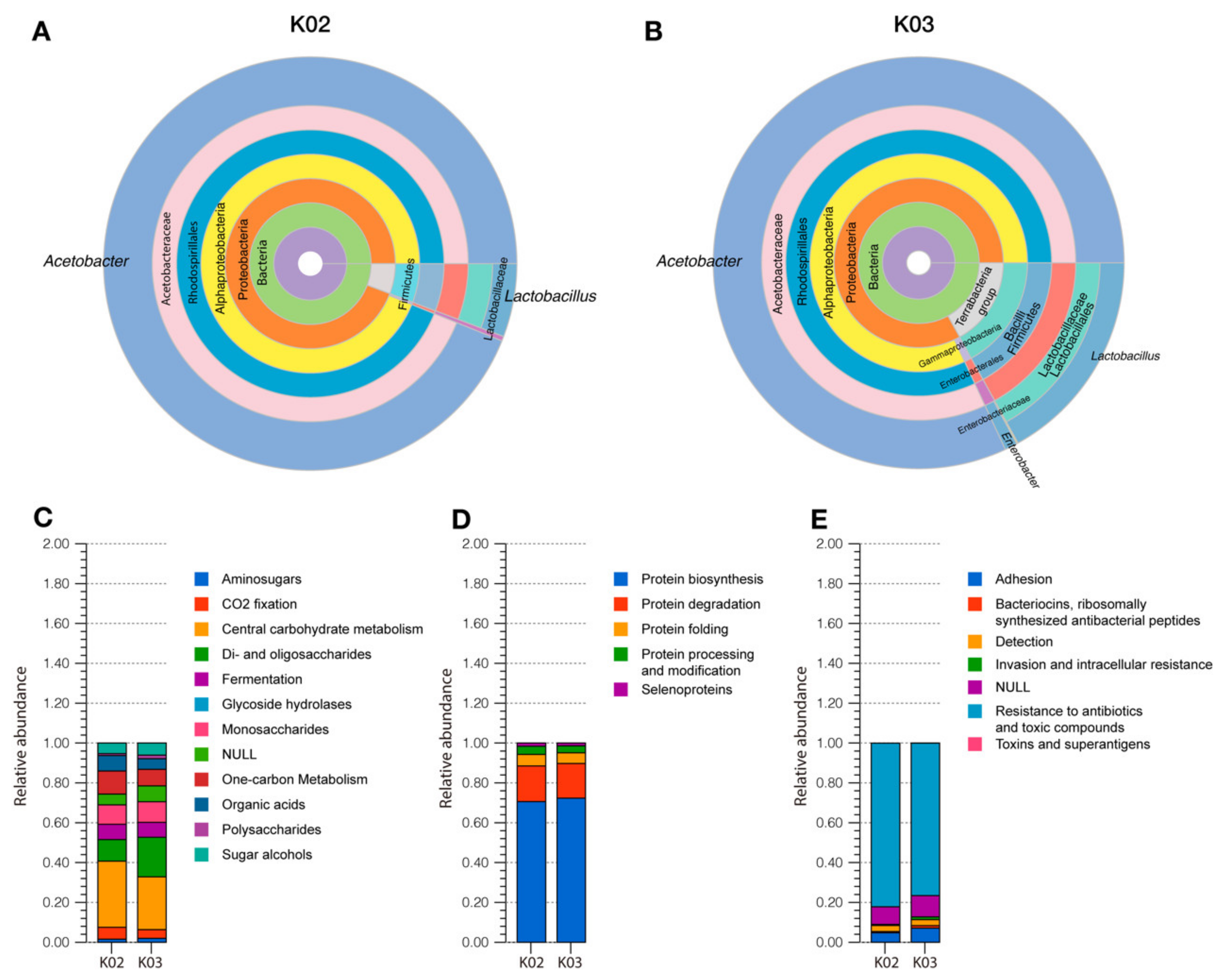
3.2. Taxonomic Analysis of Kefir Beverages

The analysis of operational taxonomic units (OTUs) from shotgun metagenomic sequencing showed that Bacteria was the most abundant domain with 98.82% and 99.65% in K02 and K03, respectively, while Eukarya represented a 1.11% (K02) and a 0.25% (K03). The taxonomic analysis of the Eukarya domain revealed that Kazachstania, Saccharomyces, and Citeromyces were the most abundant genera. Bacterial taxonomic analysis, performed through amplicon-based sequencing and analysis of the 16S rDNA (Statistics of the assembly are detailed in Supplementary Table S2), indicated that the main genera in kefir beverages were Acetobacter (93.43% in K02 and 80.99% in K03) and Lactobacillus (5.72% in K02 and 16.75% in K03) (Figure 1A,B). OTUs of bacteria belonging to the Enterobacteriaceae family were also detected; however, the relative abundances of these microorganisms were lower than 2.2% (0.37% in K02 and 2.18% in K03).

3.3. Functional Characterization of Kefir Beverages

The functional characterization of kefir beverages was performed by two different approaches: metagenomic analyses and confirmation of the functionality of these genes through chemical, biochemical and microbiological assays.
For shotgun metagenomic sequencing, a total of 24,349 and 33,094 genes were predicted in K02 and K03, respectively, and the statistics of the analysis are detailed in Supplementary Table S3.

3.3.1. Carbohydrate Metabolism

According to functional annotation using subsystem analysis of K02 and K03 metagenomes, carbohydrates represented about 12% in both samples (Supplementary Figure S2). From this, the genes involved in di- and oligosaccharide metabolism represented 10.8% in K02 and 19.8% in K03 (Figure 1C, Supplementary Figure S3A), where maltose, maltodextrin, lactose, and β-glucosides metabolism were the most abundant in both samples (Supplementary Figure S3B). Genes encoding β-galactosidase enzymes, lacZ, and lacG, were identified in both kefir samples through KEGG Orthology-based analysis and were attributed to the class Bacilli. Monosaccharide metabolism represented 9.7% and 10.3% of the carbohydrate subsystem in K02 and K03 metagenomes, respectively (Figure 1C, Supplementary Figure S3C). Fermentative metabolism was also identified through subsystem analysis, being acetoin and butanediol metabolism, lactic fermentation, and mixed acid fermentation the most abundant within this group of genes (Supplementary Figure S3D). Regarding lactic fermentation, ldh and ldhA, encoding L-lactate and D-lactate dehydrogenases, responsible for the production of L-lactate and D-lactate from pyruvate, respectively, were identified in both beverages. D-lactate biosynthesis was associated with bacteria belonging to Firmicutes, Alphaproteobacteria, Gammaproteobacteria, and Ascomycetes, while genes involved in L-lactate biosynthesis were attributed to Firmicutes.
Related to mixed acid fermentation, the complete genetic pathways to produce acetate and propanoate, two short-chain fatty acids (SCFA), were identified. Chemical analyses of the kefir beverages revealed that both samples contained acetic acid and propionic acid produced by glucose fermentation, while butyric acid was not detected (Table 1). Likewise, the evaluation of the enzymatic activity of kefir beverages revealed that K02 and K03 presented β-galactosidase activity and anaerobically metabolized glucose to produce acetoin (Table 2). On the other hand, kefir whey of K02 (K02W) and K03 (K03W) metabolized glucose, mannitol, inositol, sorbitol, rhamnose, sucrose, melibiose, amygdalin, and arabinose.

3.3.2. Protein Metabolism

Protein metabolism was the third most abundant subsystem in kefir beverages metagenomes (Figure 1D, Supplementary Figure S4A), where biosynthesis (70%) and degradation (17%) were the main subgroups (Supplementary Figure S4B). Genes belonging to the degradation of proteins included caseinolytic proteases (ClpPs) in both samples (Supplementary Figure S4C) and were mainly attributed to Bacilli and Alphaproteobacteria. The evaluation of the caseinase activity on modified skim milk agar confirmed the proteolytic activity of both beverages (Figure 2). No significant differences between both kefir beverages (p > 0.05) were observed in this parameter at all the evaluated times.

3.3.3. Microbial Resistance to Antimicrobials: Heavy Metals and Antibiotics

In K02 and K03, the Virulence, Disease, and Defense subsystem represented 3.28% and 2.91%, respectively (Supplementary Figure S2), in which antibiotic and toxin resistance genes were the most abundant (83.73% in K02 and 83.63% in K03) (Figure 1E, Supplementary Figure S5A). Regarding antibiotic resistance, multidrug resistance efflux pumps were the most abundant subgroup with 28.14% in K02 and 20.51% in K03 (Supplementary Figure S5B). Other genetic markers that confer resistance to β-lactams, fluoroquinolones, erythromycin, and fosfomycin were also detected on kefir metagenomes.
To evaluate bacterial resistance to antibiotics in kefir beverages, the susceptibility of these microorganisms to ampicillin, chloramphenicol, fosfomycin, and kanamycin was determined. In K02, the microbial count on skim milk agar was significatively reduced in the presence of chloramphenicol and fosfomycin (p < 0.05) (Figure 3A). On the other hand, ampicillin, chloramphenicol, and fosfomycin significantly reduced acetic acid bacteria and lactic acid bacteria counts (p < 0.05) in this beverage. In K03, no significant changes in the microbial count were produced in the presence of the evaluated antibiotics (p > 0.05) on skim milk agar, while a decreased bacterial count of acetic acid microorganisms was observed on GYC agar supplemented with fosfomycin (p < 0.05) (Figure 3B). Additionally, lactic acid bacteria counts were decreased on MRS agar supplemented with ampicillin, chloramphenicol, and fosfomycin.

3.3.4. Microbial Interaction in Kefir Beverages: Biofilm Formation and Host Interaction

Bioinformatic analyses of K02 and K03 metagenomes revealed the presence of genes involved in biofilm initiation and biofilm dispersal. Regarding biofilm initiation, the csgBA and csgDEFG operons responsible for the formation of curli, a β-amyloid protein, were detected. The presence of curli in kefir beverages was tested with Congo Red (CR) and curcumin staining. The results showed that K02 and K03 were labeled when plated on CR agar (Figure 4C,D). CR can stain other bacterial products, therefore, complementary assays using curcumin staining and a calcofluor-supplemented agar were used to detect curli and cellulose, specifically, confirming that K02 and K03 contained curli (Figure 4G,H) and that cellulose is not produced in kefir beverages (Supplementary Figure S6).
Related to biofilm regulation, the following genes were identified in this study: biofilm regulators bssR and bssS, toxin-antitoxin biofilm protein tabA, biofilm growth-associated repressor bigR, and biofilm dispersal mediator bdcA. In this context, the effect of kefir beverages on biofilm formation of opportunistic and environmental bacteria Pseudomonas aeruginosa PAO1 and Cobetia marina, respectively, was evaluated using cell-free kefir (K02 and K03), kefir whey (K02W and K03W), and denaturized kefir whey (K02W-dn and K03W-dn). The results demonstrate a 60% decrease in biofilm formation of P. aeruginosa in the presence of K02, K03, K02W and K02W-dn, while K03W and K03-dn reduced biofilm formation by 50% approximately (Figure 5A). On the other hand, C. marina biofilm formation was significantly reduced in the presence of all samples excluding K02. The highest effect was observed with K02W-dn with a reduction close to 80% (Figure 5B). No significant differences (p > 0.05) in biofilm formation of P. aeruginosa and C. marina were observed between K02 and K03. Differences between K02W and K03W were only observed in P. aeruginosa, while in C. marina, denaturized whey from both kefir beverages showed significant differences (p < 0.05). In both cases, K02 derived whey (K02W and K02W-dn) showed the highest biofilm inhibition.

4. Discussion

The microbial composition of kefir varies among different geographical regions, substrates, and fermentation conditions, with lactic acid bacteria being the most common type of microorganism in this dairy product. In this study, the taxonomic analysis of these beverages’ microbiota revealed that the dominant genus is Acetobacter. In both kefir samples, these acetic acid bacteria (AAB) represent more than 80% of the bacterial community, with K02 showing the higher proportion (93.43%). Acetobacter has been identified in different fermented foods like kombucha [66], cocoa beans [67], palm wine [68], and kefir [69]. In contrast to lactic acid bacteria (LAB), that have been identified in all kefir beverages, the presence of acetic acid bacteria in these dairy beverages is highly variable, with relative abundance that ranges from 0% to 90% [3,4,39]. Despite this variability, currently, in most metagenomically characterized kefir beverages, this genus represents less than 50% of the total bacterial community. For example, Marsh and coworkers characterized the microbiota of 15 kefir beverages from different countries, finding that in 67% of these samples, Acetobacter corresponded to 50% or less, with 37% kefir samples containing no AAB [3]. A factor that could explain the higher abundance of Acetobacter in K02 and K03, compared to other kefir beverages, is related to the production process of this study, specifically regarding the incubation period. During the fermentation, a successional phenomenon occurs where the AAB fraction tends to increase over time [44]. In this context, our 48-h fermentation process differs from other studies with 12–24 h incubation periods and could be influencing the microbial content of kefir beverages. Additionally, the difference in the microbiota composition might be related to the geographical origin of the kefir grains used as starters for fermentation. Nevertheless, since there are no previous studies of Chilean kefir beverages at a metagenomic level, further studies focused on the microbiota and the dynamics of this microbial ecosystem during fermentation in Chilean kefir beverages are needed.
With less than 20%, LAB species represented a minor component of these Chilean kefir beverages, which differs from previous studies where LAB from the phylum Firmicutes were the most abundant microorganisms in kefir beverages [9,21,33,34,35,45,70]. Among lactic acid bacteria, the only genus identified in K02 and K03 was Lactobacillus. This result differs from previous studies showing that LAB in kefir comprise different bacterial genera, including Lactococcus, Lactobacillus, and Leuconostoc [3,34,40].
A minor proportion of the kefir beverages microbiota was represented by yeast belonging to the Saccharomycetaceae family. The genera Kazachstania and Saccharomyces, which have been previously identified in kefir beverages and grains [71,72,73], were the most abundant in both samples. Despite its low abundance in kefir beverages, it is important to highlight that the metagenomic analyses of this study showed that Saccharomycetaceae contributed to the complete biosynthetic pathway of B-complex vitamins and ergosterol, genetic pathways for protein hydrolysis, and alcoholic fermentation of glucose. These metabolic pathways of the yeast present in the Chilean beverages are known to contribute to their organoleptic and antimicrobial properties [74,75].
Kefir is considered a health-promoting food due to its content of probiotic microorganisms. In our study, the main microbial component of the kefir beverages microbiota was bacteria from the genus Acetobacter. Despite not being a classic probiotic genus, probiotic species of Acetobacter have been isolated from dairy products [76,77]. On the other hand, the genus Lactobacillus, the second most abundant in the studied, are naturally part of different human microbiotas, and several species/strains are considered probiotic microorganisms [78,79,80,81,82]. The yeast component of these beverages included Saccharomyces and Kazachstania, two genera with proven probiotic features [72,83,84,85,86]. Despite these promissory results, further strain-specific analyses of isolated bacteria and yeast are needed to confirm their probiotic features. A traditional application of probiotics is the treatment of dysbiosis induced by antibiotics; nevertheless, antimicrobial therapy can affect their viability. In this context, antibiotic resistance observed in AAB and LAB in Chilean kefir may represent an advantage for the concomitant treatment with antibiotics.
Metabolic activity in kefir beverages is also of great relevance for its beneficial effects on human health. One of the primary nutrients in bovine milk is lactose, a disaccharide formed by glucose and galactose. In kefir, lactose is hydrolyzed by the microbial community through the enzymatic activity of β-galactosidase, reducing its concentration and producing glucose and galactose, which are used to form kefiran, the main exopolysaccharide in kefir [87]. In this context, consumption of kefir has been shown to improve symptoms of lactose intolerance [24], a worldwide syndrome that affects about 70% of the adult population, which is caused by a decreased production of lactase in the small intestine [88]. In the present study, genes involved in lactose metabolism and absorption were detected, while the functionality of these genes was confirmed through enzymatic assays (Supplementary Table S3) in K02 and K03. In addition to lactose metabolism, the production of short-chain fatty acids (SCFA) was detected in kefir beverages. These compounds are usually produced in the colon by microorganisms of the gut microbiota and are used as an energy source by enterocytes. They regulate intestinal motility and have an anti-inflammatory effect [89]. Quantification of SCFA in K02 and K03 showed that acetic acid was the most abundant SCFA, followed by propionic acid (Table 1). These results agree with metagenomic studies that identified the complete genomic pathways for synthetizing these SCFA and the taxonomic studies that revealed that AAB were the most abundant in the Chilean kefir beverages microbiotas. Although genes involved in butyrate production were detected through metagenomic analyses, the genetic biosynthetic route was not complete, explaining why no butyric acid was detected. The differences in the acetic acid content between K02 and K03, where K02 showed a higher concentration of this SCFA, could be directly related to the higher relative abundance of microorganisms from the genus Acetobacter in its microbiota (93.43% vs. 80.99%) (Figure 1A,B). Acetobacter are acetic acid producing bacteria [90], and a higher proportion of these microorganisms in kefir beverages is associated with higher acetic acid content in fermented milks [4].
The microorganisms present in kefir and kefir grains form a symbiotic microbial community, where co-metabolic networks operate and allow microbial growth and, eventually, the production of this probiotic food. Metagenomic and functional analyses indicated that bacteria from the Firmicutes phylum metabolized lactose, producing glucose and galactose. Glucose is used by Firmicutes for the biosynthesis of L-lactate and D-lactate through fermentation, and yeasts can use glucose to produce acetic and propionic acid and alcohol [91]. According to metagenomic analyses, ethanol produced by yeast can be metabolized by Alphaproteobacteria to produce acetic acid by oxidation of ethanol [92]. In kefir, SCFA contribute to its organoleptic and preservative properties [93,94]. Besides its relevance for carbon and energy metabolism of the microbial ecosystem of kefir, glucose, and galactose produced by Firmicutes can be used as substrates for the biosynthesis of kefiran, the main polysaccharide of the extracellular matrix of kefir, which is essential for the formation of the macroscopic structure of kefir grain and its propagation [87]. Additionally, the biosynthetic routes for b-vitamins were identified in K02 and K03. In kefir, B-vitamins produced by yeast induce Lactobacillus growth, contributing to the metabolic activity in this microbial consortium [74].
One of the most biologically interesting aspects of kefir is that it is a complex ecosystem where microbe–microbe, microbe–substrate, and interkingdom interactions play a key role in the stability, spread, and perpetuation of this microcosm. One of the mechanisms of microbial interaction previously identified in fermented food is the quorum sensing (QS) communication system that regulates inter- and intra-species interactions [95]. In this study, genes belonging to QS were identified through metagenomic analyses. In this scenario, further studies to confirm the presence of autoinducer molecules in the Chilean kefir beverages are needed.
Besides genes encoding autoinducer molecules of the QS system, the metagenomes of kefir beverages contained genes involved in biofilm formation and dispersal. Metagenomic analyses and phenotypic characterization of K02 and K03 identified genes involved in curli biosynthesis and the production of the microbial β-amyloid (microbial curli), respectively. Curli is an adhesion protein involved in biofilm formation produced by different microorganisms like Proteobacteria, Bacteriodetes, Chloroflexi, and Actinobacteria [96,97,98]. Regarding kefir-associated microorganisms, microbial curli has been detected in species of Lactobacillus, although their role in biofilm formation has not been elucidated [99]. β-amyloid proteins are widely spread in nature with diverse biological implications. In humans, β-amyloid (human curli) is related to protein folding and processing alterations. Even though this has been directly associated with the development of neurodegenerative pathologies like Alzheimer’s disease, Parkinson’s disease, and spongiform encephalopathies [100], recent studies focused on β-amyloid-blocking agents showed that the neurodegenerative effect of these proteins is not only explained by its concentration in the nervous system [101,102,103]. On the other hand, human curli has also been associated with beneficial effects such as antiviral and antibacterial activity and the stimulation of the host defenses [104,105,106]. In bacteria, β-amyloid proteins (microbial curli) play physiological roles in adhesion, biofilm formation, and microbial resistance to the immune system and to toxins [107]. At the intestinal level, microbial curli fibers have been shown to promote intestinal barrier integrity and reduce inflammation by activating of Toll-like receptor 2 (TLR2), stimulating intestinal homeostasis [108,109]. These differences in the effect of β-amyloid fibers might be directly related to the port of entry because systemic exposure to these proteins has been shown to have detrimental systemic and nervous effects [110,111], while oral exposure promotes intestinal health, as mentioned above. In this study, microbial curli was detected in both kefir beverages (K02 and K03) by two different methods (Congo Red and curcumin staining). Considering that the production process of kefir beverages generates a ready-to-drink product, the presence of microbial curli represents an advantage due to its effect on the gut barrier and the potential application of these proteins for the treatment of gastrointestinal pathologies associated with increased intestinal barrier permeability. In this context, this field is of great interest for further studies. Moreover, it is important to highlight that this work is the first to report the presence of microbial curli in a kefir beverage.
Regarding pathogen control by this dairy beverage, genes related to biofilm dispersal were identified in this work. K02 and K03 showed an inhibitory effect on biofilm formation of Pseudomonas aeruginosa PAO1. In Cobetia marina, an inhibitory effect was observed for all the evaluated samples, except K02. Biofilm formation is a virulence factor of a wide range of pathogenic bacteria, including Klebsiella pneumoniae, Escherichia coli, and P. aeruginosa [112,113], contributing to host colonization and antibiotic resistance [114]. Previous reports have shown the ability of species of Lactobacillus to reduce bacterial biofilm formation [115,116]. Despite that K02 and K03 differed in the relative abundance of LAB (5.72% in K02 vs. 16.75%), their effect on biofilm formation did not present significant differences between both products, suggesting that this feature does not only depend on one microbial component of the kefir microbiota. On the other hand, the inhibitory effect of kefir whey was not affected by thermal treatment (95 °C), which suggests that the inhibition of biofilm formation is not enzymatically mediated.

5. Conclusions

The characterization of two Chilean kefir beverages, K02 and K03, revealed that these dairy products contain live and potentially probiotic bacteria, with the dominance of Acetobacter genus, representing a difference with other kefir beverages from different geographical origins. By their microbial structure and function, these beverages contain enzymes and biomolecules, including short-chain fatty acids, microbial curli, and inhibitors of biofilm formation, with potential biotechnological applications and health benefits. Despite their differences in the relative abundance of Acetobacter and Lactobacillus of these beverages’ microbiota, significant differences were only observed in acetic acid content, suggesting that the functional features of this type of food associated the microbial community. Finally, this is the first report of the presence of microbial curli in this type of food, opening a new area for exploring the beneficial effect of these beverages or microbial amyloid over the intestinal barrier function for the treatment of gastrointestinal pathologies involving intestinal dysfunction and inflammation.

Supplementary Materials

The following are available online at https://www.mdpi.com/article/10.3390/foods11070900/s1, Figure S1: Kefir grains. Figure S2: Subsystem analysis of kefir beverages. Figure S3: Metabolism of proteins in kefir beverages. Figure S4: Metabolism of carbohydrates in kefir beverages. Figure S5: Virulence, Disease and Defense genes in kefir beverages, Figure S6: Cellulose in kefir beverages. Table S1: Composition of non-commercial culture media. Table S2: Sequence statistics for metagenomic sequencing of 16S rDNA. Table S3: Sequence statistics for shotgun metagenomic sequencing kefir beverages.

Author Contributions

Conceptualization, C.I.-Q. and M.A.D.; methodology, C.I.-Q. and M.A.D.; investigation, C.I.-Q., K.G.-P., M.C., C.C., R.D.-V. and M.A.D.; writing—original draft preparation, C.I.-Q. and M.A.D.; writing—review and editing, C.I.-Q., O.S. and M.A.D.; visualization, C.I.-Q. and M.A.D.; funding acquisition, C.I.-Q., K.G.-P. and M.A.D. All authors have read and agreed to the published version of the manuscript.

Funding

This research was funded by the Chilean National Agency for Research and Development (ANID) (PAI79170114 and FONDEF VIU17E0112), and by the Centro de Micro-Bioinnovación, Universidad de Valparaíso (DIUV-CIDI 4/2016).

Data Availability Statement

Sequence data reported in this study have been submitted to the National Center for Biotechnology (BioProject PRJNA388572). K02 and K03 metagenomes were submitted as BioSamples under accession number SAMN09389955 and SAMN08943003, respectively. Raw sequence data of shotgun metagenomic sequencing were deposited under accession SRR8282406 (K02) and SRR7287342 (K03). Raw sequencing data of amplicon-based 16S rDNA sequencing was deposited under the following accession numbers: SRR10232092 (K02) and SRR10233296 (K03).

Acknowledgments

The authors acknowledge the contribution of Catherine Ramos and Claudia Santibañez to this study with their technical support.

Conflicts of Interest

The authors declare no conflict of interest.

References

  1. Farnworth, E.R. Kefir. J. Nutraceuticals Funct. Med Foods 1999, 1, 57–68. [Google Scholar] [CrossRef]
  2. Da Miguel, M.G.C.P.; Cardoso, P.G.; de Lago, L.A.; Schwan, R.F. Diversity of Bacteria Present in Milk Kefir Grains Using Culture-Dependent and Culture-Independent Methods. Food Res. Int. 2010, 43, 1523–1528. [Google Scholar] [CrossRef]
  3. Marsh, A.J.; O’Sullivan, O.; Hill, C.; Ross, R.P.; Cotter, P.D. Sequencing-Based Analysis of the Bacterial and Fungal Composition of Kefir Grains and Milks from Multiple Sources. PLoS ONE 2013, 8, e69371. [Google Scholar] [CrossRef] [PubMed] [Green Version]
  4. Guangsen, T.; Xiang, L.; Jiahu, G. Microbial Diversity and Volatile Metabolites of Kefir Prepared by Different Milk Types. CyTA J. Food 2021, 19, 399–407. [Google Scholar] [CrossRef]
  5. Leite, A.M.O.; Miguel, M.A.L.; Peixoto, R.S.; Ruas-Madiedo, P.; Paschoalin, V.M.F.; Mayo, B.; Delgado, S. Probiotic Potential of Selected Lactic Acid Bacteria Strains Isolated from Brazilian Kefir Grains. J. Dairy Sci. 2015, 98, 3622–3632. [Google Scholar] [CrossRef] [PubMed] [Green Version]
  6. Garrote, G.L.; Abraham, A.G.; De Antoni, G.L. Chemical and Microbiological Characterisation of Kefir Grains. J. Dairy Res. 2001, 68, 639–652. [Google Scholar] [CrossRef]
  7. Carasi, P.; Jacquot, C.; Romanin, D.E.; Elie, A.-M.; De Antoni, G.L.; Urdaci, M.C.; de los Serradell, M.A. Safety and Potential Beneficial Properties of Enterococcus Strains Isolated from Kefir. Int. Dairy J. 2014, 39, 193–200. [Google Scholar] [CrossRef]
  8. Sabir, F.; Beyatli, Y.; Cokmus, C.; Onal-Darilmaz, D. Assessment of Potential Probiotic Properties of Lactobacillus Spp., Lactococcus Spp. and Pediococcus Spp. Strains Isolated from Kefir. J. Food Sci. 2010, 75, M568–M573. [Google Scholar] [CrossRef]
  9. Simova, E.; Beshkova, D.; Angelov, A.; Hristozova, T.; Frengova, G.; Spasov, Z. Lactic Acid Bacteria and Yeasts in Kefir Grains and Kefir Made from Them. J. Ind. Microbiol. Biotechnol. 2002, 28, 1–6. [Google Scholar] [CrossRef]
  10. Vardjan, T.; Mohar Lorbeg, P.; Rogelj, I.; Čanžek Majhenič, A. Characterization and Stability of Lactobacilli and Yeast Microbiota in Kefir Grains. J. Dairy Sci. 2013, 96, 2729–2736. [Google Scholar] [CrossRef] [Green Version]
  11. TaŞ, T.K.Ö.K.; Ekinci, F.Y.; Guzel-Seydim, Z.B. Identification of Microbial Flora in Kefir Grains Produced in Turkey Using PCR. Int. J. Dairy Technol. 2012, 65, 126–131. [Google Scholar] [CrossRef]
  12. Garofalo, C.; Ferrocino, I.; Reale, A.; Sabbatini, R.; Milanović, V.; Alkić-Subašić, M.; Boscaino, F.; Aquilanti, L.; Pasquini, M.; Trombetta, M.F.; et al. Study of Kefir Drinks Produced by Backslopping Method Using Kefir Grains from Bosnia and Herzegovina: Microbial Dynamics and Volatilome Profile. Food Res. Int. 2020, 137, 109369. [Google Scholar] [CrossRef]
  13. Garrote, G.L.; Abraham, A.G.; De Antoni, G.L. Microbial Interactions in Kefir: A Natural Probiotic Drink. In Biotechnology of Lactic Acid Bacteria: Novel Applications; Wiley-Blackwell: Hoboken, NJ, USA, 2010; 327p. [Google Scholar]
  14. Cheirsilp, B.; Shoji, H.; Shimizu, H.; Shioya, S. Interactions between Lactobacillus Kefiranofaciens and Saccharomyces Cerevisiae in Mixed Culture for Kefiran Production. J. Biosci. Bioeng. 2003, 96, 279–284. [Google Scholar] [CrossRef]
  15. Rosa, D.D.; Dias, M.M.S.; Grześkowiak, Ł.M.; Reis, S.A.; Conceição, L.L.; Peluzio, M.C.G. Milk Kefir: Nutritional, Microbiological and Health Benefits. Nutr. Res. Rev. 2017, 30, 82–96. [Google Scholar] [CrossRef]
  16. Farag, M.A.; Jomaa, S.A.; Abd El-Wahed, A.; El-Seedi, H.R. The Many Faces of Kefir Fermented Dairy Products: Quality Characteristics, Flavour Chemistry, Nutritional Value, Health Benefits, and Safety. Nutrients 2020, 12, 346. [Google Scholar] [CrossRef] [Green Version]
  17. Bellikci-Koyu, E.; Sarer-Yurekli, B.P.; Akyon, Y.; Aydin-Kose, F.; Karagozlu, C.; Ozgen, A.G.; Brinkmann, A.; Nitsche, A.; Ergunay, K.; Yilmaz, E.; et al. Effects of Regular Kefir Consumption on Gut Microbiota in Patients with Metabolic Syndrome: A Parallel-Group, Randomized, Controlled Study. Nutrients 2019, 11, 2089. [Google Scholar] [CrossRef] [Green Version]
  18. Hsu, Y.-J.; Huang, W.-C.; Lin, J.-S.; Chen, Y.-M.; Ho, S.-T.; Huang, C.-C.; Tung, Y.-T. Kefir Supplementation Modifies Gut Microbiota Composition, Reduces Physical Fatigue, and Improves Exercise Performance in Mice. Nutrients 2018, 10, 862. [Google Scholar] [CrossRef] [Green Version]
  19. Kim, D.-H.; Chon, J.-W.; Kim, H.; Seo, K.-H. Modulation of Intestinal Microbiota in Mice by Kefir Administration. Food Sci. Biotechnol. 2015, 24, 1397–1403. [Google Scholar] [CrossRef]
  20. Carasi, P.; Trejo, F.M.; Pérez, P.F.; De Antoni, G.L.; de los Serradell, M.A. Surface Proteins from Lactobacillus Kefir Antagonize In Vitro Cytotoxic Effect of Clostridium Difficile Toxins. Anaerobe 2012, 18, 135–142. [Google Scholar] [CrossRef]
  21. De Montijo-Prieto, S.; Moreno, E.; Bergillos-Meca, T.; Lasserrot, A.; Ruiz-López, M.-D.; Ruiz-Bravo, A.; Jiménez-Valera, M. A Lactobacillus Plantarum Strain Isolated from Kefir Protects against Intestinal Infection with Yersinia Enterocolitica O9 and Modulates Immunity in Mice. Res. Microbiol. 2015, 166, 626–632. [Google Scholar] [CrossRef]
  22. Urdaneta, E.; Barrenetxe, J.; Aranguren, P.; Irigoyen, A.; Marzo, F.; Ibáñez, F.C. Intestinal Beneficial Effects of Kefir-Supplemented Diet in Rats. Nutr. Res. 2007, 27, 653–658. [Google Scholar] [CrossRef]
  23. De Vrese, M.; Keller, B.; Barth, C.A. Enhancement of Intestinal Hydrolysis of Lactose by Microbial β-Galactosidase (EC 3.2.1.23) of Kefir. Br. J. Nutr. 1992, 67, 67–75. [Google Scholar] [CrossRef] [PubMed] [Green Version]
  24. Hertzler, S.R.; Clancy, S.M. Kefir Improves Lactose Digestion and Tolerance in Adults with Lactose Maldigestion. J. Am. Diet. Assoc. 2003, 103, 582–587. [Google Scholar] [CrossRef] [PubMed]
  25. Chen, H.L.; Tung, Y.T.; Tsai, C.L.; Lai, C.W.; Lai, Z.L.; Tsai, H.C.; Lin, Y.L.; Wang, C.H.; Chen, C.M. Kefir Improves Fatty Liver Syndrome by Inhibiting the Lipogenesis Pathway in Leptin-Deficient Ob/ob Knockout Mice. Int. J. Obes. 2013, 38, 1172. [Google Scholar] [CrossRef] [PubMed]
  26. Chen, H.L.; Tsai, T.C.; Tsai, Y.C.; Liao, J.W.; Yen, C.C.; Chen, C.M. Kefir Peptides Prevent High-Fructose Corn Syrup-Induced Non-Alcoholic Fatty Liver Disease in a Murine Model by Modulation of Inflammation and the JAK2 Signaling Pathway. Nutr. Diabetes 2016, 6, e237. [Google Scholar] [CrossRef]
  27. Gao, J.; Gao, J.; Gu, F.; Ruan, H.; Chen, Q.; He, J.; He, G. Induction of Apoptosis of Gastric Cancer Cells SGC7901 in Vitro by a Cell-Free Fraction of Tibetan Kefir. Int. Dairy J. 2013, 30, 14–18. [Google Scholar] [CrossRef]
  28. Khoury, N.; El-Hayek, S.; Tarras, O.; El-Sabban, M.; El-Sibai, M.; Rizk, S. Kefir Exhibits Anti-proliferative and Pro-apoptotic Effects on Colon Adenocarcinoma Cells with No Significant Effects on Cell Migration and Invasion. Int. J. Oncol. 2014, 45, 2117–2127. [Google Scholar] [CrossRef]
  29. Prado, M.R.M.; Boller, C.; Zibetti, R.G.M.; de Souza, D.; Pedroso, L.L.; Soccol, C.R. Anti-Inflammatory and Angiogenic Activity of Polysaccharide Extract Obtained from Tibetan Kefir. Microvasc. Res. 2016, 108, 29–33. [Google Scholar] [CrossRef]
  30. Oryan, A.; Alemzadeh, E.; Eskandari, M.H. Kefir Accelerates Burn Wound Healing Through Inducing Fibroblast Cell Migration In Vitro and Modulating the Expression of IL-1β, TGF-β1, and bFGF Genes In Vivo. Probiotics Antimicrob. Proteins 2019, 11, 874–886. [Google Scholar] [CrossRef]
  31. Alves, E.; Gregório, J.; Baby, A.R.; Rijo, P.; Rodrigues, L.M.; Rosado, C. Homemade Kefir Consumption Improves Skin Condition—A Study Conducted in Healthy and Atopic Volunteers. Foods 2021, 10, 2794. [Google Scholar] [CrossRef]
  32. Zamberi, N.R.; Mohamad, N.E.; Yeap, S.K.; Ky, H.; Beh, B.K.; Liew, W.C.; Tan, S.W.; Ho, W.Y.; Boo, S.Y.; Chua, Y.H.; et al. 16S Metagenomic Microbial Composition Analysis of Kefir Grain Using MEGAN and BaseSpace. Food Biotechnol. 2016, 30, 219–230. [Google Scholar] [CrossRef]
  33. Ilıkkan, Ö.K.; Bağdat, E.Ş. Comparison of Bacterial and Fungal Biodiversity of Turkish Kefir Grains with High-Throughput Metagenomic Analysis. LWT 2021, 152, 112375. [Google Scholar] [CrossRef]
  34. Kazou, M.; Grafakou, A.; Tsakalidou, E.; Georgalaki, M. Zooming Into the Microbiota of Home-Made and Industrial Kefir Produced in Greece Using Classical Microbiological and Amplicon-Based Metagenomics Analyses. Front. Microbiol. 2021, 12, 64. [Google Scholar] [CrossRef]
  35. Yegin, Z.; Zafer Yurt, M.N.; Tasbasi, B.B.; Acar, E.E.; Altunbas, O.; Ucak, S.; Ozalp, V.C.; Sudagidan, M. Determination of Bacterial Community Structure of Turkish Kefir Beverages via Metagenomic Approach. Int. Dairy J. 2022, 129, 105337. [Google Scholar] [CrossRef]
  36. Kalamaki, M.S.; Angelidis, A.S. High-Throughput, Sequence-Based Analysis of the Microbiota of Greek Kefir Grains from Two Geographic Regions. Food Technol. Biotechnol. 2020, 58, 138–146. [Google Scholar] [CrossRef]
  37. Dertli, E.; Çon, A.H. Microbial Diversity of Traditional Kefir Grains and Their Role on Kefir Aroma. LWT Food Sci. Technol. 2017, 85, 151–157. [Google Scholar] [CrossRef]
  38. Zeng, X.; Wang, Y.; Jia, H.; Wang, Z.; Gao, Z.; Luo, Y.; Sheng, Q.; Yuan, Y.; Yue, T. Metagenomic Analysis of Microflora Structure and Functional Capacity in Probiotic Tibetan Kefir Grains. Food Res. Int. 2022, 151, 110849. [Google Scholar] [CrossRef]
  39. Melkonian, C.; Gottstein, W.; Blasche, S.; Kim, Y.; Abel-Kistrup, M.; Swiegers, H.; Saerens, S.; Edwards, N.; Patil, K.R.; Teusink, B.; et al. Finding Functional Differences Between Species in a Microbial Community: Case Studies in Wine Fermentation and Kefir Culture. Front. Microbiol. 2019, 10, 1347. [Google Scholar] [CrossRef] [Green Version]
  40. You, L.; Yang, C.; Jin, H.; Kwok, L.-Y.; Sun, Z.; Zhang, H. Metagenomic Features of Traditional Fermented Milk Products. LWT 2022, 155, 112945. [Google Scholar] [CrossRef]
  41. Nalbantoglu, U.; Cakar, A.; Dogan, H.; Abaci, N.; Ustek, D.; Sayood, K.; Can, H. Metagenomic Analysis of the Microbial Community in Kefir Grains. Food Microbiol. 2014, 41, 42–51. [Google Scholar] [CrossRef]
  42. Tenorio-Salgado, S.; Castelán-Sánchez, H.G.; Dávila-Ramos, S.; Huerta-Saquero, A.; Rodríguez-Morales, S.; Merino-Pérez, E.; Roa de la Fuente, L.F.; Solis-Pereira, S.E.; Pérez-Rueda, E.; Lizama-Uc, G. Metagenomic Analysis and Antimicrobial Activity of Two Fermented Milk Kefir Samples. Microbiologyopen 2021, 10, e1183. [Google Scholar] [CrossRef] [PubMed]
  43. Gao, J.; Gu, F.; He, J.; Xiao, J.; Chen, Q.; Ruan, H.; He, G. Metagenome Analysis of Bacterial Diversity in Tibetan Kefir Grains. Eur. Food Res. Technol. 2013, 236, 549–556. [Google Scholar] [CrossRef]
  44. Walsh, A.M.; Crispie, F.; Kilcawley, K.; O’Sullivan, O.; O’Sullivan, M.G.; Claesson, M.J.; Cotter, P.D.; Dutton, R.J. Microbial Succession and Flavor Production in the Fermented Dairy Beverage Kefir. mSystems 2016, 1, e00052-16. [Google Scholar] [CrossRef] [PubMed] [Green Version]
  45. Biçer, Y.; Telli, A.E.; Sönmez, G.; Turkal, G.; Telli, N.; Uçar, G. Comparison of Commercial and Traditional Kefir Microbiota Using Metagenomic Analysis. Int. J. Dairy Technol. 2021, 74, 528–534. [Google Scholar] [CrossRef]
  46. Horwitz, W.; Latimer, G. Official Methods of Analysis of AOAC International, 18th ed.; AOAC International: Gaithersburg, MA, USA, 2006. [Google Scholar]
  47. Asarat, M.; Apostolopoulos, V.; Vasiljevic, T.; Donkor, O. Short-Chain Fatty Acids Produced by Synbiotic Mixtures in Skim Milk Differentially Regulate Proliferation and Cytokine Production in Peripheral Blood Mononuclear Cells. Int. J. Food Sci. Nutr. 2015, 66, 755–765. [Google Scholar] [CrossRef]
  48. Donkor, O.N.; Henriksson, A.; Vasiljevic, T.; Shah, N.P. Probiotic Strains as Starter Cultures Improve Angiotensin-Converting Enzyme Inhibitory Activity in Soy Yogurt. J. Food Sci. 2005, 70, m375–m381. [Google Scholar] [CrossRef]
  49. Schmieder, R.; Edwards, R. Quality Control and Preprocessing of Metagenomic Datasets. Bioinformatics 2011, 27, 863–864. [Google Scholar] [CrossRef] [Green Version]
  50. Truong, D.T.; Franzosa, E.A.; Tickle, T.L.; Scholz, M.; Weingart, G.; Pasolli, E.; Tett, A.; Huttenhower, C.; Segata, N. MetaPhlAn2 for Enhanced Metagenomic Taxonomic Profiling. Nat. Methods 2015, 12, 902. [Google Scholar] [CrossRef]
  51. Langmead, B.; Salzberg, S.L. Fast Gapped-Read Alignment with Bowtie 2. Nat. Methods 2012, 9, 357. [Google Scholar] [CrossRef] [Green Version]
  52. Nurk, S.; Meleshko, D.; Korobeynikov, A.; Pevzner, P.A. metaSPAdes: A New Versatile Metagenomic Assembler. Genome Res. 2017, 27, 824–834. [Google Scholar] [CrossRef] [Green Version]
  53. Lomsadze, A.; Zhu, W.; Borodovsky, M. Ab Initio Gene Identification in Metagenomic Sequences. Nucleic Acids Res. 2010, 38, e132. [Google Scholar] [CrossRef] [Green Version]
  54. Huerta-Cepas, J.; Forslund, K.; Coelho, L.P.; Bork, P.; von Mering, C.; Szklarczyk, D.; Jensen, L.J. Fast Genome-Wide Functional Annotation through Orthology Assignment by eggNOG-Mapper. Mol. Biol. Evol. 2017, 34, 2115–2122. [Google Scholar] [CrossRef] [Green Version]
  55. Buchfink, B.; Xie, C.; Huson, D.H. Fast and Sensitive Protein Alignment Using DIAMOND. Nat. Methods 2014, 12, 59. [Google Scholar] [CrossRef]
  56. Kanehisa, M.; Sato, Y.; Morishima, K. BlastKOALA and GhostKOALA: KEGG Tools for Functional Characterization of Genome and Metagenome Sequences. J. Mol. Biol. 2016, 428, 726–731. [Google Scholar] [CrossRef] [Green Version]
  57. Meyer, F.; Paarmann, D.; D’Souza, M.; Olson, R.; Glass, E.M.; Kubal, M.; Paczian, T.; Rodriguez, A.; Stevens, R.; Wilke, A.; et al. The Metagenomics RAST Server—A Public Resource for the Automatic Phylogenetic and Functional Analysis of Metagenomes. BMC Bioinform. 2008, 9, 386. [Google Scholar] [CrossRef] [Green Version]
  58. Caporaso, J.G.; Lauber, C.L.; Walters, W.A.; Berg-Lyons, D.; Lozupone, C.A.; Turnbaugh, P.J.; Fierer, N.; Knight, R. Global Patterns of 16S rRNA Diversity at a Depth of Millions of Sequences per Sample. Proc. Natl. Acad. Sci. USA 2011, 108, 4516–4522. [Google Scholar] [CrossRef] [Green Version]
  59. Caporaso, J.G.; Kuczynski, J.; Stombaugh, J.; Bittinger, K.; Bushman, F.D.; Costello, E.K.; Fierer, N.; Peña, A.G.; Goodrich, J.K.; Gordon, J.I.; et al. QIIME Allows Analysis of High-Throughput Community Sequencing Data. Nat. Methods 2010, 7, 335. [Google Scholar] [CrossRef] [Green Version]
  60. Edgar, R.C. Search and Clustering Orders of Magnitude Faster than BLAST. Bioinformatics 2010, 26, 2460–2461. [Google Scholar] [CrossRef] [Green Version]
  61. Huson, D.H.; Auch, A.F.; Qi, J.; Schuster, S.C. MEGAN Analysis of Metagenomic Data. Genome Res. 2007, 17, 377–386. [Google Scholar] [CrossRef] [Green Version]
  62. O’Toole, G.A.; Kolter, R. Initiation of Biofilm Formation in Pseudomonas Fluorescens WCS365 Proceeds via Multiple, Convergent Signalling Pathways: A Genetic Analysis. Mol. Microbiol. 1998, 28, 449–461. [Google Scholar] [CrossRef]
  63. McCrate, O.A.; Zhou, X.; Cegelski, L. Curcumin as an Amyloid-Indicator Dye in E. Coli. Chem. Commun. 2013, 49, 4193. [Google Scholar] [CrossRef] [Green Version]
  64. Bokranz, W.; Wang, X.; Tschäpe, H.; Römling, U. Expression of Cellulose and Curli Fimbriae by Escherichia Coli Isolated from the Gastrointestinal Tract. J. Med. Microbiol. 2005, 54, 1171–1182. [Google Scholar] [CrossRef]
  65. Baba, T.; Ara, T.; Hasegawa, M.; Takai, Y.; Okumura, Y.; Baba, M.; Datsenko, K.A.; Tomita, M.; Wanner, B.L.; Mori, H. Construction of Escherichia Coli K-12 in-Frame, Single-Gene Knockout Mutants: The Keio Collection. Mol. Syst. Biol. 2006, 2, 2006.0008. [Google Scholar] [CrossRef] [Green Version]
  66. De Filippis, F.; Troise, A.D.; Vitaglione, P.; Ercolini, D. Different Temperatures Select Distinctive Acetic Acid Bacteria Species and Promotes Organic Acids Production during Kombucha Tea Fermentation. Food Microbiol. 2018, 73, 11–16. [Google Scholar] [CrossRef]
  67. Soumahoro, S.; Ouattara, H.G.; Droux, M.; Nasser, W.; Niamke, S.L.; Reverchon, S. Acetic Acid Bacteria (AAB) Involved in Cocoa Fermentation from Ivory Coast: Species Diversity and Performance in Acetic Acid Production. J. Food Sci. Technol. 2020, 57, 1904–1916. [Google Scholar] [CrossRef]
  68. Ouoba, L.I.I.; Kando, C.; Parkouda, C.; Sawadogo-Lingani, H.; Diawara, B.; Sutherland, J.P. The Microbiology of Bandji, Palm Wine of Borassus akeassii from Burkina Faso: Identification and Genotypic Diversity of Yeasts, Lactic Acid and Acetic Acid Bacteria. J. Appl. Microbiol. 2012, 113, 1428–1441. [Google Scholar] [CrossRef]
  69. Li, L.; Wieme, A.; Spitaels, F.; Balzarini, T.; Nunes, O.C.; Manaia, C.M.; Van Landschoot, A.; De Vuyst, L.; Cleenwerck, I.; Vandamme, P. Acetobacter Sicerae Sp. Nov., Isolated from Cider and Kefir, and Identification of Species of the Genus Acetobacter by dnaK, groEL and rpoB Sequence Analysis. Int. J. Syst. Evol. Microbiol. 2014, 64, 2407–2415. [Google Scholar] [CrossRef]
  70. Wang, X.; Li, W.; Xu, M.; Tian, J.; Li, W. The Microbial Diversity and Biofilm-Forming Characteristic of Two Traditional Tibetan Kefir Grains. Foods 2022, 11, 12. [Google Scholar] [CrossRef] [PubMed]
  71. Goktas, H.; Dikmen, H.; Demirbas, F.; Sagdic, O.; Dertli, E. Characterisation of Probiotic Properties of Yeast Strains Isolated from Kefir Samples. Int. J. Dairy Technol. 2021, 74, 715–722. [Google Scholar] [CrossRef]
  72. Gut, A.M.; Vasiljevic, T.; Yeager, T.; Donkor, O.N. Characterization of Yeasts Isolated from Traditional Kefir Grains for Potential Probiotic Properties. J. Funct. Foods 2019, 58, 56–66. [Google Scholar] [CrossRef]
  73. Cho, Y.-J.; Kim, D.-H.; Jeong, D.; Seo, K.-H.; Jeong, H.S.; Lee, H.G.; Kim, H. Characterization of Yeasts Isolated from Kefir as a Probiotic and Its Synergic Interaction with the Wine Byproduct Grape Seed Flour/extract. LWT 2018, 90, 535–539. [Google Scholar] [CrossRef]
  74. Stadie, J.; Gulitz, A.; Ehrmann, M.A.; Vogel, R.F. Metabolic Activity and Symbiotic Interactions of Lactic Acid Bacteria and Yeasts Isolated from Water Kefir. Food Microbiol. 2013, 35, 92–98. [Google Scholar] [CrossRef] [PubMed]
  75. Farnworth, E.R. Kefir–A Complex Probiotic. Food Sci. Technol. Bull. Fu 2006, 2, 1–17. [Google Scholar] [CrossRef] [Green Version]
  76. Haghshenas, B.; Nami, Y.; Abdullah, N.; Radiah, D.; Rosli, R.; Barzegari, A.; Yari Khosroushahi, A. Potentially Probiotic Acetic Acid Bacteria Isolation and Identification from Traditional Dairies Microbiota. Int. J. Food Sci. Technol. 2015, 50, 1056–1064. [Google Scholar] [CrossRef]
  77. Haghshenas, B.; Nami, Y.; Abdullah, N.; Radiah, D.; Rosli, R.; Khosroushahi, A.Y. Anticancer Impacts of Potentially Probiotic Acetic Acid Bacteria Isolated from Traditional Dairy Microbiota. LWT Food Sci. Technol. 2015, 60, 690–697. [Google Scholar] [CrossRef]
  78. Hammes, W.P.; Vogel, R.F. The Genus Lactobacillus. In The Genera of Lactic Acid Bacteria; Wood, B.J.B., Holzapfel, W.H., Eds.; Springer: Boston, MA, USA, 1995; pp. 19–54. ISBN 978-1-4615-5817-0. [Google Scholar]
  79. Ramos, C.L.; Thorsen, L.; Schwan, R.F.; Jespersen, L. Strain-Specific Probiotics Properties of Lactobacillus Fermentum, Lactobacillus Plantarum and Lactobacillus Brevis Isolates from Brazilian Food Products. Food Microbiol. 2013, 36, 22–29. [Google Scholar] [CrossRef] [Green Version]
  80. Maragkoudakis, P.A.; Zoumpopoulou, G.; Miaris, C.; Kalantzopoulos, G.; Pot, B.; Tsakalidou, E. Probiotic Potential of Lactobacillus Strains Isolated from Dairy Products. Int. Dairy J. 2006, 16, 189–199. [Google Scholar] [CrossRef]
  81. Bao, Y.; Zhang, Y.; Zhang, Y.; Liu, Y.; Wang, S.; Dong, X.; Wang, Y.; Zhang, H. Screening of Potential Probiotic Properties of Lactobacillus Fermentum Isolated from Traditional Dairy Products. Food Control 2010, 21, 695–701. [Google Scholar] [CrossRef]
  82. Kumar, A.; Kumar, D. Characterization of Lactobacillus Isolated from Dairy Samples for Probiotic Properties. Anaerobe 2015, 33, 117–123. [Google Scholar] [CrossRef]
  83. Fernandez-Pacheco, P.; Arévalo-Villena, M.; Bevilacqua, A.; Corbo, M.R.; Briones Pérez, A. Probiotic Characteristics in Saccharomyces Cerevisiae Strains: Properties for Application in Food Industries. LWT 2018, 97, 332–340. [Google Scholar] [CrossRef]
  84. Azhar, M.A.; Munaim, M.S.A. Identification and Evaluation of Probiotic Potential in Yeast Strains Found in Kefir Drink Samples from Malaysia. Int. J. Food Eng. 2019, 15, 20180347. [Google Scholar] [CrossRef]
  85. Agarbati, A.; Canonico, L.; Marini, E.; Zannini, E.; Ciani, M.; Comitini, F. Potential Probiotic Yeasts Sourced from Natural Environmental and Spontaneous Processed Foods. Foods 2020, 9, 287. [Google Scholar] [CrossRef] [Green Version]
  86. Silva, T.; Reto, M.; Sol, M.; Peito, A.; Peres, C.M.; Peres, C.; Malcata, F.X. Characterization of Yeasts from Portuguese Brined Olives, with a Focus on Their Potentially Probiotic Behavior. LWT Food Sci. Technol. 2011, 44, 1349–1354. [Google Scholar] [CrossRef]
  87. Zajšek, K.; Kolar, M.; Goršek, A. Characterisation of the Exopolysaccharide Kefiran Produced by Lactic Acid Bacteria Entrapped within Natural Kefir Grains. Int. J. Dairy Technol. 2011, 64, 544–548. [Google Scholar] [CrossRef]
  88. Swallow, D.M. Genetics of Lactase Persistence and Lactose Intolerance. Annu. Rev. Genet. 2003, 37, 197–219. [Google Scholar] [CrossRef]
  89. Ríos-Covián, D.; Ruas-Madiedo, P.; Margolles, A.; Gueimonde, M.; De los Reyes-Gavilán, C.G.; Salazar, N. Intestinal Short Chain Fatty Acids and Their Link with Diet and Human Health. Front. Microbiol. 2016, 7, 185. [Google Scholar] [CrossRef] [Green Version]
  90. Mamlouk, D.; Gullo, M. Acetic Acid Bacteria: Physiology and Carbon Sources Oxidation. Indian J. Microbiol. 2013, 53, 377–384. [Google Scholar] [CrossRef] [Green Version]
  91. Güzel-Seydim, Z.B.; Seydim, A.C.; Greene, A.K.; Bodine, A.B. Determination of Organic Acids and Volatile Flavor Substances in Kefir during Fermentation. J. Food Compos. Anal. 2000, 13, 35–43. [Google Scholar] [CrossRef]
  92. Adler, P.; Frey, L.J.; Berger, A.; Bolten, C.J.; Hansen, C.E.; Wittmann, C. The Key to Acetate: Metabolic Fluxes of Acetic Acid Bacteria under Cocoa Pulp Fermentation-Simulating Conditions. Appl. Environ. Microbiol. 2014, 80, 4702–4716. [Google Scholar] [CrossRef] [Green Version]
  93. Halstead, F.D.; Rauf, M.; Moiemen, N.S.; Bamford, A.; Wearn, C.M.; Fraise, A.P.; Lund, P.A.; Oppenheim, B.A.; Webber, M.A. The Antibacterial Activity of Acetic Acid against Biofilm-Producing Pathogens of Relevance to Burns Patients. PLoS ONE 2015, 10, e0136190. [Google Scholar] [CrossRef] [Green Version]
  94. Vieira, C.P.; Rosario, A.I.L.S.; Lelis, C.A.; Rekowsky, B.S.S.; Carvalho, A.P.A.; Rosário, D.K.A.; Elias, T.A.; Costa, M.P.; Foguel, D.; Conte-Junior, C.A. Bioactive Compounds from Kefir and Their Potential Benefits on Health: A Systematic Review and Meta-Analysis. Oxid. Med. Cell. Longev. 2021, 2021, 1–34. [Google Scholar] [CrossRef] [PubMed]
  95. Johansen, P.; Jespersen, L. Impact of Quorum Sensing on the Quality of Fermented Foods. Curr. Opin. Food Sci. 2017, 13, 16–25. [Google Scholar] [CrossRef]
  96. Jonas, K.; Tomenius, H.; Kader, A.; Normark, S.; Römling, U.; Belova, L.M.; Melefors, O. Roles of Curli, Cellulose and BapA in Salmonella Biofilm Morphology Studied by Atomic Force Microscopy. BMC Microbiol. 2007, 7, 70. [Google Scholar] [CrossRef] [PubMed] [Green Version]
  97. Prigent-Combaret, C.; Prensier, G.; Le Thi, T.T.; Vidal, O.; Lejeune, P.; Dorel, C. Developmental Pathway for Biofilm Formation in Curli-Producing Escherichia Coli Strains: Role of Flagella, Curli and Colanic Acid. Environ. Microbiol. 2000, 2, 450–464. [Google Scholar] [CrossRef]
  98. Larsen, P.; Nielsen, J.L.; Dueholm, M.S.; Wetzel, R.; Otzen, D.; Nielsen, P.H. Amyloid Adhesins Are Abundant in Natural Biofilms. Environ. Microbiol. 2007, 9, 3077–3090. [Google Scholar] [CrossRef]
  99. Ambalam, P.; Kondepudi, K.K.; Nilsson, I.; Wadström, T.; Ljungh, Å. Bile Stimulates Cell Surface Hydrophobicity, Congo Red Binding and Biofilm Formation of Lactobacillus strains. FEMS Microbiol. Lett. 2012, 333, 10–19. [Google Scholar] [CrossRef] [Green Version]
  100. Chiti, F.; Dobson, C.M. Protein Misfolding, Functional Amyloid, and Human Disease. Annu. Rev. Biochem. 2006, 75, 333–366. [Google Scholar] [CrossRef] [Green Version]
  101. Alonso, R.; Pisa, D.; Fernández-Fernández, A.M.; Carrasco, L. Infection of Fungi and Bacteria in Brain Tissue From Elderly Persons and Patients With Alzheimer’s Disease. Front. Aging Neurosci. 2018, 10, 159. [Google Scholar] [CrossRef] [Green Version]
  102. Higashi, T.; Nishii, R.; Kagawa, S.; Kishibe, Y.; Takahashi, M.; Okina, T.; Suzuki, N.; Hasegawa, H.; Nagahama, Y.; Ishizu, K.; et al. 18F-FPYBF-2, a New F-18-Labelled Amyloid Imaging PET Tracer: First Experience in 61 Volunteers and 55 Patients with Dementia. Ann. Nucl. Med. 2018, 32, 206–216. [Google Scholar] [CrossRef] [Green Version]
  103. Brothers, H.M.; Gosztyla, M.L.; Robinson, S.R. The Physiological Roles of Amyloid-β Peptide Hint at New Ways to Treat Alzheimer’s Disease. Front. Aging Neurosci. 2018, 10, 118. [Google Scholar] [CrossRef]
  104. Eimer, W.A.; Vijaya Kumar, D.K.; Navalpur Shanmugam, N.K.; Rodriguez, A.S.; Mitchell, T.; Washicosky, K.J.; György, B.; Breakefield, X.O.; Tanzi, R.E.; Moir, R.D. Alzheimer’s Disease-Associated β-Amyloid Is Rapidly Seeded by Herpesviridae to Protect against Brain Infection. Neuron 2018, 99, 56–63.e3. [Google Scholar] [CrossRef] [Green Version]
  105. De Lorenzi, E.; Chiari, M.; Colombo, R.; Cretich, M.; Sola, L.; Vanna, R.; Gagni, P.; Bisceglia, F.; Morasso, C.; Lin, J.S.; et al. Evidence That the Human Innate Immune Peptide LL-37 May Be a Binding Partner of Amyloid-β and Inhibitor of Fibril Assembly. J. Alzheimer’s Dis. 2017, 59, 1213–1226. [Google Scholar] [CrossRef] [Green Version]
  106. Frost, G.R.; Li, Y.-M. The Role of Astrocytes in Amyloid Production and Alzheimer’s Disease. Open Biol. 2017, 7, 170228. [Google Scholar] [CrossRef] [Green Version]
  107. Blanco, L.P.; Evans, M.L.; Smith, D.R.; Badtke, M.P.; Chapman, M.R. Diversity, Biogenesis and Function of Microbial Amyloids. Trends Microbiol. 2012, 20, 66–73. [Google Scholar] [CrossRef] [Green Version]
  108. Oppong, G.O.; Rapsinski, G.J.; Newman, T.N.; Nishimori, J.H.; Biesecker, S.G.; Tükel, Ç. Epithelial Cells Augment Barrier Function via Activation of the Toll-Like Receptor 2/Phosphatidylinositol 3-Kinase Pathway upon Recognition of Salmonella enterica Serovar Typhimurium Curli Fibrils in the Gut. Infect. Immun. 2013, 81, 478. [Google Scholar] [CrossRef] [Green Version]
  109. Oppong, G.O.; Rapsinski, G.J.; Tursi, S.A.; Biesecker, S.G.; Klein-Szanto, A.J.P.; Goulian, M.; McCauley, C.; Healy, C.; Wilson, R.P.; Tükel, C. Biofilm-Associated Bacterial Amyloids Dampen Inflammation in the Gut: Oral Treatment with Curli Fibres Reduces the Severity of Hapten-Induced Colitis in Mice. NPJ Biofilms Microbiomes 2015, 1, 15019. [Google Scholar] [CrossRef] [Green Version]
  110. Chen, S.G.; Stribinskis, V.; Rane, M.J.; Demuth, D.R.; Gozal, E.; Roberts, A.M.; Jagadapillai, R.; Liu, R.; Choe, K.; Shivakumar, B.; et al. Exposure to the Functional Bacterial Amyloid Protein Curli Enhances Alpha-Synuclein Aggregation in Aged Fischer 344 Rats and Caenorhabditis Elegans. Sci. Rep. 2016, 6, 1–10. [Google Scholar] [CrossRef]
  111. Bian, Z.; Yan, Z.-Q.; Hansson, G.K.; Thorén, P.; Normark, S. Activation of Inducible Nitric Oxide Synthase/Nitric Oxide by Curli Fibers Leads to a Fall in Blood Pressure during Systemic Escherichia Coli Infection in Mice. J. Infect. Dis. 2001, 183, 612–619. [Google Scholar] [CrossRef] [Green Version]
  112. Brindhadevi, K.; LewisOscar, F.; Mylonakis, E.; Shanmugam, S.; Verma, T.N.; Pugazhendhi, A. Biofilm and Quorum Sensing Mediated Pathogenicity in Pseudomonas Aeruginosa. Process Biochem. 2020, 96, 49–57. [Google Scholar] [CrossRef]
  113. Surgers, L.; Boyd, A.; Girard, P.-M.; Arlet, G.; Decré, D. Biofilm Formation by ESBL-Producing Strains of Escherichia Coli and Klebsiella Pneumoniae. Int. J. Med. Microbiol. 2019, 309, 13–18. [Google Scholar] [CrossRef]
  114. Uruén, C.; Chopo-Escuin, G.; Tommassen, J.; Mainar-Jaime, R.C.; Arenas, J. Biofilms as Promoters of Bacterial Antibiotic Resistance and Tolerance. Antibiotics 2021, 10, 3. [Google Scholar] [CrossRef] [PubMed]
  115. Kim, N.-N.; Kim, W.J.; Kang, S.-S. Anti-Biofilm Effect of Crude Bacteriocin Derived from Lactobacillus Brevis DF01 on Escherichia Coli and Salmonella Typhimurium. Food Control 2019, 98, 274–280. [Google Scholar] [CrossRef]
  116. Shangguan, W.; Xie, T.; Zhang, R.; Lu, C.; Han, X.; Zhong, Q. Anti-Biofilm Potential of Kefir-Derived Lactobacillus paracasei L10 against Vibrio Parahaemolyticus. Lett. Appl. Microbiol. 2021, 73, 750–758. [Google Scholar] [CrossRef] [PubMed]
Figure 1. Taxonomic and metagenomic features of kefir beverages. Bacterial taxonomic analysis of the metagenomes of kefir beverages through 16S rDNA sequencing, represented as bacterial relative abundance (A,B). Subsystem analysis of kefir beverages metagenomes: Carbohydrates (C), Protein metabolism (D), and Virulence, Disease, and Defense (E).
Foods 11 00900 g001
Figure 2. Casein hydrolysis activity of kefir beverages. Hydrolysis of casein was evaluated measuring proteolysis halo on skim milk agar. Caseinase activity was measured in triplicates and error bars represent standard deviation.
Foods 11 00900 g002
Figure 3. Bacterial susceptibility to antibiotics in kefir beverages. Count of total microbial cell, acetic acid bacteria (AAB) and lactic acid bacteria (LAB) in kefir beverages K02 (A) and K03 (B) supplemented with ampicillin (AMP) at 100 μg/mL, chloramphenicol (CLP) at 50 μg/mL, fosfomycin (FOS) at 50 μg/mL, and kanamycin (KAN) at 50 μg/mL. Kefir beverages produced in absence of antibiotics were used as controls. All microbial counts were evaluated in triplicates and error bars correspond to standard deviations. Asterisks denote statistically significant changes (p < 0.05).
Foods 11 00900 g003
Figure 4. Detection of β-amyloid protein curli in kefir beverages. Presence of curli in kefir beverages K02 (C,G) and K03 (D,H) evaluated on YESCA agar supplemented with Congo red (25 µg/mL) and Coumassie-Brilliant Blue G-250 at (1 µg/mL) (AD) and YESCA agar supplemented with curcumin (18 µg/mL) (EH). Escherichia coli strain KW1025 (ΔcsgA::Km) was used as negative control (A,E) and E. coli strain BW25113 was used as positive control for amyloid protein (curli) production (B,F).
Foods 11 00900 g004
Figure 5. Effect of kefir on biofilm formation. The effect of kefir beverages (K02 and K03), kefir whey (K02W and K03W), and denaturized kefir whey (K02W-dn and K03W-dn) on biofilm formation was evaluated for Pseudomonas aeruginosa (A) and Cobetia marina (B). Results are expressed as biofilm formation ratio compared to the control condition. Samples were evaluated in triplicates and error bars represent standard deviation. Asterisks denote statistically significant changes (p < 0.05).
Foods 11 00900 g005
Table 1. Nutritional and microbiological properties of kefir beverages.
CharacterizationParameterK02K03
NutritionalLipid (% w/w)3.41 ± 0.10 a3.15 ± 0.08 b
Protein (% w/w)3.06 ± 0.06 a3.07 ± 0.08 a
Dietary fiber (% w/w)1.71 ± 0.24 a1.28 ± 0.18 a
Carbohydrates (% w/w)1.95 ± 0.07 a2.61 ± 0.16 b
Energy (kcal/100 g)50.76 a51.07 a
Acetic acid (g/L)2.71 ± 1.01 a1.78 ± 0.32 b
Butyric acid (g/L)0.0 ± 0.0 a0.0 ± 0.0 a
Lactic acid (g/L)6.96 ± 0.55 a6.45 ± 0.60 a
Propionic acid (g/L)0.59 ± 0.20 a0.57 ± 0.15 a
MicrobiologicalTotal microorganism (CFU/mL)3 × 109 ± 3 × 10⁸ a3 × 109 ± 1 × 10⁸ a
AAB (CFU/mL)2 × 109 ± 7 × 10⁷ a3 × 109 ± 1 × 10⁸ a
LAB (CFU/mL)1 × 108 ± 3 × 10⁷ a1 × 108 ± 4 × 10⁷ a
EnterobacteriaNDND
Yeast (CFU/mL)3 × 108 ± 2 × 10⁸ a2 × 108 ± 6 × 10⁷ a
CFU: Colony forming units. ND: Not detected. Different letters denote statistically significant difference (p < 0.05).
Table 2. Enzymatic activity of kefir beverages and kefir whey.
Enzymatic ActivityK02K02WK03K03W
Beta-galactosidase++
Arginine dehydrolase
Lysine decarboxylase
Ornithine decarboxylase
Citrate utilization
H2S production
Urea hydrolysis
Deaminase
Indole production
Acetoin production++++
Gelatinase
Fermentation/Oxidation of glucose++++
Fermentation/Oxidation of mannitol++
Fermentation/Oxidation of inositol++
Fermentation/Oxidation of sorbitol++
Fermentation/Oxidation of rhamnose++
Fermentation/Oxidation of sucrose++
Fermentation/Oxidation of melibiose++
Fermentation/Oxidation of amygdalin++
Fermentation/Oxidation of arabinose++
Publisher’s Note: MDPI stays neutral with regard to jurisdictional claims in published maps and institutional affiliations.

