Article

New Hydrolyzable Tannin with Potent Antioxidant and α-Glucosidase Inhibitory Activity from Black Tea Produced from Camellia taliensis

1
State Key Laboratory of Phytochemistry and Plant Resources in West China, Kunming Institute of Botany, Chinese Academy of Sciences, Kunming 650201, China
2
University of Chinese Academy of Sciences, Beijing 100049, China
3
Zhao-Hong Gu-Shu Training Center, Lincang 675911, China
*
Author to whom correspondence should be addressed.
These authors contributed equally to this work.
Foods 2023, 12(13), 2512; https://doi.org/10.3390/foods12132512
Submission received: 30 May 2023 / Revised: 19 June 2023 / Accepted: 25 June 2023 / Published: 28 June 2023
(This article belongs to the Special Issue Utilization of Plant Foods as Functional Ingredient)

Abstract

:
Camellia taliensis (W. W. Smith) Melchior, belonging to the genus Camellia sect. Thea., is mainly distributed from northern Myanmar to western and southwestern Yunnan province of China, and its leaves have been used to make various teas by the locals of its growing regions. The chemical constituents of C. taliensis are significantly related to those of cultivated tea plants, C. sinensis and C. sinensis var. assamica. The HPLC-ESI-MS analysis of black tea prepared from the leaves of C. taliensis showed a rich existence of polyphenols. Further comprehensive chemical study led to the separation and recognition of 32 compounds (132), including one new hydrolyzable tannin, 1-O-galloyl-4,6-tetrahydroxydibenzofurandicarboxyl-β-D-glucopyranose (1), and one new natural product (24). The known compounds referred to seven hydrolyzable tannins (28), 10 flavonols and glycosides (918), and 14 simple phenolics (1932). Their structures were elucidated by comprehensive spectroscopic analyses. Among them, 20 compounds (2, 3, 6, 7, 8, 15, 17, 18, 2022, 2432) were isolated from black tea for the first time. Most isolates displayed obvious antioxidant activities on DPPH and ABTS+ assays, and the hydrolyzable tannins 1, 35, 7, and 8 exhibited stronger inhibitory activities on α-glycosidase than quercetin and acarbose (IC50 = 5.75 and 223.30 μM, respectively), with IC50 values ranging from 0.67 to 2.01 μM.

Graphical Abstract

1. Introduction

Black tea, a fully fermented tea, is usually prepared from the fresh leaves and/or buds of Camellia sinensis or C. sinensis var. assamica, through catalyzed oxidation by polyphenol oxidase (PPO) contained in the fresh tea leaves. Due to its fragrant aroma and various positive health effects, e.g., antioxidant, anti-hyperlipidemia, anti-hyperglycemic, and anti-microbial activities [1,2,3,4,5], it is increasingly attracting attention from both consumers and researchers as well as becoming a mainstream commodity in the international tea trade, accounting for about 80% of the global tea trade [6]. To date, nearly 100 compounds were separated from black tea, such as theaflavins [7,8], catechins and their derivatives [4,9], flavonols and related glycosides [3,10], flavoalkaloids [4,11], minorly hydrolyzable tannins [9], and alkaloids [12].
C. taliensis (W. W. Smith) Melchior is the most extensively distributed wild tea tree in the genus Camellia sect. Thea. [13,14], and primarily distributed from the north of Myanmar to the west and southwest of Yunnan province, China, with a scattered distribution along the Ailao Mountain and Lancang (Mekong) and Nujiang (Salween) river basins of China [14,15]. The chemical study of C. taliensis may contribute to increasing its popularity and further promote the economic development of its growing regions. Our previous study showed that C. taliensis and cultivated tea plants (C. sinensis and C. sinensis var. assamica) were alike in terms of their chemical compositions, all containing abundant flavan-3-ols and caffeine, but a slightly lower content in the case of C. taliensis. In addition, the rich hydrolyzable tannins is one of the characteristics of C. taliensis, among which 1,2-di-O-galloyl-4,6-O-(S)-hexahydroxydiphenoyl-β-D-glucopyranose was considered to be a marker component as its content in dried leaves reached up to 2.44% [13]. As the earliest and mostly used wild tea tree, C. taliensis has been used as a raw material to manufacture various teas, e.g., green tea, raw Pu-er tea, and black tea by the locals of its growing regions [16,17].
Our phytochemical study of green tea prepared from the leaves of C. taliensis revealed 33 compounds, including 12 hydrolyzable tannins, 7 flavan-3-ol derivatives, 5 flavonols and glycosides, 8 simple phenolics, and caffeine. Most of them had significant antioxidant activities [2,18]. Moreover, 91 volatile constituents with antioxidant activities were also discovered in its unprocessed leaves and green tea [16]. As a part of our ongoing chemical study of tea and its original plant, black tea produced from the leaves of C. taliensis, gathered from the Fengqing County of Yunnan province, China, was chemically investigated. The HPLC-MS combined with electrospray ionization (HPLC-ESI-MS) analysis indicated the rich presence of polyphenols. Further comprehensive chemical study on its EtOAc extract resulted in the separation and recognition of 32 phenolic compounds, which were 8 hydrolyzable tannins (18) with one new tannin (1), 10 flavonols and its glycosides (918), and 14 simple phenolics (1932) with one new natural product (24). The antioxidant activity on the DPPH and ABTS+ assays of most isolates and α-glucosidase inhibitory activity of all hydrolyzable tannins were tested. Herein, we report the study.

2. Materials and Methods

2.1. General Procedure

UV spectra were recorded using a Shimadzu UV2401A spectrophotometer (Shimadzu Co., Kyoto, Japan) with methanol as a solvent. The optical rotations in methanol were detected with a P-1020 polarimeter (JASCO, Tokyo, Japan). A chirascan V100-Applied Photophysics spectrometer (Applied Photophysics Ltd., Leatherhead, Surrey, UK) was applied to collect the electronic circular dichroism data. Bruker NMR (600 MHz and 500 MHz) spectrometers (Bruker Co., Karlsruhe, Germany) were used to record all NMR spectra in methanol-d4 (CD3OD) using solvent peaks as references. Chemical shifts were denoted by δ (ppm) and coupling constants were indicated as J (Hz). ESIMS and HRESIMS data were recorded on a Shimadzu LCMS-2020 (Shimadzu Corporation, Tokyo, Japan). LC-MS analysis was monitored on Agilent 1290 (Agilent Technologies Inc., Santa Clara, USA) equipped with COSMOSIL C-18 column (4.6 × 250 mm inner diameter, 5 μm) with DAD detection. Semi-HPLC was performed on a Hanbon HPLC (Hanbon Sci. & Tech., Huai’an, China) and equipped with a Thermo Hypersil GOLD aQ column (10 × 250 mm inner diameter, 5 μm).

2.2. Chemicals and Reagents

Diaion HP20SS (63−150 µm) and MCI-gel CHP20P (75−100 μm) (Mitsubishi Chemical Co., Tokyo, Japan), Sephadex LH-20 (25−100 μm) and TSK gel Toyopearl HW-40F (37−70 μm) (GE Healthcare Bio-Science AB, Uppsala, Sweden), and RP-18 (40−60 μm, Merck, Darmstadt, Germany) were used as the padding of column chromatography (CC). Thin-layer chromatography (TLC) was carried out using precasted silica gel F254 plates (Qingdao Haiyang Chemical Co., Ltd. Qingdao, China) using methylbenzene:ethyl formate:formic acid (Shanghai Titan Scientific Co., Ltd., Shanghai, China) (1:7:2, 1:7:1, 2:7:1, 3:6:1, 4:5:1, v/v/v) as the eluents. UV radiation was used to locate the spots by soaking with a sulfuric acid:ethanol (1:9, v/v) solution followed by heating. The deionized water and redistilled organic solvents such as acetone, chloroform (CHCl3), methanol (MeOH), ethyl acetate (EtOAc), ethanol (EtOH), and n-butanol (n-BuOH) were used for column chromatography. The chromatographic-grade acetonitrile (MeCN), formic acid (HCOOH), and deionized water (H2O) were used for HPLC.

2.3. Materials

Black tea from C. taliensis was gathered in Dasi township, Fengqing County, Yunnan Province, China, in 2021. The specimen (KIB-Z-2107B17) was stored in the State Key Laboratory of Phytochemistry and Plant Resources in West China, Kunming Institute of Botany (KIB), Chinese Academy of Sciences (CAS).

2.4. HPLC and LC-MS Analysis

The fine powder (4.0 g) of black tea from C. taliensis was ultrasonically extracted twice (20 min each time) during 12 h with 70% MeOH (150 mL) at room temperature. The extract was first dried down to produce a crude residue, which was then dissolved with water and redistributed using CHCl3 to weaken the interference from caffeine. For further HPLC and LC-MS analyses, the aqueous fraction was filtered over a 0.22 μm nylon membrane.
Then, 20 μL sample solution was applied for the HPLC analysis conducted with an Agilent Zorbax SB C-18 column (4.6 × 250 mm inner diameter, 5 μm), with a gradient elution of 4–40% (45 min) MeOH−H2O (containing 1.5‰ HCOOH) solution (1 mL/min) as a mobile phase. The chromatogram was collected at 240, 254, 280, 300, 330, and 350 nm. The temperature of the column was kept at 30 °C. The electrospray ionization (ESI) with two modes (negative and positive ionization), working with a full-scan mode (100−1500 m/z), was applied to perform MS analysis. The following operation parameters were applied: the ion spray voltage, 4 kV; temperature, 400 °C.

2.5. Extraction and Isolation
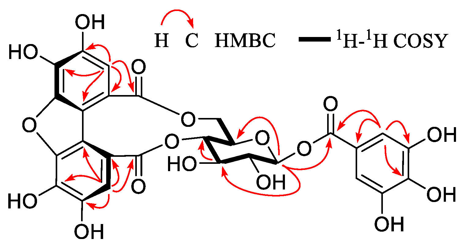
Black tea from C. taliensis (10.2 kg) was extracted using 60% aqueous acetone (40 L × 4, 1 week for each) at room temperature. After recycling acetone under reduced pressure, the yielded crude extract was dissolved with water and subsequently redistributed using CHCl3, EtOAc, and n-BuOH. The EtOAc fraction (350 g) was further fractionated by Sephadex LH-20 CC eluted with aqueous MeOH (0–100%) to give six fractions (Fr. 1–Fr. 6).
Fr. 2 (32.2 g) was treated with MCI-gel CHP20P CC (42 × 6 cm) eluted with aqueous MeOH (0−100%) to give compounds 19 (5.09 g) and 20 (0.39 g) and another 15 subfractions (Fr. 2-1–Fr. 2-15). Compound 3 (10.2 mg) was obtained from Fr. 2-1 (286.6 mg) by repeated CC over RP-18 and Toyopearl HW-40F eluted with aqueous MeOH (0–100%) and semi-HPLC (aqueous MeCN, 7%). Compound 30 (15.3 mg) and compounds 6 (11.9 mg), 21 (3.8 mg), 25 (12.2 mg), 28 (9.8 mg) were achieved from Fr. 2-3 (1.96 g) and Fr. 2-4 (1.58 g), respectively, via Sephadex LH-20 CC (aqueous MeOH, 0–100%) and further semi-HPLC (aqueous MeCN). Fr. 2-5 (1.28 g) was manipulated with Toyopearl HW-40F CC using aqueous MeOH (0–100%) as an eluent, combined with semi-HPLC (aqueous MeCN) to give 2 (1.9 mg), 4 (36.3 mg), 22 (2.0 mg), 23 (33.4 mg), 26 (10.6 mg), 27 (7.6 mg). Multiple CC over Toyopearl HW-40F, RP-18, and Sephadex LH-20 using aqueous MeOH (0−100%) as eluent, followed with semi-HPLC (aqueous MeCN) afforded 9 (15.1 mg), 11 (1.4 mg), 12 (43.0 mg), 13 (13.3 mg), 17 (10.6 mg), 18 (8.7 mg), 29 (21.5 mg) from Fr. 2-7 (9.43 g), and 14 (4.5 mg) and 15 (4.5 mg) from Fr. 2-8 (4.67 g), respectively. Fr. 2-9 (1.80 g) and Fr. 2-10 (0.99 g) were manipulated using Toyopearl HW-40F CC eluted with aqueous MeOH (0−100%), followed by semi-HPLC (aqueous MeCN with 1.5‰ HCOOH, 30% and 34%) to give 24 (13.6 mg) and 16 (9.4 mg), respectively.
Fr. 3 (121.2 g) was treated using Diaion HP20SS CC (60 × 7 cm), with aqueous MeOH (0−100%) as eluent, to afford 15 subfractions (Fr. 3-1–Fr. 3-15). Fr. 3-7 (16.93 g) was manipulated with repeated CC over Toyopearl HW-40F, RP-18, MCI-gel CHP20P, and Sephadex LH-20 eluted with a gradient system of aqueous MeOH (0–100%), followed with semi-HPLC (aqueous MeCN) to give 1 (8.4 mg), 5 (3.4 mg), 7 (3.5 mg), 8 (13.5 mg), and 31 (7.3 mg). Fr. 3-11 (3.29 g) was processed with multiple CC over Toyopearl HW-40F and Sephadex LH-20 eluted with aqueous MeOH (0–100%), followed with semi-HPLC (aqueous MeCN with 1.5‰ HCOOH, 27%) to give 10 (3.6 mg). Compound 32 (3.3 mg) was gained from Fr. 3-12 (6.70 g) through multiple CC over Toyopearl HW-40F and Sephadex LH-20 with aqueous MeOH (0–100%) as eluent, followed by semi-HPLC (aqueous MeCN, 35%).

2.6. Compound 1

Yellow-brown amorphous powder; α D 20 +34.4 (0.10, MeOH); ESI-MS: m/z 615 [M − H]; HRESI-MS: m/z 615.0625 [M − H] (calculated for C27H19O17, 615.0628); UV λmax (MeOH) (log ε): 276 (4.35), 222 (4.51) nm; 1H NMR (500 MHz, CD3OD): δH 7.20 (s, H-3″), 7.12 (s, 2H, H-2′, 6′), 7.05 (s, H-3‴), 5.73 (d, J = 8.2 Hz, H-1), 5.00 (m, H-4), 4.78 (m, H-6a), 4.01 (m, H-6b), 3.98 (m, H-5), 3.86 (t, J = 9.4 Hz, H-3), 3.65 (dd, J = 9.4, 8.2 Hz, H-2). 13C NMR (125 MHz, CD3OD): δC 171.5 (C-7‴), 170.0 (C-7″), 166.8 (C-7′), 147.7 (C-6‴), 147.6 (C-6″), 146.6 (C-3′, 5′), 145.5 (C-4‴), 145.2 (C-4″), 140.6 (C-4′), 136.1 (C-5″), 134.0 (C-5‴), 120.4 (C-1′), 119.8 (C-2‴), 117.0 (C-2″), 116.2 (C-3″), 115.9 (C-1″), 115.4 (C-1‴), 112.1 (C-3‴), 110.6 (C-2′, 6′), 95.7 (C-1), 76.2 (C-4), 74.9 (C-3), 74.2 (C-2), 71.7 (C-5), 67.6 (C-6).

2.7. Antioxidant Activity

Assays for DPPH and ABTS+ were carried out using the previous method with some modifications [19]. The DPPH assay was conducted as follows: the tested compound (final concentration: 0.2–1000 μM in EtOH, 100 μL) and DPPH solution (200 μM in EtOH, 100 μL) were sequentially added to 96-well plates at room temperature, then hid from the light for 15 min. The blank control and positive control (ascorbic acid) were set simultaneously. OD values at 517 nm were detected with a microplate reader. ABTS+ assay was carried out as follows: firstly, the ABTS+ solution was prepared using the procedure in our previous paper [19]. Then, the tested compound (final concentration: 0.2–1000 μM in EtOH, 10 μL) and ABTS+ solution (200 μL) were added to 96-well plates at room temperature in an orderly manner, and then hided from light for 6–8 min. The blank control and positive control (trolox) were set synchronously. OD values at 405 nm were then detected with a microplate reader. All the reactions were set up three replicates. The formula for calculating the scavenging percentage is as follows: inhibition percentages (%) = (ODblank − ODtest)/ODblank × 100%, and IC50 values were computed based on the Reed–Muench method [20].

2.8. α-Glucosidase Inhibitory Activity

As recorded in our prior paper [4], 4-nitrophenol-α-D-glucopyranoside (PNPG) was used as the zymolyte for the screening of enzyme inhibitors. Initially, 96-well plates with the following ingredients were added in a proper order: testing compound (50 μM), α-glucosidase solution (0.025 U/mL), buffer, and the zymolyte (1 mM), and kept at 37 °C for 50 min. Quercetin and acarbose were used as the positive control. All the reactions were set up with three replicates. OD values at 405 nm were recorded with a microplate reader. The formula for calculating the scavenging percentage is as follows: inhibition percentages (%) = (ODblank − ODtest)/ODblank × 100%, and IC50 values were computed based on the Reed–Muench method [20].

3. Results and Discussion

3.1. HPLC and LC-MS Analysis
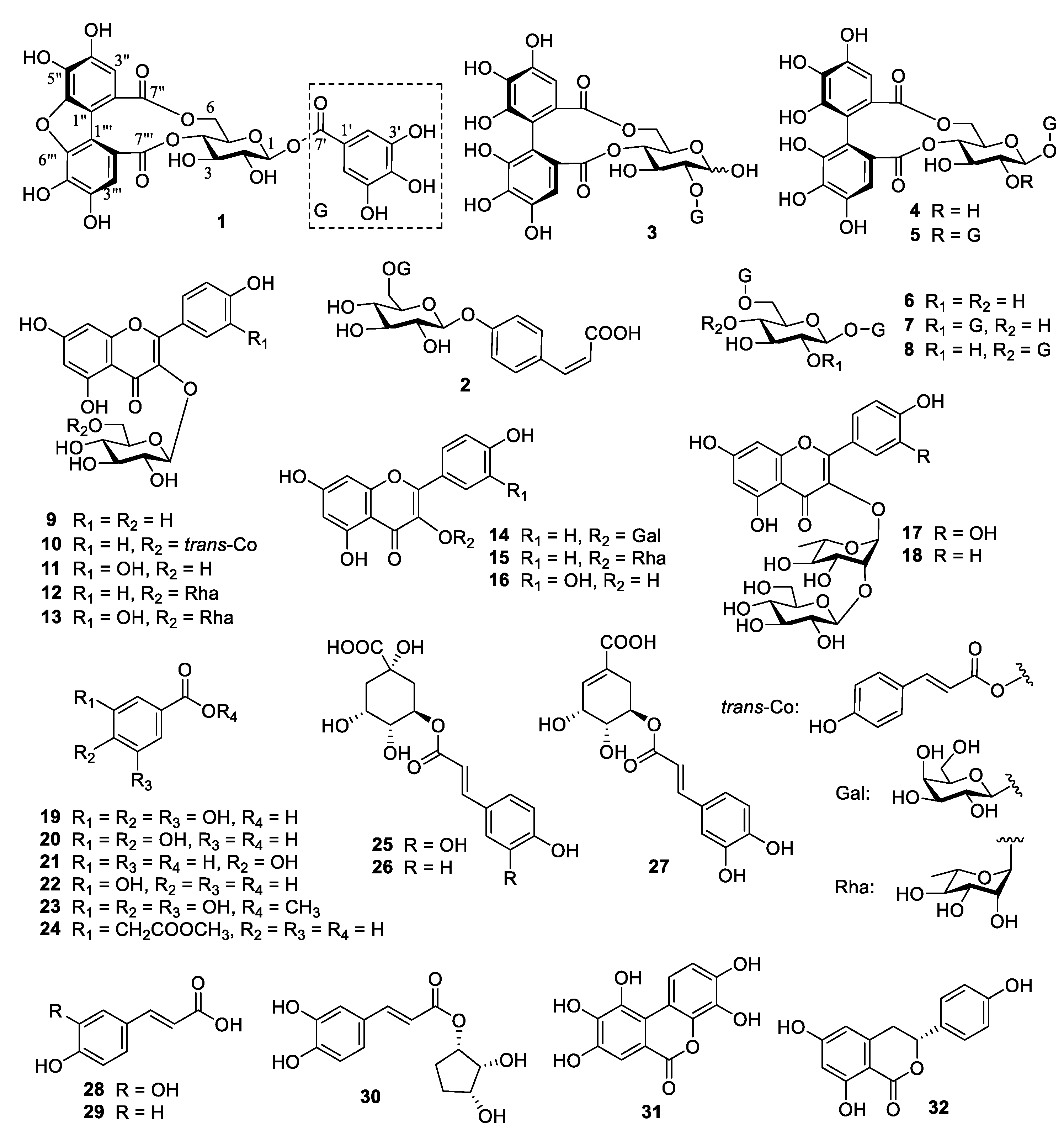
HPLC-ESI-MS analysis of the extract of black tea from C. taliensis using 70% aqueous MeOH revealed a total of 33 chemical constituents, including mainly polyphenols, e.g., 5 catechins, 3 theaflavins, 4 hydrolyzable tannins, 6 simple phenolics, 13 flavonols and their glycosides and coumarin, as well as theanine (Table 1), on the basis of their quasi-molecular ions, fragment ions, UV absorption, and retention times (tR), combined with standards obtained in our prior studies. Half of them were also isolated in further chemical study.
Catechins were found at peaks 8, 11, 12, 19, and 26 in Figure 1. Peak 8 was recognized as (+)-catechin through the [M − H] quasi-molecular ion at m/z 289, tR (15.55 min) and UV absorption (λmax 212, 274 nm), combined with those of the reference substance. Thus, (−)-epicatechin, (−)-epigallocatechin-3-O-gallate, (−)-epicatechin-3-O-gallate, and (−)-epiafzelechin 3-O-gallate were discovered as peaks 11 (tR 18.71 min; λmax 211, 274 nm), 12 (tR 19.22 min; λmax 209, 271 nm), 19 (tR 24.96 min; λmax 209, 273 nm) and 26 (tR 29.04 min; λmax 210, 278 nm) by the same token, respectively.
Peaks 32, 33, and 34 in Figure 1 were speculated to be theaflavins. Peak 33 was identified as theaflavin-3,3′-digallate according to fragment ions at m/z 715 [M − galloyl], 697 [M − H − gallic acid], and compared the tR (36.94 min) and UV absorptions (λmax 210, 276 nm) with those of the reference substance. In a similar way, peaks 32 (m/z 563 [M − galloyl]; tR 36.15 min; λmax 210, 272 nm) and 34 (m/z 563 [M − galloyl]; tR 37.07 min; λmax 210, 277 nm) were confirmed as theaflavin-3-gallate and theaflavin-3′-gallate, respectively.
Peaks 2, 6, 9, and 14 in Figure 1 were conjectured to be hydrolyzable tannins. Due to the fragment ions (m/z 331 [M − H], 169 [M − H − glucosyl]) and tR (5.43 min), peak 2 was identified as β-glucogallin. Similarly, peaks 6 (m/z 313 [M − H − gallic acid]; tR 14.17 min; λmax 230, 266, 297 nm), 9 (m/z 463 [M − H − gallic acid], 301 [463 − glucosyl]; tR 15.70 min; λmax 227, 270 nm) and 14 (m/z 483 [M − galloyl], 169 [gallic acid − H]; tR 20.36 min; λmax 223, 277 nm) were recognized as 1,6-di-O-galloyl-β-D-glucopyranose (6), strictinin (4) and 1,4,6-tri-O-galloyl-β-D-glucopyranose (8), respectively, and these were obtained during further chemical isolation.
Peaks 3, 4, 5, 7, 10, and 13 in Figure 1 were presumed to be simple phenolic compounds. Peaks 4 (m/z 125 [M − H]; tR 6.82 min) and 5 (m/z 343 [M − H]; tR 6.82 min) were identified as pyrogallol and theogallin, respectively. Similarly, gallic acid (19), methyl gallate (23), chlorogenic acid (25), and 5-O-(E)-p-coumaroylquinic acid (26) were determined at peaks 3 (m/z 169 [M − H], 125 [M − COO]; tR 6.82 min; λmax 227, 275 nm), 7 (m/z 169 [M − CH3], 125 [M − COOCH3]; tR 15.31 min; λmax 227, 270 nm), 10 (m/z 191 [quinic acid − H], 179[caffeic acid − H]; tR 16.12 min; λmax 218, 328 nm), and 13 (m/z 191 [quinic acid − H], 163 [coumaric acid − H]; tR 19.34 min; λmax 228, 307 nm), respectively. All of them were obtained during further chemical isolation.
Peaks 16, 18, 20, 21, 22, 23, 24, 25, 27, 28, 30, 31, and 35 in Figure 1 were inferred to be flavonols and their glycosides. Peak 16 was identified as rutin (13) by the fragment ions (m/z 308 [glucosyl + rhamnosyl], 301 [quercetin]), tR (23.79 min) and UV absorption (λmax 216, 257, 354 nm). Likewise, peaks 18 (m/z 301 [quercetin]; tR 24.85 min; λmax 210, 257, 354 nm), 21 (m/z 447 [M − rhamnosyl], 285 [447 − glucosyl]; tR 25.63 min, λmax 260, 302, 362 nm), 22 (m/z 302 [quercetin]; tR 26.31 min), 23 (m/z 593 [M − H]; tR 26.62 min; λmax 210, 257, 350 nm), 24 (m/z 285 [M − H − glucosyl]; tR 27.16 min; λmax 226, 264, 348 nm), 25 (m/z 285 [M − H − galactosyl]; tR 28.21 min; λmax 210, 266, 347 nm), 27 (m/z 285 [M − H − rhamnosyl]; tR 31.45 min; λmax 210, 264, 343 nm), 31 (m/z 447 [M − 2H − coumaroyl], 285 [447 − glucosyl]; tR 35.95 min; λmax 210, 266, 315 nm) and 35 (m/z 301 [M − H]; tR 37.24 min; λmax 210, 256, 371 nm) were recognized as quercetin 3-O-β-D-glucopyranoside (11), kaempferol 3-O-rutinoside (12), quercetin 3-(2″-β-D-glucosy-l)-α-L-rhamnopyranoside (17), kaempferol 3-(2″-β-D-glucosyl)-α-L-rhamnopyranoside (18), kaempherol 3-O-β-D-glucopyranoside (9), kaempherol 3-O-β-D-galacopyranoside (14), kaempherol 3-O-α-L-rhamnopyranoside (15), kaempferol 3-O-(6″-trans-p-coumaroy-l)-β-D-glucopyranoside (10), and quercetin (16), respectively, and they were also isolated from further chemical study. In addition, peaks 20 (m/z 463 [M − H]; tR 25.25 min), 28 (m/z 755 [M − H − rhamnosyl], 447 [M − H − glucosyl − coumaroyl]; tR 33.19 min), 30 (m/z 739 [M − H − rhamnosyl], 577 [739 − glucosyl], 431 [577 − H − coumaroyl], 285 [431 − rhamnosyl]; tR 34.28 min), were speculated to be quercetin 3-O-β-D-galactopyranoside, quercetin-3-O-coumaroyl-rhamnosyl-glucosyl-rhamnopyranoside, and kaempferol-3-O-coumaroyl-rhamnosyl-glucosyl-rhamnopyranoside, respectively.
In addition, two other types of compounds were also identified. Compared the quasi-molecular ion of m/z 173 [M − H] and fragment ions (m/z 127, 87, 45) with those of the standard sample, peak 1 was inferred to be theanine. Similarly, peak 15 (m/z 177 [M − H], 132, 109, 89, 65; tR 21.24 min; λmax 210, 255, 328 nm) was identified as 5,7-dihydroxycoumarin.

3.2. Identification of Compounds 132

The extract of black tea from C. taliensis using 60% aqueous acetone was dissolved with water and successively redistributed with CHCl3, EtOAc, and n-BuOH. The EtOAc extract was further isolated by various CC on Diaion HP20SS, RP-18, Toyopearl HW-40F, MCI-gel CHP20P, and Sephadex LH-20, to yield 32 compounds, including one undescribed hydrolyzable tannin (1) and one new natural product (24). The compounds, which had were in advance, were recognized as seven hydrolyzable tannins (28) including 4-O-(6′-O-galloyl-β-D-glucopyranosyl)-cis-p-coumaric acid (2) [21], 2-O-galloyl-4,6-O-(S)-hexahydroxydiphenoyl-β-D-glucopyranose (3) [22], 1-O-galloyl-4,6-O-(S)-hexahydroxydiphenoyl-β-D-glucopyranose (4) [23], 1,2-di-O-galloyl-4,6-O-(S)-hexahydroxydiphenoyl-β-D-glucopyranose (5) [24], 1,6-di-O-galloyl-β-D-glucopyranose (6) [25], 1,2,6-tri-O-galloy-β-D-glucopyranose (7) [26], and 1,4,6-tri-O-galloyl-β-D-glucopyranose (8) [27], 10 flavonol and its glycosides (918) including kaempferol 3-O-β-D-glucopyranoside (9) [28], kaempferol 3-O-(6″-trans-p-coumaroyl)-β-D-glucopyranoside (10) [29], quercetin 3-O-β-D-glucopyranoside (11) [30], kaempferol 3-O-rutinoside (12) [23], rutin (13) [31], kaempferol 3-O-β-D-galactopyranoside (14) [32], kaempferol 3-O-α-L-rhamnopyranoside (15) [33], quercetin (16) [34], quercetin 3-(2″-β-D-glucosyl)-α-L-rhamnopyranoside (17) [35], and kaempferol 3-(2″-β-D-glucosyl)-α-L-rhamnopyranoside (18) [35], and 14 simple phenolics (1932) including gallic acid (19) [36], 3,4-dihydroxybenzoic acid (20) [37], p-hydroxybenzoic acid (21) [38], m-hydroxybenzoic acid (22) [39], methyl gallate (23) [40], 3-(2-methoxy-2-oxoethyl) benzoic acid (24) [41], chlorogenic acid (25) [42], 5-O-(E)-p-coumaroylquinic acid (26) [43], 5-O-caffeoylshikimic acid (27) [44], caffeic acid (28) [45], E-p-hydroxycinnamic acid (29) [46], 1-(3′,4′-dihydroxycinnamoyl) cyclopentane-2,3-diol (30) [47], 3,4,8,9,10-pentahydroxydibenzo[b,d]pyran-6-one (31) [48], and (3R)-thunberginol C (32) [49], respectively, based on previously reported MS, and NMR spectroscopic data (Figure 2). Before this study, the 20 compounds (2, 3, 6, 7, 8, 15, 17, 18, 2022, 2432) were never been separated from black tea.
Compound 1 was obtained as a yellow-brown amorphous powder with α D 20 +34.4 (c 0.10, MeOH) (Figure S9). Its molecular formula, C27H20O17, was deduced from HRESI-MS at m/z 615.0625 [M − H] (calculated for 615.0628) (Figure S7), demonstrating 18 degrees of unsaturation. Detailed analysis of the NMR data revealed that 1 had a great semblable structure to that of strictinin (4) (Figures S1 and S2). The existence of one glucosyl (δC 95.7, 76.7, 74.9, 74.2, 71.7, 67.6; δH 5.73, 1H, d, J = 8.2 Hz) with C1 form proved by the study on the conformation of ellagitannins (ETs) [50] and the large coupling constant (J = 8.2 Hz) of anomeric proton (δH 5.73) [51], one galloyl (δC 146.6 (2C), 140.6, 120.4, 110.6 (2C), 166.8; δH 7.12, 2H, s), and one HHDP-related acyl (δC 147.7, 147.6, 145.5, 145.2, 136.1, 134.0, 119.8, 117.0, 116.2, 115.9, 115.4, 112.1, 171.5, 170.0; δH 7.20, 7.05. each 1H, s) groups were deduced apparently from the 1H and 13C NMR spectra (Table 2). The HMBC correlation from the anomeric proton (δH 5.73) to galloyl carboxyl carbon (δC 166.8) clarified that the galloyl group was connected at glucosyl C-1. The obvious lower field shift of glucosyl C-6 (δC 67.6), H-4 (δH 5.00), and H-6 (δH a, 4.78; b, 4.01) compared with those of 1-O-galloyl-β-D-glucopyranose [52], suggested the HHDP-related acyl group should be attached to the glucosyl C-4 and C-6 positions, analogous to those of 4. However, 1H and 13C NMR data assignable the HHDP-related acyl group in 1 were very different to those of the HHDP group in 4, and the molecular formula of 1 was differed from 4 by one H2O, indicating that 1 should be a dehydrated derivative of 4. The HHDP-related acyl group at glucosyl C-6 and C-4 in 1 was determined to be 1,1′-(3,3′,4,4′-tetrahydroxy) dibenzofurandicarboxyl group, compared with the NMR data with those in mallotusinin, the first compound with a 1,1′-(3,3′,4,4′-tetrahydroxy) dibenzofurandicarboxyl group isolated from Mallotus japonicus [53]. Other 2D NMR correlations (Figure 3 and Figure S3–S6) confirmed the structure of 1. Hence, compound 1 was confirmed to be 1-O-galloyl-4,6-tetrahydroxydibenzofurandicarboxyl-β-D-glucopyranose, as shown in Figure 2.
Hydrolyzable tannins are segmented into two major classes based on their structures: gallotannins (GTs) and ETs. GTs are esters with only a galloyl group bounded to glucose, while ETs are esters with hexahydroxydiphenoyl (HHDP) or its similar acyl groups commonly bounded to glucose [50]. Isolates 68 were classified as GTs, and 1, 3, 4, and 5 were classified as ETs. ETs are often present as an equilibrium mixture of two isomers with a different configuration at anomeric carbon [50]. For example, compound 3 was isolated as a mixture, due to the chirality of anomeric carbon. It was reported that the original product of the oxidative coupling of two galloyl groups in ETs was the dehydrohexahydroxydiphenoyl (DHHDP) group, which was then reduced to the HHDP group [54], as shown in Figure S10. The DHHDP group in dehydroellagitannins usually comes simultaneously in six-membered and five-membered hemiacetal rings. However, due to the influence of Gibbs free energy, the stability of the six-membered ring structure of DHHDP is better than that of the five-membered ring structure. Meanwhile, the pressure load carried by ester carbonyl carbons and the flexibility of the macrocylic lactone ring in ETs make a difference to the stability of DHHDP [54]. Consequently, the structure, molecule size, and location of acyl group are all related to the structural diversity of ETs, which will affect the stability of DHHDP. A study found that the DHHDP group, created with pyridine in acetonitrile, was disproportionated by redox reaction to yield the 1,1′-(3,3′,4,4′-tetrahydroxy) dibenzofurandicarboxyl group as a reduction product [55]. Thus, compound 1 could be a reduction product, reduced through the two galloyl groups of 4 which were converted into DHHDP by the oxidative coupling reaction, followed by redox disproportionation.
Urolithins, as natural metabolites of ETs with better gastrointestinal absorption, were reported to have inhibitory effects on the proliferation of prostate and colon cancer cells as well as anti-inflammation activity [56]. It is reported that ETs were hydrolyzed by intestinal bacteria to produce ellagic acid, which were subsequently converted into urolithins. The presence of compound 31 (resembled a urolithin) demonstrated a possible occurrence of the ester hydrolysis of ET acyl groups and further decarboxylation during the fermentation of black tea made from the leaves of C. taliensis.

3.3. Antioxidant Activity

The antioxidant activities through DPPH and ABTS+ assays for all of the hydrolyzable tannins (18), flavonol and its glycosides (918), and simple phenolic compounds (1923, 2532) isolated from C. taliensis black tea, were assessed. The results, shown in Table 3, indicated that the amount of phenolic hydroxyl group was clearly correlated with their antioxidant activities, and the sequence of activity was hydrolyzable tannins > simple phenolics > flavonol and its glycosides.
All eight hydrolyzable tannins (18) showed a stronger free radical scavenging activities than ascorbic acid and trolox. The sequence of activity for inhibiting the DPPH radical was 8 > 6 > 5 > 4 > 1 > 2 > 7 > 3 > ascorbic acid, and the sequence of activity for inhibiting the ABTS+ radical was 5 > 8 > 4 > 6 > 3 > 1 > 7 > 2 > trolox. In hydrolyzable tannins, galloyl group at C-2 of glucose could weaken the radical scavenging effects from the weaker results of 3 and 7 than those of 4 and 8.
Part of the flavonol and its glycosides (11, 13, 16, 17) showed equivalent effects to the positive control (ascorbic acid and trolox). The sequence of activity for inhibiting the DPPH radical was 13 > 16 > 11 > ascorbic acid > 17, and the sequence of activity for inhibiting the ABTS+ radical was 16 > 13 > 11 > trolox > 17. The p-coumaryl group has a little positive influence on the antioxidant effects of flavonol and its glycosides, which could be found from 10 (6.28%) and 9 (0.97%), respectively.
Part of the simple phenolic compounds (19, 23, 25, 28, 30, 31) showed stronger activity than the positive control (ascorbic acid and trolox). The sequence of activity for the inhibiting DPPH radical was 19 > 23 > 31 > 25 > 28 > 30 > ascorbic acid, and the sequence of activity for inhibiting the ABTS+ radical was 31 > 23 > 28 > 19 > 25 > 30 > trolox. The p-coumaric acid derivatives showed little radical scavenging effects, weaker than caffeic acid derivatives from the order of activity (25 > 30 > 27 > 26 > 29), which was consistent with reports in the literature [57].

3.4. α-Glucosidase Inhibitory Activity

The inhibitory activities of hydrolyzable tannins 18 and gallic acid (19) on α-glucosidase were investigated. At a concentration of 50 μM, 1, 35, 7 and 8 with three or more galloyl groups showed a higher inhibition ratio (>50%) on α-glucosidase. Their IC50 values were further evaluated, and as shown in Table 4, all showed a stronger inhibitory activity than quercetin and acarbose (IC50 = 5.75 and 223.30 μM, respectively), with IC50 values ranging from 0.67 to 2.01 μM. Their activity order was 5 > 3 > 1 > 4 > 7 > 8 > quercetin > acarbose. Compounds 2 and 6 with one or two galloyl groups showed equivalent inhibitory effects to acarbose on α-glucosidase at a concentration of 50 μM, while gallic acid (19) showed almost no inhibitory effect. The results revealed that the number of phenolic hydroxyl group plays a positive role on the α-glucosidase inhibitory activities of hydrolyzable tannins, probably due to their hydrophobic association with α-glucosidase [58].

4. Conclusions

C. taliensis, with similar chemical constituents to the extensively cultivated tea plants, C. sinensis and its variety assamica, is the earliest, most used and widely distributed wild tea tree, whose leaves were utilized by the locals of its growing regions to manufacture various types of tea. A comprehensive chemical study of black tea from C. taliensis resulted in the separation and recognition of one new hydrolyzable tannin, 1-O-galloyl-4,6-tetrahydroxydibenzofurandicarboxyl-β-D-glucopyranose (1), together with 31 known compounds, comprising seven hydrolyzable tannins (28), 10 flavonols and their glycosides (918), and 14 simple phenolics (1932) with 24 as a new natural product. It is noted that 15 compounds (2, 3, 15, 17, 18, 21, 22, 24, 2632) were obtained from C. taliensis and 20 compounds (2, 3, 6, 7, 8, 15, 17, 18, 2022, 2432) were isolated from black tea for the first time. The isolation of hydrolyzable tannins, especially new ones from fermented tea, is challenging, because hydrolyzable tannins are easily hydrolyzed during fermentation process. Nonetheless, one new hydrolyzable tannin was isolated and identified in the study. As far as we know, the hydrolyzable tannin with 1,1′-(3,3′,4,4′-tetrahydroxy) dibenzofurandicarboxyl group was discovered from tea for the first time. Most of the isolates showed significant antioxidant and α-glucosidase inhibitory activities, which were positively correlated with the amount of phenolic hydroxyl group. However, more antioxidant and hypoglycemic activity experiments could be carried out. In short, C. taliensis is also an ideal material to produce black tea with mild bitter and astringent flavor and rich polyphenols with significant antioxidant and α-glucosidase inhibitory activities, which indicated that the black tea from C. taliensis has the potential to be developed as health products.

Supplementary Materials

The following supporting information can be downloaded at: https://www.mdpi.com/article/10.3390/foods12132512/s1, Figure S1: 1H NMR spectrum of compound 1 in CD3OD; Figure S2: 13C NMR spectrum of compound 1 in CD3OD; Figure S3: HSQC spectrum of compound 1 in CD3OD; Figure S4: HMBC spectrum of compound 1 in CD3OD; Figure S5: COSY spectrum of compound 1 in CD3OD; Figure S6: ROESY spectrum of compound 1 in CD3OD; Figure S7: HRESI-MS spectrum of compound 1; Figure S8: CD and UV spectra of compound 1 in MeOH; Figure S9: OR of compound 1 in MeOH; Figure S10: Previous studies on the oxidative coupling of galloyl groups; Figure S11: 1H NMR spectrum of compound 2 in CD3OD; Figure S12: 13C NMR spectrum of compound 2 in CD3OD; Figure S13: 1H NMR spectrum of compound 3 in CD3OD; Figure S14: 13C NMR spectrum of compound 3 in CD3OD; Figure S15: 1H NMR spectrum of compound 4 in CD3OD; Figure S16: 13C NMR spectrum of compound 4 in CD3OD; Figure S17: 1H NMR spectrum of compound 5 in CD3OD; Figure S18: 13C NMR spectrum of compound 5 in CD3OD; Figure S19: 1H NMR spectrum of compound 6 in CD3OD; Figure S20: 13C NMR spectrum of compound 6 in CD3OD; Figure S21: 1H NMR spectrum of compound 7 in CD3OD; Figure S22: 13C NMR spectrum of compound 7 in CD3OD; Figure S23: 1H NMR spectrum of compound 8 in CD3OD; Figure S24: 13C NMR spectrum of compound 8 in CD3OD.

Author Contributions

M.C.: writing—original draft, investigation, methodology. N.L.: writing—original draft, investigation, methodology, funding acquisition. H.-T.Z. and M.Z.: project administration. Z.-H.D.: resources. D.W.: formal analysis. C.-R.Y.: conceptualization. Y.-J.Z.: supervision, conceptualization, writing—review and editing, funding acquisition. All authors have read and agreed to the published version of the manuscript.

Funding

This work was supported by the Postdoctoral Directional Training Foundation of Yunnan Province, Industrial Innovation Talents of Rejuvenating Yunnan talents support plan of Yunnan province, Industrial-Research Talent Project of CAS K.C.Wong Leading Talent Plan, and the National Natural Science Foundation of China (No. 21672223).

Data Availability Statement

Data are contained within the article or supplementary material.

Acknowledgments

We are grateful to the staffs of the analytical and bioactivity screening group at the State Key Laboratory of Phytochemistry and Plant Resources in West China, KIB, CAS, for measuring the spectroscopic data and α-glucosidase inhibitory activity.

Conflicts of Interest

The authors declare no conflict of interest.

References

  1. Li, J.-Q.; He, M.-Z.; Wang, R.-Y.; Zhou, D.-P.; Liu, X.-H.; Li, J.-H.; Yang, G.-R. An overview of Camellia taliensis. J. Anhui Agric. Sci. 2022, 50, 12–17. (In Chinese) [Google Scholar]
  2. Zhu, L.-F.; Xu, M.; Zhu, H.-T.; Wang, D.; Yang, S.-X.; Yang, C.-R.; Zhang, Y.-J. New flavan-3-ol dimer from green tea produced from Camellia taliensis in the Ai-Lao mountains of southwest China. J. Agric. Food Chem. 2012, 60, 12170–12176. [Google Scholar] [CrossRef] [PubMed]
  3. Li, N.; Chen, M.; Zhu, H.-T.; Zhang, M.; Wang, D.; Yang, C.-R.; Zhang, Y.-J. Theaflavoids A-C, new flavan-3-ols with potent α-glucosidase inhibitory activity from Yunnan black tea ‘Jin-Ya’. LWT-Food Sci. Technol. 2022, 168, 113918. [Google Scholar] [CrossRef]
  4. Li, N.; Zhu, H.-T.; Wang, D.; Zhang, M.; Yang, C.-R.; Zhang, Y.-J. New flavoalkaloids with potent α-glucosidase and acetylcholinesterase inhibitory activities from Yunnan black tea ‘Jin-Ya’. J. Agric. Food Chem. 2020, 68, 7955–7963. [Google Scholar] [CrossRef] [PubMed]
  5. Indrawati, R.; Zubaidah, E.; Sutrisno, A.; Limantara, L.; Yusuf, M.M.; Brotosudarmo, T.H.P. Visible light-induced antibacterial activity of pigments extracted from dregs of green and black teas. Scientifica 2021, 2021, 5524468. [Google Scholar] [CrossRef]
  6. Tanaka, T.; Matsuo, Y. Production mechanisms of black tea polyphenols. Chem. Pharm. Bull. 2020, 68, 1131–1142. [Google Scholar] [CrossRef]
  7. Collier, P.D.; Bryce, T.; Mallows, R.; Thomas, P.E.; Frost, D.J.; Korver, O.; Wilkins, C.K. The theaflavins of black tea. Tetrahedron 1973, 29, 125–142. [Google Scholar] [CrossRef]
  8. Bryce, T.; Collier, P.D.; Mallows, R.; Thomas, P.E.; Frost, D.J.; Wilkins, C.K. Three new theaflavins from black tea. Tetrahedron Lett. 1972, 13, 463–466. [Google Scholar] [CrossRef]
  9. Shii, T.; Tanaka, T.; Watarumi, S.; Matsuo, Y.; Miyata, Y.; Tamaya, K.; Tamaru, S.; Tanaka, K.; Matsui, T.; Kouno, I. Polyphenol composition of a functional fermented tea obtained by tea-rolling processing of green tea and loquat leaves. J. Agric. Food Chem. 2011, 59, 7253–7260. [Google Scholar] [CrossRef] [PubMed]
  10. Zhou, H.; Li, H.-M.; Du, Y.-M.; Yan, R.-A.; Ou, S.-Y.; Chen, T.-F.; Wang, Y.; Zhou, L.-X.; Fu, L. C-Geranylated flavanones from YingDe black tea and their antioxidant and α-glucosidase inhibition activities. Food Chem. 2017, 235, 227–233. [Google Scholar] [CrossRef] [PubMed]
  11. Tanaka, T.; Watarumi, S.; Fujieda, M.; Kouno, I. New black tea polyphenol having -ethyl-2 pyrrolidinone moiety derived from tea amino acid theanine: Isolation, characterization and partial synthesis. Food Chem. 2005, 93, 81–87. [Google Scholar] [CrossRef]
  12. Wang, W.; Tang, X.; Hua, F.; Ling, T.-J.; Wan, X.-C.; Bao, G.-H. Camellimidazole A–C, three methylene-bridged dimeric imidazole alkaloids from Keemun black Tea. Org. Lett. 2018, 20, 2672–2675. [Google Scholar] [CrossRef]
  13. Yang, C.-R.; Zhang, Y.-J.; Gao, D.-F.; Chen, K.-K.; Jiang, H.-J. The evaluation of germplasm resource of Camellia taliensis and origin of cultivated Camellia sinensis var. assamica. Tea Sci. Technol. 2008, 1–4. (In Chinese) [Google Scholar]
  14. Ming, T.-L. A revision of Camellia Sect. Thea. Acta Bot. Yunnanica 1992, 14, 115–132. (In Chinese) [Google Scholar]
  15. Jiang, H.-B.; Wang, Y.-G.; Tang, Y.-C.; Song, W.-X.; Li, Y.-Y.; Ji, P.-Z.; Huang, X.-Q. Investigation of wild tea plant (Camellia taliensis) germplasm resource from Yunnan, China. Southwest China J. Agric. Sci. 2009, 22, 1153–1157. (In Chinese) [Google Scholar]
  16. Zhu, L.-F.; Dong, H.-Z.; Yang, S.-X.; Zhu, H.-T.; Xu, M.; Zeng, S.-F.; Yang, C.-R.; Zhang, Y.-J. Chemical compositions and antioxidant activity of essential oil from green tea produced from Camellia taliens·is (Theaceae) in Yuanjiang, southwestern China. Plant Divers. Resour 2012, 34, 409–416. (In Chinese) [Google Scholar] [CrossRef]
  17. Zhao, D.-W.; Yang, J.-B.; Yang, S.-X.; Kato, K.; Luo, J.-P. Genetic diversity and domestication origin of tea plant Camellia taliensis (Theaceae) as revealed by microsatellite markers. BMC Plant Biol. 2014, 14, 14. [Google Scholar] [CrossRef] [Green Version]
  18. Gao, D.-F.; Zhang, Y.-J.; Yang, C.-R.; Chen, K.-K.; Jiang, H.-J. Phenolic antioxidants from green tea produced from Camellia taliensis. J. Agric. Food Chem. 2008, 56, 7517–7521. [Google Scholar] [CrossRef] [PubMed]
  19. Meng, X.-H.; Zhu, H.-T.; Yan, H.; Wang, D.; Yang, C.-R.; Zhang, Y.-J. C-8 N-ethyl-2-pyrrolidinone-substituted flavan-3-ols from the leaves of Camellia sinensis var. pubilimba. J. Agric. Food Chem. 2018, 66, 7150–7155. [Google Scholar] [CrossRef] [PubMed]
  20. Haggett, P.; Gunawardena, K.A. Determination of population thresholds for settlement functions by the reed-muench method. Prof. Geogr. 1964, 16, 6–9. [Google Scholar] [CrossRef]
  21. Peng, Z.-C.; Hao, J.; Pan, X.-G.; Ye, X.-S.; Li, X.-X.; Yin, W.-F.; Zhang, W.-K.; Xu, J.-K. Isolation and identification of chemical constituents from fruit of Cornus officinalis. Chin Trad Herb Drugs 2021, 52, 4480–4486. (In Chinese) [Google Scholar]
  22. Santos, S.C.; Pereira, M.O.A.; Santos, K.B.; Ferri, P.H. Bioactive compounds of fruit parts of three Eugenia uniflora biotypes in four ripening stages. Chem. Biodivers. 2021, 18, e2100704. [Google Scholar] [CrossRef] [PubMed]
  23. Feng, W.-S.; Hao, Z.-Y.; Zheng, X.-K.; Kuang, H.-X. Chemical constituents from leaves of Celastrus gemmatus Loes. Yao Xue Xue Bao Acta Pharm. Sin. 2007, 42, 625–630. (In Chinese) [Google Scholar]
  24. Yagi, K.; Goto, K.; Nanjo, F. Identification of a major polyphenol and polyphenolic composition in leaves of Camellia irrawadiensis. Chem. Pharm. Bull 2009, 57, 1284–1288. [Google Scholar] [CrossRef] [Green Version]
  25. Li, N.; Li, X.; Fang, Z.-G.; Masayuki, Y. Chemical constituents from Canavalia gladiata. J. Shenyang Pharm. Univ. 2007, 24, 676–678. (In Chinese) [Google Scholar]
  26. Liang, Y.; Xu, M.; Tang, C.; Fu, K.; Li, X.; Song, Y.; Zhang, J.; Wang, Z. The study of chemical components in Qishiwei Zhenzhu pills and its anti-apoptotic mechanism in cerebral ischemic based on LC-MS and network pharmacology. J. Ethnopharmacol. 2023, 302, 115891. [Google Scholar] [CrossRef]
  27. Li, C.-W.; Dong, H.-J.; Cui, C.-B. The synthesis and antitumor activity of twelve galloyl glucosides. Molecules 2015, 20, 2034–2060. [Google Scholar] [CrossRef] [Green Version]
  28. Guo, H.-L.; Zhou, J.-Y. Isolation and struture elucidation of glycosides in n-butanol extracts from rhizome of Periploca calophylla. China J. Chin. Mater Med. 2005, 30, 44–46. (In Chinese) [Google Scholar]
  29. Zhang, Y.; Deng, S.; Li, X.-X.; Chen, Y.; Han, L.-F.; Wang, T. Isolation and identification of constituents from Leonurus japonicus II. Chin J. Med. Chem. 2013, 23, 480–485. (In Chinese) [Google Scholar]
  30. Kazuma, K.; Noda, N.; Suzuki, M. Malonylated flavonol glycosides from the petals of Clitoria ternatea. Phytochemistry 2003, 62, 229–237. [Google Scholar] [CrossRef]
  31. Zang, E.-H.; Chen, Z.-W.; Zhang, C.-H.; Li, M.-H. Chemical constituents of Physochlaina physaloides (L.) G. Don (Solanaceae). Biochem. Syst. Ecol. 2021, 98, 104332. [Google Scholar] [CrossRef]
  32. Castillo-Muñoz, N.; Gómez-Alonso, S.; García-Romero, E.; Hermosín-Gutiérrez, I. Flavonol profiles of Vitis vinifera red grapes and their single-cultivar wines. J. Agric. Food Chem. 2007, 55, 992–1002. [Google Scholar] [CrossRef] [PubMed]
  33. Wang, G.J.; Tsai, T.H.; Lin, L.C. Prenylflavonol, acylated flavonol glycosides and related compounds from Epimedium sagittatum. Phytochemistry 2007, 68, 2455–2464. [Google Scholar] [CrossRef]
  34. Tao, L.; Huang, J.; Zhao, Y.; Li, C. Chemical constituents in Buddleja albiflora. China J. Chin. Mater. Medica 2009, 34, 3043–3046. (In Chinese) [Google Scholar]
  35. Hasler, A.; Gross, G.A.; Meier, B.; Sticher, O. Complex flavonol glycosides from the leaves of Ginkgo biloba. Phytochemistry 1992, 31, 1391–1394. [Google Scholar] [CrossRef] [PubMed]
  36. Watanabe, M.; Devkota, H.P. Antioxidant phenolic constituents from the leaves of Acer ginnala var aidzuense. J. Nat. Remedies 2017, 17, 9–12. [Google Scholar] [CrossRef] [Green Version]
  37. Li, K.; Li, X.-M.; Wang, B.-G. Chemical constituents from herb of Gueldenstaedtia stenophylla. China J. Chin. Mater Med. 2008, 33, 1711–1713. (In Chinese) [Google Scholar]
  38. Zhao, A.-H.; Zhao, Q.-S.; Li, R.-T.; Sun, H.-D. Chemical constituents from Clerodendranthus spicatus. Acta Bot. Yunnanica 2004, 26, 563–568. (In Chinese) [Google Scholar]
  39. Miao, C.-P.; Hu, J.; Zhai, Y.-Z.; Song, F.; Xuan, Q.-C.; Chen, Y.-W.; Wu, S.-H. Identification and secondary metabolites of endophytic fungus PR20 from Paeonia delavayi. Nat. Prod. Res. Dev. 2012, 24, 1339–1442. (In Chinese) [Google Scholar]
  40. Ahmad, I.; Bashir, A.; Janbaz, K.; Uzair, M.; Ashraf, M. Urease inhibitors and antioxidants from Vernonia cinerascens. J. Chem. Soc. Pak. 2011, 33, 14–117. [Google Scholar]
  41. Balo, T.; Sapi, A.; Kiss, A.; Raimbaud, E.; Paysant, J.; Cattin, M.-E.; Berger, S.; Kotschy, A.; Faucher, N. Synthesis of thieno[2,3-c]pyridine derived GRK2 inhibitors. Monatsh Chem. 2022, 1–19. [Google Scholar] [CrossRef]
  42. Lee, E.J.; Kim, J.S.; Kim, H.P.; Lee, J.H.; Kang, S.S. Phenolic constituents from the flower buds of Lonicera japonica and their 5-lipoxygenase inhibitory activities. Food Chem. 2010, 120, 134–139. [Google Scholar] [CrossRef]
  43. Kuczkowiak, U.; Petereit, F.; Nahrstedt, A. Hydroxycinnamic acid derivatives obtained from a commercial crataegus extract and from authentic Crataegus spp. Sci. Pharm. 2014, 82, 835–846. [Google Scholar] [CrossRef] [PubMed] [Green Version]
  44. Liu, C.; Zou, K.; Guo, Z.; Zhao, Y.; Cheng, F.; Zhang, H. Chemical constituents from leaves of Boehmeria nivea. China J. Chin. Mater. Medica 2010, 35, 1432–1434. (In Chinese) [Google Scholar]
  45. Wu, Z.-J.; Ouyang, M.-A.; Yang, C.-R. Polyphenolic constituents of Salvia sonchifolia. Acta Bot. Yunnanica 1999, 21, 393–398. (In Chinese) [Google Scholar]
  46. Bergman, M.; Varshavsky, L.; Gottlieb, H.E.; Grossman, S. The antioxidant activity of aqueous spinach extract: Chemical identification of active fractions. Phytochemistry 2001, 58, 143–152. [Google Scholar] [CrossRef]
  47. Cardoso, C.L.; Silva Bolzani, V.D.; Siqueira Silva, D.H.; Ishii, H.; Berova, N.; Nakanishi, K. The absolute configuration of 1-(3′,4′-dihydroxycinnamoyl)cyclopentane-2,3-diol from the amazonian tree Chimarrhis turbinata. J. Nat. Prod. 2006, 69, 1046–1050. [Google Scholar] [CrossRef]
  48. Pfundstein, B.; El Desouky, S.K.; Hull, W.E.; Haubner, R.; Erben, G.; Owen, R.W. Polyphenolic compounds in the fruits of Egyptian medicinal plants (Terminalia bellerica, Terminalia chebula and Terminalia horrida): Characterization, quantitation and determination of antioxidant capacities. Phytochemistry 2010, 71, 1132–1148. [Google Scholar] [CrossRef]
  49. Park, S.; Na Kim, Y.; Kwak, H.J.; Jeong, E.J.; Kim, S.H. Estrogenic activity of constituents from the rhizomes of Rheum undulatum Linné. Bioorganic Med. Chem. Lett. 2018, 28, 552–557. [Google Scholar] [CrossRef]
  50. Matsuo, Y.; Iki, M.; Okubo, C.; Saito, Y.; Tanaka, T. Conformationally flexible ellagitannins: Conformational analysis of davidiin and punicafolin via DFT calculation of 1H NMR coupling constants. ChemRxiv 2022. [Google Scholar] [CrossRef]
  51. Jiang, Z.-H.; Tanaka, T.; Iwata, H.; Sakamoto, S.; Hirose, Y.; Kouno, I. Ellagitannins and lignan glycosides from Balanophora japonica (Balanophoraceae). Chem. Pharm. Bull. 2005, 53, 339–341. [Google Scholar] [CrossRef] [Green Version]
  52. Li, C.-H.; Yang, X.-D.; Zhao, J.-F.; Li, L. The chemical constituents of Breynia rostrata. Yao Xue Xue Bao Acta Pharm. Sin. 2006, 41, 125–127. (In Chinese) [Google Scholar]
  53. Saijo, R.; Nonaka, G.I.; Nishioka, I. Tannins and related compounds. LXXXIV. Isolation and characterization of five new hydrolyzable tannins from the bark of Mallotus japonicus. Chem. Pharm. Bull. 1989, 37, 2063–2070. [Google Scholar] [CrossRef] [PubMed] [Green Version]
  54. Yamashita, T.; Matsuo, Y.; Saito, Y.; Tanaka, T. Formation of dehydrohexahydroxydiphenoyl esters by oxidative coupling of galloyl esters in an aqueous medium involved in ellagitannin biosynthesis. Chem.Asian J. 2021, 16, 1735–1740. [Google Scholar] [CrossRef] [PubMed]
  55. Tanaka, T.; Nonaka, G.I.; Nishioka, I. Tannins and related compounds. C. Reaction of dehydrohexahydroxydiphenic acid esters with bases, and its application to the structure determination of pomegranate tannins, granatins A and B. Chem. Pharm. Bull. 1990, 38, 2424–2428. [Google Scholar] [CrossRef] [Green Version]
  56. Yang, X.; Tomas-Barbera, F.A. Tea is a significant dietary source of ellagitannins and ellagic acid. J. Agric. Food Chem. 2019, 67, 5394–5404. [Google Scholar] [CrossRef]
  57. Kilani-Jaziri, S.; Mokdad-Bzeouich, I.; Krifa, M.; Nasr, N.; Ghedira, K.; Chekir-Ghedira, L. Immunomodulatory and cellular anti-oxidant activities of caffeic, ferulic, and p-coumaric phenolic acids: A structure-activity relationship study. Drug Chem. Toxicol. 2016, 40, 416–424. [Google Scholar] [CrossRef]
  58. Tanaka, T.; Zhang, H.; Jiang, Z.-H. Relationship between hydrophobicity and structure of hydrolyzable tannins, and association of tannins with crude drug constituents in aqueous solution. Chem. Pharm. Bull. 1997, 45, 1891–1897. [Google Scholar] [CrossRef] [PubMed] [Green Version]
Figure 1. HPLC chromatogram for the extract of black tea from C. taliensis using 70% MeOH.
Foods 12 02512 g001
Figure 2. Compounds 132 isolated from black tea from C. taliensis.
Foods 12 02512 g002
Figure 3. Key 2D NMR correlations of compound 1.
Foods 12 02512 g003
Table 1. LC-MS/MS identified compounds in black tea from C. taliensis.
PeaktR (min)MWMSMS2−MS+Compounds
14.04174173[M − H]127, 87, 45 Theanine
25.43332331[M − H]241, 211, 169[M − H − glu], 151 β-Glucogallin
36.82170169[M − H]125[M − COO] 19
46.82126125[M − H] Pyrogallol
56.82344343[M − H] 345[M + H]+Theogallin
614.17484483[M − H]357, 313[M − H − gallic acid], 271, 210, 169, 125[169 − COO] 6
715.31184183[M − H]169[M − CH3], 125[M − COOCH3] 23
815.55290289[M − H]245[M − H − COO], 203[245 − C2H2O], 137[1,3A], 125[1,4A], 85 (+) Catechin
915.70634633[M − H]577, 463[M − H − gallic acid], 425, 387, 301[463 − glu], 274, 232, 169[gallic acid − H] 4
1016.12354353[M − H]191[quinic acid − H], 179[caffeic acid − H], 173[191 − H2O], 135, 69 25
1118.71290289[M − H]245[M − H − COO], 205[M − H – 2 · C2H2O], 179[M − H − C6H6O2], 151[179 − CO], 125[1,4A] (−)-Epicatechin
1219.22458457[M − H]411, 331, 305[M − H − C7H4O4], 287[M − H − C7H6O5], 233, 199, 169[gallic acid − H], 125[287(1,4A)] (−)-Epigallocatechin-3-O-gallate
1319.34338337[M − H]191[quinic acid − H], 173[191 − H2O], 163[coumaric acid − H], 155, 137, 119, 93361[M + Na]+26
1420.36636635[M − H]483[M − galloyl], 465[M − H − gallic acid], 411, 313[465 − H − galloyl], 169[gallic acid − H], 125[169 − COO] 8
1521.24178177[M − H]132, 109, 89, 65 5,7-Dihydroxycoumarin
1623.79610609[M − H]466, 308[glu + rha], 301[quercetin], 300, 190, 113 13
1724.37302301[M − H]185, 79 Not identified
1824.85464463[M − H]301[quercetin], 271, 191, 169 11
1924.96442441[M − H]331[M − H − C6H6O2], 289[M − galloyl], 271[M − H − gallic acid], 193[331 − C7H6O3], 169[gallic acid − H], 125[169 − COO]443[M + H]+(−)-Epicatechin-3-O-gallate
2025.25464463[M − H] 465[M + H]+Quercetin 3-O-β-D-galactopyranoside
2125.63594593[M − H]447[M − rha], 285[447 − glu] 12
2226.31610609[M − H]302[quercetin] 17
2326.62594593[M − H]561, 494, 453, 413, 365, 321, 285, 230, 159, 125, 93595[M + H]+18
2427.16448447[M − H]327, 285[M − H − glu], 255, 227, 174, 151, 127449[M + H]+9
2528.21448447[M − H]327, 285[M − H − gal], 255, 227, 198, 151, 93449[M + H]+14
2629.04426425[M − H]273[M − galloyl], 255[M − H − gallic acid], 229[273 − COO], 169[gallic acid − H], 151, 125[255(1,4A)] (−)-Epiafzelechin 3-O-gallate
2731.45432431[M − H]285[M − H − rha], 255, 227 15
2833.19902901[M − H]755[M − H − rha], 603, 447[755 − H − glu-cou], 416, 327, 301[447 − rha] 903[M + H]+Quercetin-3-O-cou-rha-glu-rhamnopyranoside
2933.90580579[M − H] Not identified
3034.28886885[M − H]739[M − H − rha], 609, 577[739 − glu], 521, 431[577 − H − cou], 408, 285[431 − rha] Kaempferol-3-O-cou-rha-glu-rhamnopyranoside
3135.95594593[M − H]447[M − 2H − cou], 285[447 − glu], 148 10
3236.15716715[M − H]563[M − galloyl], 545[M − H − gallic acid], 502, 407, 319 Theaflavin-3-gallate
3336.94868867[M − H]715[M − galloyl], 697[M − H − gallic acid], 571, 545[715 − gallic acid], 483, 441, 372, 257, 169 Theaflavin-3,3′-digallate
3437.07716715[M − H]679, 601, 563[M − galloyl], 545[M − H − gallic acid], 316 Theaflavin-3′-digallate
3537.24302301[M − H]257, 179, 151, 107 16
Note: cou, rha, glu, and gal refer to coumaroyl, rhamnosyl, glucosyl, and galactosyl, respectively.
Table 2. 13C (125 MHz) and 1H (500 MHz) NMR spectroscopic data of 1 and 4 in CD3OD (δ in ppm, J in Hz).
No.14
δC, TypeδH (J in Hz)δC, TypeδH (J in Hz)
Glucose-195.7, d5.73, d, J = 8.2 Hz96.2, s5.69, d, J = 8.1 Hz
274.2, d3.65, dd, J = 9.4, 8.2 Hz74.7, t3.63, dd, J = 9.4, 8.1 Hz
374.9, d3.86, t, J = 9.4 Hz76.0, d3.74, t, J = 9.4 Hz
476.2, d5.00, m73.2, t4.87, t, J = 9.4 Hz
571.7, d3.98, m73.7, d4.06, dd, J = 9.4, 6.5 Hz
667.6, ta 4.78, m64.3, ta 5.24, dd, J = 13.3, 6.5 Hz
b 4.01, m b 3.83, dd, J = 13.3, 1.2 Hz
Galloyl-1′120.4, s 120.5, s
2′,6′110.6, d7.12, s110.5, d7.16, s
3′,5′146.6, s 146.6, s
4′140.6, s 140.5, s
7′166.8, s 166.8, s
Acyl-1″115.9, s 116.8, s
2″117.0, s 126.3, s
3″116.2, s7.20, s108.6, d6.71, s
4″145.2, s 145.9, s
5″136.1, s 137.6, s
6″147.6, s 144.9, s
7″170.0, s 169.6, s
Acyl-1‴115.4, s 116.6, s
2‴119.8, s 126.6, s
3‴112.1, s7.05, s108.3, d6.57, s
4‴145.5, s 145.8, s
5‴134.0, s 137.3, s
6‴147.7, s 144.8, s
7‴171.5, s 169.9, s
Table 3. Antioxidant activities of compounds 18, 918, 1923, and 2532 from black tea from C. taliensis a.
SamplesDPPHABTS+
IC50 (μM) bInhibition Ratio (%) cIC50 (μM) bInhibition Ratio (%) c
1338.9 ± 24.7 110.7 ± 2.1
2502.1 ± 14.0 400.4 ± 9.6
3941.3 ± 191.0 106.0 ± 0.5
4311.5 ± 10.8 84.8 ± 0.4
5300.8 ± 79.6 75.6 ± 2.1
6217.3 ± 27.4 99.7 ± 1.4
7532.1 ± 38.0 128.4 ± 0.9
8156.5 ± 26.1 77.2 ± 0.7
9 0.97 0.19
10 6.28 11.55
112304.0 ± 372.3 322.8 ± 0.6
12 8.58 4.24
13900.3 ± 63.4 265.8 ± 5.4
14 −0.94 4.03
15 1.04 8.00
161141.6 ± 206.8 206.2 ± 2.6
172616.0 ± 500.5 480.9 ± 4.1
18 1.08 5.89
19237.2 ± 22.6 201.7 ± 3.4
202119.3 ± 335.7 462.0 ± 9.3
21 3.32 3.21
22 7.58 7.00
23298.8 ± 32.3 187.4 ± 2.7
25455.5 ± 45.5 272.0 ± 1.6
26 6.92 6.82
27722.2 ± 43.2 545.1 ± 12.8
28478.7 ± 33.8 198.7 ± 4.1
29 1.55 8.12
30547.5 ± 61.5 306.9 ± 3.3
31335.2 ± 24.0 107.4 ± 0.5
323938.3 ± 825.2 560.3 ± 3.3
Ascorbic acid d1197.0 ± 87.7
Trolox d 404.8 ± 6.2
a Values represent the means ± SD (n = 3). b IC50 = half-maximal inhibitory concentration in μM to DPPH and ABTS+ radical. c Inhibition ratio (%) at a concentration of 500 μM. d Positive control.
Table 4. The inhibitory activities of 18 and 19 on α-glucosidase in black tea from C. taliensis a.
SamplesIC50 (μM) bInhibition Ratio (%) c
Quercetin d5.75 ± 0.7864.81 ± 3.30 e
Acarbose d223.30 ± 9.9865.02 ± 1.19 f
11.77 ± 0.05
2 18.29 ± 1.33
31.74 ± 0.03
41.96 ± 0.06
50.67 ± 0.04
6 38.42 ± 1.57
71.96 ± 0.05
82.01 ± 0.02
19 3.01 ± 1.66
a Values represent means ± SD (n = 3). b IC50 = half-maximal inhibitory concentration to α-glucosidase. c Inhibition ratio (%) at a concentration of 50 μM. d Positive control. e Tested concentration: 10 μM. f Tested concentration: 400 μM.
Disclaimer/Publisher’s Note: The statements, opinions and data contained in all publications are solely those of the individual author(s) and contributor(s) and not of MDPI and/or the editor(s). MDPI and/or the editor(s) disclaim responsibility for any injury to people or property resulting from any ideas, methods, instructions or products referred to in the content.

