Article

Microbiome of Free-Living Amoebae (FLA) Isolated from Fresh Organic Produce: Potential Risk to Consumers?

by
Lara Soler
1,
Yolanda Moreno
1,*,
Laura Moreno-Mesonero
1,2,
Inmaculada Amorós
1,
José Luís Alonso
1 and
María Antonia Ferrús
2
1
Research Institute of Water and Environmental Engineering (IIAMA), Universitat Politècnica de València, Camino de Vera s/n, 46022 Valencia, Spain
2
Biotechnology Department, Universitat Politècnica de València, Camino de Vera s/n, 46022 Valencia, Spain
*
Author to whom correspondence should be addressed.
Foods 2023, 12(16), 3102; https://doi.org/10.3390/foods12163102
Submission received: 18 July 2023 / Revised: 11 August 2023 / Accepted: 13 August 2023 / Published: 18 August 2023
(This article belongs to the Section Food Microbiology)

Abstract

:
In response to growing global interest in organic agriculture, this study delves into the microbial landscape of organically grown raw produce with a focus on food safety. Vegetables that are consumed raw are potential vehicles for the transmission of any type of microorganism capable of causing human disease. Free-living amoebae (FLA) are ubiquitous protozoa found in many ecosystems and can serve as hosts to pathogenic bacteria. So far, data regarding the FLA bacterial microbiome in fresh produce remain scarce and are non-existent for those of organic origin. Thus, the aim of this preliminary work is to characterize the microbiome of FLA in commonly consumed raw vegetables to know their possible implications for consumers. A total of 40 organic cabbage, lettuce, spinach, and strawberry samples were analyzed. FLA were found in all samples, and their bacterial microbiome was obtained via amplicon sequencing using the Illumina MiSeq platform and pair-end protocol. Acanthamoeba spp. and Vermamoeba vermiformis were identified via qPCR in 65.0% and 25.0% of the samples, respectively. Regarding the bacterial microbiome of FLA, the most abundant genera were Pseudomonas (1.8–17.8%) and Flavobacterium (1.7–12.6%). Bacteria not previously related to FLA, such as Prosthecobacter or Cellvibrio, are described in this work. Importantly, several bacterial genera found within the FLA microbiome were identified as potential human pathogens, including Pseudomonas, Flavobacterium, Arcobacter, Klebsiella, Mycobacterium, Salmonella and Legionella. This is the first work in which FLA microbiome isolated from organic products has been characterized, underscoring the significance of understanding FLA’s role as carriers of pathogenic bacteria in the context of organic food safety.

1. Introduction

There is currently a growing global interest in organic agricultural production in search of sustainability and the improvement of public health. In addition, interest in organic food has grown markedly as consumers and merchants react to the effects of pesticides on human health, the environment, and food safety [1].
In other words, due to environmental awareness, health concerns, interest in improving quality of life, safety, and ethical and political reasons, the population consumes plant-based organic products [2,3]. These are produced from organic agriculture, a production system that promotes biodiversity and the biological cycle of the soil, crops and livestock [4]. In comparison, conventional methods use synthetic pesticides and fertilizers, which cause infertility in the soil and can be carcinogenic to humans [3].
In Spain, as in most developed countries, the most advanced organic primary production sector is that of vegetable origin. In the same way, both fruits and vegetables are the most consumed organic foods [5]. Vegetables that are consumed raw are potential vehicles for the transmission of any type of pathogen [6]. However, it should be noted that there is a knowledge gap in some aspects of the microbiological quality and safety of organic food [5]. The main risk of organic food consumption lies in bacterial contamination.
Some established sources of microbiological contamination of fresh produce include soil, livestock, wildlife, contaminated manure, and the use of fertilizers or irrigation, the last two being the main risk factors [7]. Pathogens which are found in soil can also contaminate crops via direct contact with contaminated manure, vectors, heavy rains or spray watering [8].
Irrigation water has been identified as an important reservoir and a potential vehicle for foodborne pathogens, as these can remain viable and survive for several months. Indeed, the relationship between some parasites, such as protozoa, belonging to the genera Cryptosporidium or Giardia, which are present in water, and the contamination of vegetables has been shown [9].
Free-living protozoa such as free-living amoebae (FLA) are ubiquitous in many ecosystems such as surface water, wastewater, or drinking water as bacterial predators [10]. However, some bacteria can survive within FLA and then would also be able to survive immune cell phagocytosis [11]. Although some FLA species can act as hosts to pathogenic bacteria (the so-called “Trojan horses”) that could cause a threat to public health [12], there is little information about their presence in vegetables. These are often not included in microbiological surveillance, as they wrongly tend to be considered harmless microorganisms. Consequently, data regarding FLA in food remain scarce and, as far as we are concerned, there is only one previous work in which the presence of FLA in lettuce and the subsequent analysis of their bacterial microbiome was determined so far, thus suggesting that FLA are potential carriers of human pathogens [13].
Available studies have mainly focused on isolating FLA from a wide variety of vegetables such as lettuce, spinach, and vegetable sprouts [14,15]. FLA isolated from vegetable sprouts were found to have Acanthamoeba and Vannella as the dominant taxa. Moreover, FLA proved to be resistant to vegetable treatments such as washing and sanitation since they were also recovered from ready-to-eat lettuce [15].
Although a wide range of articles describe the interactions of FLA with amoeba-resistant bacteria (ARB), very few have studied the complete bacterial microbiome of FLA [16]. So far, only three studies have addressed this issue using next-generation sequencing (NGS) technologies, determining the microbiome of FLA isolated from drinking water sources [17], wastewater [18], and vegetables [13]. Therefore, in order to establish which bacteria tend to interact with FLA and to discover new potential ARB, further research on the bacterial microbiome of FLA is necessary.
From a public health perspective, the presence of pathogenic bacteria in FLA is of concern for several reasons. First, FLA act as hosts for foodborne pathogenic bacteria such as Campylobacter jejuni, Legionella pneumophila, or Listeria monocytogenes, among others, to which they offer protection, ensuring their survival [19]. Second, these internalized bacteria are able to develop escape routes and overcome cellular digestion mechanisms [20]. Moreover, some FLA are pathogenic themselves [21].
For this reason, in this study, a preliminary characterization of the bacterial microbiome of FLA isolated from organically grown fresh produce which are frequently eaten raw was carried out to know their implications for consumers’ health.

2. Materials and Methods

2.1. Samples and Processing

A total of 40 samples of fresh organic produce which are typically consumed raw were purchased from different local stores in the city of Valencia (Spain) from November 2020 to December 2021. The samples included 11 cabbage (Brassica oleracea var. capitata), 11 lettuce (Lactuca sativa), 11 spinach (Spinacia oleracea), and 7 strawberry (Fragaria ananassa) samples.
Fifty grams per sample (outer leaves of vegetable samples) were placed in a Stomacher sterile bag (REF.252019; Interscience, France) containing 250 mL of PAS buffer (Page’s Amoeba Saline; 1 g/L sodium citrate, 0.4 mM CaCl2, 4 mM MgSO4, 2.5 mM Na2HPO4, 2.5 mM KH2PO4, 50 μM Fe(NH4)2(SO4)2, pH 6.5). Cabbage, lettuce, and spinach samples were homogenized in a mixer (Homogenius HG 400, MAYO International, Italy) for 1 min at medium speed. Strawberry samples were gently homogenized by hand for 1 min.
Thereafter, homogenates without organic residuals were filtered through nitrocellulose filters with a pore size of 1.2 μm. Filters were then aseptically placed upside-down into non-nutrient agar (NNA; bacteriological agar dissolved in PAS (2 g/100 mL)) plates, and incubated at 28 °C. After 24 h, the filters were removed, and the plates continued at 28 °C for a week or until FLA growth was observed, for a maximum period of 30 days.

2.2. FLA Isolation

FLA growth was checked using a phase contrast microscope (40×). The contents of FLA-positive plates were recovered by adding PAS buffer to NNA plates and using a sterile cell lifter. The contents were concentrated via centrifugation (3000 rpm for 8 min) and resuspended in PBS buffer. Afterwards, sodium hypochlorite was added at a final concentration of 100 ppm to make sure to kill all bacteria non-internalized into FLA. The solution was kept in darkness for an hour at room temperature. Thereafter, sodium hypochlorite was eliminated via centrifugation (3000 rpm for 8 min), and the sediment was resuspended in 500 μL of PBS and subsequently treated with propidium monoazide (PMA; Biotium, Inc., Fremont, CA, USA) at a final concentration of 50 µM. PMA was used to eliminate dead cells and free DNA fragments present outside the FLA as previously explained [13]. Then, samples were frozen at −20 °C until DNA purification was performed.

2.3. DNA Purification and Amplicon Sequencing

DNA was purified using the GeneJET™ Genomic DNA Purification Kit (Thermo Scientific, Bremen, Germany), following the manufacturer’s instructions for cultured mammalian cells, although the incubation period at 56 °C was extended until 30 min as previously performed [22]. Amplicon sequencing was performed in Illumina MiSeq via a 2 × 300 bp paired-end run at FISABIO sequencing service (Valencia, Spain). Amplicon libraries were generated using the recommended set of primers and conditions specified by the 16S Metagenomic Sequencing Library Preparation guide, which target the 16S rRNA V3-V4 region and creates a single amplicon of approximately 460 bp (Part # 15044223 Rev. B).

2.4. Data Analysis

Raw data was analyzed using Quantitative Insights into Microbial Ecology 2 (QIIME2) v2020.11 software (https://qiime2.org/ (accessed on 15 June 2023)) [23]. Sequences were first imported. Then, forward and reverse sequences were merged, quality and chimera-checked, and clustered into Amplicon Sequence Variants (ASVs) defined at 99% sequence similarity using the DADA2 algorithm [24]. Sequences with a length of less than 200 bp and with a minimum phred Q30 quality value were discarded. Thereafter, ASVs underwent taxonomic assignment using the “classify-consensus-blast” plugin [25] and SILVA SSU v138.1 database [26]. Subsequently, ASVs assigned to chloroplasts and mitochondria were filtered out. Then, samples were rarefied to 38,287 sequences per sample so that comparisons among them could be performed. Alpha-diversity was evaluated using observed features and Faith’s Phylogenetic Diversity indices [27]. Beta diversity on both unweighted and weighted UniFrac [28] were estimated using q2-diversity after samples were rarefied (subsampled without replacement) and visualized using principal coordinate analysis (PCoA). Beta diversity analysis was computed to establish the differences between microbial communities between the distinct types of fresh organic produce (cabbage, lettuce, spinach, or strawberry). Moreover, ASVs of the genera containing pathogenic species were aligned against BLAST database using the blastn suite (https://blast.ncbi.nlm.nih.gov/Blast.cgi (accessed on 16 June 2023)). Plots were generated using the ggplot2 package [29] in R Software v4.2.2 [30].

2.5. Acanthamoeba spp. and Vermamoeba vermiformis qPCR Identification

Acanthamoeba spp. identification was conducted using the primers AcantF900 and AcantR1100 to amplify a 180 bp 18S rRNA fragment and the TaqMan probe AcantP1000 [31]. TaqMan qPCR reactions were performed in a final volume of 20 µL which contained 0.8 µL of each primer (10 µM), 0.4 µL of the probe (10 µM), 4 µL of LightCycler® TaqMan® Master Reaction mix (Roche Applied Science, Barcelona, Spain), 0.4 µL of BSA (1 mg/mL), and 3 µL of DNA template. Reactions were run in a LightCycler 2.0 with an initial DNA denaturation step at 95 °C for 10 min, followed by 40 cycles of: 95 °C for 10 s, 63 °C for 8 s and 72 °C for 7 s. A positive control with Acanthamoeba castellanii DNA and a negative control without DNA were included in all assays. Amplifications were conducted in duplicate.
Vermamoeba vermiformis identification was conducted using the primers Hv1227F and Hv1728R, which amplify a 502 bp 18S rRNA fragment [10]. SYBR Green qPCR was performed in a final volume of 20 µL which contained 0.4 µL of each primer (10 µM), 2 µL of LightCycler® FastStart DNA Master SYBR Green I (Roche Applied Science, Spain), 2.4 µL of MgCl2, 2 µL of BSA (4 mg/mL) and 4 µL of DNA template. The cycling conditions conducted in LightCycler 2.0 were an initial denaturation step at 95 °C for 10 min, followed by 40 cycles of: 95 °C for 10 s, 56 °C for 10 s and 72 °C for 25 s. A positive control with V. vermiformis DNA and a negative control without DNA were included in all assays. Amplifications were also conducted in duplicate. The characteristic melting temperature (Tm) for V. vermiformis amplicon was 88.3 ± 0.6 °C.
Standard curves for each qPCR were constructed with tenfold serial dilutions of DNA from each protozoan. Triplicate analyses were run for each DNA dilution.

3. Results and Discussion

With the aim of contributing crucial information to the field of food safety by shedding light on the microbial dynamics in organically grown raw vegetables and their implications for public health, characterization of FLA bacterial microbiome from organic raw produces have been analyzed in this work.
Few studies have reported the presence of FLA in neither vegetables nor fruit so far. Our group has previously identified the presence of FLA in lettuce samples (100%), but in that case, vegetables were conventionally grown [13]. Similarly, in the present work, FLA growth was observed in all processed fresh organic produce (40/40, 100%), this being the first study in which organic vegetables have been checked for the presence of FLA. Moreover, other authors studied the presence of free-living protozoa, a group in which FLA are included, and all samples contained this group of microorganisms [15]. These authors concluded that washing or rinsing lettuce leaves followed by spin-drying in a household salad spinner reduced the protozoan numbers only by a maximum 1 log unit. They concluded that it is currently unknown to what extent industrial washing and sanitation affect FLA populations on produce surfaces, probably because they are very resistant to different stress conditions in their cyst form. Chavatte et al. [14] analyzed the presence of FLA in vegetable sprouts, finding them in 79% of the samples. In this case, Acanthamoeba spp. and Vannella spp. were the dominant genera. In addition, other authors studied the prevalence of protozoa in lettuce and spinach samples [32]. However, in contrast with our results, a low presence of FLA was reported. These authors concluded that FLA presence was underrepresented in their work, probably because the outer leaves were trimmed off the vegetables prior purchasing them, thus leaving the most-colonized leaves out of the analysis. In this sense, in the current work, as it has been mentioned, the outer leaves of the organic vegetable samples were selected for the analyses to recover the most diversity of the FLA present in the samples.
More recently, the presence of Acanthamoeba spp. in different fresh vegetables (garden cress, chives, mint, parsley, and basil) has been studied [33]. In this study, 25.7% of the vegetable samples were positive for Acanthamoeba spp. growth based on morphology criteria. In the present study, the presence of both Acanthamoeba spp. and V. vermiformis has been evaluated via qPCR. Results indicated the occurrence of Acanthamoeba spp. and V. vermiformis in 65.0% and 25.0% of the samples, respectively (Table 1). To our knowledge there are no previous reports about the identification of these FLA in vegetables or fruits, neither conventional nor organic using qPCR technique.
To elucidate the microbiome of FLA isolated from organic cabbage, lettuce, spinach, and strawberry samples, demonstrating their role as Trojan horses for potential pathogenic bacteria, 16S rRNA amplicon sequencing technique was applied after a treatment to kill non-internalized bacteria and block their DNA from amplification. After amplicon sequencing with Illumina MiSeq platform, 5,708,286 raw reads were obtained. A total of 3,386,019 reads remained after quality filtering, chimera removal and chloroplasts and mitochondria filtering out. Samples’ reads were rarefied to 38,287 reads per sample to compare them. Remaining reads were clustered into 9625 ASVs.
The most abundant bacterial phyla were Proteobacteria and Bacteroidota (also known as Bacteroidetes), which both represented an average of 80.84% of the bacterial microbiome of FLA among the samples (Figure 1A). The phylum Proteobacteria was the most abundant in all types of samples (i.e., cabbage, lettuce, spinach, and strawberry). Similarly, Bacteroidota was the second most abundant phylum in all cases. However, the third most abundant phylum differed in each type of fresh produce, Myxococcota being the third most abundant phylum in organic lettuce, Verrucomicrobiota in both organic spinach and cabbage samples, and Firmicutes in strawberry samples (Figure 1A, Table S1). Moreno-Mesonero et al. [13] studied the microbiome of 20 conventionally grown lettuce samples and found that the most represented phyla were Proteobacteria and Bacteroidota, thus coinciding with the results obtained in the present study at phylum level. So far, this is the first publication which has reported on the study of FLA microbiome from fresh organic produce. Other authors also characterized FLA microbiome via amplicon sequencing, but in those cases, FLA were isolated from water sources [17,18].
The most abundant classes of bacteria associated with FLA in the present study were Gammaproteobacteria, Bacteroidia, Alphaproteobacteria and Verrucomicrobiae, which accounted for 87.13% of the diversity of the samples. The remaining classes were found in an average relative abundance of less than 5% (Figure 1B). When analyzed individually, the most abundant classes (>5%) in each type of sample were also Gammaproteobacteria, Bacteroidia, and Alphaproteobacteria, but in different relative abundances. Moreover, Polyaginia was also among the most abundant classes in organic lettuce samples (Figure 1B, Table S2). In all types of samples, the bacterial class Gammaproteobacteria had the highest abundance. This class predominated in organic cabbage, lettuce, and strawberry samples, while in organic spinach samples its abundance was similar to that of Bacteroidia class.
The distribution at the genus level showed quite heterogeneous results. The most abundant bacterial genera (>2%) considering all samples were Pseudomonas (11.53%), Flavobacterium (7.23%), Prosthecobacter (4.64%), Stenotrophomonas (3.78%), Pedobacter (3.37%), Cellvibrio (3.29%), Allorhizobium (2.93%), Sphingobium (2.74%), Massilia (2.58%), Achromobacter (2.44%), an unclassified genus of the family Alcaligenaceae (2.26%), Aquabacterium (2.22%), and Variovorax (2.01%), which accounted for 51.03% of the bacterial FLA microbiome. Among these abundant genera detected as part of the microbiome of FLA isolated from fresh organic produce, Massilia, Flavobacterium, Stenotrophomonas and Achromobacter were also among the most abundant genera of the bacterial microbiome of FLA isolated from conventional lettuce samples [13]. Beta diversity analysis demonstrated significant differences in microbial communities among the various fresh organic produce types, emphasizing the complexity and variability of microbial interactions within FLA-associated ecosystems (Figure 2, Table S3). Pseudomonas was the predominant genus in both organic lettuce and cabbage samples and the second most abundant genus in organic strawberry samples. However, this genus was not among the most abundant in organic spinach samples. The predominant genera in organic spinach and strawberry samples were Flavobacterium and Sphingobium, respectively.
The genera Pseudomonas and Flavobacterium have been previously described as ARB [34,35], and the genus Sphingobium has been previously found to be part of the FLA microbiome [18]. Moreover, in agreement with Moreno-Mesonero et al. [13], the genera Massilia, Flavobacterium, Pseudorhodoferax, Stenotrophomonas, and Delftia stood out among the most abundant genera in lettuce samples.
Among the identified FLA microbiome, some abundant bacterial genera not previously related to FLA were detected as part of the FLA microbiome: Prosthecobacter, Pedobacter, Cellvibrio, Nannocystis, Polyangium, and Dyadobacter.
Alpha diversity analysis was used to reflect the richness of bacterial communities detected as part of the FLA microbiome. Sample richness can be observed in rarefaction curves, which showed that the number of observed features in spinach samples was the highest, followed by lettuce, cabbage, and strawberry samples (Figure 3A). Faith’s Phylogenetic Diversity index, which indicates phylogenetic richness, showed a significant difference among the type of sample (Kruskal–Wallis test, p = 0.0012, Table S4). Pairwise comparisons showed that the diversity of spinach samples was significantly different to that of cabbage (Kruskal–Wallis test, p = 0.0001) and strawberry (Kruskal–Wallis test, p = 0.0043) samples (Figure 3B, Table S4). Beta diversity analysis indicated that there were significant differences in the bacterial communities of the FLA microbiome depending on the type of sample, both with and without considering the taxonomy’s relative abundance (unweighted UniFrac, Permanova test, p = 0.001, Figure 3C, Table S5; weighted UniFrac, Permanova test, p = 0.001, Figure 3D, Table S6). Pairwise comparisons showed that, when considering only the bacterial composition (unweighted UniFrac), all types of samples were different from each other. However, when considering both bacterial composition and its abundance (weighted UniFrac), all types of samples were different from each other (Permanova test, p < 0.05) except for the pair lettuce and cabbage samples (Permanova test, p = 0.053).
The study of the FLA microbiome of vegetables which are frequently consumed raw is of foremost importance to know their possible role in the potential transmission of bacterial human pathogens. In this concern, some of the identified genera in this study are of interest to public health because they contain species that are potentially pathogenic to humans. Many of the bacterial genera of public health importance which have been detected as part of the FLA microbiome in this work have been previously identified as ARB [36,37,38]. Out of them, Pseudomonas had the highest relative abundance. This bacterium has previously been linked to FLA and has been identified as an ARB [17]. Pseudomonas mendocina, identified in this work, is an opportunistic species with the ability to adapt to a wide variety of environments that causes nosocomial infections, most of them associated with hosts with compromised immune systems. The second relevant identified genus was Flavobacterium. This genus contains pathogenic species, among which Flavobacterium meningosepticum is found, which causes different nosocomial infections such as meningitis or endocarditis [39]. Flavobacterium psychrophilum is also a human pathogen that has been occasionally detected in water samples, biofilms, or river sediments that receive outflow water from infected fish farms [40]. Moreover, both Pseudomonas and Flavobacterium have been previously detected as part of the FLA microbiome isolated from conventional lettuce and water sources [13,17,18].
Other relevant genera found as part of the FLA microbiome in low relative abundances are Acinetobacter, Aeromonas, Arcobacter, Bacillus, Brevundimonas, Klebsiella, Legionella, Mycobacterium, and Salmonella (Table S7). Legionella is one of the most studied genera associated with FLA [41]. Among its pathogenic species, L. pneumophila is a common ARB, causing severe pneumonia that can be fatal. In the present study, the genus Legionella represented almost 0.5% of the sequences, and even in two lettuce and one cabbage sample, it had greater abundance values (6.18% and 5.21% in lettuce samples and 3.56% in a cabbage sample). It has been previously demonstrated that this human pathogen can escape the digestion of FLA and even multiply inside amoebae such as A. castellanii or V. vermiformis [19]. The presence of Aeromonas, Arcobacter, Legionella, Mycobacterium and Salmonella inside FLA isolated from non-organic vegetables has been previously reported [9]. However, other genera which contain pathogenic species such as Acinetobacter, Bacillus, Brevundimonas, or Klebsiella have been identified for the first time as part of the FLA microbiome isolated from fresh organic produce in the current study, although they were previously found as part of the microbiome of FLA from water sources [17,18].
To identify and corroborate the presence of pathogenic species as part of the FLA microbiome, the sequence of each ASV of the relevant genera was confronted against the NCBI database using the BLAST tool. It was confirmed that ASVs contained some species classified in risk group 2, such as Arcobacter cryaerophilus, Bacillus thuringiensis, L. pneumophila, P. mendocina, Salmonella enterica, Staphylococcus epidermidis, or Stenotrophomonas maltophilia. That is, they could cause human disease, but there is generally little chance of posing a threat to laboratory personnel, the population, or the environment. Once pathogenic bacteria have been confirmed as a part of FLA microbiome, other specific studies to show their viability and infection potential could be addressed using other techniques such as PMA-qPCR (propidium monoazide-qPCR) or DVC-FISH (Direct Viable Count-Fluorescent in situ Hybridization) [13,42].

4. Conclusions

This is the first work in which the presence of FLA and their bacterial microbiome has been studied in fresh raw organic produce to highlight the critical need to comprehend the intricate relationship between FLA and bacterial communities in these produces. It has been demonstrated that FLA are ubiquitous in these organic vegetables, and the specific FLA Acanthamoeba spp. and V. vermiformis were detected in 65.0% and 25.0%, respectively, of the samples via qPCR. The bacterial microbiome of FLA was significantly different depending on the type of fresh organic produce. Some bacteria detected in this work, such as Pseudomonas or Flavobacterium, have been previously described as ARB; however, the presence of other bacteria which have been described as related to FLA for the first time in this work, such as Prosthecobacter, Pedobacter or Cellvibrio, underscore the complexity of these ecosystems and the need for comprehensive studies to unveil their significance. Moreover, among the FLA microbiome from fresh organic produce, human pathogenic bacteria, such as A. cryaerophilus, B. thuringiensis, L. pneumophila or S. enterica were detected, thus suggesting that FLA could be carriers of such potential pathogens and can act as transmission vehicles, possibly causing public health issues. Hence, it would be necessary to carry out further specific research (e.g., PMA- qPCR or DVC-FISH) to demonstrate the viability and infectivity of the different identified bacteria to determine the actual risk for consumers.
In conclusion, this study contributes valuable insights into the microbial ecology of FLA in raw organic produce. The implications of FLA as carriers of pathogenic bacteria necessitate continued research to elucidate the extent of their impact on food safety. Addressing these aspects is crucial for ensuring the health and well-being of consumers who rely on organic produce for its nutritional benefits.

Supplementary Materials

The following supporting information can be downloaded at: https://www.mdpi.com/article/10.3390/foods12163102/s1, Table S1: Relative abundance (%) of the most abundant bacterial phyla identified as part of the FLA microbiome isolated from fresh organic produce; Table S2: Relative abundance (%) of the most abundant bacterial classes identified as part of the FLA microbiome isolated from fresh organic produce; Table S3: Relative abundance (%) of the most abundant bacterial genera (>2% in at least one type of sample) identified as part of the FLA microbiome isolated from fresh organic produce; Table S4: Kruskal–Wallis test on all groups and pairwise comparation of the alpha diversity index Faith’s Phylogenetic Diversity between types of samples.; Table S5: Permanova test on all groups and pairwise comparation of the unweighted beta diversity index between types of samples; Table S6: Permanova test on all groups and pairwise comparation of the weighted beta diversity index between types of samples; Table S7: Relative abundance (%) of relevant bacterial genera identified in low abundances as part of the FLA microbiome isolated from fresh organic produce.

Author Contributions

Conceptualization, Y.M. and L.M.-M.; methodology, L.M.-M. and I.A.; software, L.S.; validation, J.L.A.; formal analysis, L.S., Y.M. and L.M.-M.; investigation, L.S.; resources, Y.M. and M.A.F.; data curation, L.S.; writing—original draft preparation, L.M.-M., Y.M. and L.S.; writing—review and editing, Y.M. and M.A.F.; visualization, L.M.-M.; supervision, Y.M.; project administration, Y.M. and M.A.F.; funding acquisition, Y.M. and M.A.F. All authors have read and agreed to the published version of the manuscript.

Funding

This research was funded by the Spanish Ministerio de Ciencia, Innovación y Universidades, grant number PID2019-105691RB-I00.

Data Availability Statement

The data presented in this study are available on request from the corresponding author.

Conflicts of Interest

The authors declare no conflict of interest.

References

  1. Hughner, R.S.; McDonagh, P.; Prothero, A.; Shultz, C.J.; Stanton, J. Who are organic food consumers? A compilation and review of why people purchase organic food. J. Consum. Behav. 2007, 6, 94–110. [Google Scholar] [CrossRef]
  2. Merlini, V.V.; Pena, F.L.; da Cunha, D.T. Microbiological Quality of Organic and Conventional Leafy Vegetables. J. Food Qual. 2018, 2018, 4908316. [Google Scholar] [CrossRef]
  3. Kuan, C.H.; Rukayadi, Y.; Ahmad, S.H.; Wan Mohamed Radzi, C.W.J.; Thung, T.Y.; Premarathne, J.M.K.J.K.; Chang, W.S.; Loo, Y.Y.; Tan, C.W.; Ramzi, O.B.; et al. Comparison of the Microbiological Quality and Safety between Conventional and Organic Vegetables Sold in Malaysia. Front. Microbiol. 2017, 8, 1433. [Google Scholar] [CrossRef]
  4. Maffei, D.F.; Batalha, E.Y.; Landgraf, M.; Schaffner, D.W.; Franco, B.D. Microbiology of organic and conventionally grown fresh produce. Braz. J. Microbiol. 2016, 47 (Suppl. S1), 99–105. [Google Scholar] [CrossRef] [PubMed]
  5. MAPA (Ministerio de Agricultura, Pesca y Alimentación). Análisis de la Caracterización y Proyección de la Producción Ecológica en España en 2019; Secretaría General Técnica: Madrid, Spain, 2020; NIPO: 00321083X. [Google Scholar]
  6. Lynch, M.F.; Tauxe, R.V.; Hedberg, C.W. The growing burden of foodborne outbreaks due to contaminated fresh produce: Risks and opportunities. Epidemiol. Infect. 2009, 137, 307–315. [Google Scholar] [CrossRef]
  7. Alegbeleye, O.O.; Singleton, I.; Sant’ana, A.S. Sources and contamination routes of microbial pathogens to fresh produce during field cultivation: A review. Food Microbiol. 2018, 73, 177–208. [Google Scholar] [CrossRef] [PubMed]
  8. Rajwar, A.; Srivastava, P.; Sahgal, M. Microbiology of Fresh Produce: Route of Contamination, Detection Methods, and Remedy. Crit. Rev. Food Sci. Nutr. 2016, 56, 2383–2390. [Google Scholar] [CrossRef]
  9. Amorós, I.; Alonso, J.L.; Cuesta, G. Cryptosporidium oocysts and Giardia cysts on salad products irrigated with contaminated water. J. Food Prot. 2010, 73, 1138–1140. [Google Scholar] [CrossRef]
  10. Bornier, F.; Zas, E.; Potheret, D.; Laaberki, M.-H.; Coupat-Goutaland, B.; Charpentier, X. Environmental Free-Living Amoebae Can Predate on Diverse Antibiotic-Resistant Human Pathogens. Appl. Environ. Microbiol. 2021, 87, e0074721. [Google Scholar] [CrossRef]
  11. Samba-Louaka, A.; Robino, E.; Cochard, T.; Branger, M.; Delafont, V.; Aucher, W.; Wambeke, W.; Bannantine, J.P.; Biet, F.; Héchard, Y. Environmental Mycobacterium avium subsp. Paratuberculosis Hosted by Free-Living Amoebae. Front. Cell. Infect. Microbiol. 2018, 8, 28. [Google Scholar] [CrossRef]
  12. Barker, J.; Brown, M.R.W. Trojan Horses of the microbial world: Protozoa and the survival of bacterial pathogens in the environment. Microbiology 1994, 140, 1253–1259. [Google Scholar] [CrossRef] [PubMed]
  13. Moreno-Mesonero, L.; Hortelano, I.; Moreno, Y.; Ferrús, M.A. Evidence of viable Helicobacter pylori and other bacteria of public health interest associated with free-living amoebae in lettuce samples by next generation sequencing and other molecular techniques. Int. J. Food Microbiol. 2020, 318, 108477. [Google Scholar] [CrossRef] [PubMed]
  14. Chavatte, N.; Lambrecht, E.; Van Damme, I.; Sabbe, K.; Houf, K. Abundance, diversity and community composition of free-living protozoa on vegetable sprouts. Food Microbiol. 2016, 55, 55–63. [Google Scholar] [CrossRef] [PubMed]
  15. Vaerewijck, M.J.; Sabbe, K.; Baré, J.; Houf, K. Occurrence and diversity of free-living protozoa on butterhead lettuce. Int. J. Food Microbiol. 2011, 147, 105–111. [Google Scholar] [CrossRef] [PubMed]
  16. Janda, W.M. Amoeba-Resistant Bacteria: Their Role in Human Infections. Clin. Microbiol. Newsl. 2010, 32, 177–184. [Google Scholar] [CrossRef]
  17. Delafont, V.; Brouke, A.; Bouchon, D.; Moulin, L.; Héchard, Y. Microbiome of free-living amoebae isolated from drinking water. Water Res. 2013, 47, 6958–6965. [Google Scholar] [CrossRef]
  18. Moreno-Mesonero, L.; Ferrús, M.A.; Moreno, Y. Determination of the bacterial microbiome of free-living amoebae isolated from wastewater by 16S rRNA amplicon-based sequencing. Environ. Res. 2020, 190, 109987. [Google Scholar] [CrossRef]
  19. Vaerewijck, M.J.M.; Baré, J.; Lambrecht, E.; Sabbe, K.; Houf, K. Interactions of Foodborne Pathogens with Free-living Protozoa: Potential Consequences for Food Safety. Compr. Rev. Food Sci. Food Saf. 2014, 13, 924–944. [Google Scholar] [CrossRef]
  20. Siddiqui, R.; Khan, N.A. Biology and pathogenesis of Acanthamoeba. Parasit. Vectors 2012, 5, 6. [Google Scholar] [CrossRef]
  21. Visvesvara, G.S.; Moura, H.; Schuster, F.L. Pathogenic and opportunistic free-living amoebae: Acanthamoeba spp., Balamuthia mandrillaris, Naegleria fowleri, and Sappinia diploidea. FEMS Immunol. Med. Microbiol. 2007, 50, 1–26. [Google Scholar] [CrossRef]
  22. Moreno-Mesonero, L.; Moreno, Y.; Alonso, J.L.; Ferrús, M.A. DVC-FISH and PMA-qPCR techniques to assess the survival of Helicobacter pylori inside Acanthamoeba castellanii. Res. Microbiol. 2016, 167, 29–34. [Google Scholar] [CrossRef]
  23. Bolyen, E.; Rideout, J.R.; Dillon, M.R.; Bokulich, N.A.; Abnet, C.C.; Al-Ghalith, G.A.; Alexander, H.; Alm, E.J.; Arumugam, M.; Asnicar, F.; et al. Reproducible, Interactive, Scalable and Extensible Microbiome Data Science using QIIME 2. Nat. Biotechnol. 2019, 37, 852–857. [Google Scholar] [CrossRef]
  24. Callahan, B.J.; Mcmurdie, P.J.; Rosen, M.J.; Han, A.W.; Johnson, A.J.A.; Holmes, S.P. DADA2: High-resolution sample inference from Illumina amplicon data. Nat. Methods 2016, 13, 581–583. [Google Scholar] [CrossRef] [PubMed]
  25. Camacho, C.; Coulouris, G.; Avagyan, V.; Ma, N.; Papadopoulos, J.; Bealer, K.; Madden, T.L. BLAST+: Architecture and applications. BMC Bioinform. 2009, 10, 421. [Google Scholar] [CrossRef]
  26. Quast, C.; Pruesse, E.; Yilmaz, P.; Gerken, J.; Schweer, T.; Yarza, P.; Peplies, J.; Glöckner, F.O. The SILVA Ribosomal RNA Gene Database Project: Improved Data Processing and Web-Based Tools. Nucleic Acids Res. 2013, 41, D590–D596. [Google Scholar] [CrossRef]
  27. Faith, D.P. Conservation evaluation and phylogenetic diversity. Biol. Conserv. 1992, 61, 1–10. [Google Scholar] [CrossRef]
  28. Lozupone, C.A.; Hamady, M.; Kelley, S.T.; Knight, R. Quantitative and Qualitative β Diversity Measures Lead to Different Insights into Factors That Structure Microbial Communities. Appl. Environ. Microbiol. 2007, 73, 1576–1585. [Google Scholar] [CrossRef]
  29. Wickham, H. ggplot2: Elegant Graphics for Data Analysis. In Use R! Springer: New York, NY, USA, 2016. [Google Scholar] [CrossRef]
  30. R Core Team. R: A Language and Environment for Statistical Computing; R Foundation for Statistical Computing: Vienna, Austria, 2022; Available online: https://www.R-project.org/ (accessed on 17 June 2023).
  31. Qvarnstrom, Y.; Visvesvara, G.S.; Sriram, R.; da Silva, A.J. Multiplex Real-Time PCR Assay for Simultaneous Detection of Acanthamoeba spp., Balamuthia mandrillaris, and Naegleria fowleri. J. Clin. Microbiol. 2006, 44, 3589–3595. [Google Scholar] [CrossRef] [PubMed]
  32. Gourabathini, P.; Brandl, M.T.; Redding, K.S.; Gunderson, J.H.; Berk, S.G. Interactions between Food-Borne Pathogens and Protozoa Isolated from Lettuce and Spinach. Appl. Environ. Microbiol. 2008, 74, 2518–2525. [Google Scholar] [CrossRef]
  33. Fatemi, M.; Niyyati, M.; Rouhani, S.; Karamati, S.A.; Mirjalali, H.; Karanis, P. Contamination of fresh vegetables in municipal stores with pathogenic Acanthamoeba genotypes; a public health concern. Int. J. Environ. Health Res. 2022, 33, 1010–1021. [Google Scholar] [CrossRef]
  34. Corsaro, D.; Wylezich, C.; Walochnik, J.; Venditti, D.; Michel, R. Molecular identification of bacterial endosymbionts of Sappinia strains. Parasitol. Res. 2017, 116, 549–558. [Google Scholar] [CrossRef] [PubMed]
  35. Garcia, A.; Goñi, P.; Cieloszyk, J.; Fernandez, M.T.; Calvo-Beguería, L.; Rubio, E.; Fillat, M.F.; Peleato, M.L.; Clavel, A. Identification of Free-Living Amoebae and Amoeba-Associated Bacteria from Reservoirs and Water Treatment Plants by Molecular Techniques. Environ. Sci. Technol. 2013, 47, 3132–3140. [Google Scholar] [CrossRef]
  36. Goñi, P.; Fernández, M.T.; Rubio, E. Identifying endosymbiont bacteria associated with free-living amoebae. Environ. Microbiol. 2014, 16, 339–349. [Google Scholar] [CrossRef] [PubMed]
  37. Balczun, C.; Scheid, P.L. Free-Living Amoebae as Hosts for and Vectors of Intracellular Microorganisms with Public Health Significance. Viruses 2017, 9, 65. [Google Scholar] [CrossRef] [PubMed]
  38. Samba-Louaka, A.; Delafont, V.; Rodier, M.-H.; Cateau, E.; Héchard, Y. Free-living amoebae and squatters in the wild: Ecological and molecular features. FEMS Microbiol. Rev. 2019, 43, 415–434. [Google Scholar] [CrossRef]
  39. Ratner, H. Topics in Clinical Microbiology Flavobacterium Meningosepticum. Infect. Control. Hosp. Epidemiol. 1984, 5, 237–241. [Google Scholar] [CrossRef]
  40. Duchaud, E.; Boussaha, M.; Loux, V.; Bernardet, J.-F.; Michel, C.; Kerouault, B.; Mondot, S.; Nicolas, P.; Bossy, R.; Caron, C.; et al. Complete genome sequence of the fish pathogen Flavobacterium psychrophilum. Nat. Biotechnol. 2007, 25, 763–769. [Google Scholar] [CrossRef]
  41. Magnet, A.; Peralta, R.H.S.; Gomes, T.S.; Izquierdo, F.; Fernandez-Vadillo, C.; Galvan, A.L.; Pozuelo, M.J.; Pelaz, C.; Fenoy, S.; Del Águila, C. Vectorial role of Acanthamoeba in Legionella propagation in water for human use. Sci. Total Environ. 2015, 505, 889–895. [Google Scholar] [CrossRef]
  42. García-Hernández, J.; Hernández, M.; Moreno, Y. Combination of Direct Viable Count and Fluorescent In Situ Hybridization (DVC-FISH) as a Potential Method for Identifying Viable Vibrio parahaemolyticus in Oysters and Mussels. Foods 2021, 10, 1502. [Google Scholar] [CrossRef]
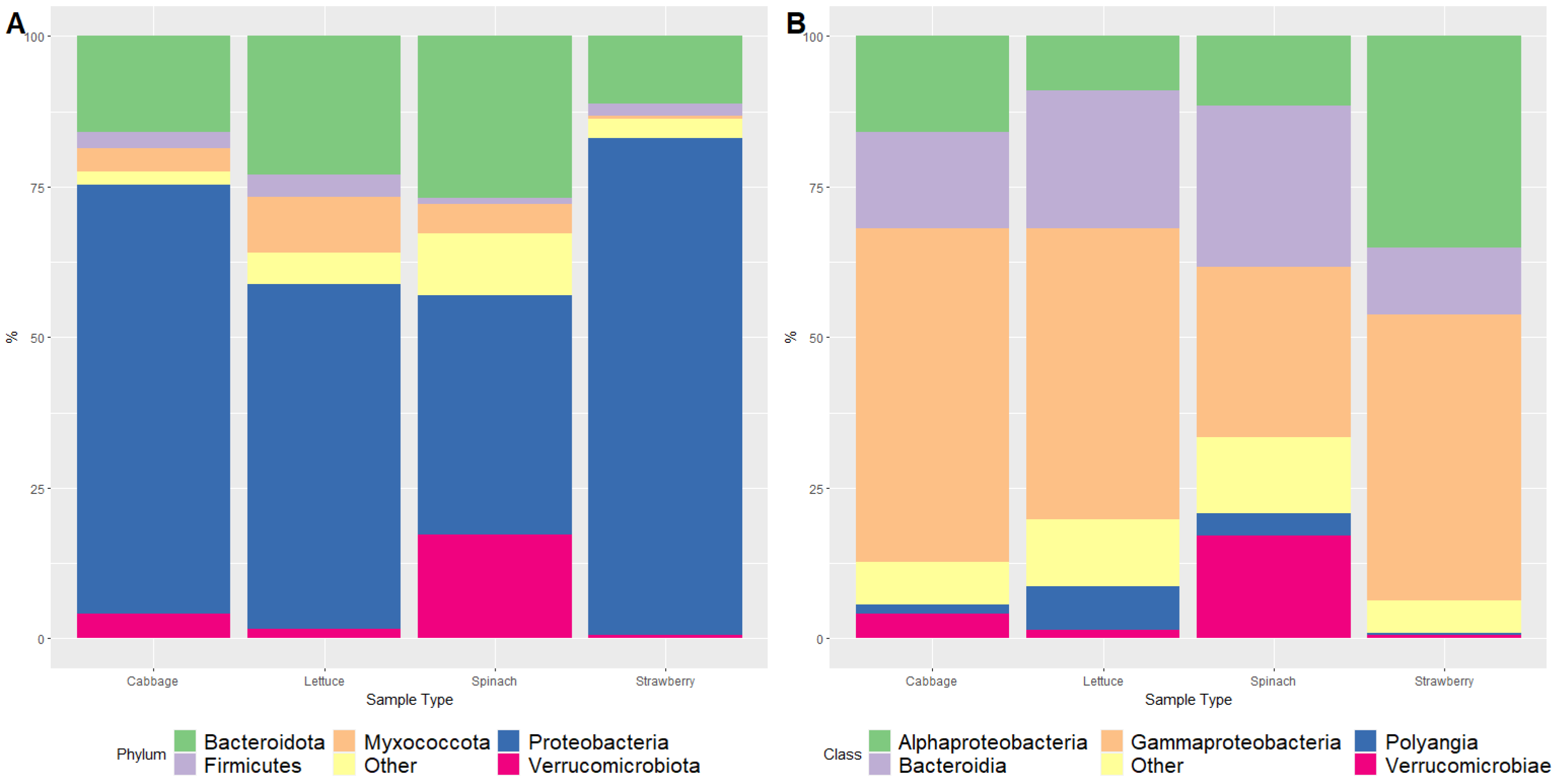
Figure 1. Relative abundance (%) of bacterial phyla (A) and classes (B) identified as part of the FLA microbiome isolated from fresh organic produce.
Foods 12 03102 g001
Figure 2. Heatmap of the relative abundance (%) of the most abundant bacterial genera (>2% in at least one type of sample) identified as part of the FLA microbiome isolated from fresh organic produce.
Foods 12 03102 g002
Figure 3. Microbial diversity analysis. (A) Rarefaction curve. (B) Faith’s Phylogenetic Diversity index. (C) Two-dimensional principal coordinate analysis (PCoA) plots based on unweighted UniFrac distance matrices. (D) Two-dimensional principal coordinate analysis (PCoA) plots based on weighted UniFrac distance matrices.
Foods 12 03102 g003
Table 1. Detection of Acanthamoeba spp. and Vermamoeba vermiformis via qPCR in fresh organic produce.
Acanthamoeba spp.V. vermiformis
Lettuce4/11 (36.4%)1/11 (9.1%)
Cabbage11/11 (100.0%)4/11 (36.4%)
Spinach4/11 (36.4%)4/11 (36.4%)
Strawberry7/7 (100.0%)1/7 (14.3%)
Total26/40 (65.0%)10/40 (25.0%)
Disclaimer/Publisher’s Note: The statements, opinions and data contained in all publications are solely those of the individual author(s) and contributor(s) and not of MDPI and/or the editor(s). MDPI and/or the editor(s) disclaim responsibility for any injury to people or property resulting from any ideas, methods, instructions or products referred to in the content.