Communication

Bacteriocin-Producing Enterococcus faecium OV3-6 as a Bio-Preservative Agent to Produce Fermented Houttuynia cordata Thunb. Beverages: A Preliminary Study

by
Thiwanya Choeisoongnern
1,
Chaiyavat Chaiyasut
2,
Bhagavathi Sundaram Sivamaruthi
2,3,
Netnapa Makhamrueang
3,4,
Sartjin Peerajan
5,
Sasithorn Sirilun
2,4,* and
Phakkharawat Sittiprapaporn
1,*
1
Neuropsychological Research Laboratory, Neuroscience Research Center, School of Anti-Aging and Regenerative Medicine, Mae Fah Luang University, Bangkok 10110, Thailand
2
Innovation Center for Holistic Health, Nutraceuticals and Cosmeceuticals, Faculty of Pharmacy, Chiang Mai University, Chiang Mai 50200, Thailand
3
Office of Research Administration, Chiang Mai University, Chiang Mai 50200, Thailand
4
Department of Pharmaceutical Sciences, Faculty of Pharmacy, Chiang Mai University, Chiang Mai 50200, Thailand
5
Health Innovation Institute, Chiang Mai 50200, Thailand
*
Authors to whom correspondence should be addressed.
Foods 2023, 12(19), 3520; https://doi.org/10.3390/foods12193520
Submission received: 22 August 2023 / Revised: 20 September 2023 / Accepted: 20 September 2023 / Published: 22 September 2023
(This article belongs to the Section Food Microbiology)

Abstract

:
Microbial contamination affects the quality of the fermented Houttuynia cordata Thunb. (H. cordata) beverage (FHB). The present study aimed to assess the bio-preservative property of Enterococcus faecium OV3-6 (E. faecium OV3-6) during the production of FHB. The antimicrobial activity against Escherichia coli, Salmonella, Bacillus cereus, and Staphylococcus aureus and the survival of E. faecium OV3-6 were studied. Then, FHB fermentation was performed with different preservatives (non-preservative, E. faecium OV3-6, cell-free supernatant of E. faecium OV3-6, and nisin) with and without representative pathogens. The maximum antimicrobial activity against S. aureus and B. cereus was observed after 18 h of cultivation in an MRS medium. E. faecium OV3-6 was used as a starter to produce the FHB, and the strain survived up to 48 h in the fermented beverage. E. faecium OV3-6 and its cell-free supernatant inhibited the growth of E. coli, Salmonella, B. cereus, and S. aureus in the stimulated FHB. The non-preservatives and nisin-containing FHB showed inhibition against Gram-positive pathogens. The FHB treated with E. faecium OV3-6 was rich in lactic acid bacteria, and the product was at an acceptable level of pH (less than 4.3). Certain limitations were identified in the study, such as lack of nutritional, metabolomics analysis, and safety and consumer acceptability of FHB. The results suggested that E. faecium OV3-6 could be used as a bio-preservative to produce fermented plant beverages (FPBs).

1. Introduction

Safety and product quality are the major concerns of fermented beverages. Preferably, FPBs are prepared through the lactic acid bacteria (LAB)-mediated fermentation process [1]. FPBs are consumed in large quantities in Asian countries [2,3]. Many studies have reported the benefits of FPB consumption, including disease prevention, cognitive improvement, and anti-aging [2,4,5]. FPBs are prepared using various raw materials such as fruits, vegetables, cereals, and herbs [3]. Houttuynia cordata Thunb., known as Plu-Khao in Thai, is a popular plant used to produce FPBs, mainly because of its bioactive properties [6,7]. The reports proved that H. cordata has several pharmacological activities, including antiviral [8], antibacterial [9,10,11], antioxidant [12], anti-inflammatory [13], and anticancer [6] activities.
The traditional fermentation process (natural fermentation without preservatives or starter cultures) has also been used to produce some FPBs. During fermentation, microbial contamination can occur, which causes spoilage. Many standard techniques, such as thermal process and chemical preservation, have improved the microbial safety of FPBs and increased their shelf life [14,15]. However, these techniques have disadvantages, including altering nutritional values, residual toxic substances (e.g., sulfites), and changes in the taste of the product [16].
LABs act as probiotics and aid in preventing microbial contamination via antimicrobial agents like bacteriocin [17]. Several bacteriocin-producing LAB strains have been reported, such as Enterococcus faecium [18,19], Lactobacillus rhamnosus [20], Lactobacillus plantarum [21,22], Lactococcus lactis subsp. lactis (L. lactis) [23,24], and Leuconostoc citreum GJ7 [25]. LAB starters could act as bio-preservatives in fermentation [26].
Nisin, produced by L. lactis, is a well-studied bio-preservative commercially used as an additive in the food and beverage industry [27,28]. A recent study stated that nisin could inhibit Gram-positive bacteria [29,30]. In addition, nisin is safe for consumption and easily destroyed by human digestion [29]. However, nisin is limited by its specific usage for acid food and low heat resistance under neutral pH [31].
Previously, we have reported the safety and probiotic properties of the bacteriocin-producing E. faecium OV3-6 [32]. A detailed report on the bio-preservative nature of E. faecium OV3-6 is not available. Knowing the efficiency of E. faecium OV3-6 is essential to ensure that it is a bio-preservative agent to produce FHBs. Therefore, the primary objective of this study was to evaluate the efficiency of E. faecium OV3-6 as a bacteriocin-producing bio-preservative bacteria to produce FHBs.

2. Materials and Methods

2.1. Microbial Strains Used in the Study

E. faecium OV3-6 (GenBank accession no: MN453594) isolated from the FHBs was used in the study. Staphylococcus aureus CIP 76.25 was obtained from the Nantes-Atlantic National College of Veterinary Medicine, Food Science and Engineering, Nantes, France. Bacillus cereus ATCC 11778, Escherichia coli ATCC 25922, and Salmonella Typhi DMST 22842 were obtained from the culture collection of the Faculty of Pharmacy, Chiang Mai University.

2.2. Plant Used in the Study

The H. cordata plant was obtained from a farm in Chiang Mai, Thailand. The plant was identified with the help of plant specimens (number: 023239) of the Faculty of Pharmacy, Chiang Mai University.

2.3. Antimicrobial Activity of E. faecium OV3-6

An overnight culture of E. faecium OV3-6 strain was inoculated into de Man Rogosa and Sharpe (MRS) broth medium (HiMedia, Mumbai, India) and cultivated with moderate stirring at 37 °C for 48 h [33]. The cell-free supernatants (CFS) were collected every 6 h of the culturing process. The antimicrobial activity of E. faecium OV3-6 was determined using an agar well diffusion assay [32]. The occurrence of a clear zone around the well was observed in the optimal growth condition of indicator strains (B. cereus ATCC 11778 and S. aureus CIP 76.25), which were cultured in Brain Heart Infusion (BHI) medium (HiMedia, Mumbai, India). The antimicrobial activity was stated in arbitrary units per milliliter (AU/mL) [32].

2.4. Assessment of E. faecium OV3-6 Growth in MRS Medium

E. faecium OV3-6 strain was cultured in MRS broth as detailed (refer to Section 2.3). The number of bacterial cells was estimated using optical density (OD) measurement [34]. The samples (at 0, 6, 12, 18, 24, 30, 36, 42, and 48 h) were analyzed with a multi-mode microplate reader (model Beckman Coulter DTX 880, Fullerton, CA, USA) at a wavelength of 595 nm.

2.5. Assessment of pH Changes during E. faecium OV3-6 Growth

E. faecium OV3-6 strain was cultured in MRS broth as detailed (refer to Section 2.3). The pH changes in the samples (at 0, 6, 12, 18, 24, 30, 36, 42, and 48 h) were measured using a pH meter (model HQ40d, Hach, Loveland, CO, USA) at room temperature.

2.6. Survival of E. faecium OV3-6 in the Fermented Beverage (FB) with and without H. cordata

The starter culture (E. faecium OV3-6) was cultured in MRS broth at suitable conditions (with maximum antimicrobial activity) obtained from the above experiment.
Two fermentation batches (with H. cordata and without H. cordata) were performed to study the viability of E. faecium OV3-6 and its bio-preservative properties. 1: The FB without H. cordata was a mixture of sugar cane and water at a ratio of 1:11 (w/v). 2: The FB with H. cordata was prepared by mixing H. cordata, sugar cane, and water at 1:1:10 (w/w/v) [35,36]. The mixtures were pasteurized, and E. faecium OV3-6 was inoculated (10%) as a starter for the fermentation process. The fermentation process was conducted at 30 ± 2 °C for 48 h. The MRS broth with starter culture (E. faecium OV3-6) was used as a control. The bacterial load of the samples was determined using the plate count method on MRS agar [37], and the results were reported as log colony forming unit per milliliters (log CFU/mL).

2.7. Assessment of the Bio-Preservative Property of E. faecium OV3-6 in Simulated FHB

FHBs were prepared with or without pathogens and preservatives (E. faecium OV3-6, CFS of E. faecium OV3-6, and nisin). No preservatives or 10% (v/v) E. faecium OV3-6 or 10% CFS of E. faecium OV3-6, or 5 mg/kg nisin of the sample, were added to the FHBs.
Regarding pathogens, a pathogen mixture containing E. coli ATCC 5922, S. typhi DMST 22842, S. aureus CIP 76.25, and B. cereus ATCC 11778 was used. The pathogens were cultured in Brain Heart Infusion (BHI) medium (HiMedia, Mumbai, India) at 37 °C for 18 h. The culture was serially diluted in phosphate buffer saline (PBS), and cell count was determined using the plate count method [38,39]. Then, the appropriate dilatation with 107 CFU/mL of pathogen was selected for the study, and the pathogenic mixture was inoculated in the FHB. The fermentation process was carried out for 180 days, and samples were collected at different points (0, 0.5, 1, 2, 3, 5, 7, 10, 15, 30, 60, 90, 120, 150, and 180 days), and stored at 30 ± 2 °C until further analysis. The experimental batches are detailed in Table 1.
The fermented beverage’s microbial load (LAB, E. coli, Salmonella, S. aureus, and B. cereus) was assessed using the serial dilution and plate count technique [40]. MRS agar was used for culturing LAB strains. E. coli, Salmonella, S. aureus, and B. cereus were cultured in a selective medium such as Eosin Methylene Blue (EMB) agar, Salmonella Shigella (SS) agar, Baird–Parker agar, and Phenol Red Egg Yolk Polymyxin agar, respectively. The culture plates were incubated at 37 °C for 24 h. The colonies were counted and expressed as log CFU/mL. The changes in pH during fermentation were measured using a pH meter at room temperature.

2.8. Statistics Analysis

All experiments were performed in triplicate. The analytical data were expressed as mean ± standard deviations. The data were analyzed with analysis of variance (ANOVA) using SPSS (version 17, SPSS Inc., Chicago, IL, USA). Tukey tests were used to determine significant differences between the mean values at a significant level of 95% (p < 0.05).

3. Results

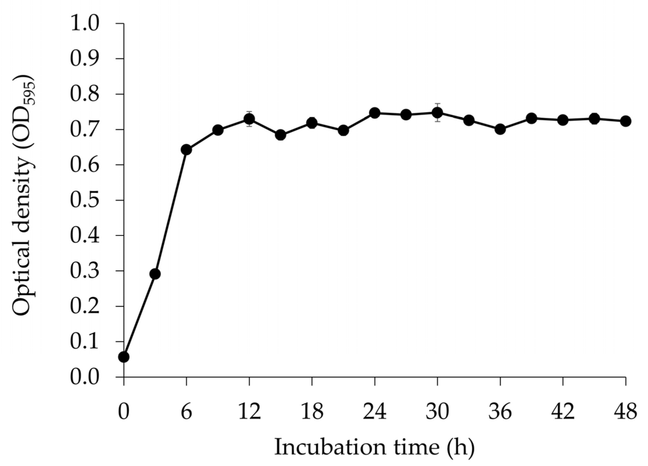
3.1. Microbial Growth of E. faecium OV3-6 Count in MRS Medium

E. faecium OV3-6 count in MRS medium was assessed to find out the growth curve of the strain and represented as OD. The microbial growth was rapid, and the OD reached 0.70 ± 0.005 after 9 h of culturing from 0.06 ± 0.002. The growth was relatively stable after 9 h. The maximum OD of 0.75 ± 0.003 was observed after 30 h of incubation (Figure 1).

3.2. pH Change during the Growth of E. faecium OV3-6 in MRS Medium

The assessment of pH change provides information about the utilization of sugars in the medium and fermentation. pH of the medium during E. faecium OV3-6 cultivation was exhibited in the range of 4.34 ± 0.04 to 6.08 ± 0.08. During E. faecium OV3-6 cultivation, the pH value of the medium gradually decreased. The lowest (4.34 ± 0.04) and highest (6.08 ± 0.08) pH values were observed at 48 and 0 h of the cultivation, respectively (Figure 2).

3.3. Antimicrobial Activity of E. faecium OV3-6

The antimicrobial activity was presented as a growth inhibitory property of E. faecium OV3-6 against S. aureus CIP 76.25 and B. cereus ATCC 11778 (Figure 3). The highest inhibitory activity against S. aureus CIP 76.25 and B. cereus ATCC 11778 was found to be 800 AU/mL (at 18 h of incubation time) and 200 AU/mL (at 18, 24, 30, 36, and 42 h of incubation time), respectively. The lowest inhibitory activity was 200 AU/mL at 6 and 48 h for S. aureus CIP 76.25 and 100 AU/mL at 6, 12, and 48 h for B. cereus ATCC 11778. There were no changes observed in the experimental triplicates.

3.4. Cell Number of E. faecium OV3-6 in FB with and without H. cordata

The measurement of the cell number of E. faecium helps to know whether the strain is growing appropriately without any inhibitions from the plant material used. The alterations of bacterial cell numbers in FBs with and without H. cordata were exhibited during 48 h of fermentation time (Figure 4). Cell counts ranged from 4.92 ± 0.17 to 8.44 ± 0.05 log CFU/mL in FB with H. cordata and 4.87 ± 0.22 to 8.31 ± 0.09 log CFU/mL in FB without H. cordata.
The cell number of E. faecium OV3-6 rapidly increased during 9–24 h of fermentation in FBs with and without H. cordata. The maximum cell number of FBs with and without H. cordata was observed after 24 h of fermentation. After 24 h of fermentation, the E. faecium OV3-6 cell number trend was stable in both beverages (Figure 4). E. faecium OV3-6 was cultured in MRS broth and served as a control. The result showed that the E. faecium OV3-6 cell number gradually increased and reached 10.39 ± 0.08 log CFU/mL after 24 h of incubation; then, there was no significant change in the cell number (Figure 4).

3.5. Microbial Load in FHBs with Different Preservative Treatments

The analysis of microbial load provides the microbial content of the FHB. The LAB load of treatments 3 and 4 were 2.85 ± 0.51 to 8.64 ± 0.35 log CFU/mL and 2.10 ± 0.17 to 8.49 ± 0.20 log CFU/mL, respectively. The maximum number of total LAB in treatments 3 and 4 were observed at day 0.5 (8.64 ± 0.35 log CFU/mL) and day 1 (8.49 ± 0.20 log CFU/mL), respectively. Then, the growth gradually decreased until day 15 (Figure 5).
Total E. coli in FHBs with different preservatives was studied. On day 0, the total E. coli in treatments 2, 4, 6, and 8 were 5.17 ± 0.35, 5.57 ± 0.06, 5.12 ± 0.42, and 5.07 ± 0.10 log CFU/mL, respectively. After 12 h (0.5 days), no E. coli were detected in treatments 4 and 6. The total E. coli in treatments 2 and 8 ranged from 4.85 ± 0.17 to 7.69 ± 0.08 log CFU/mL and 4.31 ± 0.39 to 8.02 ± 0.14 log CFU/mL, respectively. The maximum load of E. coli was found after 0.5 days of the fermentation process in treatments 2 and 8. Then, a gradual reduction was observed, and after 7 days, no E. coli were detected (Table 2). The total Salmonella in treatments 2 and 8 were 8.47 ± 0.31 log CFU/mL and 3.04 ± 0.04 to 8.23 ± 0.18 log CFU/mL, respectively. The maximum load of Salmonella was found during 0.5 to 1st day of the fermentation process in treatments 2 and 8. Then, a gradual reduction was observed, and after 15 days, no E. coli was detected. Initially (0 days), in treatments 4 and 6, 5.57 ± 0.06 and 5.10 ± 0.44 log CFU/mL of Salmonella was detected, respectively, then it was not detected. The initial observation of E. coli and Salmonella in treatments 4 and 6 was due to the forced inoculation of the pathogen in the fermentation medium (Table 2).
Initially, the total S. aureus in treatments 2, 4, 6, and 8 were 5.47 ± 0.26, 5.06 ± 0.09, 5.55 ± 0.07, and 5.32 ± 0.22 log CFU/mL, respectively. While total B. cereus in treatments 2, 4, 6, and 8 were 5.30 ± 0.06, 5.17 ± 0.20, 5.09 ± 0.27, and 5.25 ± 0.21 log CFU/mL, respectively. After 12 h (0.5 days), no S. aureus and B. cereus were detected. The amount of S. aureus and B. cereus in the different treatments was not significantly different. Also, the total LAB, E. coli, Salmonella, S. aureus, and B. cereus were not detected in treatments 1, 3, 5, and 7 during the fermentation, indicating that the fermentation processes were performed in sterile conditions (Table 2).

3.6. pH Changes in FHB with Different Preservative Treatments

The changes in pH of the different preservative treatments in FHBs are displayed in Figure 6. The treatments with pathogens showed high pH compared to non-pathogenic treatments. The pH profile of all experimental treatments guardedly decreased during 180 days of fermentation. After the fermentation process (180 days), treatments 5 (4.23 ± 0.01) and 6 (4.42 ± 0.01) showed high pH, whereas treatments 3 (3.83 ± 0.01) and 4 (3.79 ± 0.01) showed relatively low pH.

4. Discussion

The growth of E. faecium OV3-6 in MRS broth was measured (Figure 1), and the pH was reduced from 6.08 to 4.63 (Figure 2). During the growth, the strain could convert glucose to lactic acid through homofermentative pathways [41,42]. The decrease in pH and increase in acidity were more pronounced during dextrose fermentation, likely due to the higher production of lactic acid. When a probiotic strain grows in MRS broth, pH changes in the culture medium are primarily caused by the production of organic acids, especially lactic acid. Various factors, including unique characteristics of the bacterial strain and the composition of the medium, can influence the rate and extent of these pH changes. Monitoring these pH changes is essential for understanding how probiotic strains grow and metabolize nutrients, and it helps optimize the conditions for their cultivation [43,44].
The lower pH may provide suitable ionic strength; after a point, the pH reduction could inhibit the bacteria’s growth [45,46]. Many studies have reported the relationship between bacterial growth and bacteriocin production [47,48]. Bacteriocin-like inhibitory substances from Gram-positive bacteria (BGPBs) exhibit antimicrobial properties primarily by disrupting the structural integrity of bacterial membranes. These antimicrobial peptides employ diverse mechanisms to achieve this goal. For instance, certain BGPBs, like nisins, interact directly with lipid II, a specific component of the bacterial membrane, inducing the formation of pores. These pores compromise membrane integrity, leading to increased permeability and cell death. In contrast, microbisporicin disrupts the bacterial cell wall biosynthesis process, accumulating cell wall precursors within the bacterial cell. This accumulation perturbs the membrane and contributes to cell demise. Class II bacteriocins, including pediocin PA-1, target the Man-PTS system in bacteria, causing constant receptor opening and uncontrolled efflux of intracellular molecules, leading to cell damage. Subclass IIb bacteriocins such as lactococcin Q and lactococcin G are thought to create membrane pores, potentially through interactions with specific membrane proteins. In summary, BGPBs employ a range of mechanisms to destabilize bacterial membranes or disrupt essential bacterial processes, culminating in the death of the targeted bacteria [49,50,51,52,53,54]. Regarding BGNB (Bacteriocin-Producing Gram-Negative Bacteria) bacteriocins, microcins demonstrate their antibacterial effects through one of two distinct mechanisms: (i) the creation of pores within the inner bacterial membrane, as seen with microcins E492, M, and H47; (ii) the targeting of intracellular enzymes, exemplified by microcin J25, B17, and C. Unlike BGPBs, microcins necessitate entry into the targeted bacterial cell to exert their antimicrobial activity. Once inside, they utilize specific receptors located on the outer membrane of susceptible strains. These receptors are associated with iron uptake and outer membrane porin functions [54,55,56].
Valledor et al. [57] and Barman et al. [58] observed that a high level of bacteriocin production occurred at the late log phase and early stationary phase. The bacteriocins are microbial secondary metabolites, supplementary compounds produced by the microbes for interaction or competition with other organisms or the environment [59]. The early stationary phase of microbial growth is optimal for producing bacteriocin by probiotic strains [60].
Favaro et al. [61] studied the production of bacteriocins by E. faecium ST209GB, ST278GB, ST315GB, and ST711GB isolated from Bulgarian homemade white brine cheese. The study suggested that bacteriocin production was observed in logarithmic growth, and a high level of bacteriocin was displayed in stationary growth. Similarly, Abdel–Hamidet et al. [33] reported the relationship between the growth curve and bacteriocin production in E. faecium ER-3M (isolated from cattle raw milk). The peak of bacteriocins activity was observed in the early stationary phase of E. faecium ER-3M growth.
In the present study, the highest microbial inhibition was observed against B. cereus ATCC 11778 and S. aureus CIP 76.25 after 18 h of the incubation period, which was in the range of the early stationary phase of E. faecium OV3-6 (Figure 3). The study found that 18 h of culturing of E. faecium OV3-6 in an MRS medium could provide peak bacteriocin activity. The bacteriocin of E. faecium OV3-6 was also noticeable at the end of the incubation time. The activity against B. cereus ATCC 11778 and S. aureus CIP 76.25 slightly decreased after 48 h incubation (Figure 3). The proteolytic degradation of bacteriocin may be related to the reduction in its bioactivity after 48 h [62]. The enzymes could destroy the bacteriocin that E. faecium OV3-6 produces during incubation. In addition, some literature reported that the loss of bacteriocin is related to the reabsorption to the surface of the producer cell at a low pH [63].
It is necessary to confirm that the bio-active properties of raw materials should not affect the starter cultures used for the fermentation process. An essential oil isolated from the aerial parts and underground stem of H. cordata showed antimicrobial activities against S. aureus, Streptococcus mutans, Mycobacterium smegmatis, E. faecalis, Candida albicans, and Candida kefyr [11]. Sekita et al. [10] demonstrated that the ethanolic extract of H. cordata poultice showed antimicrobial activity against Gram-positive bacteria, such as Streptococcus epidermidis, Streptococcus pyogenes, Streptococcus mitis, and E. faecalis.
In the present study (Figure 4), the cell number of E. faecium OV3-6 was higher in MRS broth, which acted as a control since the MRS broth has all the essential nutrients for bacteria growth [64,65]. The trend in changes in cell number of E. faecium OV3-6 cultured in FBs with and without H. cordata was the same until 48 h of fermentation time, which indicated that H. cordata could be used as a raw material to produce fermented beverages using E. faecium OV3-6 as a starter culture.
Many bio-preservative methods have been used to enhance safety and extend the shelf life of food products using natural agents [66]. LAB is used as a natural preservative in various food products due to its antimicrobial (via bacteriocins) and metabolic activity (hydrogen peroxide and lactic acid) [67]. LAB bacteriocins are considered acceptable bio-preservative agents because they are non-toxic, non-immunogenic, thermo-resistant, and have extensive bactericidal activity [68,69].
The most commercial LAB bacteriocin is nisin [28,70], which is widely used in the food industry. Nisin is safe, and its use has been approved by the Food and Drug Administration [24,27,30]. Thus, we used nisin as one of the bio-preservative agents in FHBs.
Treatments 3 and 4 showed their LAB content until 15 days of fermentation. In the presence of pathogens, the growth of E. faecium OV3-6 was not affected in FHBs (Figure 5). The study reported that bacteriocin-producing L. plantarum LPL-1 could change the microbial community in low-salt fermented sausages [21]. However, LAB’s efficiency in fermentation depends on various factors such as nutrient composition, time, pH, temperature, material ingredients, and antagonistic microorganisms [71,72]. Additionally, most LAB strains are probiotics with health-promoting properties [73]. We previously reported that E. faecium OV3-6 has in vitro probiotic activity [32].
The pathogenic strains (E. coli, Salmonella, S. aureus, and B. cereus) were observed in FHBs on day 0. Total E. coli was observed during 0–7 days of the fermentation period. Total Salmonella was observed during 0–15 days of the fermentation period. Gram-positive pathogens (S. aureus and B. cereus) were not detected after 0.5 days of fermentation (Table 2). Treatments with E. faecium OV3-6 (treatment 4) and CFS of E. faecium OV3-6 (treatment 6) could have bacteriocin, which inhibits both Gram-positive and Gram-negative pathogenic bacteria.
Waheed et al. [74] reported that the growth of Gram-negative pathogens, E. coli SABA3 and S. typhi SABA10, was reduced by 85% and 50%, respectively, by E. faecium SANA1. Anyogu et al. [75] also suggested that CFS of E. faecium CL02 was able to inhibit both Gram-positive pathogenic bacteria (B. cereus and S. aureus) and Gram-negative pathogenic bacteria (E. coli and S. typhimurium) in vitro. Mostly, bacteriocins destroy Gram-positive bacteria by inhibiting peptidoglycan synthesis and pore formation in the cell membrane, resulting in a loss of potential membrane and cell death [76]. Some bacteriocins control the Gram-negative bacterial growth by pore formation in the cell membrane and enzyme interference, such as DNA gyrase, RNA polymerase, and aspartyl-tRNA synthetase [77].
Treatment 2 (non-preservative) and treatment 8 (with nisin) inhibited the Gram-positive pathogens throughout fermentation. Nisin inhibits Gram-positive pathogens via its action on the outer peptidoglycan layer of Gram-positive bacteria [78,79]. It has been proven that medicinal plants have antimicrobial properties against Gram-positive pathogens [10,80,81]. Accordingly, the antimicrobial effects against Gram-positive pathogens (B. cereus and S. aureus) in treatment 2 (non-preservative) are possibly due to the antimicrobial activity of the raw material (H. cordata) used in the study (Table 2). Treatment 8 (with nisin) showed antimicrobial activity against Gram-positive pathogens [30,82] but not against Gram-negative bacteria due to the protective outer membrane of the bacterium [83]. Therefore, nisin has some limitations; E. faecium OV3-6 and its metabolites are effective alternative bio-preservatives to produce fermented FHBs.
The pH level could indicate the safety and quality of the fermented product. According to Thai Community Product Standard (TCPS)—No. 481/2004, the acceptable pH level in FPBs is less than 4.3 [84]. In this study, treatments 3 and 4 (with E. faecium OV3-6) showed a pH value of less than 4.3 after 0.5 days (Figure 6). Similarly, Liu et al. [37] reported that the pH of Paocai (lactic acid-fermented vegetable), which uses E. faecium Y31 as an adjunct culture, was less than 4.3 after 2.5 days of fermentation. The FHBs with non-preservative E. faecium OV3-6 and nisin (treatments 1, 2, 3, 4, 7, and 8) were in the pH range of TCPS acceptance level at the end of fermentation. A few studies reported that the production of organic acids (lactic and acetic acids) could affect the pH of the fermented product [85,86]. However, treatments 5 and 6 (containing CFS of E. faecium OV3-6) showed a pH higher than 4.3 throughout fermentation (Figure 6).
Many reports have indicated that the metabolites in CFS of LAB have efficacy in inhibiting microbial pathogens [87,88,89]. The secreted metabolites in CFS of E. faecium OV3-6 might inhibit the growth of the other epiphytic microflora [90,91,92], which might affect organic acid production. Thus, the pH value of treatments 5 and 6 was substantially higher than the other treatments. The above statements need to be confirmed through detailed studies.
This study has some limitations, as detailed below. Even though we reported the presence of the entP gene in E. faecium OV3-6 that involves the production of bacteriocin called enterocin P, further detailed characterization of the bacteriocins-like substances was not studied. The metabolomic profile may provide detailed information on nutritional changes of the FHB, but the present study lacks that. Also, studies are required to determine the effect of H. cordata on the bacteriocin production or microbial activity of E. faecium OV3-6. The in vitro and in vivo evaluation of bioactivities and health effects of FHBs is necessary to claim that FHBs are functional food. Finally, the consumer acceptance survey also required us to market FHBs.

5. Conclusions

The study showed that E. faecium OV3-6 could produce bacteriocin-like substances during its late log phase and the early stationary phase in MRS broth. Maximum bacteriocin-like substances were produced after 18 h of incubation in MRS broth, which was confirmed through the antimicrobial activity against the representative pathogens. The survival of E. faecium OV3-6 in FBs with and without H. cordata indicated that E. faecium OV3-6 could be potent starter culture to produce FHBs. The pH value of FHBs with E. faecium OV3-6 was in the acceptable range (less than 4.3). The LAB load in FHBs was higher during fermentation with E. faecium OV3-6; it could be considered that the strain E. faecium OV3-6 improved the LAB and probiotics in FHBs. The preliminary study results indicated that E. faecium OV3-6 and CFS of E. faecium OV3-6 could be bio-preservatives to control undesirable pathogens in FPBs. Biobased preservatives could be the better option to protect customers from the adverse side effects of chemicals.
The study has some limitations, including the safety of the FHB not being studied, changes in the metabolomics were not assessed in the FHB, and the health effects of the FHB were not studied using laboratory models. Also, the FHB needs to be evaluated for consumer acceptance. These limitations block the strong claim that E. faecium OV3-6 is a potent bio-preservative to produce FHBs. Further extensive studies on the abovementioned aspects of FHBs are needed to overcome the restrictions in E. faecium OV3-6-based bio-preservatives.

Author Contributions

Conceptualization, C.C., S.S. and P.S.; methodology, C.C., T.C. and N.M.; software, T.C.; formal analysis, T.C. and N.M.; investigation, T.C.; resources, C.C. and S.P.; data curation, T.C.; writing—original draft preparation, C.C., T.C., S.S. and B.S.S.; writing—review and editing, C.C., S.S. and B.S.S.; supervision, P.S. and C.C.; project administration, S.P. and C.C.; funding acquisition, C.C. All authors have read and agreed to the published version of the manuscript.

Funding

This project was partially supported by Chiang Mai University, Chiang Mai, Thailand.

Data Availability Statement

All the data are available within the manuscript.

Acknowledgments

Authors gratefully acknowledge Mae Fah Luang University, Thailand, and Chiang Mai University, Thailand, for their support. T.C. wishes to acknowledge the support of the Postdoctoral Fellowship from Mae Fah Luang University, Thailand.

Conflicts of Interest

The authors declare no conflict of interest.

References

  1. Woraharn, S.; Lailerd, N.; Sivamaruthi, B.S.; Wangcharoen, W.; Sirisattha, S.; Peerajan, S.; Chaiyasut, C. Evaluation of factors that influence the L-glutamic and γ-aminobutyric acid production during Hericium erinaceus fermentation by lactic acid bacteria. CyTA-J. Food 2016, 14, 47–54. [Google Scholar] [CrossRef]
  2. Chaiyasut, C.; Sivamaruthi, B.S.; Makhamrueang, N.; Peerajan, S.; Kesika, P. A survey of consumer’s opinion about consumption and health benefits of fermented plant beverages in Thailand. Food Sci. Technol. 2017, 38, 299–309. [Google Scholar] [CrossRef]
  3. Sõukand, R.; Pieroni, A.; Biró, M.; Dénes, A.; Dogan, Y.; Hajdari, A.; Kalle, R.; Reade, B.; Mustafa, B.; Nedelcheva, A. An ethnobotanical perspective on traditional fermented plant foods and beverages in Eastern Europe. J. Ethnopharmacol. 2015, 170, 284–296. [Google Scholar] [CrossRef]
  4. Sivamaruthi, B.S.; Kesika, P.; Chaiyasut, C. Impact of fermented foods on human cognitive function-A review of outcome of clinical trials. Sci. Pharm. 2018, 86, 22. [Google Scholar] [CrossRef]
  5. Das, G.; Paramithiotis, S.; Sivamaruthi, B.S.; Wijaya, C.H.; Suharta, S.; Sanlier, N.; Shin, H.S.; Patra, J.K. Traditional fermented foods with anti-aging effect: A concentric review. Food Res. Int. 2020, 134, 109269. [Google Scholar] [CrossRef]
  6. Senawong, T.; Khaopha, S.; Misuna, S.; Komaikul, J.; Senawong, G.; Wongphakham, P.; Yunchalard, S. Phenolic acid composition and anticancer activity against human cancer cell lines of the commercially available fermentation products of Houttuynia cordata. Sci. Asia 2014, 40, 420–427. [Google Scholar] [CrossRef]
  7. Satthakarn, S.; Chung, W.; Promsong, A.; Nittayananta, W. Houttuynia cordata modulates oral innate immune mediators: Potential role of herbal plant on oral health. Oral Dis. 2015, 21, 512–518. [Google Scholar] [CrossRef]
  8. Chiow, K.; Phoon, M.; Putti, T.; Tan, B.K.; Chow, V.T. Evaluation of antiviral activities of Houttuynia cordata Thunb. extract, quercetin, quercetrin and cinanserin on murine coronavirus and dengue virus infection. Asian Pac. J. Trop. Med. 2016, 9, 1–7. [Google Scholar] [CrossRef]
  9. Řebíčková, K.; Bajer, T.; Šilha, D.; Houdková, M.; Ventura, K.; Bajerová, P. Chemical Composition and Determination of the Antibacterial Activity of Essential Oils in Liquid and Vapor Phases Extracted from Two Different Southeast Asian Herbs-Houttuynia cordata (Saururaceae) and Persicaria odorata (Polygonaceae). Molecules 2020, 25, 2432. [Google Scholar] [CrossRef]
  10. Sekita, Y.; Murakami, K.; Yumoto, H.; Mizuguchi, H.; Amoh, T.; Ogino, S.; Matsuo, T.; Miyake, Y.; Fukui, H.; Kashiwada, Y. Anti-bacterial and anti-inflammatory effects of ethanol extract from Houttuynia cordata poultice. Biosci. Biotechnol. Biochem. 2016, 80, 1205–1213. [Google Scholar] [CrossRef]
  11. Verma, R.S.; Joshi, N.; Padalia, R.C.; Singh, V.R.; Goswami, P.; Kumar, A.; Iqbal, H.; Verma, R.K.; Chanda, D.; Chauhan, A. Chemical Composition and Allelopathic, Antibacterial, Antifungal, and Antiacetylcholinesterase Activity of Fish-mint (Houttuynia cordata Thunb.) from India. Chem. Biodivers. 2017, 14, e1700189. [Google Scholar] [CrossRef]
  12. Pakyntein, C.L.; Syiem, D.; Thabah, D.; Sunn, S.E. Antioxidant, anti-inflammatory and anti-hyperglycemic activity of aqueous and methanolic extract of Houttuynia cordata: An in vitro and in vivo study. GSC Biol. Pharm. Sci. 2021, 16, 145–154. [Google Scholar] [CrossRef]
  13. Woranam, K.; Senawong, G.; Utaiwat, S.; Yunchalard, S.; Sattayasai, J.; Senawong, T. Anti-inflammatory activity of the dietary supplement Houttuynia cordata fermentation product in RAW264. 7 cells and Wistar rats. PLoS ONE 2020, 15, e0230645. [Google Scholar] [CrossRef]
  14. Pinto, T.; Vilela, A.; Cosme, F. Chemical and Sensory Characteristics of Fruit Juice and Fruit Fermented Beverages and Their Consumer Acceptance. Beverages 2022, 8, 33. [Google Scholar] [CrossRef]
  15. El-Saadony, M.T.; Elsadek, M.F.; Mohamed, A.S.; Taha, A.E.; Ahmed, B.M.; Saad, A.M. Effects of chemical and natural additives on cucumber juice’s quality, shelf life, and safety. Foods 2020, 9, 639. [Google Scholar] [CrossRef]
  16. Silva, M.M.; Lidon, F. Food preservatives—An overview on applications and side effects. Emir. J. Food Agric. 2016, 28, 366–373. [Google Scholar] [CrossRef]
  17. Raj, T.; Chandrasekhar, K.; Kumar, A.N.; Kim, S.H. Recent biotechnological trends in lactic acid bacterial fermentation for food processing industries. Syst. Microbiol. Biomanuf. 2022, 2, 14–40. [Google Scholar] [CrossRef]
  18. Aspri, M.; O’Connor, P.M.; Field, D.; Cotter, P.D.; Ross, P.; Hill, C.; Papademas, P. Application of bacteriocin-producing Enterococcus faecium isolated from donkey milk, in the bio-control of Listeria monocytogenes in fresh whey cheese. Int. Dairy J. 2017, 73, 1–9. [Google Scholar] [CrossRef]
  19. Nascimento, M.S.; Moreno, I.; Kuaye, A.Y. Applicability of bacteriocin-producing Lactobacillus plantarum, Enterococcus faecium and Lactococcus lactis ssp. lactis as adjunct starter in Minas Frescal cheesemaking. Int. J. Dairy Technol. 2008, 61, 352–357. [Google Scholar] [CrossRef]
  20. Prezzi, L.E.; Lee, S.H.; Nunes, V.M.; Corassin, C.H.; Pimentel, T.C.; Rocha, R.S.; Ramos, G.L.; Guimarães, J.T.; Balthazar, C.F.; Duarte, M.C.K. Effect of Lactobacillus rhamnosus on growth of Listeria monocytogenes and Staphylococcus aureus in a probiotic Minas Frescal cheese. Food Microbiol. 2020, 92, 103557. [Google Scholar] [CrossRef]
  21. Zhang, Y.; Qin, Y.; Wang, Y.; Huang, Y.; Li, P.; Li, P. Lactobacillus plantarum LPL-1, a bacteriocin producing strain, changed the bacterial community composition and improved the safety of low-salt fermented sausages. LWT 2020, 128, 109385. [Google Scholar] [CrossRef]
  22. Obadina, A.; Oyewole, O.; Sanni, L.O.; Tomlins, K.I. Bio-preservative activities of Lactobacillus plantarum strains in fermenting Casssava ‘fufu’. Afr. J. Biotechnol. 2006, 5, 620–623. [Google Scholar]
  23. Siroli, L.; Patrignani, F.; D’Alessandro, M.; Salvetti, E.; Torriani, S.; Lanciotti, R. Suitability of the Nisin Z-producer Lactococcus lactis subsp. lactis CBM 21 to be Used as an Adjunct Culture for Squacquerone Cheese Production. Animals 2020, 10, 782. [Google Scholar] [CrossRef]
  24. Hassan, H.; St-Gelais, D.; Gomaa, A.; Fliss, I. Impact of Nisin and Nisin-Producing Lactococcus lactis ssp. lactis on Clostridium tyrobutyricum and Bacterial Ecosystem of Cheese Matrices. Foods 2021, 10, 898. [Google Scholar] [CrossRef]
  25. Chang, J.Y.; Chang, H.C. Growth inhibition of foodborne pathogens by kimchi prepared with bacteriocin-producing starter culture. J. Food Sci. 2011, 76, M72–M78. [Google Scholar] [CrossRef]
  26. Zinoviadou, K.G.; Gougouli, M.; Biliaderis, C.G. Innovative Biobased Materials for Packaging Sustainability. In Innovation Strategies in the Food Industry: Tools for Implementation, 1st ed.; Galanakis, C.M., Ed.; Elsevier Inc.: Amsterdam, The Netherlands, 2016; pp. 167–189. [Google Scholar]
  27. EFSA Panel on Food Additives and Nutrient Sources added to Food (ANS); Younes, M.; Aggett, P.; Aguilar, F.; Crebelli, R.; Dusemund, B.; Filipic, M.; Frutos, M.J.; Galtier, P.; Gundert-Remy, U. Safety of nisin (E 234) as a food additive in the light of new toxicological data and the proposed extension of use. EFSA J. 2017, 15, e05063. [Google Scholar]
  28. Khelissa, S.; Chihib, N.E.; Gharsallaoui, A. Conditions of nisin production by Lactococcus lactis subsp. lactis and its main uses as a food preservative. Arch. Microbiol. 2021, 203, 465–480. [Google Scholar] [CrossRef]
  29. Müller-Auffermann, K.; Grijalva, F.; Jacob, F.; Hutzler, M. Nisin and its usage in breweries: A review and discussion. J. Inst. Brew. 2015, 121, 309–319. [Google Scholar] [CrossRef]
  30. Özel, B.; Şimşek, Ö.; Akçelik, M.; Saris, P.E. Innovative approaches to nisin production. Appl. Microbiol. Biotechnol. 2018, 102, 6299–6307. [Google Scholar] [CrossRef]
  31. Davies, E.; Bevis, H.; Potter, R.; Harris, J.; Williams, G.; Delves-Broughton, J. Research note: The effect of pH on the stability of nisin solution during autoclaving. Lett. Appl. Microbiol. 1998, 27, 186–187. [Google Scholar] [CrossRef]
  32. Choeisoongnern, T.; Sirilun, S.; Waditee-Sirisattha, R.; Pintha, K.; Peerajan, S.; Chaiyasut, C. Potential Probiotic Enterococcus faecium OV3-6 and Its Bioactive Peptide as Alternative Bio-Preservation. Foods 2021, 10, 2264. [Google Scholar] [CrossRef]
  33. Abdel-Hamid, M.S.; Anis, A.; Elbawab, R.H.; Mohammed, A.A.; Orabi, S.H.; Fathalla, S.I. Distinctive antagonistic role of new Enterococcus faecium ER-3M strain and its bacteriocin effect against Staphylococcus aureus Pneumonia. Rend. Lincei. Sci. Fis. Nat. 2018, 29, 675–690. [Google Scholar] [CrossRef]
  34. Valledor, S.J.D.; Bucheli, J.E.V.; Holzapfel, W.H.; Todorov, S.D. Exploring beneficial properties of the bacteriocinogenic Enterococcus faecium ST10Bz strain isolated from boza, a Bulgarian cereal-based beverage. Microorganisms 2020, 8, 1474. [Google Scholar] [CrossRef] [PubMed]
  35. Chaiyasut, C.; Kusirisin, W.; Lailerd, N.; Lerttrakarnnon, P.; Suttajit, M.; Srichairatanakool, S. Effects of phenolic compounds of fermented Thai indigenous plants on oxidative stress in streptozotocin-induced diabetic rats. Evid. -Based Complement. Altern. Med. 2011, 2011, 749307. [Google Scholar] [CrossRef]
  36. Peerajan, S.; Chaiyasut, C.; Sirilun, S.; Chaiyasut, K.; Kesika, P.; Sivamaruthi, B.S. Enrichment of nutritional value of Phyllanthus emblica fruit juice using the probiotic bacterium, Lactobacillus paracasei HII01 mediated fermentation. Food Sci. Technol. 2016, 36, 116–123. [Google Scholar] [CrossRef]
  37. Liu, W.; Zhang, L.; Shi, J.; Yi, H.; Zhang, Y.; Zhang, S.; Gao, W.; Du, M.; Han, X.; Yu, X. Assessment of the safety and applications of bacteriocinogenic Enterococcus faecium Y31 as an adjunct culture in North-eastern Chinese traditional fermentation paocai. Food Control 2015, 50, 637–644. [Google Scholar] [CrossRef]
  38. Association of Official Analytical Chemists. Official Methods of Analysis, 15th ed.; AOAC: Arlington, VA, USA, 1990. [Google Scholar]
  39. American Public Health Association. Compendium of Methods for the Microbiological Examination of Foods, 2nd ed.; APHA: Washington, DC, USA, 1984. [Google Scholar]
  40. Wang, C.L.; Shi, D.J.; Gong, G.L. Microorganisms in Daqu: A starter culture of Chinese Maotai-flavor liquor. World J. Microbiol. Biotechnol. 2008, 24, 2183–2190. [Google Scholar] [CrossRef]
  41. Lancetti, R.; Sciarini, L.; Pérez, G.T.; Salvucci, E. Technological performance and selection of lactic acid bacteria isolated from Argentinian grains as starters for wheat sourdough. Curr. Microbiol. 2021, 78, 255–264. [Google Scholar] [CrossRef]
  42. Sun, W.; Liu, J.; Xu, H.; Li, W.; Zhang, J. L-Lactic acid fermentation by Enterococcus faecium: A new isolate from bovine rumen. Biotechnol. Lett. 2015, 37, 1379–1383. [Google Scholar] [CrossRef]
  43. Guan, X.; Xu, Q.; Zheng, Y.; Qian, L.; Lin, B. Screening and characterization of lactic acid bacterial strains that produce fermented milk and reduce cholesterol levels. Braz. J. Microbiol. 2017, 48, 730–739. [Google Scholar] [CrossRef]
  44. García-Núñez, I.M.; Santacruz, A.; Serna-Saldívar, S.O.; Hernandez, S.L.; Guerra, C.A. Assessment of Potential Probiotic and Synbiotic Properties of Lactic Acid Bacteria Grown In Vitro with Starch-Based Soluble Corn Fiber or Inulin. Foods 2022, 11, 4020. [Google Scholar] [CrossRef] [PubMed]
  45. Reis, J.; Paula, A.; Casarotti, S.; Penna, A. Lactic acid bacteria antimicrobial compounds: Characteristics and applications. Food Eng. Rev. 2012, 4, 124–140. [Google Scholar] [CrossRef]
  46. Lambert, R.; Stratford, M. Weak-acid preservatives: Modelling microbial inhibition and response. J. Appl. Microbiol. 1999, 86, 157–164. [Google Scholar] [CrossRef] [PubMed]
  47. Todorov, S.D.; Wachsman, M.; Tomé, E.; Dousset, X.; Destro, M.T.; Dicks, L.M.T.; de Melo Franco, B.D.G.; Vaz-Velho, M.; Drider, D. Characterisation of an antiviral pediocin-like bacteriocin produced by Enterococcus faecium. Food Microbiol. 2010, 27, 869–879. [Google Scholar] [CrossRef]
  48. Hadji-Sfaxi, I.; El-Ghaish, S.; Ahmadova, A.; Batdorj, B.; Le Blay-Laliberté, G.; Barbier, G.; Haertlé, T.; Chobert, J.M. Antimicrobial activity and safety of use of Enterococcus faecium PC4. 1 isolated from Mongol yogurt. Food Control 2011, 22, 2020–2027. [Google Scholar] [CrossRef]
  49. McAuliffe, O.; Ross, R.P.; Hill, C. Lantibiotics: Structure, biosynthesis and mode of action. FEMS Microbiol. Rev. 2001, 25, 285–308. [Google Scholar] [CrossRef]
  50. Wiedemann, I.; Böttiger, T.; Bonelli, R.R.; Wiese, A.; Hagge, S.O.; Gutsmann, T.; Seydel, U.; Deegan, L.; Hill, C.; Ross, P.; et al. The mode of action of the lantibiotic lacticin 3147—A complex mechanism involving specific interaction of two peptides and the cell wall precursor lipid II. Mol. Microbiol. 2006, 61, 285–296. [Google Scholar] [CrossRef]
  51. Castiglione, F.; Lazzarini, A.; Carrano, L.; Corti, E.; Ciciliato, I.; Gastaldo, L.; Candiani, P.; Losi, D.; Marinelli, F.; Selva, E.; et al. Determining the Structure and Mode of Action of Microbisporicin, a Potent Lantibiotic Active Against Multi resistant Pathogens. Chem. Biol. 2008, 15, 22–31. [Google Scholar] [CrossRef]
  52. Nishie, M.; Nagao, J.-I.; Sonomoto, K. Antibacterial Peptides “Bacteriocins”: An Overview of Their Diverse Characteristics and Applications. Biocontrol Sci. 2012, 17, 1–16. [Google Scholar] [CrossRef]
  53. Cavera, V.L.; Arthur, T.D.; Kashtanov, D.; Chikindas, M.L. Bacteriocins and their position in the next wave of conventional antibiotics. Int. J. Antimicrob. Agents 2015, 46, 494–501. [Google Scholar] [CrossRef]
  54. Simons, A.; Alhanout, K.; Duval, R.E. Bacteriocins, antimicrobial peptides from bacterial origin: Overview of their biology and their impact against multidrug-resistant bacteria. Microorganisms 2020, 8, 639. [Google Scholar] [CrossRef]
  55. Duquesne, S.; Destoumieux-Garzón, D.; Peduzzi, J.; Rebuffat, S. Microcins, gene-encoded antibacterial peptides from enterobacteria. Nat. Prod. Rep. 2007, 24, 708–734. [Google Scholar] [CrossRef] [PubMed]
  56. Mathavan, I.; Beis, K. The role of bacterial membrane proteins in the internalization of microcin MccJ25 and MccB17. Biochem. Soc. Trans. 2012, 40, 1539–1543. [Google Scholar] [CrossRef] [PubMed]
  57. Valledor, S.J.D.; Dioso, C.M.; Bucheli, J.E.V.; Park, Y.J.; Suh, D.H.; Jung, E.S.; Kim, B.; Holzapfel, W.H.; Todorov, S.D. Characterization and safety evaluation of two beneficial, enterocin-producing Enterococcus faecium strains isolated from kimchi, a Korean fermented cabbage. Food Microbiol. 2022, 102, 103886. [Google Scholar] [CrossRef] [PubMed]
  58. Barman, S.; Ghosh, R.; Mandal, N.C. Production optimization of broad spectrum bacteriocin of three strains of Lactococcus lactis isolated from homemade buttermilk. Ann. Agrar. Sci. 2018, 16, 286–296. [Google Scholar] [CrossRef]
  59. Sansinenea, E.; Ortiz, A. Secondary metabolites of soil Bacillus spp. Biotechnol. Lett. 2011, 33, 1523–1538. [Google Scholar] [CrossRef]
  60. Zangeneh, M.; Khorrami, S.; Khaleghi, M. Bacteriostatic activity and partial characterization of the bacteriocin produced by L. plantarum sp. isolated from traditional sourdough. Food Sci. Nutr. 2020, 8, 6023–6030. [Google Scholar] [CrossRef]
  61. Favaro, L.; Basaglia, M.; Casella, S.; Hue, I.; Dousset, X.; de Melo Franco, B.D.G.; Todorov, S.D. Bacteriocinogenic potential and safety evaluation of non-starter Enterococcus faecium strains isolated from homemade white brine cheese. Food Microbiol. 2014, 38, 228–239. [Google Scholar] [CrossRef]
  62. Nešuta, O.; Buděšínský, M.; Hadravová, R.; Monincová, L.; Humpoličková, J.; Čeřovský, V. How proteases from Enterococcus faecalis contribute to its resistance to short α-helical antimicrobial peptides. Pathog. Dis. 2017, 75, ftx091. [Google Scholar] [CrossRef]
  63. Herranz, C.; Martinez, J.; Rodriguez, J.; Hernandez, P.; Cintas, L. Optimization of enterocin P production by batch fermentation of Enterococcus faecium P13 at constant pH. Appl. Microbiol. Biotechnol. 2001, 56, 378–383. [Google Scholar] [CrossRef]
  64. De Man, J.C.; Rogosa, M.; Sharpe, M.E. A medium for the cultivation of lactobacilli. J. Appl. Bacteriol. 1960, 23, 130–135. [Google Scholar] [CrossRef]
  65. Calderon, M.; Loiseau, G.; Guyot, J.P. Nutritional requirements and simplified cultivation medium to study growth and energetics of a sourdough lactic acid bacterium Lactobacillus fermentum Ogi E1 during heterolactic fermentation of starch. J. Appl. Microbiol. 2001, 90, 508–516. [Google Scholar] [CrossRef]
  66. Rai, M.; Pandit, R.; Gaikwad, S.; Kövics, G. Antimicrobial peptides as natural bio-preservative to enhance the shelf-life of food. J. Food Sci. Technol. 2016, 53, 3381–3394. [Google Scholar] [CrossRef]
  67. Agriopoulou, S.; Stamatelopoulou, E.; Sachadyn-Król, M.; Varzakas, T. Lactic acid bacteria as antibacterial agents to extend the shelf life of fresh and minimally processed fruits and vegetables: Quality and safety aspects. Microorganisms 2020, 8, 952. [Google Scholar] [CrossRef]
  68. Ibrahim, S.A.; Ayivi, R.D.; Zimmerman, T.; Siddiqui, S.A.; Altemimi, A.B.; Fidan, H.; Esatbeyoglu, T.; Bakhshayesh, R.V. Lactic acid bacteria as antimicrobial agents: Food safety and microbial food spoilage prevention. Foods 2021, 10, 3131. [Google Scholar] [CrossRef]
  69. Vesković-Moračanin, S.M.; Đukić, D.A.; Memiši, N.R. Bacteriocins produced by lactic acid bacteria: A review. Acta Period. Technol. 2014, 45, 271–283. [Google Scholar] [CrossRef]
  70. Woraprayote, W.; Malila, Y.; Sorapukdee, S.; Swetwiwathana, A.; Benjakul, S.; Visessanguan, W. Bacteriocins from lactic acid bacteria and their applications in meat and meat products. Meat Sci. 2016, 120, 118–132. [Google Scholar] [CrossRef] [PubMed]
  71. Serrazanetti, D.I.; Gottardi, D.; Montanari, C.; Gianotti, A. Dynamic stresses of lactic acid bacteria associated to fermentation processes. In Lactic Acid Bacteria-R & D for Food, Health and Livestock Purposes, 1st ed.; Kongo, J.M., Ed.; IntechOpen: London, UK, 2013; pp. 539–570. [Google Scholar]
  72. Syrokou, M.K.; Themeli, C.; Paramithiotis, S.; Mataragas, M.; Bosnea, L.; Argyri, A.A.; Chorianopoulos, N.G.; Skandamis, P.N.; Drosinos, E.H. Microbial ecology of Greek wheat sourdoughs, identified by a culture-dependent and a culture-independent approach. Foods 2020, 9, 1603. [Google Scholar] [CrossRef]
  73. Sadiq, M.B. Lactic acid bacteria as potential probiotics. In Probiotics, Prebiotics and Synbiotics: Technological Advancements towards Safety and Industrial Applications, 1st ed.; Panesar, P.S., Anal, A.K., Eds.; Wiley: Hoboken, NJ, USA, 2022; pp. 57–72. [Google Scholar]
  74. Waheed, S.; Rasool, M.H.; Aslam, B.; Muzammil, S.; Waseem, M.; Shahid, M.; Saqib, M.; Hayat, S.; Naeem, M.; Taj, Z.; et al. Antagonistic Potential of Dairy Origin Enterococcus faecium Against Multidrug-Resistant Foodborne Pathogens. Rom. Biotechnol. Lett. 2021, 26, 2406–2415. [Google Scholar] [CrossRef]
  75. Anyogu, A.; Awamaria, B.; Sutherland, J.; Ouoba, L. Molecular characterisation and antimicrobial activity of bacteria associated with submerged lactic acid cassava fermentation. Food Control 2014, 39, 119–127. [Google Scholar] [CrossRef]
  76. Héchard, Y.; Sahl, H.G. Mode of action of modified and unmodified bacteriocins from Gram-positive bacteria. Biochimie 2002, 84, 545–557. [Google Scholar] [CrossRef] [PubMed]
  77. Cotter, P.D.; Ross, R.P.; Hill, C. Bacteriocins- A viable alternative to antibiotics? Nat. Rev. Microbiol. 2013, 11, 95–105. [Google Scholar] [CrossRef] [PubMed]
  78. Beveridge, T.J. Structures of gram-negative cell walls and their derived membrane vesicles. J. Bacteriol. 1999, 181, 4725–4733. [Google Scholar] [CrossRef] [PubMed]
  79. Tavares, T.D.; Antunes, J.C.; Padrão, J.; Ribeiro, A.I.; Zille, A.; Amorim, M.T.P.; Ferreira, F.; Felgueiras, H.P. Activity of specialized biomolecules against gram-positive and gram-negative bacteria. Antibiotics 2020, 9, 314. [Google Scholar] [CrossRef]
  80. Palombo, E.A.; Semple, S.J. Antibacterial activity of traditional Australian medicinal plants. J. Ethnopharmacol. 2001, 77, 151–157. [Google Scholar] [CrossRef]
  81. Nostro, A.; Germano, M.; D’angelo, V.; Marino, A.; Cannatelli, M. Extraction methods and bioautography for evaluation of medicinal plant antimicrobial activity. Lett. Appl. Microbiol. 2000, 30, 379–384. [Google Scholar] [CrossRef]
  82. Shin, J.M.; Gwak, J.W.; Kamarajan, P.; Fenno, J.C.; Rickard, A.H.; Kapila, Y.L. Biomedical applications of nisin. J. Appl. Microbiol. 2016, 120, 1449–1465. [Google Scholar] [CrossRef]
  83. Li, Q.; Montalban-Lopez, M.; Kuipers, O.P. Increasing the Antimicrobial Activity of Nisin-Based Lantibiotics against Gram-Negative Pathogens. Appl. Environ. Microbiol. 2018, 84, e00052-18. [Google Scholar] [CrossRef]
  84. TISI. Thai Community Products Standard (TCPS) No. 481/2004. Available online: https://www.tisi.go.th/home/en (accessed on 7 July 2022).
  85. Jiang, F.G.; Cheng, H.J.; Liu, D.; Wei, C.; An, W.J.; Wang, Y.F.; Sun, H.T.; Song, E.L. Treatment of Whole-Plant Corn Silage With Lactic Acid Bacteria and Organic Acid Enhances Quality by Elevating Acid Content, Reducing pH, and Inhibiting Undesirable Microorganisms. Front. Microbiol. 2020, 11, 593088. [Google Scholar] [CrossRef]
  86. Choi, Y.J.; Yong, S.; Lee, M.J.; Park, S.J.; Yun, Y.R.; Park, S.H.; Lee, M.A. Changes in volatile and non-volatile compounds of model kimchi through fermentation by lactic acid bacteria. LWT 2019, 105, 118–126. [Google Scholar] [CrossRef]
  87. Mariam, S.H.; Zegeye, N.; Tariku, T.; Andargie, E.; Endalafer, N.; Aseffa, A. Potential of cell-free supernatants from cultures of selected lactic acid bacteria and yeast obtained from local fermented foods as inhibitors of Listeria monocytogenes, Salmonella spp. and Staphylococcus aureus. BMC Res. Notes 2014, 7, 606. [Google Scholar] [CrossRef] [PubMed]
  88. Ballén, V.; Ratia, C.; Cepas, V.; Soto, S.M. Enterococcus faecalis inhibits Klebsiella pneumoniae growth in polymicrobial biofilms in a glucose-enriched medium. Biofouling 2020, 36, 846–861. [Google Scholar] [CrossRef] [PubMed]
  89. Hasan, F.B.; Reza, M.; Al Masud, H.A.; Uddin, M.K.; Uddin, M.S. Preliminary characterization and inhibitory activity of bacteriocin like substances from Lactobacillus casei against multi-drug resistant bacteria. Bangladesh J. Microbiol. 2019, 36, 1–6. [Google Scholar] [CrossRef]
  90. Nongkhlaw, F.M.W.; Joshi, S. Distribution pattern analysis of epiphytic bacteria on ethnomedicinal plant surfaces: A micrographical and molecular approach. J. Microsc. Ultrastruct. 2014, 2, 34–40. [Google Scholar]
  91. Nout, M.; Rombouts, F. Fermentative preservation of plant foods. J. Appl. Microbiol. 1992, 73, 136S–147S. [Google Scholar] [CrossRef]
  92. War Nongkhlaw, F.M.; Joshi, S. Epiphytic and endophytic bacteria that promote growth of ethnomedicinal plants in the subtropical forests of Meghalaya, India. Rev. Biol. Trop. 2014, 62, 1295–1308. [Google Scholar] [CrossRef]
Figure 1. The growth (represented in OD) of E. faecium OV3-6 in MRS broth. Error bars represent the standard deviation of experimental triplicates.
Foods 12 03520 g001
Figure 2. The changes in the pH of MRS broth during E. faecium OV3-6 growth. Error bars represent the standard deviation of experimental triplicates.
Foods 12 03520 g002
Figure 3. Antimicrobial activity (represented as AU/mL) of E. faecium OV3-6 against selected pathogens. Error bars represent the standard deviation of experimental triplicates.
Foods 12 03520 g003
Figure 4. The growth of E. faecium OV3-6 in fermented beverage (FB) with and without H. cordata and MRS broth. Error bars represent the standard deviation of experimental triplicates.
Foods 12 03520 g004
Figure 5. The lactic acid bacteria (LAB) profile in FHBs with different preservatives. LAB was not detected in treatments 1, 2, 5–8. Also, LAB was not detected in any treatments after 15 days of fermentation.
Foods 12 03520 g005
Figure 6. The change in pH values in FHBs with different preservatives.
Foods 12 03520 g006
Table 1. Experimental setup with or without preservatives in FHBs.
Treatment NumbersFactors
Bio-PreservativesPathogens *
1No preservative-
2No preservative+
3E. faecium OV3-6-
4E. faecium OV3-6+
5CFS of E. faecium OV3-6-
6CFS of E. faecium OV3-6+
7Nisin-
8Nisin+
* A mixture of E. coli ATCC 5922, S. typhi DMST 22842, S. aureus CIP 76.25, and B. cereus ATCC 11778; at the concentration of 107 CFU/mL each strain; +: Presence of pathogens; -: Absence of pathogens.
Table 2. The representative pathogens load during the fermentation of FHB with different preservatives.
TreatmentsE. coli (Log CFU/mL)
0 Day0.5 Day1 Day2 Days3 Days5 Days7 Days10 Days15 Days
25.17 ± 0.35 a7.69 ± 0.08 a7.43 ± 0.01 a7.30 ± 0.15 b6.91 ± 0.13 b6.03 ± 0.40 a4.85 ± 0.17 bNDND
45.57 ± 0.06 aNDNDNDNDNDNDNDND
65.12 ± 0.42 aNDNDNDNDNDNDNDND
85.07 ± 0.10 a8.02 ± 0.14 b7.81 ± 0.18 b6.37 ± 0.39 a6.03 ± 0.02 a5.72 ± 0.01 a4.31 ± 0.39 aNDND
Total Salmonella (Log CFU/mL)
25.18 ± 0.15 a8.47 ± 0.31 b8.22 ± 0.01 a6.95 ± 0.10 a6.59 ± 0.15 a6.38 ± 0.29 a6.30 ± 0.15 b5.95 ± 0.05 a2.97 ± 0.07 a
45.57 ± 0.06 a, bNDNDNDNDNDNDNDND
65.10 ± 0.44 aNDNDNDNDNDNDNDND
85.85 ± 0.10 b8.12 ± 0.14 a8.23 ± 0.18 a6.96 ± 0.04 a6.72 ± 0.24 a6.93 ± 0.01 b5.55 ± 0.10 a6.00 ± 0.03 a3.04 ± 0.04 a
S. aureus (Log CFU/mL)
25.47 ± 0.26 bNDNDNDNDNDNDNDND
45.06 ± 0.09 aNDNDNDNDNDNDNDND
65.55 ± 0.07 bNDNDNDNDNDNDNDND
85.32 ± 0.22 a, bNDNDNDNDNDNDNDND
B. cereus (Log CFU/mL)
25.30 ± 0.06 aNDNDNDNDNDNDNDND
45.17 ± 0.20 aNDNDNDNDNDNDNDND
65.09 ± 0.27 aNDNDNDNDNDNDNDND
85.25 ± 0.21 aNDNDNDNDNDNDNDND
Data are shown as means ± SD of triplicate determinations. The letters indicate that the values are significantly different (p ˂ 0.05) with an individual column. No microbial load was found in treatments 1, 3, 5, and 7. ND: Not detected.
Disclaimer/Publisher’s Note: The statements, opinions and data contained in all publications are solely those of the individual author(s) and contributor(s) and not of MDPI and/or the editor(s). MDPI and/or the editor(s) disclaim responsibility for any injury to people or property resulting from any ideas, methods, instructions or products referred to in the content.

