Article

Supercritical CO2 Extraction of Terpenoids from Indocalamus latifolius Leaves: Optimization, Purification, and Antioxidant Activity

1
The Key Laboratory of State Forest Food Resources Utilization and Quality Control, Department of Forest Foods, Zhejiang Academy of Forestry, Hangzhou 310023, China
2
Department of Food Science and Technology, College of Light Industry and Food Engineering, Nanjing Forestry University, Nanjing 210037, China
*
Authors to whom correspondence should be addressed.
Foods 2024, 13(11), 1719; https://doi.org/10.3390/foods13111719
Submission received: 30 April 2024 / Revised: 21 May 2024 / Accepted: 27 May 2024 / Published: 30 May 2024
(This article belongs to the Section Food Engineering and Technology)

Abstract

:
This study aimed to investigate the efficacy of supercritical CO2 (SC-CO2) extraction in enhancing the extraction rate, purity, and antioxidant activity of Indocalamus latifolius (Keng) McClure (Poaceae) leaf terpenoids (ILLTs). Crude extracts obtained from leaves were subjected to qualitative and quantitative analyses, revealing neophytadiene, phytol, β-sitosterol, β-amyrone, squalene, and friedelin as the primary terpenoid constituents, identified through gas chromatography–mass spectrometry (GC-MS). Compared with steam distillation extraction (SD), simultaneous distillation extraction (SDE), ultra-high pressure-assisted n-hexane extraction (UHPE-Hex), ultra-high pressure-assisted ethanol extraction (UHPE-EtOH), ultrasound-assisted n-hexane extraction (UE-Hex), and ultrasound-assisted ethanol extraction (UE-EtOH), SC-CO2 exhibited a superior ILLT extraction rate, purity, and antioxidant activity. Scanning electron microscopy (SEM) observations of the residues further revealed more severe damage to both the residues and their cell walls after SC-CO2 extraction. Under optimal parameters (4.5 h, 26 MPa, 39 °C, and 20% ethyl alcohol), the ILLT extraction rate with SC-CO2 reached 1.44 ± 0.12 mg/g, which was significantly higher than the rates obtained by the other six methods. The subsequent separation and purification using WelFlash C18-l, BUCHI-C18, and Sephadex LH-20 led to an increase in the purity of the six terpenoid components from 12.91% to 93.34%. Furthermore, the ILLTs demonstrated cytotoxicity against HepG2 cells with an IC50 value of 148.93 ± 9.93 μg/mL. Additionally, with increasing concentrations, the ILLTs exhibited an enhanced cellular antioxidant status, as evidenced by reductions in both reactive oxygen species (ROS) and malondialdehyde (MDA) levels.

1. Introduction

Indocalamus latifolius (Keng) McClure (Poaceae) is native to China, and is renowned for its distinctive pleasant aroma [1]. Due to their broad leaves, they are frequently utilized as packaging materials during the traditional Chinese Dragon Boat Festival. Studies have demonstrated that these leaves possess medicinal properties such as heat-clearing, hemostatic, detoxifying, and anti-inflammatory effects [2]. Previous phytochemical studies on I. latifolius leaves have isolated and identified active polysaccharides, flavonoids [3], terpenoids, and other secondary metabolites [1]. Terpenoids, a super family of structurally diverse natural products [4], are found in animals, plants, fungi, algae, corals, and others, which play an important role in antiviral activity [5], anti-inflammatory effects [6], antitumor activity [7], and antifungal activity [8], and have garnered significant attention. However, there has been no systematic investigation into the various types of diterpenes that can be isolated from I. latifolius leaves, and the physicochemical properties of the terpenoids derived from these leaves remain poorly understood.
Given the impact of the extraction process, the pre-treatments, and the choice of solvent on the chemical composition and biological activities of the extracts [9], selecting an optimization system to enhance yield is crucial. Prolonged extraction times associated with conventional methods (such as stirring, heat reflux, or a Soxhlet apparatus) result in increased degradation of thermolabile and photosensitive compounds [10]. Supercritical carbon dioxide (SC-CO2) is the most common solvent utilized in supercritical fluid extraction, offering numerous advantageous characteristics including enhanced selectivity, a greater purity of extracts [11], preservation of heat-sensitive compounds and natural flavors, and it is an environmentally friendly technology [12]. This method has been widely employed for the extraction and enrichment of flavonoids [13], phenolic compounds [14], phytosterols [15], polysaccharides [16], and diterpenes [10]. Barbosa et al. [17] utilized SC-CO2 to extract essential oils, including kahweol, cafestol, and 16-O-methylcafestol from spent coffee grounds, resulting in a 212–410% increase in diterpene concentration. According to the research of Glisic et al. [18], a lower pressure and reduced CO2 consumption were found to be more conducive to the extraction of low molecular weight compounds.
Currently, SC-CO2 has been applied to extract polyphenols from bamboo leaves [19]. However, there is a scarcity of studies investigating the extraction process, cytotoxicity, and cellular antioxidant capacity of I. latifolius leaf terpenoids (ILLTs). Thus, this study aimed to identify the most suitable extraction method for ILLTs, comparing the extraction rates, purities, and extract antioxidant activities of different extraction methods. The optimization of extraction conditions was conducted using single-factor and response surface experimental methods to establish a green extraction process for ILLTs, aiming to preserve the activity of terpenoid compounds and achieve high extraction rates. Under the optimal conditions, crude extracts of I. latifolius leaves were obtained and subsequently purified to obtain terpenoid extracts with a higher purity. Finally, the antiproliferative activity of the ILLTs on HepG2 cells and their protective ability against TBHP-induced oxidative damage in HepG2 cells were investigated.

2. Materials and Methods

2.1. Materials and Chemicals

I. latifolius leaves, identified by researcher Liu Bentong, were gathered from the Bamboo Botanical Garden of Zhejiang Academy of Forestry. Carbon dioxide (CO2, 99.0% purity) was purchased from Hangzhou Yuetong Co., Ltd. (Hangzhou, China); petroleum ether, dichloromethane, isopropyl alcohol, methanol, ethyl acetate (HPLC grade), and DPPH were bought from Xilong Scientific Co., Ltd. (Shantou, China) WelFlash C18-l, C18 Bio-sep, and Sephadex LH-20 were bought from Welch Materials (Shanghai) Co., Ltd. (Shanghai, China)Standards for phytol and β-sitosterol (≥95% purity) were delivered from Shanghai Acmec Biochemical Co., Ltd. (Shanghai, China). The standard for β-amyrone (98% purity) was purchased from J&K Scientific Co., Ltd. (San Jose, CA, USA) Standards for friedelin, squalene, neophytadiene (≥90% purity), and Vc were purchased from Aladdin Biochemical Technology Co., Ltd. (Shanghai, China) Dulbecco’s modified Eagle’s medium (DMEM), penicillin–streptomycin mixed solution (10,000 units/mL and 10 mg/mL, respectively), trypsin–EDTA, fetal bovine serum (FBS), and the human hepatoma carcinoma HepG2 cell line were bought from Shanghai Fuheng Biotechnology Co., Ltd. (Shanghai, China). Cell counting kit-8 (CCK-8) and test kits were purchased from Beyotime Biotechnology (Shanghai, China). TBHP was obtained from Alfa Aesar (Shanghai, China).

2.2. GC-MS Analysis

A gas chromatography–mass spectrometry (GC-MS) apparatus (GCMS-QP2020 NX, Shimadzu Global Laboratory Consumables Co., Tokyo, Japan) was used to analyze the crude extract and diterpene content (neophytadiene, phytol, β-sitosterol, β-amyrone, squalene, and friedelin). The chromatographic column employed was an SH-I-5il MS (30 m × 0.25 mm × 0.25 μm film thickness), with high-purity helium used as the gas carrier at a flow rate of 1 mL/min. The injector temperature was maintained at 250 °C. The injection volume was 1.0 mL with unsplit stream sampling. The temperature program commenced at 50 °C (held for 1 min), subsequently ramping up to 160 °C at a rate of 8 °C per minute (held for 1 min), and then ramping up to 300 °C at a rate of 5 °C per minute (held for 30 min). The MS ion source temperature was maintained at 230 °C, while the inlet line temperature was set to 280 °C. The scan range encompassed 35–600 amu at an electron energy of 70 eV, with a solvent delay of 4 min. The obtained chromatographic ion fragmentation maps were cross-referenced with ion fragmentation in NIST 20 (National Institute of Standards and Technology) for target compound characterization.

2.3. Quantification of Diterpenes

Quantification of the diterpenes: Neophytadiene, phytol, β-sitosterol, β-amyrone, squalene, and friedelin were dissolved and diluted with methyl alcohol to form 2.5 μg/mL, 5 μg/mL, 10 μg/mL, 20 μg/mL, and 40 μg/mL standard solutions, which were then analyzed by GC-MS as detailed in Section 2.2 after passing through a 0.45 mm nylon membrane. The standard curve for diterpenes was calculated as follows:
Phytol: y = 1 × 10⁸x − 69,112 (R2 = 0.9995); neophytadiene: y = 5 × 10⁸x − 67,1704 (R2 = 0.9992); β-sitosterol: y = 15,423x − 2466.6 (R2 = 0.9997); β-amyrone: y = 1 × 10⁸x − 90,468 (R2 = 0.9992); friedelin: y = 3 × 10⁷x − 8826.5 (R2 = 0.9996); squalene: y = 7 × 10⁸x − 28,5411 (R2 = 0.9996).
In addition, the absolute content of other compounds was based on the content of phytol (mp), the relative content of phytol (CP), and the relative content of compounds (CX) detected by GC-MS. The formula is as follows:
Absolute   content   of   X   mg = m p   ×   C X C p

2.4. Extraction Methods

2.4.1. Supercritical Carbon Dioxide Extraction (SC-CO2)

The experimental assays were performed using supercritical extracting equipment (Spe-ed SFE, Applied Separations Inc., Allentown, PA, USA). The extractor was packed with 20 g of I. latifolius leaf powder. The thermostatic bath was adjusted to 5 ± 1 °C and the pressure was adjusted by pumping supercritical carbon dioxide (SC-CO2). The product separated from the fluid was collected into amber-colored glass bottles. After dissolving in 10 mL of an ethanol solution, the product was passed through a 0.45 μm organic filter membrane and analyzed by GC-MS.

2.4.2. Steam Distillation (SD)

Approximately 40 g powder of I. latifolius leaf powder was added to 500 mL of distilled water, followed by 4 h of distillation. The distillation process was repeated twice using n-hexane. The n-hexane solution was collected and rotary evaporated until the solvent completely evaporated. The residue was weighed, dissolved in 5 mL of n-hexane, and treated with anhydrous sodium sulfate. Then, it was filtered through a 0.45 μm organic filter membrane and analyzed by GC-MS.

2.4.3. Simultaneous Distillation Extraction (SDE)

Approximately 40 g powder of I. latifolius leaf powder and 500 mL of distilled water were added to a 1000 mL round-bottom flask, which was placed in the light phase end of a simultaneous distillation extraction device with electric heat jacket heating. On the other side, 50 mL of dichloromethane was placed in the thermostatic bath (45 ± 1) °C. After 2.5 h, the mixture was rotary evaporated at 30 °C until the dichloromethane completely evaporated. The residue was weighed, dissolved in 5 mL of n-hexane, and treated with anhydrous sodium sulfate. Then, it was passed through a 0.45 μm organic filter membrane and analyzed by GC-MS.

2.4.4. Ultrasound-Assisted Extraction (UE)

Approximately 20 g of I. latifolius leaf powder was placed in a conical flask and 200 mL of n-hexane or ethanol was added. The temperature was set to 40 °C, and ultrasound was applied (25/45 kHz, 600 W) for 20 min using an ultrasonic cleaning machine (SB25-12DTS, Nanjing Safer Biotech Co., Nanjing, China). The supernatant was collected, and this process was repeated three times. After the third ultrasound extraction, the mixture was centrifuged at 3000 rpm for 10 min at room temperature, and the supernatant was collected. The supernatants were combined, rotary evaporated at 30 °C until the solvent completely evaporated, and the residue was weighed. Then, 10 mL of n-hexane was added, and an appropriate amount of anhydrous sodium sulfate was added. The solution was filtered through a 0.45 μm organic membrane filter and analyzed by GC-MS.

2.4.5. Ultra-High Pressure-Assisted Extraction (UHPE)

Approximately 20 g of I. latifolius leaf powder and 500 mL of an n-hexane or ethanol solution were placed in a vacuum-sealed bag. The sealed bag was placed in the processing chamber of an ultra-high-pressure apparatus and treated at 400 MPa for 10 min. After centrifugation at 3000 rpm for 10 min at room temperature, the supernatant was collected and rotary evaporated until the solvent evaporated completely. The residue was weighed, and then 10 mL of n-hexane was added along with anhydrous sodium sulfate. The solution was filtered through a 0.45 μm organic membrane filter and analyzed by GC-MS.

2.5. Measurement of Scanning Electron Microscopy (SEM)

The morphological changes of I. latifolius leaves before and after the treatments were observed by scanning electron microscopy (S–3400 N, HITACHI, Tokyo, Japan) at 12.5 kV. Images of typical morphologies were captured at 500× magnification.

2.6. Determination of In Vitro Antioxidant Activity of ILLTs by Different Methods

2.6.1. DPPH Radical Scavenging Activity Assay

Following the method of reference [20] with modifications, ILLT crude extracts from the different extraction methods were prepared into sample solutions with concentrations ranging from 0.4 to 1.2 mg/mL. After mixing according to Table 1, the mixture was placed in the dark for 30 min, and the absorbance was measured at 517 nm using a spectrophotometer. Vc was used as a positive control. The DPPH radical scavenging activity was calculated according to Formula (2):
DPPH   radical   scavenging   activity   % = 1   A sample   A blank A control   ×   100

2.6.2. Hydroxyl Radical Scavenging Activity Assay

ILLT crude extracts from the different extraction methods were prepared into sample solutions with concentrations ranging from 0.4 to 1.2 mg/mL. Then, the samples (1 mL) were incubated with a salicylic acid ethanol solution (1 mL, 9.0 mmol/L), FeCl2 (1 mL, 9.0 mmol/L), and H2O2 (1 mL, 8.8 mmol/L) for 30 min at 37 °C in the dark. The absorbance of the reaction mixture was measured at 536 nm using the multifunctional enzyme marker (Varioskan Flash, Thermo Fisher Scientific, Waltham, MA, USA). The hydroxyl radical scavenging rate was calculated based on Equation (3):
Hydroxyl   radical   scavenging   activity   % = 1   A 1   A 2 A 0   ×   100
where A0 is the absorbance of the control sample, A1 is the absorbance of the blank sample prepared by replacing nanoliposome samples with ultrapure water, and A2 is the absorbance of the sample prepared without adding salicylic acid ethanol solution.

2.7. Optimization of SC-CO2 Extraction

2.7.1. Single-Factor Test

The effects of four different factors on diterpene extraction were investigated, which were the extraction stress (10, 20, 30, 40, and 50 MPa), extraction temperature (32, 37, 42, 47, and 52 °C), cosolvent concentration (0, 10, 20, 30, and 40% v/w), and extraction time (1, 2, 3, 4, 5, and 6 h). The average value was tested three times to determine the appropriate experimental conditions.

2.7.2. Optimization of Experimental Design

RSM based on the Box–Behnken design was applied to evaluate the effect of the four variables (extraction stress, extraction temperature, extraction time, and cosolvent) on the response of ILLT yield. According to the results of the single-factor experiment, a central composite design with three levels of each factor was utilized to optimize the SC-CO2 extraction conditions (Table 2).

2.8. Calculation of Diterpene Yield and Purity

The diterpene yield and purity were quantified according to the Formulas (4) and (5) based on the mass ratio between the extract (Mext) and raw material (M) and between diterpenes (MITLD) and the products (Mpur).
Diterpene   Yield   mg / g = M ext M   ×   100
Diterpene   Purity   % = M ITLD M pur   ×   100

2.9. Isolation and Purification

Using crude extracts from SC-CO2 as raw materials, the method referred to in references [21,22,23] was employed for the isolation and purification with adjustments. The steps were as follows: (1) The crude extract was dissolved in n-hexane at a mass/volume ratio of 1:10. After filtration through a 0.45 μm organic membrane, the filtrate was collected and rotary evaporated until the solvent completely evaporated. Then, petroleum ether (60–90 °C) was added to ensure complete dissolution. (2) The above petroleum ether extract was separated by WelFlash C18-l column chromatography using petroleum ether/dichloromethane (7:3) as the eluent, obtaining 20 fractions. Each fraction was concentrated and analyzed by GC-MS. Fractions containing a higher content of triterpenoids were combined and concentrated. (3) The concentrated solution obtained above was chromatographed on a BUCHI-C18 column using a gradient elution of petroleum ether/isopropanol (94:6-88:12-75:25), yielding 12 fractions. After concentration, each fraction was weighed and analyzed by GC-MS. The purity was calculated using the differential method, and fractions with a higher purity were collected. (4) The above fractions were concentrated and further purified using Sephadex LH-20 chromatography (70% MeOH/ethyl acetate, 4:1-1:1-1:4), resulting in 25 fractions (F1-25). The purity of each fraction was calculated using the same method as above, and each fraction was analyzed by GC-MS.

2.10. Cell Culture

HepG2 cells were cultured in DMEM, supplemented with 10% FBS and 1% penicillin and streptomycin. The cells were maintained within a humidified cell incubator at 37 °C and 5% CO2 atmosphere.

2.10.1. Cell Viability

Cell viability was assessed using the CCK-8 assay [24]. The cells were seeded at a density of 1 × 10⁵ cells per well within 96-well plates for 24 h. Subsequently, the cells were exposed to the ILLTs (80–250 μg/mL) and a TBHP solution (5–100 μg/mL) for 24 h. The culture medium was removed from each well, and the treated cells underwent two washes with PBS. A serum-free DMEM medium (90 mL) and CCK-8 solution (10 mL) were introduced to each well. The cultures were then incubated for an additional 0.5 h at 37 °C. Using a multifunctional enzyme marker (Varioskan Flash, Thermo Fisher Scientific), the absorbance was measured at 450 nm. Cell viability was calculated using Formula (6):
Cell   viability   ( % ) = A 1   A 0 A 2   A 0   ×   100
where A0 is the absorbance of the blank group, A1 is the absorbance of the treated group, and A2 is the absorbance of the control group.

2.10.2. Determination of ROS and MDA Levels in Cells

The contents of ROS and MDA were determined as described by Wang et al. [25] with slight modifications. Firstly, digested HepG2 cells were inoculated at a density of 1 × 10⁵ cells/well in 96-well cell culture plates featuring a black periphery and transparent bottom for 24 h. The initial culture medium was discarded. Then, 80–90 μg/mL of the ILLTs was introduced into the wells. After 24 h, this was replaced with 5 g/mL of a TBHP solution for another 24 h. Subsequently, 10 µM DCFH-DA was added and the plate was incubated for 0.5 h, followed by a PBS wash. Utilizing a multifunctional microplate reader, the fluorescence intensity of the 96-well plate was measured with excitation at 485 nm and emission at 528 nm. Intracellular ROS levels in the experimental group were expressed as a percentage of the negative control group and were calculated according to Formula (7):
ROS   level   % = F F 0   ×   100
where F is the fluorescence intensity of the injury model group or drug intervention group, and F0 is the fluorescence intensity of the blank control group.
For the assessment of the MDA content, digested HepG2 cells (6 × 10⁵) were separately pipetted into 6-well cell culture dishes and cultured for 24 h. After following the above steps, the MDA content was measured using the corresponding test kits.

2.10.3. The Production of ROS Was Observed by Microscopy

The digested cells were inoculated into 6-well plates at a density of 3 × 10⁵ cells per well and incubated for 24 h. The subsequent steps were described in Section 2.10.2. The observation of fluorescence production within the cells was performed using a fluorescence microscope (DM6B, Leica, Heidelberg, Germany) equipped with a FITC fluorescence channel.

2.11. Statistical Analysis

All data were reported as the mean ± standard deviation, with each experiment repeated three times. Statistical analyses were performed using SPSS software (Versions 19, Armonk, NY, USA) and Origin 2022 (Origin Lab Co., Northampton, MA, USA). To assess differences, one-way analysis of variance (ANOVA) and Duncan’s multiple comparisons were conducted. The different letters indicate significant differences (p < 0.05) among samples within the same experimental group.

3. Results and Discussion

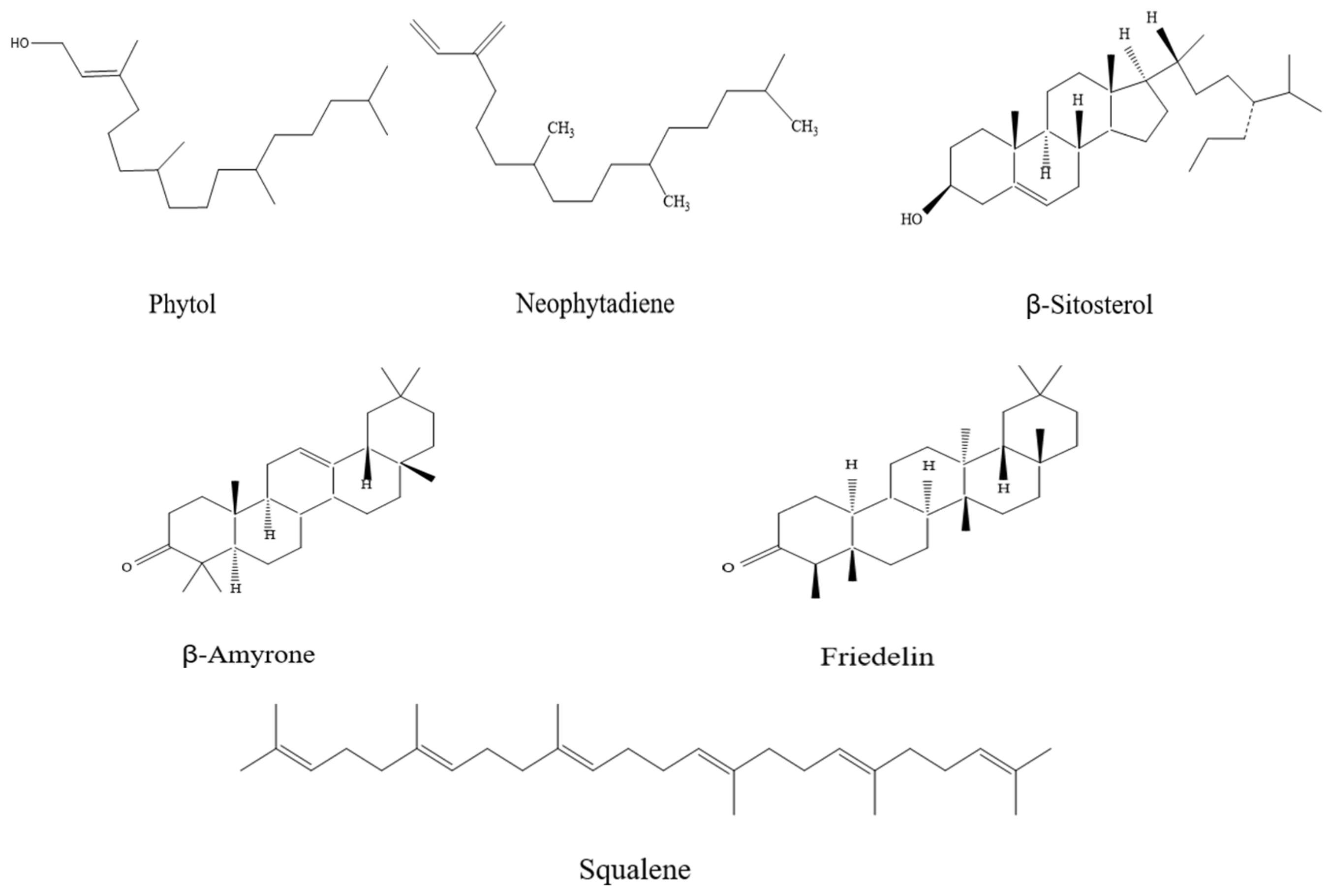
3.1. Impact of Different Extraction Methods on the Composition of I. latifolius Leaf Extracts

Various extraction methods, such as SD, SDE, UHPE-Hex, UHPE-EtOH, UE-Hex, UE-EtOH, and SC-CO2, were utilized for the extraction of terpenoids from I. latifolius leaves. The total ion chromatogram is depicted in Figure 1, while Table 3 illustrates the composition of the extracts, identifying a total of 111 components. The total absolute contents of the components were identified as follows: 10.360 mg (SD), 7.347 mg (SDE), 30.686 mg (UHPE-Hex), 4.682 mg (UHPE-EtOH), 13.749 mg (UE-EtOH), 35.957 mg (UE-Hex), and 28.160 mg (SC-CO2).
The GC-MS analysis of the UHPE-EtOH extract revealed the presence of only seven compounds, suggesting that the ethanol–ultrasound conditions might not be optimal for extracting active ingredients from I. latifolius leaves. Conversely, SD, SDE, and UHPE-Hex yielded relatively fewer components, with 18, 28, and 24 compounds, respectively, predominantly comprising terpenoids and hydrocarbons. The UE-Hex, UE-EtOH, and SC-CO2 extraction methods yielded 42, 27, and 38 components, respectively, predominantly terpenoids, including phytol, neophytadiene, β-amyrone, squalene, friedelin, β-sitosterol, campesterol, stigmasterol, sclareol, α-ionone, D-friedoolean-14-en-3-one, phytone, β-ionone, lup-20(29)-en-3-one, vitamin E, and γ-tocopherol. However, the UE extracts contained a large number of ester compounds.
The number and total absolute content of terpenoids in the SC-CO2 extracts (16, 25.207 mg) were significantly higher than those in the UE-Hex (12, 23.880 mg) and UE-EtOH (13, 9.665 mg) extracts. Notably, neophytadiene, phytol, β-sitosterol, β-amyrone, squalene, and friedelin exhibited relatively high contents in both the SC-CO2 and ultrasound extracts. The chemical structures of these compounds are depicted in Figure 2. Thus, these six terpenoid compounds were regarded as the primary terpenoids in I. latifolius leaves.

3.2. Influence of Different Extraction Methods on the Extraction Rate of I. latifolius Leaf Terpenoids (ILLTs)

The samples underwent GC-MS analysis to determine the contents of neophytadiene, phytol, β-sitosterol, β-amyrone, squalene, and friedelin, which were calculated using standard curves. Figure 3 displays the extraction rate and purity of the ILLTs using seven different extraction methods (SD, SDE, UHPE-Hex, UHPE-EtOH, UE-Hex, UE-EtOH, and SC-CO2). As shown in the figure, there were significant differences in the total extraction rate and purity of the six terpenoids obtained by different extraction methods. The SC-CO2 extraction rate exhibited the highest extraction yield and purity (1.18 ± 0.03 mg/g ILLTs, 8.67 ± 0.23%), followed by UE-Hex (1.12 ± 0.05 mg/g ILLTs, 4.44 ± 0.19%) and UE-EtOH (0.43 ± 0.06 mg/g ILLTs, 3.02 ± 0.19%). Tang et al. [26] discovered that supercritical fluid extraction technology was beneficial for the extraction of terpenoids. Moreover, the studies of He et al. [27] and Glisic et al. [28] demonstrated that compared to ultrasound-assisted extraction, supercritical extraction can obtain higher extraction rates and purity of flavonoids or essential oils from plants. The extraction efficiency of bioactive components is correlated with solvent polarity [29]. Ethanol, used as a solvent in ultrasound-assisted extraction, exhibited a better extraction efficiency than n-hexane, albeit with inferior purity. Luca et al. [30] and Zhang et al. [31] reported that an ethanol solution aids in extracting total triterpenoids, and it also facilitates the dissolution of other active ingredients. Although the extraction rates of SC-CO2 and UE-Hex were similar, SC-CO2 exhibited a significantly higher purity. Additionally, both UE and UHPE suffer from drawbacks such as high consumption of organic solvents, solvent residues, and increased impurities, thereby complicating subsequent purification steps. The lower extraction rates of the two distillation methods might result from compound decomposition during prolonged high-temperature processes and large polarity differences [32].

3.3. SEM Imaging of I. latifolius Leaves during Extraction Using Different Extraction Methods

SEM can be employed to observe the microstructural changes of I. latifolius leaves following extraction using the various extraction methods to elucidate the impact of these methods on the physical structure of the leaves. As depicted in Figure 4A, the surface of the leaves prior to the treatment appeared smooth, with regular microcavity structures visible along the edges. In Figure 4B–D, leaf residues treated by SD, SDE, and UHPE-EtOH demonstrated partially regular microcavity structures, exhibiting relatively smooth surfaces and fewer cracks. Figure 4E–H illustrate the residues treated by UHPE-Hex, UE-EtOH, UE-Hex, and SC-CO2, respectively, displaying rough surfaces, loose structures, and notable cracks and fragments. The employment of the UE and SC-CO2 extraction methods, particularly SC-CO2, led to more severe fragmentation of the leaves, resulting in greater damage to the cell walls and enhanced release of terpenoid compounds [33]. Ultrasonic action can enhance the cavitation effect of the liquid medium, thereby increasing the diffusion coefficient of the solutes (active ingredients) [34]. In addition, organic solvents have certain dissolution and softening effects on the cell wall structure, exacerbating the damage to microcavities [35]. The SEM results further supported the effectiveness of the UE and SC-CO2 extraction methods in promoting the yield of terpenoid compounds from Indocalamus latifolius leaves.

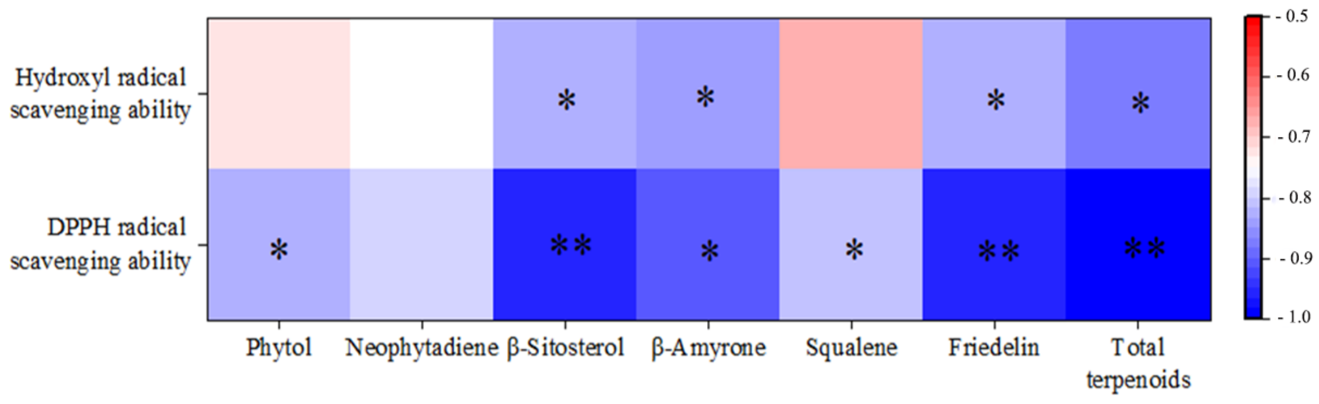
3.4. Study on the Antioxidant Activity of Extracts by Different Methods

The ability of potential antioxidants to transfer an electron to a reducing compound can be assessed by calculating the DPPH free radical scavenging rate [14]. As shown in Figure 5A, the extracts obtained from the seven extraction methods (SD, SDE, UHPE-Hex, UHPE-EtOH, UE-EtOH, UE-Hex, and SC-CO2) all exhibited DPPH free radical scavenging ability, and this ability was concentration-dependent. Within the concentration range of 0.4–1.2 mg/mL, the ability of the UE-EtOH, UE-Hex, and SC-CO2 extracts to scavenge DPPH free radicals increased with the concentration of the I. latifolius leaf extract. At a concentration of 0.8 mg/mL, the DPPH free radical scavenging rates of the three extracts gradually approached that of the control Vc, with the DPPH free radical scavenging rate of SC-CO2 extract slightly higher than that of the UE-EtOH and UE-Hex extracts. However, the DPPH free radical scavenging ability of the SD, SDE, and UHPE-Hex extracts gradually increased with concentration, while the scavenging rate was consistently lower than that of the other three extracts. As illustrated in Figure 5B, the ability of the seven extracts to scavenge hydroxyl radicals gradually increased with concentration. Compared with SD, SDE, UHPE-Hex, UE-EtOH, and UE-Hex, the SC-CO2 extract exhibited a stronger ability to scavenge hydroxyl radicals.
Studies have demonstrated that neophytadiene, phytol, β-sitosterol, β-amyrone, squalene, and friedelin, isolated from various plant organs, significantly contribute to the scavenging of DPPH free radicals, hydroxyl free radicals, superoxide anions, and carbon monoxide [22,36,37,38,39,40]. This highlights the remarkable antioxidant properties of these compounds across a range of reactive oxygen species. Thus, the correlation analysis conducted using SPSS revealed a significant association between the content of terpenoid compounds and antioxidant activity. As indicated in Figure 6, a significant correlation was observed between the two antioxidant methods (p < 0.05). Notably, there was an extremely significant negative correlation between the total content of terpenoids and the DPPH radical scavenging ability (IC50 value) (p < 0.01), as well as a significant negative correlation with the hydroxyl radical scavenging ability (IC50 value) (p < 0.05). Apart from neophytadiene, there was a highly significant (p < 0.01) or significant (p < 0.05) negative correlation between the DPPH radical scavenging ability (IC50 value) and the content of five terpenoid compounds. Meanwhile, a significant negative correlation was observed between the hydroxyl radical scavenging ability (IC50 value) and β-amyrone, β-sitosterol, and friedelin contents (p < 0.05). This suggested a potential synergistic effect among these compounds in enhancing antioxidant activity, warranting further investigation into their therapeutic potential.

3.5. Effects of Operating Conditions of SC-CO2 on the Extraction of ILLTs

The results of all the single-factor experiments are presented in Figure 6. The extraction yield and selectivity of the compound are influenced by the density, diffusivity, and viscosity of the fluid [41]. As shown in Figure 7A, the yield of the ILLTs notably increased when the extraction pressure was increased from 10 MPa to 20 MPa. A higher pressure may facilitate the diffusion of the solute and its contact with the supercritical CO2, sequentially increasing the yield [42]. However, a pressure exceeding 20 MPa led to the reverse trend on account of the highly compressed CO2, resulting in solute–solvent repulsion, as reported by Gong et al. [16]. Therefore, the pressure range of 10–30 MPa was deemed more suitable for RSM studies.
As indicated in Figure 7B, the extraction rate of the ILLTs showed an initial increase followed by a decrease with the temperature rise, similar to the findings of [42]. Above 37 °C, the curve gently declined. Further increases in temperature might lead to the degradation of terpenoids [43], resulting in a decrease in extraction yield. Thus, the temperature range of 32–42 °C was chosen for the CCD design.
Due to the nonpolar SC-CO2, polar modifiers facilitate the rapid and efficient solubility of polar molecules [44]. Initially, the yield of the ILLTs increased, reaching a maximum, but decreased when the cosolvent concentration exceeded 10% (v/w), as depicted in Figure 7C. According to González-Hernández et al. [45], less significant non-polar terpene hydrocarbons gradually transform into oxygenated terpenes with an increase in the cosolvent amount, demonstrating the cosolvent’s function. Subsequently, a cosolvent concentration of 10% (v/w) was selected as the CCD central point.
Under a constant extraction temperature (40 °C), extraction pressure (30 MPa), and ethanol content (10%, v/w), the impact of extraction time (2, 3, 4, 5, and 6 h) on ILLT yield was assessed. As shown in Figure 7D, the yield of the ILLTs increased with the extraction time until it reached 4 h, after which, a decreasing trend was observed. Gasparini et al. [46] discovered that longer extraction times and higher extraction pressures resulted in a decrease in diterpene selectivity, consistent with the findings mentioned above. Therefore, an extraction time of 4 h was selected as the central point of the CCD design.

3.6. Optimization of Extraction Conditions by CCD

The trial results and analysis of variance (ANOVA) of the experimental design tests are summarized in Table 4 and Table 5. The responses were fully explained by the predictive model established by the second-order polynomial Equation (8):
Y = 1 . 26 + 0 . 1431   ×   A + 0 . 0621   ×   B + 0 . 1070   ×   C + 0 . 0549   ×   D + 0 . 1035   ×   A   ×   B + 0 . 0395   ×   A   ×   C + 0 . 0457   ×   A   ×   D     0 . 0206   ×   B   ×   C + 0 . 0245   ×   B   ×   D + 0 . 0619   ×   C   ×   D     0 . 2252   ×   A 2 0 . 1173   ×   B 2 + 0 . 008   ×   C 2   0 . 1708   ×   D 2
The yields of ITLDs ranged from 0.6904 mg/g mg/g to 1.3107 mg/g. The determination coefficients of the models (R2) and the adjusted determination coefficient (R2adj) were 0.9738 and 0.9455, respectively, approaching 1, indicating that the model fit the experimental results [46]. The p-value of the model (<0.01) and the lack of fit p-value (>0.05) indicated that the model was statistically reliable [28]. The order of influence on the ILLT yield was as follows: extraction pressure > cosolvent concentration > extraction temperature > extraction time. Table 5 illustrates that the four linear coefficients (A, B, C, D), interaction coefficients (AB, AC, AD, CD), and quadratic term coefficients (A2, B2, D2) had significant effects on the yield of ITLDs (p < 0.05).
Figure 8 depicts the plots of the response surface and contour to more clearly reflect the influence relationship among the interactions of extraction temperature, extraction pressure, cosolvent concentration, and extraction time on the yield of ILLTs. As can be seen in Figure 8, the steepest slope of the AB response surface indicates that the interaction between extraction temperature and extraction pressure had the most significant impact on the extraction yield of ILLTs [47]. This result was consistent with the results of the ANOVA. The contour (Figure 8) indicates that the ILLT extraction yield initially improved and then declined with increasing extraction temperature, extraction pressure, or extraction time. Furthermore, there were positive effects of the cosolvent concentration on the extraction yield below 20%.
The response surface optimization model yielded the following optimal theoretical parameters: extraction pressure of 25.6 MPa, extraction temperature of 39.35 °C, cosolvent concentration of 20%, and extraction time of 4.52 h. Considering the experimental equipment conditions, the extraction time was set to 4.5 h, extraction pressure to 26 MPa, extraction temperature to 39 °C, and cosolvent concentration to 20%. Under these optimized conditions, the experimental yield of ILLTs was 1.44 ± 0.12 mg/g, which closely approximated the predicted value of 1.46 mg/g. Additionally, the yield of ITLDs extracted by supercritical CO2 fluid extraction was notably higher than that extracted by other techniques. In the research of Zulkafli et al. [19], β-siosterol, β-amyrene, and friedelin were likewise identified in bamboo leaf extracts using the SC-CO2 extraction method. Luca et al. [30] observed that lower pressures (90–110 bar) and lower temperatures (40–50 °C) contributed to improved extraction of the three hemp terpenes. A similar optimized condition (19 MPa/55 °C/5% EtOH) to that was employed in this study to enrich diterpenes from spent coffee grounds [17].

3.7. Purification Results of Terpenoid Compounds from I. latifolius Leaves

The crude extract obtained using the optimized conditions underwent purification, and the purified samples were subsequently analyzed by GC-MS. Following purification through WelFlash C18-l, BUCHI-C18, and Sephadex LH-20, the crude extract of I. latifolius leaves exhibited an increased purity of components F14 and F17. The GC-MS results are presented in Figure 9. The gas chromatogram of the samples showed distinct peaks at 22, 27, 38, 44, 45, and 48 min, corresponding to standards for neophytadiene, phytol, β-sitosterol, β-amyrone, squalene, and friedelin. A comparison of the mass spectra of the six major compounds in the sample with those of known compounds and standards in the GC-MS NIST 20 library (Figures S1–S6) confirmed a high degree of agreement between the six major purified compounds and the standards. The purified F17 fraction also showed trace amounts of campesterol and stigmasterol, which are plant sterols belonging to triterpenoids, in addition to β-sitosterol. The purity of the crude extract of terpenoid compounds from I. latifolius leaves obtained under the optimized conditions increased from 12.91% to 93.34%.

3.8. Cytotoxicity Effect of ILLTs on HepG2 Cells

High doses of plant extracts can be toxic and harmful to normal physiological systems. But, if the concentration is too low, it will not be effective [46]. Therefore, a toxicity analysis was performed to ensure that the extract was safe for the treatment of HepG2 cells. The direct effect of the ILLTs on HepG2 cell viability was studied in the concentration range of 0–250 μg/mL. The results in Figure 10A show that treatment with 80–250 μg/mL of the ILLTs for 24 h started to exhibit cytotoxicity in a dose-dependent manner (92.64–9.71%). However, at a concentration of 40 μg/mL, the cytotoxicity was negligible. Furthermore, the ILLTs exhibited an IC50 of 148.93 ± 9.93 μg/mL. Under the condition of a high cell number, we selected a concentration of 80–90 μg/mL for further experiments. Nguyen et al. [48] successfully isolated nine sesquiterpenes and two diterpenes from Curcuma zedoaroides (Zingiberaceae) rhizomes which displayed significant activity against A549, MCF-7, MDA-MB231, HL-60, and HepG2 cells, with IC50 values ranging from 3.13 μM to 30.10 μM. As depicted in Figure 10B, at a concentration of 10 g/mL of TBHP, the cell viability decreased to 73.67%, indicating that significant oxidative damage was induced by TBHP. Therefore, 10 mg/mL of TBHP was chosen as the experimental concentration to establish models of oxidative damage.

3.9. Effects of ILLTs on ROS and MDA Content of HepG2 Cells

Currently, there are little data on the antioxidant effects of plant terpenoids in HepG2 cells. To determine the protective effects of ILLTs against oxidative stress, the well-known inducer TBHP was used to generate excessive ROS and MDA levels in HepG2 cells. From the images shown in Figure 10D, TBHP increased the intracellular ROS level by 399.78% compared to the control group. Cells that were treated with 80 μg/mL and 90 μg/mL of the ILLTs significantly reduced the intracellular ROS concentration, which dropped by 299.70% and 239.61%, respectively. These findings were consistent with the fluorescence microscopy results (Figure 10C), where TBHP-stimulated cells exhibited intense green fluorescence due to ROS.
ROS produced by cells can react with polyunsaturated fatty acids in biological membranes, leading to lipid peroxidation and the generation of MDA [49]. The groups treated with TBHP exhibited similar effects, regarding the accumulation of MDA (Figure 10D). When the cells were treated with 80–90 μg/mL of the ILLTs, the MDA content decreased by 15.31–26.07%. The data indicate that luteolin diminished the accumulation of intracellular ROS and MDA in a dose-dependent manner.

4. Conclusions

This study developed a rapid qualitative analysis method for the components of I. latifolius leaves and conducted a quantitative analysis of six major terpenoid compounds using GC-MS. A qualitative analysis was performed on crude extracts obtained from I. latifolius leaves using various extraction methods including SD, SDE, UHPE-Hex, UHPE-EtOH, UE-Hex, UE-EtOH, and SC-CO2 extraction methods. A total of 121 components were identified, with the total absolute contents of components from SD (18), SDE (28), UHPE-Hex (24), UHPE-EtOH (7), UE-Hex (42), UE-EtOH (27), and SC-CO2 (38) extracts being 10.360 mg, 7.347 mg, 30.686 mg, 4.682 mg, 35.957 mg,13.749 mg, and 28.160 mg, respectively. Neophytadiene, phytol, β-sitosterol, β-amyrone, squalene, and friedelin were identified as the main terpenoid compounds in I. latifolius leaves (ILLTs). The effects of the seven different extraction methods on the extraction rate, purity, and antioxidant activity of the ILLTs were investigated. The experimental results demonstrated that the SC-CO2 extraction method could significantly enhance the extraction rate and purity of terpenoid compounds from I. latifolius leaves. The SEM results also revealed substantial damage to the leaf structure caused by the SC-CO2 extraction method, facilitating solute solubilization.
Additionally, the crude extract obtained by SC-CO2 exhibited significant advantages in terms of antioxidant activity. The correlation analysis suggested a potential synergistic effect among these compounds in enhancing antioxidant activity, warranting further investigation into their therapeutic potential. The optimal conditions for extracting ILLTs were determined through single-factor experiments and orthogonal experiments, resulting in an extraction time of 4.5 h, extraction pressure of 26 MPa, extraction temperature of 39 °C, and cosolvent concentration of 20%. Under these conditions, the actual extraction rate for the ILLTs reached 1.44 ± 0.12 mg/g, significantly higher than the other six methods. The ILLTs were demonstrated to have cytotoxic effects on HepG2 cells with an IC50 value of 148.93 ± 9.93 μg/mL. Moreover, the ILLTs were able to improve the cellular antioxidant status in a dose-dependent manner and protect HepG2 cells from oxidative damage by inhibiting ROS and MDA production. These terpenoid compounds have been shown to possess a potential synergistic effect in enhancing antioxidant activity, holding promising prospects for treating diseases caused by oxidative stress. In order to have a more comprehensive understanding of the processing properties and other functional properties of Indocalamus latifolius leaf terpenoids, further studies are needed.

Supplementary Materials

The following supporting information can be downloaded at: https://www.mdpi.com/article/10.3390/foods13111719/s1, Figure S1: Mass spectrum of purified samples and standards of neophytadiene; Figure S2: Mass spectrum of purified samples and standards of phytol; Figure S3: Mass spectrum of purified samples and standards of squalene; Figure S4: Mass spectrum of purified samples and standards of β-amyrone; Figure S5: Mass spectrum of purified samples and standards of β-sitosterol; Figure S6: Mass spectrum of purified samples and standards of friedelin.

Author Contributions

Y.C., conceptualization, methodology, investigation, formal analysis, writing—original draft, writing—review and editing; Y.W., conceptualization, funding acquisition, resources, formal analysis, methodology, investigation, writing—review and editing; L.H., resources, formal analysis, methodology, investigation; L.W., formal analysis, investigation, writing—review and editing; J.Z., resources, formal analysis, methodology, investigation; Z.Y., resources, formal analysis, methodology, investigation; Q.L., resources, formal analysis, methodology, investigation; R.S., conceptualization, resources, supervision, writing—review and editing. All authors have read and agreed to the published version of the manuscript.

Funding

This work was supported by a research fellowship from the Key Laboratory of State Forest Food Resources Utilization and Quality Control and financial grants from the Project of Zhejiang Provincial and Academy Corporation (No. 2022SY07) and the Project of Science and Technology of Zhejiang Government, China (No. 2024F1065-3).

Institutional Review Board Statement

Not applicable.

Informed Consent Statement

Not applicable.

Data Availability Statement

The original contributions presented in the study are included in the article/Supplementary Materials, further inquiries can be directed to the corresponding authors.

Conflicts of Interest

The authors declare no conflicts of interest.

References

  1. Chen, C.; Ding, Y.; Elmahadi, E.A.; Zhou, J.; Xu, H. Study on the isolation, purification and physicochemical properties of polysaccharides from Indocalamus tesselatus. Biomed. Chromatogr. 1999, 13, 11–14. [Google Scholar] [CrossRef]
  2. Sun, J.; Xun, H.; Yu, J.; Tang, F.; Yue, Y.-D.; Guo, X.-F. Chemical Constituents and Antibacterial Properties of Indocalamus latifolius McClure Leaves, the Packaging Material for “Zongzi”. Molecules 2015, 20, 15686–15700. [Google Scholar] [CrossRef] [PubMed]
  3. Zhang, Y.; He, L.; Li, Q.; Cheng, J.; Wang, Y.; Zhao, J.; Yuan, S.; Chen, Y.; Shi, R. Optimization of ultrasonic-assisted deep eutectic solvent for the extraction of polysaccharides from Indocalamus tessellatus leaves and their biological studies. Sustain. Chem. Pharm. 2022, 30, 100855. [Google Scholar] [CrossRef]
  4. Tu, L.; Cai, X.; Zhang, Y.; Tong, Y.; Wang, J.; Su, P.; Lu, Y.; Hu, T.; Luo, Y.; Wu, X.; et al. Mechanistic analysis for the origin of diverse diterpenes in Tripterygium wilfordii. Acta Pharm. Sin. B 2022, 12, 2923–2933. [Google Scholar] [CrossRef] [PubMed]
  5. Wardana, A.P.; Aminah, N.S.; Rosyda, M.; Abdjan, M.I.; Kristanti, A.N.; Tun, K.N.W.; Choudhary, M.I.; Takaya, Y. Potential of diterpene compounds as antivirals, a review. Heliyon 2021, 7, e07777. [Google Scholar] [CrossRef] [PubMed]
  6. Liu, Y.; Liu, Y.; Wang, X.; Qiao, Z.; Yu, X.; Zhu, Y.; Xie, L.; Qiang, L.; Fu, Y. Bioactive daphnane diterpenes from Wikstroemia chuii with their potential anti-inflammatory effects and anti-HIV activities. Bioorg. Chem. 2020, 105, 104388. [Google Scholar] [CrossRef]
  7. Jiang, C.; Yang, M.; Zhao, C.; Liu, F.; Yang, H.; Li, Z.; Han, T.; Lin, B.; Li, D.; Hua, H. Abietane diterpenes from the twigs and leaves of Cephalotaxus oliveri Mast. with antitumor activity. Phytochemistry 2022, 199, 113187. [Google Scholar] [CrossRef] [PubMed]
  8. Paulous, R.; Zakaria, Z.; Awang, J.; Kamada, T.; Vairappan, C.S. Antifungal diterpene from Rhizome of wild bornean ginger, Hornstedtia havilandii (Zingiberaceae). Biochem. Syst. Ecol. 2022, 105, 104546. [Google Scholar] [CrossRef]
  9. Herrera, J.G.; Ramos, M.P.; de Lima Albuquerque, B.N.; de Oliveira Farias de Aguiar, J.C.R.; Agra Neto, A.C.; Guedes Paiva, P.M.; do Amaral Ferraz Navarro, D.M.; Pinto, L. Multivariate evaluation of process parameters to obtain essential oil of Piper corcovadensis using supercritical fluid extraction. Microchem. J. 2022, 181, 107747. [Google Scholar] [CrossRef]
  10. Pizani, R.S.; Viganó, J.; de Souza Mesquita, L.M.; Contieri, L.S.; Sanches, V.L.; Chaves, J.O.; Souza, M.C.; da Silva, L.C.; Rostagno, M.A. Beyond aroma: A review on advanced extraction processes from rosemary (Rosmarinus officinalis) and sage (Salvia officinalis) to produce phenolic acids and diterpenes. Trends Food Sci. Technol. 2022, 127, 245–262. [Google Scholar] [CrossRef]
  11. Guta, M.; Tan, H.; Zhao, Y. Optimization of supercritical CO2 extraction of incensole-enriched oil from Boswellia papyrifera resin using response surface methodology. J. Supercrit. Fluids 2024, 205, 106154. [Google Scholar] [CrossRef]
  12. Pereira, G.S.L.; Magalhães, R.d.S.; Fraga, S.; de Souza, P.T.; de Lima, J.P.; Meirelles, A.J.d.A.; Sampaio, K.A. Extraction of bioactive compounds from Butia capitata fruits using supercritical carbon dioxide and pressurized fluids. J. Supercrit. Fluids 2023, 199, 105959. [Google Scholar] [CrossRef]
  13. Vinitha, U.G.; Sathasivam, R.; Muthuraman, M.S.; Park, S.U. Intensification of supercritical fluid in the extraction of flavonoids: A comprehensive review. Physiol. Mol. Plant Pathol. 2022, 118, 101815. [Google Scholar] [CrossRef]
  14. Melloul, S.; Zehioua, R.; Meniai, A.-H. Supercritical CO2 extraction of bioactive compounds from local Peganum Harmala plant seeds and optimization of the extraction yield and the antioxidant activities. Sustain. Chem. Pharm. 2022, 28, 101815. [Google Scholar] [CrossRef]
  15. Alvarez-Henao, M.V.; Cardona, L.; Hincapié, S.; Londoño-Londoño, J.; Jimenez-Cartagena, C. Supercritical fluid extraction of phytosterols from sugarcane bagasse: Evaluation of extraction parameters. J. Supercrit. Fluids 2022, 179, 105427. [Google Scholar] [CrossRef]
  16. Gong, T.; Liu, S.; Wang, H.; Zhang, M. Supercritical CO2 fluid extraction, physicochemical properties, antioxidant activities and hypoglycemic activity of polysaccharides derived from fallen Ginkgo leaves. Food Biosci. 2021, 42, 101153. [Google Scholar] [CrossRef]
  17. Barbosa, H.M.A.; de Melo, M.M.R.; Coimbra, M.A.; Passos, C.P.; Silva, C.M. Optimization of the supercritical fluid coextraction of oil and diterpenes from spent coffee grounds using experimental design and response surface methodology. J. Supercrit. Fluids 2014, 85, 165–172. [Google Scholar] [CrossRef]
  18. Glisic, S.; Ivanovic, J.; Ristic, M.; Skala, D. Extraction of sage (Salvia officinalis L.) by supercritical CO2: Kinetic data, chemical composition and selectivity of diterpenes. J. Supercrit. Fluids 2010, 52, 62–70. [Google Scholar] [CrossRef]
  19. Zulkafli, Z.D.; Wang, H.; Miyashita, F.; Utsumi, N.; Tamura, K. Cosolvent-modified supercritical carbon dioxide extraction of phenolic compounds from bamboo leaves (Sasa palmata). J. Supercrit. Fluids 2014, 94, 123–129. [Google Scholar] [CrossRef]
  20. Muala, W.C.B.; Desobgo, Z.S.C.; Jong, N.E. Optimization of extraction conditions of phenolic compounds from Cymbopogon citratus and evaluation of phenolics and aroma profiles of extract. Heliyon 2021, 7, e06744. [Google Scholar] [CrossRef]
  21. Claro-Cala, C.M.; Grao-Cruces, E.; Toscano, R.; Millan-Linares, M.C.; Montserrat-de la Paz, S.; Martin, M.E. Acyclic Diterpene Phytol from Hemp Seed Oil (Cannabis sativa L.) Exerts Anti-Inflammatory Activity on Primary Human Monocytes-Macrophages. Foods 2022, 11, 2366. [Google Scholar] [CrossRef] [PubMed]
  22. Itoh, T.; Ono, A.; Kawaguchi, K.; Teraoka, S.; Harada, M.; Sumi, K.; Ando, M.; Tsukamasa, Y.; Ninomiya, M.; Koketsu, M.; et al. Phytol isolated from watermelon (Citrullus lanatus) sprouts induces cell death in human T-lymphoid cell line Jurkat cells via S-phase cell cycle arrest. Food Chem. Toxicol. 2018, 115, 425–435. [Google Scholar] [CrossRef]
  23. He, B.; Chen, L.; Chen, C.; Zeng, J.; Xu, J.; He, X.; Wang, Y. Anti-inflammatory terpenoids from the seeds of Eucommia ulmoides. Phytochem. Lett. 2024, 61, 44–51. [Google Scholar] [CrossRef]
  24. Zhang ZDai, X. Preparation of alginate oligosaccharide nanoliposomes and an analysis of their inhibitory effects on Caco-2 cells. IET Nanobiotechnol. 2018, 12, 946–950. [Google Scholar] [CrossRef]
  25. Wang, Y.; Wang, X.; Hao, X.; Yan, Y.; Hong, M.; Wei, S.; Zhou, Y.; Wang, Q.; Cheng, Y.; Liu, Y. Ethanol Extract of Centipeda minima Exerts Antioxidant and Neuroprotective Effects via Activation of the Nrf2 Signaling Pathway. Oxidative Med. Cell. Longev. 2019, 2019, 9421037. [Google Scholar] [CrossRef] [PubMed]
  26. Tang, J.; Zhao, N.; Gao, Y.; Han, R.; Wang, X.; Tian, J.; Gao, J. Phytosterol profiles and iridoids of the edible Eucommia ulmoides Oliver seeds and their anti-inflammatory potential. Food Biosci. 2021, 43, 101295. [Google Scholar] [CrossRef]
  27. He, D.; Wang, S.; Fang, G.; Zhu, Q.; Wu, J.; Li, J.; Shi, D.; Lian, X. LXRs/ABCA1 activation contribute to the anti-inflammatory role of phytosterols on LPS-induced acute lung injury. J. Funct. Foods 2022, 89, 104966. [Google Scholar] [CrossRef]
  28. Glisic, S.B.; Ristic, M.; Skala, D.U. The combined extraction of sage (Salvia officinalis L.): Ultrasound followed by supercritical CO2 extraction. Ultrason. Sonochem. 2011, 18, 318–326. [Google Scholar] [CrossRef]
  29. Chamali, S.; Bendaoud, H.; Bouajila, J.; Camy, S.; Saadaoui, E.; Condoret, J.-S.; Romdhane, M. Optimization of accelerated solvent extraction of bioactive compounds from Eucalyptus intertexta using response surface methodology and evaluation of its phenolic composition and biological activities. J. Appl. Res. Med. Aromat. Plants 2023, 35, 100464. [Google Scholar] [CrossRef]
  30. Luca, S.V.; Kittl, T.; Minceva, M. Supercritical CO2 extraction of hemp flowers: A systematic study to produce terpene-rich and terpene-depleted cannabidiol fractions. Ind. Crops Prod. 2022, 187, 115395. [Google Scholar] [CrossRef]
  31. Zhang, K.; Ding, Z.; Mo, M.; Duan, W.; Bi, Y.; Kong, F. Essential oils from sugarcane molasses: Chemical composition, optimization of microwave-assisted hydrodistillation by response surface methodology and evaluation of its antioxidant and antibacterial activities. Ind. Crops Prod. 2020, 156, 112875. [Google Scholar] [CrossRef]
  32. Bajer, T.; Surmová, S.; Eisner, A.; Ventura, K.; Bajerová, P. Use of simultaneous distillation-extraction, supercritical fluid extraction and solid-phase microextraction for characterisation of the volatile profile of Dipteryx odorata (Aubl.) Willd. Ind. Crops Prod. 2018, 119, 313–321. [Google Scholar] [CrossRef]
  33. Xie, Y.; Wang, F.; Ke, B.; Zhang, L.; Yan, X.; Deng, G. Sequential two-stage extraction of Xanthoceras sorbifolia seed oil using supercritical CO2 and CO2-expanded ethanol. J. Supercrit. Fluids 2023, 200, 105977. [Google Scholar] [CrossRef]
  34. Zhao, F.; Wang, X.; Liu, H.; Qian, J. In vitro and in vivo anti-inflammatory models demonstrate oligopeptides play a significant role in anti-inflammatory properties of white tea. J. Funct. Foods 2024, 112, 105983. [Google Scholar] [CrossRef]
  35. Díaz-Viciedo, R.; Hortelano, S.; Girón, N.; Massó, J.M.; Rodriguez, B.; Villar, A.; de las Heras, B. Modulation of inflammatory responses by diterpene acids from Helianthus annuus L. Biochem. Biophys. Res. Commun. 2008, 369, 761–766. [Google Scholar] [CrossRef] [PubMed]
  36. Sunil, C.; Duraipandiyan, V.; Ignacimuthu, S.; Al-Dhabi, N.A. Antioxidant, free radical scavenging and liver protective effects of friedelin isolated from Azima tetracantha Lam. leaves. Food Chem. 2013, 139, 860–865. [Google Scholar] [CrossRef]
  37. Wang, G.; Zhang, X.; Wu, Q.; Jin, Y.; Ning, C.; Wang, R.; Mao, J.; Chen, M. The hepatoprotective effects of Sedum sarmentosum extract and its isolated major constituent through Nrf2 activation and NF-κB inhibition. Phytomedicine 2019, 53, 263–273. [Google Scholar] [CrossRef] [PubMed]
  38. Khan, Z.; Nath, N.; Rauf, A.; Emran, T.B.; Mitra, S.; Islam, F.; Chandran, D.; Barua, J.; Khandaker, M.U.; Idris, A.M.; et al. Multifunctional roles and pharmacological potential of β-sitosterol: Emerging evidence toward clinical applications. Chem.-Biol. Interact. 2022, 365, 110117. [Google Scholar] [CrossRef] [PubMed]
  39. Sahoo, S.; Rath, D.; Kar, D.M.; Pattanaik, S. Hepatoprotective potency of Litsea glutinosa (L.) C.B. Rob. leaf methanol extract on H2O2-induced toxicity in HepG2 cells. J. Ethnopharmacol. 2023, 304, 116076. [Google Scholar] [CrossRef] [PubMed]
  40. Dumandan, N.G.; Kagaoan, A.C.T.; Acda, R.D.P.; Tumambing, C.R.; Pham, L.J.; Mohamed, S.A. Extraction, Profiling, and Characterization of Phytosterols and Triterpenoids from Pili (Canarium ovatum Engl.) Pulp Oil Exhibiting Antioxidant and Antibacterial Properties. Biochem. Res. Int. 2022, 2022, 6604984. [Google Scholar] [CrossRef]
  41. Marzlan, A.A.; Muhialdin, B.J.; Zainal Abedin, N.H.; Mohammed, N.K.; Abadl, M.M.T.; Mohd Roby, B.H.; Meor Hussin, A.S. Optimized supercritical CO2 extraction conditions on yield and quality of torch ginger (Etlingera elatior (Jack) R.M. Smith) inflorescence essential oil. Ind. Crops Prod. 2020, 154, 112581. [Google Scholar] [CrossRef]
  42. Wang, H.; Liu, Y.; Wei, S.; Yan, Z. Application of response surface methodology to optimise supercritical carbon dioxide extraction of essential oil from Cyperus rotundus Linn. Food Chem. 2012, 132, 582–587. [Google Scholar] [CrossRef] [PubMed]
  43. Yousefi, M.; Rahimi-Nasrabadi, M.; Pourmortazavi, S.M.; Wysokowski, M.; Jesionowski, T.; Ehrlich, H.; Mirsadeghi, S. Supercritical fluid extraction of essential oils. TrAC Trends Anal. Chem. 2019, 118, 182–193. [Google Scholar] [CrossRef]
  44. Rout, P.K.; Naik, S.N.; Rao, Y.R.; Jadeja, G.; Maheshwari, R.C. Extraction and composition of volatiles from Zanthoxylum rhesta: Comparison of subcritical CO2 and traditional processes. J. Supercrit. Fluids 2007, 42, 334–341. [Google Scholar] [CrossRef]
  45. González-Hernández, R.A.; Valdez-Cruz, N.A.; Trujillo-Roldán, M.A. Factors that influence the extraction methods of terpenes from natural sources. Chem. Pap. 2024, 78, 2783–2810. [Google Scholar] [CrossRef]
  46. Gasparini, A.; Ferrentino, G.; Angeli, L.; Morozova, K.; Zatelli, D.; Scampicchio, M. Ultrasound assisted extraction of oils from apple seeds: A comparative study with supercritical fluid and conventional solvent extraction. Innov. Food Sci. Emerg. Technol. 2023, 86, 103370. [Google Scholar] [CrossRef]
  47. Hu, Y.; Yang, L.; Liang, Z.; Chen, J.; Zhao, M.; Tang, Q. Comparative analysis of flavonoids extracted from Dendrobium chrysotoxum flowers by supercritical fluid extraction and ultrasonic cold extraction. Sustain. Chem. Pharm. 2023, 36, 101267. [Google Scholar] [CrossRef]
  48. Nguyen, T.T.; Tran, T.H.; Nguyen, T.H.; Do, T.H. Cytotoxic sesquiterpenes and diterpenes from the rhizomes of Curcuma zedoaroides Chaveer. & Tanee. Biochem. Syst. Ecol. 2024, 112, 104781. [Google Scholar]
  49. Tao, L.; Gu, F.; Liu, Y.; Yang, M.; Wu, X.-Z.; Sheng, J.; Tian, Y. Preparation of antioxidant peptides from Moringa oleifera leaves and their protection against oxidative damage in HepG2 cells. Front. Nutr. 2022, 9, 1062671. [Google Scholar] [CrossRef]
Figure 1. Total ion compound chromatograms (TICs) of I. latifolius leaf extracts.
Foods 13 01719 g001
Figure 2. The chemical structures of terpenoids.
Foods 13 01719 g002
Figure 3. Main effect plot showing the effect of each factor on the yield (mg/g). Different letters indicate significant differences (p < 0.05). The black and red letters for the statistical analysis represent the significance of the extraction rate and purity differences, respectively.
Foods 13 01719 g003
Figure 4. SEM of I. latifolius leaf powder before (A) and after extraction by SD (B), SDE (C), UHPE-EtOH (D), UHPE-Hex (E), UE-EtOH (F), UE-Hex (G), and SC-CO2 (H).
Foods 13 01719 g004
Figure 5. DPPH radical scavenging activity (A) and hydroxyl radical scavenging activity (B) of ILLTs extracted by different methods.
Foods 13 01719 g005
Figure 6. Correlation analysis of terpenoid content and antioxidant activity in I. latifolius leaf extracts. ** highly significant, p < 0.01; * significant, 0.01 < p < 0.05.
Foods 13 01719 g006
Figure 7. Effect of various factors on the extraction yield of ILLTs. Effect of extraction pressure (A), extraction temperature (B), cosolvent content (C), time (D) on extraction yield.
Foods 13 01719 g007
Figure 8. Response surface plots showing interactive effects of two-factor interactions on the yield of ITLDs. (AF) contour map, (af) 3D surface map.
Foods 13 01719 g008
Figure 9. Gas chromatogram of purified samples (F14, F17) and standards.
Foods 13 01719 g009
Figure 10. Effects of different concentrations of ILLTs (A) and TBHP (B) on cell viability of HepG2 cells; fluorescence images (C) of effects of different concentrations of ILLTs on TBHP-induced ROS formation in HepG2 cells; effects of different concentrations of ILLTs on TBHP-induced ROS and MDA production in HepG2 cells (D). Different letters indicate significant differences (p < 0.05).
Foods 13 01719 g010
Table 1. DPPH free radical scavenging experiment reagent volumes.
GroupSamples, VcEthyl AlcoholDPPH Ethanol Solution (0.1 mmol/L)
Asample1 mL-1 mL
Ablank1 mL1 mL-
Acontrol-1 mL1 mL
Table 2. The design and results of central composite design (CCD).
FactorUnitsLevel of Factor
−101
A: Extraction stressMPa102030
B: Extraction temperature°C323742
C: Cosolvent% (v/w)01020
D: Timeh345
Table 3. Volatile composition of I. latifolius leaves expressed as the compound percentages based on GC-MS analysis.
No.CompoundMolecular FormulaMolecular WeightCASAbsolute Content * (mg)
SDSDEUHPE-EtOHUHPE-HexUE-EtOHUE-HexSC-CO2
1IsophytolC20H40O297505-32-80.069
± 0.001
0.036
± 0.001
2PhytolC20H40O297150-86-70.677
± 0.043
0.438
± 0.061
0.219
± 0.017
1.553
± 0.083
2.932
± 0.096
5.38
± 0.089
5.745
± 0.073
3HeneicosanolC21H44O3133381-26-8 1.948
± 0.101
0.086
± 0.060
4OctacosanolC28H58O411557-61-9 0.148
± 0.001
0.167
± 0.001
0.071
± 0.029
5LignocerolC24H50O355506-51-4 0.094
± 0.031
6CampesterolC28H48O401474-62-4 0.196
± 0.037
0.346
± 0.052
0.422
± 0.047
7StigmasterolC29H48O41383-48-7 0.104
± 0.018
0.171
± 0.046
0.269
± 0.051
8β-SitosterolC29H50O4155779-62-4 0.019
± 0.006
3.094
± 0.122
8.686
± 0.141
9.909
± 0.901
9LupeolC30H50O427545-47-1 0.074
± 0.061
102-(Octadecyloxy)-ethanolC20H42O25332136-72-3 0.013
± 0.041
112-Ethyl-1-decanolC12H26O18621078-65-9 0.011
± 0.001
12Tetrahydrofurfuryl alcoholC5H10O210297-99-4 0.061
± 0.008
131-DocosanolC22H46O326661-19-8 0.285
± 0.067
14SclareolC20H36O2308515-03-7 0.227
± 0.001
0.084
± 0.033
15(E, E)-10,12-Hexadecadien-1-ol acetatC16H30O238765-19-5 0.033
± 0.012
16HeptacosanolC27H56O3972004-39-9 0.124
± 0.092
17NonacosanolC29H60O42525154-56-7 0.279
± 0.082
18Glutina-5-ene-3β-olC30H50O427545-24-4 0.072
± 0.041
19GlycerinC3H8O39230918-77-5 12.36
± 1.073
20HexadecanalC16H32O240629-80-10.064
± 0.005
0.124
± 0.076
21OctadecanalC18H36O268638-66-44.050
± 0.475
0.036
± 0.018
0.041
± 0.021
22HexanalC6H12O10066-25-1 1.18
± 0.089
232-HexenalC6H10O98505-57-7 0.220
± 0.074
24(E, E)-2,4-HeptadienalC7H10O1104313-3-5 0.061
± 0.023
25BenzeneacetaldehydeC8H8O120122-78-1 1.648
± 0.001
26NonanalC9H18O142124-19-6 0.916
± 0.149
27β-CyclocitralC10H16O152432-25-7 0.102
± 0.013
28(Z)-7-TetradecenalC14H26O21065128-96-3 0.127
± 0.016
29α-IononeC13H20O192127-41-30.003
± 0.001
0.491
± 0.165
0.024
± 0.007
30β-Apo-13-carotenoneC18H26O25817974-57-1 0.097
± 0.011
0.072
± 0.031
0.059
± 0.023
312-NonadecanoneC19H38O282629-66-3 0.057
± 0.009
32DotriacontanalC32H64O46557878-00-9 2.016
± 0.822
332-NonacosanoneC29H58O42317600-99-6 0.099
± 0.021
34PhytoneC18H36O268502-69-20.709
± 0.173
0.211
± 0.054
0.032
± 0.007
35Trans-β-IononeC13H20O19214901-07-6 0.032
± 0.008
0.070
± 0.011
36HydroxyacetoneC3H6O274116-09-6 9.461
± 0.363
37D-Friedoolean-14-en-3-oneC30H48O425514-07-8 0.062
± 0.013
0.296
± 0.047
384-(1,5-Dihydroxy-2,6,6-trimethylcyclohex-2-enyl) but-3-en-2-oneC13H20O320838963-41-6 0.035
± 0.014
394-(3-Hydroxybutyl)-3,5,5-trimethyl-2-cyclohexen-1-oneC13H22O221036151-02-7 0.041
± 0.016
40β-AmyroneC30H48O425638-97-1 0.517
± 0.001
1.583
± 0.891
1.591
± 0.131
41Lup-20(29)-en-3-oneC30H48O4251617-70-5 0.061
± 0.012
0.071
± 0.019
42FriedelinC30H50O427559-74-0 0.062
± 0.018
1.405
± 0.521
6.699
± 1.091
4.100
± 0.941
432,3-Dihydro-3,5-dihydroxy-6-methyl-4(H)-pyran-4-oneC6H8O414428564-83-2 6.588
± 0.873
4417-PentatriaconteneC15H30O2262345-28-0 0.050
± 0.016
452-Methoxy-4-vinylphenolC9H10O21507786-61-0 0.256
± 0.068
463,5-Di-tert-butylphenolC14H22O2061138-52-9 0.114
± 0.070
472,4-Di-t-butylphenolC14H22O20696-76-4 0.059
± 0.017
0.024
± 0.013
482,2′-Methylenebis(6-tert-butyl-4-methylphenol)C23H32O2340119-47-1 0.405
± 0.026
0.452
± 0.095
492,6-Di-tert-butylphenolC14H22O206128-39-2 0.561
± 0.064
50γ-TocopherolC28H48O241754-28-4 0.042
± 0.015
0.080
± 0.021
51(Z, Z)-9,12-Octadecadienoic acidC20H36O2424544-35-4 0.546
± 0.088
525,6,7,7a-Tetrahydro-4,4,7a-trimethyl-2(4H)-benzofuranoneC11H16O218015356-74-8 0.024
± 0.006
53Hexadecanoic acid, methyl esterC17H34O2270112-39-00.132
± 0.025
1.329
± 0.471
0.357
± 0.081
54Ethyl palmitateC18H36O2284628-97-7 0.096
± 0.041
0.717
± 0.092
55Octacosyl acetateC30H60O245218206-97-80.012
± 0.001
56Methyl salicylateC8H8O3152119-36-8 0.104
± 0.019
57Benzyl salicylateC14H12O3228118-58-1 0.036
± 0.017
581-Heneicosyl formateC22H44O234177899-03-7 0.013
± 0.003
59(Z)-7-Hexadecenoic acid, methyl esterC17H32O226856875-67-3 0.074
± 0.015
609-Hexadecenoic acid, ethyl esterC18H34O228254546-22-4 0.118
± 0.027
61Heptadecanoic acid, ethyl esterC19H38O229914010-23-2 0.044
± 0.009
0.046
± 0.011
62Elaidic acid ethyl esterC20H38O23116114-18-7 0.763
± 0.039
63Ethyl icosanoateC22H44O234118281-05-5 0.060
± 0.017
0.103
± 0.005
641-Hexadecanol, acetateC18H36O2284629-70-9 0.072
± 0.033
65ButyrolactoneC4H6O2863068-88-0 1.402
± 0.107
66Ethyl stearateC20H40O2313111-61-5 0.035
± 0.007
67Methyl octadeca-9,12-dienoateC19H34O22942566-97-4 0.162
± 0.017
681,2,3-Trielaidoyl glycerolC57H104O6885537-39-3 0.496
± 0.065
69Phytyl acetateC22H42O233910236-16-5 0.044
± 0.013
70β-Sitosterol acetateC31H52O2457915-05-9 0.086
± 0.014
71α-Tocopheryl acetateC31H52O34737695-91-2 0.059
± 0.005
72Ethyl linolenateC20H34O23061191-41-9 1.007
± 0.085
73Linoleic acid ethyl esterC20H36O2309544-35-4 0.579
± 0.053
0.702
± 0.074
742,2-Dimethyl-3-(3,7,12,16,20-pentamethyl-3,7,11,15,19-heneicosapentaenyl)-oxiraneC30H50O4277200-26-2 0.053
± 0.006
0.056
± 0.011
751-PentadeceneC15H3021013360-61-7 0.130
± 0.025
76(E)-3-EicoseneC20H4028174685-33-9 0.077
± 0.012
77(E)-5-EicoseneC20H4028174685-30-6 0.159
± 0.034
781-DocoseneC22H443091599-67-3 0.084
± 0.013
0.433
± 0.058
79SqualeneC30H50411111-02-4 0.006
± 0.001
0.075
± 0.009
0.054
± 0.005
0.144
± 0.012
801,19-EicosadieneC20H3827914811-95-1 0.020
± 0.001
0.380
± 0.016
818-HeptadeceneC17H342382579-4-6 0.438
± 0.081
827-Methyl-6-TrideceneC14H2819624949-42-6 0.149
± 0.036
83(Z)-9-TricoseneC23H4632327519-02-4 0.246
± 0.056
84NeophytadieneC20H38279504-96-10.002
± 0.001
0.016
± 0.002
0.383
± 0.027
0.633
± 0.036
0.073
± 0.008
0.122
± 0.015
85NonadecaneC19H40269629-92-50.247
± 0.062
0.027
± 0.004
86HeptacosaneC27H56381593-49-70.080
± 0.017
0.197
± 0.016
87TriacontaneC30H62423638-68-60.224
± 0.078
88HeneicosaneC21H44297629-94-71.960
± 0.106
0.099
± 0.012
0.187
± 0.009
0.289
± 0.017
89PentacosaneC25H52353629-99-2 0.036
± 0.011
0.050
± 0.007
0.342
± 0.029
0.209
± 0.071
90TricosaneC23H48325638-67-50.062
± 0.008
0.443
± 0.058
91UndecaneC11H241561120-21-4
92HexadecaneC16H34226544-76-3 0.034
± 0.016
93DocosaneC22H46311629-97-0 0.027
± 0.002
94TetratetracontaneC44H906197098-22-8 0.049
± 0.007
95HentriacontaneC31H64437630-04-6 1.074
± 0.006
962-MethylhexacosaneC27H563811561-02-0 0.292
± 0.021
973,5,24-Trimethyl-tetracontaneC43H8860555162-61-3 0.017
± 0.008
982,6,10,15-Tetramethyl-heptadecaneC21H4429754833-48-6 0.013
± 0.002
995,14-DibutyloctadecaneC26H5436755282-13-8 0.022
± 0.002
1007-HexyltridecaneC19H402697225-66-31.458
± 0.075
0.022
± 0.003
1018-Hexyl-pentadecaneC21H4429713475-75-70.499
± 0.035
0.210
± 0.048
102DotriacontaneC32H66451544-85-40.049
± 0.006
103TetracosaneC24H50339646-31-10.062
± 0.014
0.084
± 0.022
0.044
± 0.006
0.215
± 0.061
104OctadecaneC18H38254593-45-3 0.016
± 0.003
0.192
± 0.047
1052,6,10,15-Tetramethyl heptadecaneC21H4429754833-48-6 0.054
± 0.007
106PhytonadioneC31H46O245184-80-0 0.092
± 0.011
107CholesterolC27H46O38757-88-5 0.136
± 0.038
1082-PalmitoylglycerolC19H38O433123470-00-0 0.304
± 0.068
109HexadecanamideC16H33NO255629-54-9 0.968
± 0.056
110(Z)-9-OctadecenamideC18H35NO281301-02-0 0.706
± 0.090
0.084
± 0.029
0.115
± 0.024
111Vitamin EC29H50O24312074-53-5 0.166
± 0.012
0.448
± 0.034
0.531
± 0.078
* Absolute content means the content in 20 g of raw material.
Table 4. Response surface experiment results.
RunA 1B 1C 1D 1Yield of Diterpenes (mg/g)
ExperimentalPredicted
100000.750.76
2−1−1000.280.31
3001−10.580.57
400−1−10.490.48
500000.770.76
61−1000.420.40
711000.740.72
8−11000.190.23
900−110.470.47
1000110.810.80
1110010.600.61
1200000.780.76
13−100−10.220.21
1401−100.630.61
1501100.780.78
160−1−100.470.44
17100−10.400.40
18−10010.240.23
1900000.720.76
200−1100.720.70
2110−100.520.52
22−10100.490.45
2300000.750.76
2400000.770.76
25−10−100.350.31
26010−10.470.45
270−1010.430.44
280−10−10.370.38
2910100.830.81
3001010.630.61
1 A, extraction stress; B, extraction temperature; C, cosolvent concentration; D, extraction time.
Table 5. ANOVA for response surface quadratic model.
SourceSum of SquaresdfMean SquareF-Valuep-Value
Model1.09140.0777117.88<0.0001 **
A0.245810.2458372.87<0.0001 **
B0.046410.046470.32<0.0001 **
C0.137410.1374208.49<0.0001 **
D0.036110.036154.84<0.0001 **
AB0.042810.042864.97<0.0001 **
AC0.006210.00629.480.0081
AD0.008410.008412.70.0031 *
BC0.001710.00172.580.1271
BD0.002410.00243.660.0744
CD0.015310.015323.210.0003
A20.347810.3478527.59<0.0001 **
B20.094310.0943143.02<0.0001 **
C20.000410.00040.66090.4245
D20.199910.1999303.31<0.0001 **
Residual0.0086130.0007
Lack of Fit0.0079100.00083.80.2791
Pure Error0.000630.0002
Cor Total1.129 117.88
R2 = 0.9738 R2adj = 0.94552.560 × 10−4
** highly significant, p < 0.01; * significant, 0.01 < p < 0.05.
Disclaimer/Publisher’s Note: The statements, opinions and data contained in all publications are solely those of the individual author(s) and contributor(s) and not of MDPI and/or the editor(s). MDPI and/or the editor(s) disclaim responsibility for any injury to people or property resulting from any ideas, methods, instructions or products referred to in the content.

