Article

The Contribution of Trichoderma viride and Metallothioneins in Enhancing the Seed Quality of Avena sativa L. in Cd-Contaminated Soil

by
Wiktoria Konieczna
1,2,
Sena Turkan
1,
Marzena Warchoł
3,
Edyta Skrzypek
3,
Grażyna B. Dąbrowska
1 and
Agnieszka Mierek-Adamska
1,2,*
1
Department of Genetics, Faculty of Biological and Veterinary Sciences, Nicolaus Copernicus University in Toruń, Lwowska 1, 87-100 Toruń, Poland
2
Centre for Modern Interdisciplinary Technologies, Nicolaus Copernicus University in Toruń, Wileńska 4, 87-100 Toruń, Poland
3
The Franciszek Górski Institute of Plant Physiology, Polish Academy of Sciences, Niezapominajek 21, 30-239 Kraków, Poland
*
Author to whom correspondence should be addressed.
Foods 2024, 13(15), 2469; https://doi.org/10.3390/foods13152469
Submission received: 2 July 2024 / Revised: 28 July 2024 / Accepted: 1 August 2024 / Published: 5 August 2024
(This article belongs to the Section Plant Foods)

Abstract

:
Pollution of arable land with heavy metals is a worldwide problem. Cadmium (Cd) is a toxic metal that poses a severe threat to humans’ and animals’ health and lives. Plants can easily absorb Cd from the soil, and plant-based food is the main means of exposure to this hazardous element for humans and animals. Phytoremediation is a promising plant-based approach to removing heavy metals from the soil, and plant growth-promoting micro-organisms such as the fungi Trichoderma can enhance the ability of plants to accumulate metals. Inoculation of Avena sativa L. (oat) with Trichoderma viride enhances germination and seedling growth in the presence of Cd and, in this study, the growth of 6-month-old oat plants in Cd-contaminated soil was not increased by inoculation with T. viride, but a 1.7-fold increase in yield was observed. The content of Cd in oat shoots depended on the Cd content in the soil. Still, it was unaffected by the inoculation with T. viride. A. sativa metallothioneins (AsMTs) participate in plant–fungi interaction, however, their role in this study depended on MT type and Cd concentration. The inoculation of A. sativa with T. viride could be a promising approach to obtaining a high yield in Cd-contaminated soil without increasing the Cd content in the plant.

Graphical Abstract

1. Introduction

Urbanisation, industrialisation, and agricultural activities increase heavy metal (HM) contamination in soil and water worldwide, resulting in increased accumulation of HM in plants and food [1]. Among various HMs, cadmium (Cd) is easily absorbed by plants. Due to the usage of phosphate fertilisers, sewage sludge, and atmospheric deposition, this toxic element is widely spread on agricultural land [2]. Cd causes plant cell damage through leaf rolling and chlorosis, and reduces root and shoot length and biomass [3,4,5]. This element has properties similar to the essential micronutrient zinc (Zn) and can compete with it in biological processes [6], leading to, e.g., oxidative stress and damage to photosystems [7]. The EU regulation 2023/915 sets the maximum levels of HM in food. For example, the maximum level of Cd for wheat germ is 0.2 mg kg−1, 0.05 mg kg−1 for barley and rye, and 0.1 mg kg−1 for other cereals, including oat [8]. For non-smokers, plant-based food is the primary route of Cd exposure. Multiple studies have shown higher amounts of Cd than regulatory threshold levels in edible plant parts, e.g., durum wheat [9] and rice [10]. Heavy metals have been persistent in the environment for centuries or even millennia. They can disperse to distant areas and accumulate in biotic and abiotic components of ecosystems. This is a potential threat to human health because HM can enter the food chain through bioaccumulation in the tissues of plants and animals [2]. Cadmium causes severe health problems, including congenital disabilities and osteomalacia, and affects the functioning of the kidneys, respiratory system, circulatory system, and central nervous system. The best-known example of Cd toxicity is Itai-Itai disease. This Cd poisoning occurs among inhabitants of the Jinzu River in Japan and is mainly characterised by severe pain as a result of osteomalacia [11]. Therefore, the remediation of Cd-contaminated arable land is essential for food security.
Plants have evolved several mechanisms to maintain the homeostasis of micronutrients and detoxify non-essential HMs, including metal transporters and metal-binding proteins, peptides, and low molecular weight ligands. Metallothioneins (MTs) are small proteins that maintain metal (Zn and Cu) homeostasis and detoxify hazardous metals (Cd) by binding metal ions via cysteine residues. In plants, four MT types (MT1-4) differ in the number and arrangement of the cysteines. Several lines of evidence suggest that different MT types fulfil different roles [12,13,14]. MTs can act as antioxidants due to the presence of the sulfhydryl groups of cysteine residues [3,15]. This could be one of the reasons why MT expression is activated in plants’ responses to various stress-inducing factors [3,16,17,18,19]. In silico analyses of promoters of MT genes in canola (Brassica napus L.), Arabidopsis thaliana (L.) Heynh., rice (Oryza sativa L.), maize (Zea mays L.), and oat (Avena sativa L.) showed the presence of cis-regulatory elements (CREs) involved in the response to light, phytohormones, drought, and other abiotic stresses [3,17,20,21,22]. Moreover, the expression of MTs can be affected by microorganisms. For example, MT expression was determined in Festuca arundinacea (Schreb.) Darbysh inoculated or not inoculated with the fungus Epichloë coenophiala and subjected to nickel (Ni) stress. In non-inoculated plants, the MT expression was higher and increased with an increase in Ni concentration. In inoculated samples, the level of MT was similar in most of the tested Ni concentrations [23].
In soil, microorganisms maintain ecological balance. They are responsible for up to 90% of all processes in soils; without them, the soil becomes lifeless [24]. They can interact with each other and other living organisms, including plants [25]. HM-degraded areas often have low micro-organism activity, and to restore the degraded soil, it is crucial to re-establish microorganism populations [26]. Fungi belonging to the genus Trichoderma are fast-growing and omnipresent in the environment, and they are found in soil, water, and air. Several species belonging to Trichoderma can promote plant growth and development; some of them have been shown to increase plant growth by up to 300% [27]. They can inhibit the growth of some fungal plant pathogens, e.g., Botrytis cinerea, Colletotrichum sp., and Fusarium culmorum [28]. Trichoderma spp. produce many secondary metabolites, including indole-3-acetic acid and other auxin analogues that promote the growth of plant roots [29]. Moreover, they secrete organic acids like citric acid, gluconic acid, and fumaric acid, reducing soil pH, which increases the bioavailability of soil macroelements such as phosphorus for plants [30]. They can also increase the uptake of microelements by plants via solubilisation of, e.g., Cu, Zn, manganese (Mn), and iron (Fe) [31,32]. By lowering the soil pH, Trichoderma can also increase the bioavailability of hazardous HM [33]. Fungi belonging to Trichoderma were found to be highly tolerant to high concentrations of various elements, including micronutrients like nickel (Ni), copper (Cu), and zinc (Zn), but also non-essential elements like lead (Pb) and arsenic (As) [34,35,36,37,38]. Therefore, it was proposed that Trichoderma could be used to increase the phytoextraction of HM. For example, Brassica juncea (L.) Czern. plants treated with Trichoderma atroviride F6 accumulated more Cd and Ni than non-inoculated plants [33]. Trichoderma also increased the uptake of Cd, chromium (Cr), Cu, Zn, and Ni by plants like Miscanthus x giganteus J.M. Greef, Salix sp. L., Phalaris arundinacea L., and Panicum virgatum L. [26]. Moreover, fungi belonging to Trichoderma can accumulate HM. For example, Trichoderma viride bioaccumulated Cd and Pb, and the bioaccumulation efficacy increased with the increasing HM metal concentration in the medium [39]. Interestingly, the biomass production of Trichoderma simmonsii (UTFC 10063) increased by 46.1% when the fungus was cultured in a medium containing Cd. Still, the bioaccumulation efficacy of Cd decreased with increased Cd concentration [40].
The interactions of saprophytic fungi Trichoderma with plants are widely described in the literature [41,42,43,44]. Oat (Avena sativa L.) belongs to mycorrhizal plants, and most of the research has been conducted on mycorrhizal fungi and their effects on oat growth and yield [45]. There is little data on the interactions of saprophytic fungi with oat. Therefore, the potential role of saprophytic fungi T. viride in promoting the growth of A. sativa in Cd-contaminated soils and the possible molecular mechanisms underneath these interactions were evaluated in this study. Oat is the world’s sixth most important food, feed, and industrial cereal [46]. The importance of oats in the human diet increases constantly [47]. Therefore, it is crucial to understand the mechanisms underlying the uptake, transport, and accumulation of HM in organs of this plant. In addition, the well-developed root system [48,49,50] and the ability to accumulate toxic HM, including Cd and Pb [51,52], make this plant potentially suitable for phytoremediation. This study aimed to assess the ability of T. viride to increase oat’s tolerance to Cd and, at the same time, increase Cd accumulation. Moreover, based on data from the literature, we hypothesised that oat MTs are involved in Cd detoxification, accumulation, and interaction with T. viride. Therefore, the in vivo metal-binding ability of oat MT1-4 was verified via heterologous expression in bacteria cells and AsMTs expression was investigated in the early stages of oat growth in Cd-contaminated soil. Our results suggest that inoculating oat seeds with T. viride could increase oat yield in Cd-contaminated soils without increasing Cd-accumulation in above-ground parts of plants.

2. Materials and Methods

2.1. Microorganisms

Six previously identified T. viride strains of known plant-promoting properties were used in this study (NCBI GenBank accession numbers: T1—OL221590.1, T2—OL221591.1, T3—OL221592.1, T4—OL221593.1, T5—OL221594.1, T6—OL221595.1) [53]. The fungi were grown in liquid potato dextrose media or on potato dextrose agar (PDA) (Biocorp, Warsaw, Poland) at 23 °C. The fungi were kept on PDA slants at 4 °C for stock culture.

2.2. Metal Resistance of T. viride and Minimal Inhibitory Concentration

Cu, Zn, and Cd ions were added to the PDA medium separately at increasing concentrations from 0 to 29.8 mM for Zn, 2.6 mM for Cu, and 3.7 mM for Cd. The PDA plates were then inoculated with a mycelial disk of 7 mm diameter and grown for 7 days at 23 °C. The Minimal Inhibitory Concentration (MIC) was defined as the lowest concentration of metal that wholly inhibited fungi growth [54]. The experiment was repeated three times.

2.3. Growth of A. sativa in the Presence of Fungi

Seeds of Avena sativa L. cultivar Bingo (Plant Breeding Strzelce Ltd., PBAI Group, Strzelce, Poland) were sterilised with a mixture of 30% hydrogen peroxide and 96% ethanol (1:1, v:v) for 1 min. The seeds were then rinsed six times with sterile distilled water. Sterile seeds were then suspended in T. viride T5 spore suspension. To obtain spore suspension, sterile distilled water was poured on the PDA plate with a one-week-old fungi culture, and spores were suspended using a cell spreader. The suspension was then filtered using sterile MiraCloth (Calbiochem, Merck, Darmstadt, Germany), and the number of spores in the filtrate was counted using a hemocytometer. The solution was diluted to the final concentrations of 106, 104, and 102 spores mL−1 and used on the same day. Sterile oat seeds were inoculated with spore-suspension by incubation for 15 min, with shaking at room temperature. The inoculated seeds were placed in glass Petri dishes lined with filter paper moistened with 3.5 mL of sterile distilled water. The control was non-inoculated seeds. The seeds were kept in darkness at 23 °C for six days. The germinated seeds were counted every day, and on the 6th day, the lengths and fresh and dry (moisture analyser MA 110.R, RADWAG, Radom, Poland) biomass of shoots and roots were measured. Germination parameters, i.e., germination percentage (G), germination index (GI), mean germination time (MGT), mean germination rate (MGR), and the coefficient velocity of germination (CVG) [55] were calculated according to the formulas provided in the cited literature. The experiment was repeated three times.

2.4. Effect of Heavy Metals on the Germination and Growth of A. sativa Seedlings

Seeds were prepared as described above, then placed on Petri dishes lined with filter paper moistened with 3.5 mL of sterile distilled water (control) or solutions of 25, 80, 150, or 245 µM Cd (as CdSO4 solution). The seeds were kept in darkness at 23 °C for six days. Every day, the number of germinated seeds was counted, and on the 6th day, the lengths and fresh and dry (Moisture analyser MA 110.R, RADWAG, Radom, Poland) biomass of shoots and roots were measured. The experiment was repeated three times.

2.5. Effect of T. viride on the Growth of A. sativa in the Presence of Heavy Metals

Seeds inoculated with T. viride T5 spore suspension (102 spores mL−1) were placed on Petri dishes lined with filter papers moistened with 3.5 mL of sterile distilled water or solution of 25, 80, 150 or 245 µM Cd (as CdSO4 solution). Non-inoculated seeds served as a control. The seeds were kept in darkness at 23 °C for six days. Every day, the number of germinated seeds was counted, and on the 6th day, the lengths and fresh and dry biomass of shoots and roots were measured. The experiment was repeated three times.

2.6. Pot Experiment

For the pot experiment, a mixture of autoclaved soil and sand (5:1, v:v), amended with CdSO4 solution to the final concentration of 1, 5, 10, or 20 mg Cd kg−1 of soil, was used. To ensure the binding of Cd ions to the soil particles, the soil–Cd mixture was incubated for two weeks before seed sowing. Oat seeds were inoculated with 106 spores mL−1 solution of T. viride T5, as described above, and 5 seeds per pot were sown (for each condition, 4 pots were used). The control was non-inoculated seeds. The plants were watered with tap water twice a week, and once a month, they were watered with Hoagland solution. After two weeks, leaves were collected and frozen in liquid nitrogen for gene expression analyses. After 6 months, shoot and root length and fresh and dry (moisture analyser MA 110.R, RADWAG, Radom, Poland) biomass were measured, and the number of leaves, seeds, and panicles was counted.

2.7. Level of Heavy Metals in A. sativa L. Plants

Dry shoot biomass was ground using a mortar and pestle, and the content of Cu, Cd, and Zn was analysed by ICP-MS 7500 CX (Agilent Technologies, Santa Clara, CA, USA) in the Instrumental Analysis Laboratory, Department of Chemistry, Nicolaus Copernicus University in Toruń. The analyses were performed in three biological replicates.

2.8. Identification of Metal-Responsive Elements in the Promoters of A. sativa Metallothioneins

A 1500 bp region upstream of the ATG codon for all AsMT genes was downloaded from the GrainGenes database (https://wheat.pw.usda.gov/, accessed on 20 April 2023). Metal response elements (MRE) and copper response elements (CuRE) were identified in the promoters of A. sativa MTs using the following sequences as well as their reverse and complementary sequences: 5′-TGCAGGC-3′ [56], 5′-TGCRCNC-3′ [56,57], 5′-TGCAACC-3′, 5′-TGCACCCC-3′, 5′-GAGAGCA-3′ [58] and 5′GTAC-3′ [59].

2.9. Functional Analysis of AsMT1-4 in E. coli

Expression constructs of AsMT1-3 were prepared as described previously [16]. The coding region of AsMT4 was amplified with sequence-specific primers containing restriction sites for NdeI in the forward primer 5′-AAACATATGGGCTGCGACGACAAGTG-3′ and XhoI in the reverse primer 5′-AACTCGAGTCAGGCGGTGGAG-3′. The PCR products were digested with NdeI and XhoI, and ligated into a pET21a(+) expression vector (Novagen, Darmstadt, Germany) to be later transformed into E. coli DH5α. The plasmids were isolated (Gene MATRIX Plasmid Miniprep DNA Purification Kit; EURx, Gdańsk, Poland) and sequenced to confirm the presence of the correct open reading frame (Genomed, Warsaw, Poland). The constructs were named pET-AsMT1-4.
For functional analysis, the E. coli Rosetta (DE3) cells (Novagen, Darmstadt, Germany) were transformed with an empty pET21a vector (control) or pET-AsMT1-4 constructs using the heat shock method [53]. Overnight cultures of transformed bacterial cells were diluted (1:100, v:v) in LB medium with antibiotics (50 μg mL−1 ampicillin and 34 μg mL−1 chloramphenicol) to OD600 ≈ 0.2. To induce heavy metal stress, the cultures were supplemented with solutions of ZnSO4 or CdSO4 to final concentrations of 0.25 mM or 0.5 mM ZnSO4 and 0.1 mM or 0.25 mM CdSO4. The expression of MTs was induced using isopropyl-β-D-1-thiogalacto-pyranoside (IPTG) at the final concentration of 0.1 mM, avoiding high transgene overexpression. The controls were cultures without HM. The bacteria were incubated for 7 h (37 °C, 180 rpm), and OD600 (Implen OD600 DiluPhotometer, München, Germany) was measured every hour. The analysis was performed in three technical replicates for each of the three biological replicates. The growth rate of E. coli cultures was expressed as the slope of a linear proportion of the growth curve and was calculated using Microsoft Excel.

2.10. Gene Expression

Plant tissues were ground in liquid nitrogen, and 100 mg of the ground tissue was used for RNA isolation using an RNeasy kit (QIAGEN, Hilden, Germany). The quality and quantity of the isolated total RNA were checked via spectrophotometric measurement using a NanoDropTM Lite Spectrophotometer (Thermo Fisher Scientific, Waltham, MA, USA) and agarose gel electrophoresis stained with EtBr. The RNA was then treated with 1 U of DNase (Thermo Fisher Scientific, Waltham, MA, USA) to remove DNA contamination. The cDNA was synthesised from 1 µg of RNA using a mixture of 2.5 μM oligo(dT)20 primer and 0.2 μg of random hexamers with an NG dART RT Kit (EURx, Gdańsk, Poland), according to the manufacturer’s protocol. The reaction was performed at 25 °C for 10 min, followed by 50 min at 50 °C. The cDNA was stored at −20 °C.
The RT-qPCR reaction mixture included 4 μL of 1/30 diluted cDNA, 0.5 μM of gene-specific primers (Table 1), and 5 μL of LightCycler 480 SYBR Green I Master (Roche, Penzberg, Germany) for a total volume of 10 μL. Eukaryotic Initiation Factor 4A-3 (EIF4A) was a reference gene [60]. The reactions were performed in three technical replicates using LightCycler 480 Instrument II (Roche, Penzberg, Germany). The thermal cycling conditions were as follows: 95 °C for 5 min, 95 °C for 10 s, 60 °C for 20 s, and 72 °C for 20 s over 40 cycles. The SYBR Green I fluorescence signal was recorded at the end of the extension step in each cycle. The melt curve analysis confirmed the assay’s specificity, i.e., increasing the temperature from 55 to 95 °C at a ramp rate of 0.11 °C/s. The fold change in gene expression was calculated using LightCycler 480 Software, release 1.5.1.62 (Roche, Penzberg, Germany) [3,16,17].

2.11. Statistical Analysis

Statistical analyses were conducted using Microsoft Excel and RStudio [61]. The results are expressed as mean values with error bars representing standard error (SE). The one-way ANOVA (post hoc Tukey and Dunn’s tests), or the Kruskal–Wallis (post hoc Mann–Whitney test), were conducted based on sample type, normality, and homogeneity. Correlations were calculated using the Pearson correlation coefficient.

3. Results

3.1. Tolerance of T. viride to Cd, Cu, and Zn

The tolerance of six T. viride fungi to Zn, Cu, and Cd was tested using the minimal inhibitory concentration (MIC) method. MIC is defined as the lowest concentration of metal that completely inhibits fungi growth [54]. All six fungi could survive in tested metal concentrations (Table 2). The highest tolerance to Cd was observed for Trichoderma strain T1 (3.6 mM), but the same fungi had the lowest tolerance to Zn (22.3 mM). Strain T6 could tolerate Cu in concentrations lower than 2.5 mM, but strain T5 could not grow in 1.6 mM Cu. Based on these results, T. viride T5 was chosen for further experiments because it had a high tolerance to both Cd and Zn.

3.2. Seed Germination and Seedling Growth of A. sativa in the Presence of T. viride

To examine the effect of the inoculation of oat seeds with the spores of T. viride T5 on seed germination and early seedling growth, we inoculated oat seeds with fungal spores at concentrations 102, 104, and 106 spores mL−1 (Table 3). The highest germination percentage, which reflects the viability of the seed population, was observed for seeds inoculated with 104 spores. In contrast, treatment with 106 spore concentrations significantly decreased germination percentage (G) compared to non-inoculated seeds. Inoculation of seeds with 104 spores also increased the germination index (GI) (a measure of germination percentage and speed) and other tested parameters; however, the differences were not statistically significant. The highest GI was observed for 106 spore concentration, i.e., a 1.1-fold significant increase. Moreover, mean germination time (MGT), mean germination rate (MGR), i.e., a reciprocal of MGT that measures the time it takes for the seed to germinate, and coefficient velocity of germination (CVG), which is an indicator of the rapidity of germination, were increased by the inoculation with spores at the concentration of 106 (Table 3).
On the other hand, we observed that higher concentrations of T. viride spores (104 and 106) did not positively affect the growth of oat seedlings (Table 4). For the spore concentration 106, shoot length and biomass were the same as control plants, but roots had 1.4- and 2.0 times lower fresh and dry biomass, respectively. For plants inoculated with spores at a concentration of 104, the shoots were 1.2 times shorter, but the roots’ growth was unaffected compared to the control (Table 4). On the other hand, slight growth stimulation of oat seedlings was observed only when seeds were inoculated with 102 spores, i.e., a 1.1-time increase in shoot dry biomass. Interestingly, a 1.4-time decrease in fresh root biomass after inoculation with spores at 102 was also noticed (Table 4).

3.3. Effect of Cadmium and T. viride on the Seed Germination and Seedling Growth of A. sativa

Further, the effect of Cd and simultaneous Cd and T. viride T5 treatment on oat seed germination (Table 5) and seedling growth (Table 5) was tested. Cd treatment did not only affect the total number of germinated seeds (G)—all other parameters, reflecting the germination speed, were negatively affected by Cd treatment (Table 5). For example, GI was 1.2-fold and MGR 1.4-fold lower for seeds germinated in the presence of 245 μM Cd than the control. The inhibitory effect of Cd on germination was dependent on Cd concentration. After inoculation with T. viride spores, the negative impact of Cd on germination was also observed; however, the differences were not statistically significant (Table 5). Moreover, non-inoculated seeds in the presence of 150 and 245 µM Cd germinated more slowly (as shown by higher GI, MGR, and CVG, and lower MGT) than inoculated seeds. Interestingly, inoculation with T. viride spores decreased the total number of germinated seeds (Table 5).
Table 6 shows the morphological parameters of 6-day-old oat seedlings that grew in the presence of Cd and/or T. viride spores. The increase in Cd concentration decreased the length of shoots and roots, and fresh and dry biomass, of both inoculated and non-inoculated samples. The most noticeable decrease in shoot length was observed for plants treated with 150 µM Cd—1.8 and 1.6 times shorter for non-inoculated and inoculated samples, respectively, than the control. Root length was the most affected by 245 µM Cd, i.e., a 3.2 and 2.7 times reduction for non-inoculated and inoculated samples, respectively, compared to the control. Seedlings grown in the highest Cd concentration had the lowest fresh and dry shoot biomass in both non-inoculated and inoculated samples. The exception was the dry shoot biomass of non-inoculated plants, which was 1.1 times higher than in seedlings grown in control conditions. Inoculation with T. viride spores increased the growth of roots in high Cd concentrations, i.e., the length of roots was 1.2-fold higher in inoculated seedlings than in non-inoculated ones, both with 150 and 245 μM Cd (Table 6).

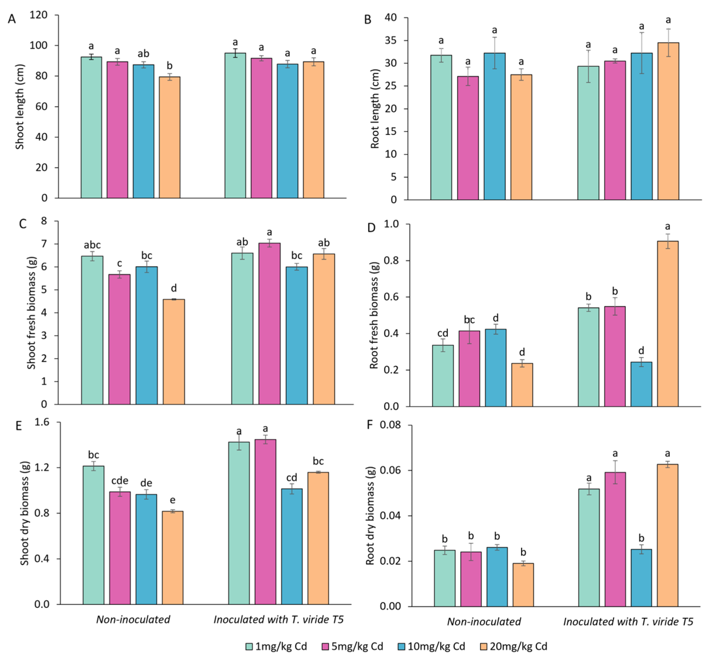
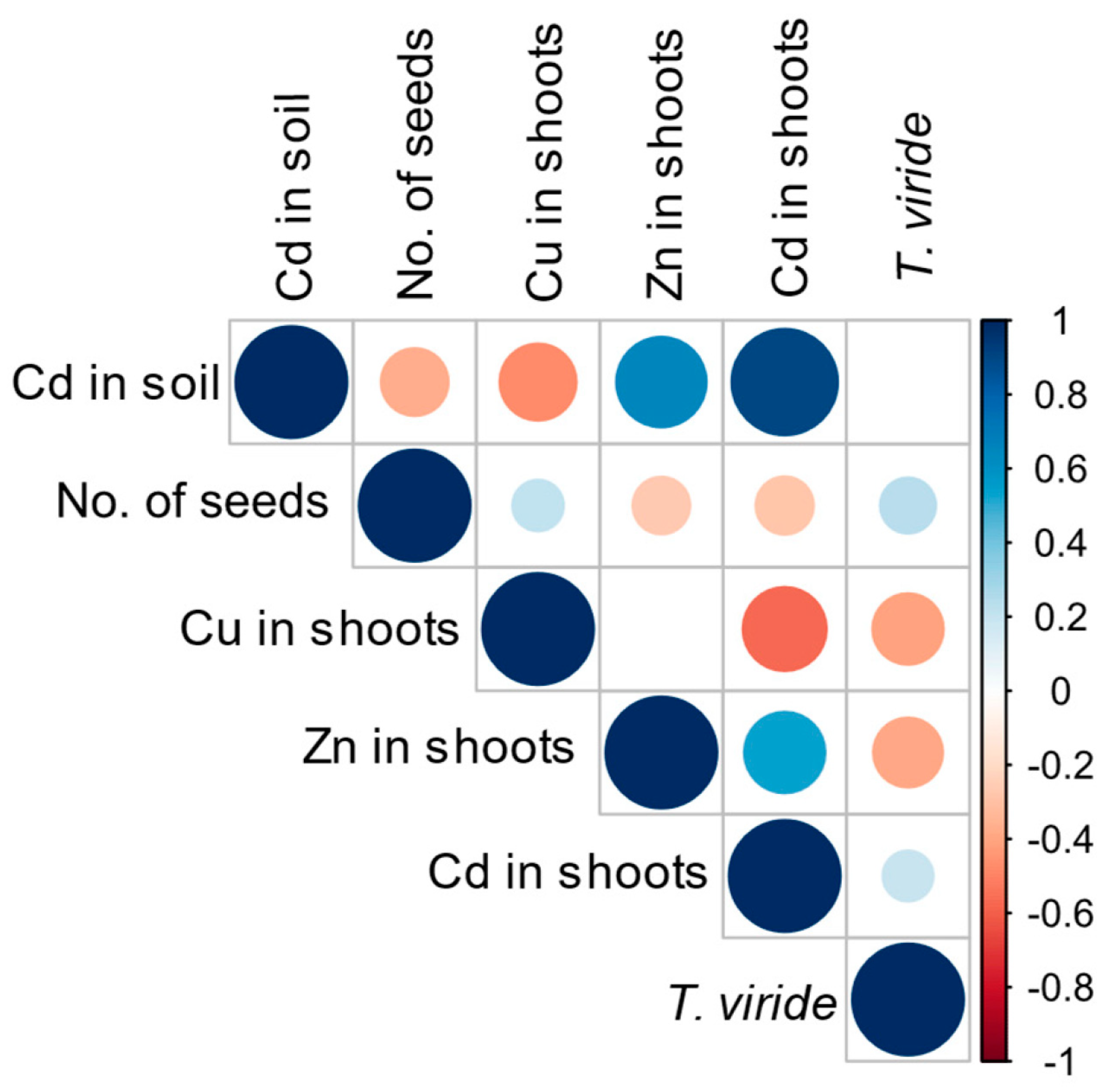
3.4. Effect of T. viride on the Growth and Yield of A. sativa Plants Grown in the Presence of Cd and on the Level of Cd Phytoextraction

The following experiment was conducted to verify the effect of oat seed inoculation with T. viride T5 on mature plant growth and yield in Cd-contaminated soil. After six months of growth, the length of the oat roots was affected neither by inoculation with T. viride nor by cadmium treatment (Figure 1B). Fresh and dry root biomass in non-inoculated samples was not affected by cadmium. Inoculation caused an increase in fresh root biomass, i.e., in the presence of 1, 5, and 20 mg Cd kg−1 soil, fresh root biomass was respectively 1.6, 1.4, and 3.8 times higher when compared to non-inoculated samples (Figure 1D). Similarly, in inoculated plants grown in soil containing 1, 5, and 20 mg Cd kg−1, dry root biomass was respectively 2, 2.5, and 3.3 times higher when compared to non-inoculated samples (Figure 1F). In non-inoculated plants, the shoot length decreased with increased Cd concentration in soil—shoots of plants treated with 20 mg Cd were 1.2 times shorter than those treated with only 1 mg Cd. However, when inoculated with T. viride T5, the shoot length remained the same across all tested Cd concentrations (Figure 1A). Shoot fresh and dry biomass of non-inoculated plants decreased with increased Cd concentrations, with plants treated with 20 mg Cd having their fresh and dry biomass 1.4 and 1.5 times lower, respectively, compared to plants treated with only 1 mg Cd (Figure 1C,E). In inoculated samples, the fresh shoot biomass was similar across all Cd concentrations. Dry shoot biomass of inoculated plants treated with 1, 5, and 20 mg Cd was respectively 1.2, 1.5, and 1.4 times higher when compared to non-inoculated plants (Figure 1E).
Treatment with cadmium significantly affected the yield, i.e., the number of panicles was 1.7-fold lower, and the number of seeds was 2.2-fold lower in plants grown in soil containing 20 mg kg−1 of Cd compared to plants grown in soil containing 1 mg kg−1 of Cd (Table 7). Similar observations were made for plants treated with T. viride T5; however, for Cd concentration, the number of panicles and seeds was higher in inoculated plants compared to in non-inoculated ones. For example, plants inoculated with T. viride grown in soil containing 20 mg kg−1 of Cd produced 1.7-fold more seeds and 1.3-fold more panicles than non-inoculated plants grown in the same Cd concentration (Table 7).
To assess the potential of A. sativa for Cd phytoextraction and the potential involvement of T. viride in this process, Cu, Cd, and Zn content was analysed in the above-ground parts of 6-month-old plants (Table 8). As expected, the concentration of Cd in the oat shoots increased with an increase in Cd concentration in the soil, i.e., plants grown in soil with 20 mg kg−1 of Cd had 6.6 times (non-inoculated) and 5.3 times (inoculated) higher Cd concentration in shoots than plants grown in soil with 1 mg kg−1 of Cd. Inoculation with T. viride T5 spores did not significantly increase the Cd uptake by oat. The content of copper in oat shoots was affected neither by Cd concentration in soil nor by inoculation with T. viride T5 spores. Interestingly, the level of Zn in shoots of oat plants was the highest in plants growing in soil containing 20 mg Cd kg−1, and it was 1.6 and 1.8 times higher in non-inoculated than in inoculated plants, respectively, compared to plants growing in soil contaminated with 1 mg Cd kg−1. Inoculation did not increase the Zn uptake regardless of Cd concentration in the soil (Table 8).
Positive correlations were observed between the amount of cadmium added to the soil and the level of Zn and Cd in oat plants. In contrast, the level of Cd in soil was negatively correlated with the level of Cu in oat shoots (Figure 2). Cadmium application was also negatively correlated with shoot length, fresh and dry biomass, and number of panicles and seeds (Supplementary Figure S3). A positive correlation was observed between inoculation with T. viride T5 spores and root length, fresh and dry biomass, and shoots’ fresh and dry biomass (Supplementary Figure S3). Interestingly, a negative correlation between inoculation with T. viride T5 spores and the levels of Cu and Zn, and a positive between inoculation with T. viride spores and the level of Cd, were observed (Figure 2).

3.5. Functional Analysis of A. sativa Metallothioneins (AsMT1-4) in Bacteria Cells

To verify the metal-binding ability of oat MT1-4, the proteins were expressed in E. coli cells in the presence of Zn and Cd (Figure 3). Bacteria transformed with plasmids carrying AsMT3 and AsMT4 grew faster under control and stress conditions caused by metal ions than bacteria transformed with an empty pET vector. The highest difference in growth rates between bacteria transformed with pET_AsMT3 and pET_AsMT4, and bacteria bearing an empty pET vector (6 and 5 times higher growth rates, respectively) was observed in medium supplemented with 0.25 mM Zn. The expression of AsMT1 and AsMT2 in bacterial cells did not increase bacteria growth. To verify the possible adverse effect of IPTG on bacteria growth, the experiment was also performed without the addition of IPTG (Supplementary Figure S1). Only bacteria transformed with pET_AsMT4 had a faster growth rate (both under control conditions and in the presence of Zn and Cd ions) than bacteria transformed with the empty pET vector.

3.6. Expression of A. sativa AsMT1-4 in Plants Growing in Cd-Contaminated Soil

To give insight into molecular mechanisms underlying Cd detoxification and interaction with T. viride in oat plants, gene expression of AsMT1-4 was analysed (Figure 4). The possibility that heavy metals induce the expression of oat MTs was verified by in silico analysis of promoter regions of AsMT1-4 genes (Supplementary Table S1). In total, 4, 4, 8, and 11 MRE were found in AsMT1, AsMT2, AsMT3, and AsMT4 promoters, respectively. The most common motif was the CuRE motif 5′-GTAC-3′, which appeared 20 times in the promoter sequences of AsMT1-4. The second most abundant motif was 5′-TGCRCNC-3′, found five times (Supplementary Table S1).
In inoculated plants, the expression of AsMT1, AsMT2, and AsMT3 in the presence of 1 mg Cd was over two times higher than in non-inoculated samples. However, in plants growing in soil containing 5, 10, and 20 mg of Cd per kg of soil, the AsMT1-3 expression in both variants, i.e., non-inoculated and inoculated, was on a similar level. The exception was AsMT2, where the expression in inoculated seedlings growing in 20 mg/kg of Cd was 1.5 times higher than in non-inoculated plants. The expression of AsMT4 was the highest in samples inoculated with T. viride in seedlings grown in the presence of 10 mg/kg of Cd (almost 2-fold higher than in non-inoculated plants). In other variants, the AsMT4 expression in both inoculated and non-inoculated samples was comparable (Figure 4).
Correlation analyses showed high positive correlations among AsMT1-3 but not between AsMT1-3 and AsMT4 (Supplementary Figure S4). Positive correlations were also observed between T. viride inoculation and expression of AsMT1-3, but negative correlations were noted between AsMT1-3 expression and the level of Cd in soil. Neither the inoculation with T. viride nor the level of Cd in the soil was correlated with the expression of AsMT4 (Supplementary Figure S4).

4. Discussion

Anthropogenic activities, like mining, the metallurgic industry, fossil fuel extraction, global transport, and agriculture, contribute to the increasing concentration of heavy metals in soil [62,63]. Even low concentrations of HM can become hazardous since they accumulate in the food chain [64,65]. Thus, it is essential to ensure that HM levels in soils and crops meet regulatory standards [66,67]. Since soil worldwide is contaminated with HM, there is an urgent need for practical, eco-friendly, and cost-effective remediation methods [68]. Phytoremediation is an eco-friendly and cost-effective method of removing hazardous pollutants, including HM, by plants. The effectiveness of this method can be increased by applying microorganisms that can interact with plants to counteract stressful environmental conditions and improve the plant’s capacity to absorb pollutants. Understanding how microorganisms and plants respond to HM in their environment is crucial for developing this remediation method [69,70]. Among several microorganisms, fungi belonging to Trichoderma are considered suitable for phytoremediation due to their ability to use various materials as a carbon source, including plastics [71,72], their ability to promote plant growth and development [18,73,74,75], and their resistance to xenobiotics [76]. Analysis of the Trichoderma harzianum transcriptome in response to Cd treatment revealed the up-regulation of cellular homeostasis, vesicle-mediated transport, and RNA processing. Moreover, sulfur-compound biosynthesis and glutathione metabolism were induced [77]. T. viride strains tested in this study differed in Cu, Cd, and Zn tolerance. Strain T1 exhibited the highest tolerance towards Cd and the lowest towards Zn, and the opposite was observed for strain T6. The tolerance of Trichoderma to HM, as reported in the literature, is variable. For example, for Zn, the concentration that inhibited the growth was reported to be four mM for unclassified Trichoderma strains [78] and 11.47 mM for Trichoderma atrioviride [35]. Those values range from 1.8 mM [79] to 2.67 mM [35] for Cd. This comparison showed that the strains of T. viride analysed in this study were highly tolerant to Zn, whereas tolerance to Cd was similar to other Trichoderma species and isolates. In contrast, the tolerance to Cu of T. viride T1-6 was relatively low since T. harzianum and T. virens tolerated Cu up to 12 mM [35,80]. Due to the similar physicochemical properties of Zn and Cd, the T. viride strain T5 was selected for further experiments because this stain had a high tolerance to both cadmium and zinc.
A seed coat is a rigid structure that protects the embryo from soil pollutants, including HM. During germination, it ruptures, and Cd content increases in seeds [81]. The negative impact of Cd on germination was shown for bean (Phaseolus vulgaris L.) [81], Sorghum bicolor (L.) Moench [82], and wheat (Triticum aestivum L.) [83]. The amount of Cd needed to inhibit germination differs from species to species and within one species from cultivar to cultivar [84]. Interestingly, it was also shown that low levels of Cd might positively affect seed germination and seedling growth [85]. In this study, Cd inhibited oat seed germination and further seedling growth, and the negative effect was more substantial for higher cadmium concentrations. In uncontaminated soil, the mean value of Cd is 0.36 mg/kg, although the Cd concentrations greatly depend on continent, country, and soil types [86]. The mean concentration of Cd in European agricultural soil is 0.15 mg/kg; in the wide-ranging analysis, croplands containing as much as 52.99 mg/kg of Cd were detected [87]. The EU risk assessment predicted no effective Cd concentration in soil of 1.1 mg per kg of dry soil based on the toxicity for plants, invertebrates, and animals [87]. Therefore, this study used soil containing 1 mg/kg Cd as a control. The yield (as shown by the number of panicles and seeds) of oat plants was significantly decreased by cadmium. For example, plants grown in soil containing 20 mg/kg Cd produced less than half of the seeds produced by plants grown in the presence of 1 mg/kg Cd. Crop plants significantly differ in their tolerance to Cd contamination, and there are also substantial differences in Cd tolerance among cultivars of the same species. A significant decrease in rice yield in soil contaminated with 1 mg/kg and 3 mg/kg of Cd was observed; however, the number of seeds produced also depends on the tested cultivar [88]. Compared to other grasses, oat is relatively tolerant to Cd stress [89]. Similar to our observations, Cd in soil up to 25 mg/kg did not significantly affect the growth of oat plants, but the yield was reduced [90].
Fungi belonging to Trichoderma are known for their plant growth-promoting properties [28,42,44,74,75]. Our previous studies show that T. viride can promote the growth of B. napus [28,74]. Barley plants inoculated with Trichoderma have up to 20% higher dry biomass than non-inoculated plants [43]. Similar reports are available for rice [91], sunflower [92], and maize [93]. The positive effect of microbial inoculation is often visible only in stress conditions [94]. For example, in control conditions, inoculation with Trichoderma did not improve wheat growth. Still, under severe water stress, inoculated wheat plants had higher dry biomass, downregulated water stress-related genes, and lower levels of proline, hydrogen peroxide, and malondialdehyde compared to non-inoculated plants [95]. In this study, seed germination and seedling growth were not improved by inoculation with T. viride in control conditions. Still, in the presence of cadmium, inoculated seeds germinated quicker, and the growth of seedling roots was enhanced in the presence of 150 and 254 μM Cd. The growth of plants was improved in soil containing 20 mg/kg Cd by inoculation with T. viride. The most significant effect was the increase in yield by T. viride inoculation observed in all Cd concentrations. The improved growth of plants in the presence of cadmium by inoculation with T. harzianum [96] and T. atrioviride [33] was shown for B. juncea. Plant growth promotion by fungi in HM-contaminated environments may be caused by increasing root absorption area and nutrient uptake [33,96]. The growth of Cicer arietinum was enhanced by inoculation with Trichoderma sp.; however, the effect was more substantial when plants were co-inoculated with Trichoderma sp. and Pseudomonas fluorescence [97]. The authors further highlighted that mechanisms that allow micro-organisms to adapt to and survive in HM-contaminated environments include binding HM to the cell wall and using siderophores to stop the HM from entering the cell, metalloproteases that bind and sequester HM in the cell, efflux pumps that eliminate HM from the cells, and antioxidant systems that reduce the negative effect of HM [97]. Combined with the growth-promoting properties of fungi belonging to Trichoderma (i.e., production of auxin analogues, organic acids, and siderophores), an increased tolerance to HM stress in inoculated plants was observed [29,30,96,97]. Interestingly, the inoculation with T. harzianum did not improve the growth of barley [98], and inoculation with Trichoderma sp. did not increase the growth and yield [90] in Cd-contaminated soil. Our recent study demonstrated a significant increase in B. napus yield by inoculation with T. viride in a field experiment [73]. Those results indicate that the improvement of plant growth and yield depends on fungi species/strain, plant species/cultivar, and the condition of the experiments.
Some crop plants are considered Cd-hyperaccumulators, e.g., several species belonging to Brassicaceae, some legumes, and some cereals [99]. For example, B. juncea accumulated more than 400 μg/g dry weight in leaves [100] and wheat up to 18 mg/kg dry weight [101]. Interestingly, Cd tolerance and accumulation are not usually related [99]. For example, wheat Cd-sensitive cultivars accumulated more Cd than Cd-tolerant ones [102]. Also, for oat, low and high Cd-accumulating cultivars were described [103]. The amount of Cd accumulated in crops and the location of Cd within plants are crucial in terms of nutrition. The World Health Organization recommends consuming no more than 25 μg of Cd monthly per kg of body weight. Oat can survive in soil polluted with heavy metals by extracting the metals from it and transferring them to above-ground parts [52,89,90,104]. The increased concentration of Cd in the soil led to an increased concentration of Cd in oat shoots. It is a widely observed phenomenon that the application of fungi belonging to Trichoderma increases the amount of extracted heavy metals, and this effect was observed for Cd but not for Cu and Zn in this study. In a study by Cao et al. [33], B. juncea inoculated with T. atroviride extracted 24% more Ni and 8% more Cd from the soil than non-inoculated plants did. Applying Trichoderma increased Cd content in shoots of maize plants growing in a Cd-contaminated soil by 38%, compared to non-inoculated plants [38]. A study showed that applying T. harzianum positively affected Cd uptake in barley (H. vulgare L.) [98]. The application of Trichoderma improved the solubility of heavy metals and, as a result, increased their uptake by Miscanthus x giganteus J.M. Greef, Panicum virgatum L., Phalaris arundinacea L., and Salix sp. [26]. A. sativa has excellent potential to be used in phytoremediation since it can grow on low-quality soil, can tolerate higher concentrations of toxic metal ions, and has higher biomass than hyperaccumulators, making the whole process more efficient [89]. Cadmium uptake influences the uptake of micronutrients since Cd enters the root using micronutrient transporters such as transporters belonging to ZIP (zinc-regulated, iron-regulated transporter-like protein) [105,106] and NRAMP (natural resistance-associated macrophage protein) [107]. In this study, the increased Cd content in the shoots was positively correlated with the Zn content in the shoots but negatively with the content of Cu. Zn and Cd have similar physicochemical properties, and usually, the higher the Cd concentration in the soil, the lower the Zn content in plants [108,109]. However, various external factors, such as pH, affect the interplay between Cd and micronutrients in soil [110]. Moreover, within plants, Cd interacts with micronutrients. For example, high zinc reduces Cd transport to shoots, whereas, in low Zn conditions, zinc translocation to shoots is increased by Cd [111].
Metallothioneins’ primary and firmly documented role in all living organisms is the homeostasis of micronutrients, mainly zinc and copper, and the detoxification of toxic metals, mostly cadmium [112]. In mammals, the expression of MTs in response to heavy metals is regulated by transcription factor MTF-1 (MRE-binding transcription factor-1) that binds to conserved regulatory motif MRE (metal response element) 5′-TGCRCNC-3′ present in MT promoter sequences. MTF-1 contains zinc finger domains and recognises MRE upon Zn metallation [113]. In yeast, the expression of metallothionein CUP1 is regulated by copper-sensing transcription factor ACE1 that binds to regulator element 5′-HTHNNGCTGD-3′ [114]. Relatively little is known about the molecular mechanisms underlying the induction of the expression of MTs by cadmium; however, it was demonstrated that Cd induces the expression of MT in animals [115], plants [116], and bacteria [117]. Databases used for the prediction of regulatory elements in plant-promoter sequences (e.g., PlantCARE [118] and New PLACE [119]) lack plant-specific MREs; therefore, the animal MRE consensus sequence was used. The potential role of the regulatory motifs similar to animal MREs in the induction of the expression of plant MTs in response to heavy metals has been confirmed [56,58,120]. In addition, plant-specific MRE [56] and CuRE (copper response element) identified in Chlamydomonas reinhardtii [121] were used. Multiple potential regulatory elements involved in response to biotic factors [3] and heavy metals present in analysed promoters support the hypothesis that AsMTs are engaged in plant interaction with T. viride and/or Cd response. Previously, we identified multiple abiotic stress response elements in AsMT promoters [3], and the role of AsMTs in response to drought [17] and osmotic [16] stress was demonstrated. Moreover, we suggested the role of AsMT1 and AsMT3 in Cd detoxification and the role of AsMT4 as a Zn specificity filter [3]. Heterologous expression of AsMT3 and AsMT4 in E. coli cells improved bacterial growth in the presence of Zn and Cd. Previously, it was shown that the expression of Brassica rapa L. MT types 1-3 increases the yeast tolerance to Zn, Cd, and Pb [122]. In contrast, the expression of B. napus MT4 in E. coli cells improved the growth of bacteria in the presence of Zn, decreased it in the presence of Cu, and had no effect on its growth in the presence of Cd. The positive/negative impact on bacteria growth also depended on the concentration of metals and/or IPTG [123]. Metallothioneins were shown to be involved in HM hyperaccumulation. For example, in a model plant for hyperaccumulators Thlaspi caerulescens (J. Presl & C. Presl) F.K. Mey., the expression of MT1 and MT2 was higher than in closely related Arabidopsis thaliana (L.) Heynh. [124]. Moreover, the expression of T. caerulescens MT3 was higher in a population with high Cd-accumulation and -tolerance [125,126]. A limited amount of evidence also suggests that MTs are involved in the interaction between plants and microorganisms. For instance, the expression of canola MTs types 1-3 was higher in seedlings inoculated with plant growth-promoting fungi T. viride. However, the enhanced expression was observed only for some T. viride strains, whereas for others, the MT1-3 expression was unaffected [73]. On the other hand, inoculation of B. napus with spores of arbuscular mycorrhizal (AM) fungi increased the expression of BnMT2 when plants were grown in soil without indigenous micro-organisms. In contrast, when indigenous microbes were present, the inoculation with AM spores decreased the expression of BnMT2 [18]. Inoculation of willow (Salix viminalis L.), growing in heavy metal-contaminated soil with rhizosphere bacteria Bacillus cereus, increased the expression of MT1. No increase in MT1 expression was observed after inoculation with the fungi Hebeloma mesophaeum [127]. In this study, Cd did not affect AsMT expression, but the inoculation with T. viride increased the expression of AsMT1-3 in soil containing 1 mg/kg Cd and AsMT4 in soil containing 10 mg/kg Cd. The response of MTs to metals depends not only on metal, plant species, and type of MT but also on plant organs and the amount of metal [128].

5. Conclusions

Inoculating A. sativa with T. viride might be a promising approach to increase the yield in Cd-contaminated soil without increasing the Cd content in plant tissues. Using Trichoderma in agriculture may improve the quantity and quality of produced food. This could be due to the fungi’s ability to produce auxin analogues, thus improving plant root growth. Moreover, the secretion of organic acids and siderophores affects the bioavailability of micronutrients, and toxic HM could be bound to fungi cell walls. This is of great importance because it could limit the inclusion of Cd in the food chain and thus improve the health of animals and humans. Meat consumption worldwide is declining for financial, environmental, and ethical reasons, thus, the importance of crops such as oats in nutrition is constantly increasing. There is an urgent need to develop strategies to enhance crop yields in contaminated soils without reducing food quality.

Supplementary Materials

The following supporting information can be downloaded at: https://www.mdpi.com/article/10.3390/foods13152469/s1, Table S1: cis-regulatory elements of AsMT1-4 promoters; Figure S1: comparison of the growth of E. coli cells transformed with empty pET21a(+) vector and pET21a(+) vectors harbouring coding regions of AsMT1-4 in the presence of Zn and Cd ions. The relative growth rate is expressed as a slope of bacterial growth curves obtained by plotting optical density against time. Media were supplemented with two concentrations of Zn and Cd ions without IPTG. The results obtained for a given condition were compared, and distinct letters indicate significant differences between E. coli carrying different plasmids (Kruskal–Wallis, Mann–Whitney; p < 0.05).; Figure S2: photographs of oat plants inoculated and not inoculated with Trichoderma viride T5 growing in soil containing Cd after 6 months of growth.; Figure S3: Pearson correlation between shoot and root length, fresh and dry biomass, number of leaves, panicles and seeds, levels of Cu, Zn, and Cd, the amount of cadmium added to the soil, and the inoculation of oat seeds with Trichoderma viride T5 spores. Only significant correlations are shown. Figure S4: Pearson correlation between AsMT1-4 expression (MT1-4), Trichoderma viride T5 inoculation, and the level of Cd in soil. Only significant correlations are shown.

Author Contributions

Conceptualization, A.M.-A.; methodology, A.M.-A.; validation, G.B.D. and A.M.-A.; formal analysis, W.K.; investigation, W.K., S.T., A.M.-A., E.S. and M.W.; resources, G.B.D. and A.M.-A.; data curation, W.K. and A.M.-A.; writing—original draft preparation, W.K. and A.M.-A.; writing—review and editing, A.M.-A. and G.B.D.; visualization, A.M.-A. and W.K.; supervision, A.M.-A. and G.B.D.; project administration, A.M.-A.; funding acquisition, W.K., A.M.-A., G.B.D., E.S. and M.W. All authors have read and agreed to the published version of the manuscript.

Funding

1. Grants for NCU Students 6th edition 90-SIDUB.6102.65.2023.G4NCUS6. 2. Grants for NCU Students 2nd edition 90-SIDUB.6102.44.2021.G4NCUS1.

Institutional Review Board Statement

Not applicable.

Informed Consent Statement

Not applicable.

Data Availability Statement

The original contributions presented in the study are included in the article/Supplementary Materials, further inquiries can be directed to the corresponding author.

Conflicts of Interest

The authors declare no conflicts of interest. The funders had no role in the design of the study, in the collection, analyses, or interpretation of data, in the writing of the manuscript, or in the decision to publish the results.

References

  1. Briffa, J.; Sinagra, E.; Blundell, R. Heavy Metal Pollution in the Environment and their Toxicological Effects on Humans. Heliyon 2020, 6, e04691. [Google Scholar] [CrossRef] [PubMed]
  2. Roberts, T.L. Cadmium and Phosphorous Fertilizers: The Issues and the Science. Procedia Eng. 2014, 83, 52–59. [Google Scholar] [CrossRef]
  3. Konieczna, W.; Mierek-Adamska, A.; Chojnacka, N.; Antoszewski, M.; Szydłowska-Czerniak, A.; Dąbrowska, G.B. Characterization of the Metallothionein Gene Family in Avena sativa L. and the Gene Expression During Seed Germination and Heavy Metal Stress. Antioxidants 2023, 12, 1865. [Google Scholar] [CrossRef] [PubMed]
  4. Rizwan, M.; Ali, S.; Rehman, M.Z.; Maqbool, A. A Critical Review on the Effects of Zinc at Toxic Levels of Cadmium in Plants. Environ. Sci. Pollut. Res. 2019, 26, 6279–6289. [Google Scholar] [CrossRef] [PubMed]
  5. Stafford, A.; Jeyakumar, P.; Hedley, M.; Anderson, C. Influence of Soil Moisture Status on Soil Cadmium Phytoavailability and Accumulation in Plantain (Plantago lanceolata). Soil Syst. 2018, 2, 9. [Google Scholar] [CrossRef]
  6. Ellen, T.P.; Costa, M. Carcinogenic Inorganic Chemicals. In Comprehensive Toxicology (Second Edition); McQueen, C.A., Ed.; Elsevier: Oxford, UK, 2010; pp. 139–160. ISBN 978-0-08-046884-6. [Google Scholar]
  7. Zulfiqar, U.; Jiang, W.; Wang, X.; Hussain, S.; Ahmad, M.; Maqsood, M.F.; Ali, N.; Ishfaq, M.; Kaleem, M.; Haider, F.U.; et al. Cadmium Phytotoxicity, Tolerance, and Advanced Remediation Approaches in Agricultural Soils; a Comprehensive Review. Front. Plant Sci. 2022, 13, 773815. [Google Scholar] [CrossRef] [PubMed]
  8. European Commission, Directorate-General for Health and Food Safety. Commission Regulation (EU) 2023/915 of 25 April 2023 on Maximum Levels for Certain Contaminants in Food and Repealing Regulation (EC) No 1881/2006. Off. J. Eur. Union 2023, 119, 103–157. [Google Scholar]
  9. Zimmerl, S.; Lafferty, J.; Buerstmayr, H. Assessing Diversity in Triticum durum Cultivars and Breeding Lines for High versus Low Cadmium Content in Seeds Using the CAPS Marker Usw47. Plant Breed. 2014, 133, 712–717. [Google Scholar] [CrossRef]
  10. Song, W.; Chen, S.; Liu, J.; Chen, L.; Song, N.; Li, N.; Liu, B. Variation of Cd Concentration in Various Rice Cultivars and Derivation of Cadmium Toxicity Thresholds for Paddy Soil by Species-Sensitivity Distribution. J. Integr. Agric. 2015, 14, 1845–1854. [Google Scholar] [CrossRef]
  11. Charkiewicz, A.E.; Omeljaniuk, W.J.; Nowak, K.; Garley, M.; Nikliński, J. Cadmium Toxicity and Health Effects—A Brief Summary. Molecules 2023, 28, 6620. [Google Scholar] [CrossRef]
  12. Freisinger, E. Structural Features Specific to Plant Metallothioneins. J. Biol. Inorg. Chem. 2011, 16, 1035–1045. [Google Scholar] [CrossRef] [PubMed]
  13. Dąbrowska, G.; Mierek-Adamska, A.; Goc, A. Characterisation of Brassica napus L. Metallothionein Genes (BnMTs) Expression in Organs and during Seed Germination. Aust. J. Crop Sci. 2013, 7, 1324–1332. [Google Scholar]
  14. Blindauer, C.A.; Schmid, R. Cytosolic Metal Handling in Plants: Determinants for Zinc Specificity in Metal Transporters and Metallothioneins. Metallomics 2010, 2, 510–529. [Google Scholar] [CrossRef] [PubMed]
  15. Huang, S.S.; Deng, J.S.; Chen, H.J.; Lin, Y.H.; Huang, G.J. Antioxidant Activities of Two Metallothionein-like Proteins from Sweet Potato (Ipomoea batatas [L.] Lam. ‘Tainong 57’) Storage Roots and Their Synthesized Peptides. Bot. Stud. 2014, 55, 64. [Google Scholar] [CrossRef] [PubMed]
  16. Konieczna, W.; Mierek-Adamska, A.; Warchoł, M.; Skrzypek, E.; Dąbrowska, G.B. The Involvement of Metallothioneins and Stress Markers in Response to Osmotic Stress in Avena sativa L. J. Agron. Crop Sci. 2023, 209, 371–389. [Google Scholar] [CrossRef]
  17. Konieczna, W.; Warchoł, M.; Mierek-Adamska, A.; Skrzypek, E.; Waligórski, P.; Piernik, A.; Dąbrowska, G.B. Changes in Physio-Biochemical Parameters and Expression of Metallothioneins in Avena sativa L. in Response to Drought. Sci. Rep. 2023, 13, 2486. [Google Scholar] [CrossRef] [PubMed]
  18. Dąbrowska, G.; Baum, C.; Trejgell, A.; Hrynkiewicz, K. Impact of Arbuscular Mycorrhizal Fungi on the Growth and Expression of Gene Encoding Stress Protein - Metallothionein BnMT2 in the Non-Host Crop Brassica napus L. J. Plant Nutr. Soil Sci. 2014, 177, 459–467. [Google Scholar] [CrossRef]
  19. Dąbrowska, G.; Hrynkiewicz, K.; Trejgell, A. Do Arbuscular Mycorrhizal Fungi Affect Metallothionein MT2 Expression in Brassica napus L. Roots? Acta Biol. Cracoviensia Ser. Bot. 2012, 54, 34–39. [Google Scholar] [CrossRef]
  20. Dąbrowska, G.; Mierek-Adamska, A.; Goc, A. Plant Metallothioneins: Putative Functions Identified by Promoter Analysis in silico. Acta Biol. Cracoviensia Ser. Bot. 2012, 54, 109–120. [Google Scholar] [CrossRef]
  21. Gao, C.; Gao, K.; Yang, H.; Ju, T.; Zhu, J.; Tang, Z.; Zhao, L.; Chen, Q. Genome-Wide Analysis of Metallothionein Gene Family in Maize to Reveal Its Role in Development and Stress Resistance to Heavy Metal. Biol. Res. 2022, 55, 1–13. [Google Scholar] [CrossRef]
  22. Yu, Q.; He, L.; Huo, C.; Jiang, X.; Chen, H.; Wang, R.; Tang, M.; Dong, L.; Chen, J.; Li, Y.; et al. Genome-Wide Identification and Expression Analysis of Heavy Metal Stress–Responsive Metallothionein Family Genes in Nicotiana tabacum. Plant Mol. Biol. Rep. 2020, 39, 443–454. [Google Scholar] [CrossRef]
  23. Mirzahossini, Z.; Shabani, L.; Sabzalian, M.R.; Sharifi-Tehrani, M. ABC Transporter and Metallothionein Expression Affected by Ni and Epichloe Endophyte Infection in Tall Fescue. Ecotoxicol. Environ. Saf. 2015, 120, 13–19. [Google Scholar] [CrossRef] [PubMed]
  24. Gilbert, J.A.; Neufeld, J.D. Life in a World Without Microbes. PLoS Biol. 2014, 12, e1002020. [Google Scholar] [CrossRef] [PubMed]
  25. Dąbrowska, G.B.; Garstecka, Z.; Trejgell, A.; Dąbrowski, H.P.; Konieczna, W.; Szyp-Borowska, I. The Impact of Forest Fungi on Promoting Growth and Development of Brassica napus L. Agronomy 2021, 11, 2475. [Google Scholar] [CrossRef]
  26. Kacprzak, M.J.; Rosikon, K.; Fijalkowski, K.; Grobelak, A. The Effect of Trichoderma on Heavy Metal Mobility and Uptake by Miscanthus giganteus, Salix sp., Phalaris arundinacea, and Panicum virgatum. Appl. Environ. Soil Sci. 2014, 2014, 506142. [Google Scholar] [CrossRef]
  27. Chacón, M.R.; Rodríguez-Galán, O.; Benítez, T.; Sousa, S.; Rey, M.; Llobell, A.; Delgado-Jarana, J. Microscopic and Transcriptome Analyses of Early Colonization of Tomato Roots by Trichoderma harzianum. Int. Microbiol. 2007, 10, 19–27. [Google Scholar] [CrossRef] [PubMed]
  28. Turkan, S.; Mierek-Adamska, A.; Kulasek, M.; Konieczna, W.B.; Dąbrowska, G.B. New Seed Coating Containing Trichoderma viride with Anti-Pathogenic Properties. PeerJ 2023, 11, e15392. [Google Scholar] [CrossRef] [PubMed]
  29. Vinale, F.; Sivasithamparam, K.; Ghisalberti, E.L.; Woo, S.L.; Nigro, M.; Marra, R.; Lombardi, N.; Pascale, A.; Ruocco, M.; Lanzuise, S.; et al. Trichoderma Secondary Metabolites Active on Plants and Fungal Pathogens. Open Mycol. J. 2014, 8, 127–139. [Google Scholar] [CrossRef]
  30. Chagas, L.F.B.; De Castro, H.G.; Colonia, B.S.O.; De Carvalho Filho, M.R.; Miller, L.D.O.; Chagas, A.F.J. Efficiency of Trichoderma spp. as a Growth Promoter of Cowpea (Vigna unguiculata) and Analysis of Phosphate Solubilization and Indole Acetic Acid Synthesis. Braz. J. Bot. 2016, 39, 437–445. [Google Scholar] [CrossRef]
  31. Aishwarya, S.; Viswanath, H.S.; Singh, A.; Singh, R. Biosolubilization of Different Nutrients by Trichoderma spp. and Their Mechanisms Involved: A Review. Int. J. Adv. Agric. Sci. Technol. 2020, 7, 34–39. [Google Scholar]
  32. Li, R.-X.; Cai, F.; Pang, G.; Shen, Q.-R.; Li, R.; Chen, W. Solubilisation of Phosphate and Micronutrients by Trichoderma harzianum and Its Relationship with the Promotion of Tomato Plant Growth. PLoS ONE 2015, 10, e0130081. [Google Scholar] [CrossRef] [PubMed]
  33. Cao, L.; Jiang, M.; Zeng, Z.; Du, A.; Tan, H.; Liu, Y. Trichoderma atroviride F6 Improves Phytoextraction Efficiency of Mustard (Brassica juncea (L.) Coss. Var. foliosa Bailey) in Cd, Ni Contaminated Soils. Chemosphere 2008, 71, 1769–1773. [Google Scholar] [CrossRef] [PubMed]
  34. Govarthanan, M.; Mythili, R.; Selvankumar, T.; Kamala-Kannan, S.; Kim, H. Myco-Phytoremediation of Arsenic- and Lead-Contaminated Soils by Helianthus annuus and Wood Rot Fungi, Trichoderma sp. Isolated from Decayed Wood. Ecotoxicol. Environ. Saf. 2018, 151, 279–284. [Google Scholar] [CrossRef] [PubMed]
  35. López Errasquín, E.; Vázquez, C. Tolerance and Uptake of Heavy Metals by Trichoderma atroviride Isolated from Sludge. Chemosphere 2003, 50, 137–143. [Google Scholar] [CrossRef] [PubMed]
  36. Mei, J.; Wang, L.; Jiang, X.; Wu, B.; Li, M. Functions of the C2H2 Transcription Factor Gene Thmea1 in Trichoderma harzianum under Copper Stress Based on Transcriptome Analysis. Biomed. Res. Int. 2018, 2018, 8149682. [Google Scholar] [CrossRef]
  37. Tansengco, M.; Tejano, J.; Coronado, F.; Gacho, C.; Barcelo, J. Heavy Metal Tolerance and Removal Capacity of Trichoderma Species Isolated from Mine Tailings in Itogon, Benguet. Environ. Nat. Resour. 2018, 16, 39–57. [Google Scholar] [CrossRef]
  38. Pehlivan, N.; Gedik, K.; Eltem, R.; Terzi, E. Dynamic Interactions of Trichoderma harzianum TS 143 from an Old Mining Site in Turkey for Potent Metal(Oid)s Phytoextraction and Bioenergy Crop Farming. J. Hazard. Mater. 2021, 403, 123609. [Google Scholar] [CrossRef] [PubMed]
  39. Sahu, A.; Mandal, A.; Thakur, J.; Manna, M.C.; Rao, A.S. Exploring Bioaccumulation Efficacy of Trichoderma viride: An Alternative Bioremediation of Cadmium and Lead. Natl. Acad. Sci. Lett. 2012, 35, 299–302. [Google Scholar] [CrossRef]
  40. Yaghoubian, Y.; Siadat, S.A.; Moradi Telavat, M.R.; Pirdashti, H.; Yaghoubian, I. Bio-Removal of Cadmium from Aqueous Solutions by Filamentous Fungi: Trichoderma Spp. and Piriformospora Indica. Environ. Sci. Pollut. Res. 2019, 26, 7863–7872. [Google Scholar] [CrossRef] [PubMed]
  41. Zhang, S.; Gan, Y.; Xu, B. Application of Plant-Growth-Promoting Fungi Trichoderma longibrachiatum T6 Enhances Tolerance of Wheat to Salt Stress through Improvement of Antioxidative Defense System and Gene Expression. Front. Plant Sci. 2016, 7, 1405. [Google Scholar] [CrossRef]
  42. Harman, G.E.; Howell, C.R.; Viterbo, A.; Chet, I.; Lorito, M. Trichoderma Species—Opportunistic, Avirulent Plant Symbionts. Nat. Rev. Microbiol. 2004, 2, 43–56. [Google Scholar] [CrossRef] [PubMed]
  43. Moya, P.; Barrera, V.; Cipollone, J.; Bedoya, C.; Kohan, L.; Toledo, A.; Sisterna, M. New Isolates of Trichoderma spp. as Biocontrol and Plant Growth–Promoting Agents in the Pathosystem Pyrenophora teres Barley in Argentina. Biol. Control 2020, 141, 104152. [Google Scholar] [CrossRef]
  44. Tyśkiewicz, R.; Nowak, A.; Ozimek, E.; Jaroszuk-Ściseł, J. Trichoderma: The Current Status of Its Application in Agriculture for the Biocontrol of Fungal Phytopathogens and Stimulation of Plant Growth. Int. J. Mol. Sci. 2022, 23, 2329. [Google Scholar] [CrossRef] [PubMed]
  45. Koide, R.; Li, M.; Lewis, J.; Irby, C. Role of Mycorrhizal Infection in the Growth and Reproduction of Wild vs. Cultivated Plants. Oecologia 1988, 77, 537–543. [Google Scholar] [CrossRef] [PubMed]
  46. Mushtaq, A.; Gul-Zaffar, G.Z.; Dar, A.D.; Mehfuza Habib, M.H. Review on Oat (Avena sativa L.) as a Dual-Purpose Crop. Sci. Res. Essays 2014, 9, 52–59. [Google Scholar] [CrossRef]
  47. Kim, I.-S.; Hwang, C.-W.; Yang, W.-S.; Kim, C.-H. Multiple Antioxidative and Bioactive Molecules of Oats (Avena sativa L.) in Human Health. Antioxidants 2021, 10, 1454. [Google Scholar] [CrossRef] [PubMed]
  48. Ehlers, W.; Khosla, B.K.; Köpke, U.; Stülpnagel, R.; Böhm, W.; Baeumer, K. Tillage Effects on Root Development, Water Uptake and Growth of Oats. Soil Tillage Res. 1980, 1, 19–34. [Google Scholar] [CrossRef]
  49. Hoad, S.; Russell, G.; Lucas, M.; Bingham, I. The Management of Wheat, Barley, and Oat Root Systems. Adv. Agron. 2001, 74, 193–246. [Google Scholar] [CrossRef]
  50. Khan, T.A.; Nadeem, F.; Gao, Y.; Yang, Y.; Wang, X.; Zeng, Z.; Hu, Y. A Larger Root System in Oat (Avena nuda L.) Is Coupled with Enhanced Biomass Accumulation and Hormonal Alterations under Low Nitrogen. Appl. Ecol. Environ. Res. 2019, 17, 4631–4653. [Google Scholar] [CrossRef]
  51. Bjerre, G.K.; Schierup, H.-H. Uptake of Six Heavy Metals by Oat as Influenced by Soil Type and Additions of Cadmium, Lead, Zinc and Copper. Plant Soil 1985, 88, 57–69. [Google Scholar] [CrossRef]
  52. Tuma, J.; Skalicky, M.; Tumova, L.; Flidr, J. Influence of Cadmium Dose and Form on the Yield of Oat (Avena sativa L.) and the Metal Distribution in the Plant. J. Elem. 2014, 19, 795–810. [Google Scholar] [CrossRef]
  53. Sambrook, J.; Russell, D.W. Molecular Cloning a Laboratory Manual; Cold Spring Harbor Laboratory Press: New York, NY, USA, 2001; ISBN 978-85-7811-079-6. [Google Scholar]
  54. Yoshida, N.; Ikeda, R.; Okuno, T. Identification and Characterization of Heavy Metal-Resistant Unicellular Alga Isolated from Soil and Its Potential for Phytoremediation. Bioresour. Technol. 2006, 97, 1843–1849. [Google Scholar] [CrossRef] [PubMed]
  55. Ranal, M.A.; de Santana, D.G. How and Why to Measure the Germination Process? Braz. J. Bot. 2006, 29, 1–11. [Google Scholar] [CrossRef]
  56. Qi, X.; Zhang, Y.; Chai, T. Characterization of a Novel Plant Promoter Specifically Induced by Heavy Metal and Identification of the Promoter Regions Conferring Heavy Metal Responsiveness. Plant Physiol. 2007, 143, 50–59. [Google Scholar] [CrossRef] [PubMed]
  57. Stuart, G.W.; Searle, P.F.; Palmiter, R.D. Identification of Multiple Metal Regulatory Elements in Mouse Metallothionein-I Promoter by Assaying Synthetic Sequences. Nature 1985, 317, 828–831. [Google Scholar] [CrossRef] [PubMed]
  58. Dong, C.-J.J.; Wang, Y.; Yu, S.-S.S.; Liu, J.-Y.Y. Characterization of a Novel Rice Metallothionein Gene Promoter: Its Tissue Specificity and Heavy Metal Responsiveness. J. Integr. Plant Biol. 2010, 52, 914–924. [Google Scholar] [CrossRef]
  59. Quinn, J.M.; Merchant, S. Two Copper-Responsive Elements Associated with the Chlamydomonas Cyc6 Gene Function as Targets for Transcriptional Activators. Plant Cell 1995, 7, 623–638. [Google Scholar] [PubMed]
  60. Yang, Z.; Wang, K.; Aziz, U.; Zhao, C.; Zhang, M. Evaluation of duplicated reference genes for quantitative real-time PCR analysis in genome unknown hexaploid oat (Avena sativa L.). Plant Methods 2020, 16, 138. [Google Scholar] [CrossRef]
  61. RStudio Team. RStudio: Integrated Development for R; RStudio, Inc.: Boston, MA, USA, 2020. [Google Scholar]
  62. Zbieralski, K.; Staszewski, J.; Konczak, J.; Lazarewicz, N.; Nowicka-Kazmierczak, M.; Wawrzycka, D.; Maciaszczyk-Dziubinska, E. Multilevel Regulation of Membrane Proteins in Response to Metal and Metalloid Stress: A Lesson from Yeast. Int. J. Mol. Sci. 2024, 25, 4450. [Google Scholar] [CrossRef] [PubMed]
  63. Khan, Z.; Elahi, A.; Bukhari, D.A.; Rehman, A. Cadmium Sources, Toxicity, Resistance and Removal by Microorganisms—A Potential Strategy for Cadmium Eradication. J. Saudi Chem. Soc. 2022, 26, 101569. [Google Scholar] [CrossRef]
  64. Guo, L.; Li, Z.; Xu, J. Effects of Cadmium Stress on Bacterial and Fungal Communities in the Whitefly Bemisia tabaci. Int. J. Mol. Sci. 2023, 24, 13588. [Google Scholar] [CrossRef]
  65. Peralta-Videa, J.R.; Lopez, M.L.; Narayan, M.; Saupe, G.; Gardea-Torresdey, J. The Biochemistry of Environmental Heavy Metal Uptake by Plants: Implications for the Food Chain. Int. J. Biochem. Cell Biol. 2009, 41, 1665–1677. [Google Scholar] [CrossRef]
  66. Khan, M.A.; Khan, S.; Khan, A.; Alam, M. Soil Contamination with Cadmium, Consequences and Remediation Using Organic Amendments. Sci. Total Environ. 2017, 601–602, 1591–1605. [Google Scholar] [CrossRef] [PubMed]
  67. Arao, T.; Ishikawa, S.; Murakami, M.; Abe, K.; Maejima, Y.; Makino, T. Heavy Metal Contamination of Agricultural Soil and Countermeasures in Japan. Paddy Water Environ. 2010, 8, 247–257. [Google Scholar] [CrossRef]
  68. Mierek-Adamska, A.; Dąbrowska, G.B.; Goc, A. Genetically modified plants and strategies of soil remediation from haevy metals. Adv. Cell Biol. 2009, 36, 649–662. [Google Scholar]
  69. Dąbrowska, G.; Hrynkiewicz, K.; Trejgell, A.; Baum, C. The Effect of Plant-Growth-Promoting Rhizobacteria on the Phytoextraction of Cd and Zn by Brassica napus L. Int. J. Phytoremediat. 2017, 19, 597–604. [Google Scholar] [CrossRef] [PubMed]
  70. Khan, A.R.; Ullah, I.; Waqas, M.; Shahzad, R.; Hong, S.J.; Park, G.S.; Jung, B.K.; Lee, I.J.; Shin, J.H. Plant Growth-Promoting Potential of Endophytic Fungi Isolated from Solanum nigrum Leaves. World J. Microbiol. Biotechnol. 2015, 31, 1461–1466. [Google Scholar] [CrossRef]
  71. Znajewska, Z.; Dąbrowska, G.B.; Hrynkiewicz, K.; Janczak, K. Biodegradation of Polycaprolactone by Trichoderma viride Fungi. Przem. Chem. 2018, 97, 1676–1679. [Google Scholar] [CrossRef]
  72. Dąbrowska, G.B.; Garstecka, Z.; Olewnik-Kruszkowska, E.; Szczepańska, G.; Ostrowski, M.; Mierek-Adamska, A. Comparative Study of Structural Changes of Polylactide and Poly(Ethylene Terephthalate) in the Presence of Trichoderma viride. Int. J. Mol. Sci. 2021, 22, 3491. [Google Scholar] [CrossRef]
  73. Garstecka, Z.; Antoszewski, M.; Mierek-Adamska, A.; Krauklis, D.; Niedojadło, K.; Kaliska, B.; Hrynkiewicz, K.; Dąbrowska, G.B. Trichoderma viride Colonizes the Roots of Brassica napus L., Alters the Expression of Stress-Responsive Genes, and Increases the Yield of Canola under Field Conditions during Drought. Int. J. Mol. Sci. 2023, 24, 15349. [Google Scholar] [CrossRef]
  74. Znajewska, Z.; Narbutt, O.; Dąbrowska, G.B.; Narbutt, O. Trichoderma viride Strains Stimulating the Growth and Development of Winter Rapeseed (Brassica napus L.). Prog. Plant Prot. 2018, 58, 264–269. [Google Scholar] [CrossRef]
  75. Antoszewski, M.; Mierek-Adamska, A.; Dąbrowska, G.B. The Importance of Microorganisms for Sustainable Agriculture—A Review. Metabolites 2022, 12, 1100. [Google Scholar] [CrossRef] [PubMed]
  76. Balcázar-López, E.; Méndez-Lorenzo, L.H.; Batista-García, R.A.; Esquivel-Naranjo, U.; Ayala, M.; Kumar, V.V.; Savary, O.; Cabana, H.; Herrera-Estrella, A.; Folch-Mallol, J.L. Xenobiotic Compounds Degradation by Heterologous Expression of a Trametes sanguineus Laccase in Trichoderma atroviride. PLoS ONE 2016, 11, e0147997. [Google Scholar] [CrossRef] [PubMed]
  77. Oshiquiri, L.H.; dos Santos, K.R.A.; Ferreira Junior, S.A.; Steindorff, A.S.; Barbosa Filho, J.R.; Mota, T.M.; Ulhoa, C.J.; Georg, R.C. Trichoderma harzianum Transcriptome in Response to Cadmium Exposure. Fungal Genet. Biol. 2020, 134, 103281. [Google Scholar] [CrossRef] [PubMed]
  78. Tripathi, P.; Singh, P.C.; Mishra, A.; Chauhan, P.S.; Dwivedi, S.; Bais, R.T.; Tripathi, R.D. Trichoderma: A Potential Bioremediator for Environmental Clean Up. Clean Technol. Environ. Policy 2013, 15, 541–550. [Google Scholar] [CrossRef]
  79. Nongmaithem, N.; Roy, A.; Bhattacharya, P.M. Screening of Trichoderma Isolates for Their Potential of Biosorption of Nickel and Cadmium. Braz. J. Microbiol. 2016, 47, 305–313. [Google Scholar] [CrossRef] [PubMed]
  80. Siddiquee, S.; Aishah, S.N.; Azad, S.A.; Shafawati, S.N.; Naher, L. Tolerance and Biosorption Capacity of Zn2+, Pb2+, Ni3+ and Cu2+ by Filamentous Fungi (Trichoderma harzianum, T. aureoviride and T. virens). Adv. Biosci. Biotechnol. 2013, 4, 570–583. [Google Scholar] [CrossRef]
  81. Sfaxi-Bousbih, A.; Chaoui, A.; El Ferjani, E. Unsuitable Availability of Nutrients in Germinating Bean Embryos Exposed to Copper Excess. Biol. Trace Elem. Res. 2010, 135, 295–303. [Google Scholar] [CrossRef] [PubMed]
  82. Kuriakose, S.V.; Prasad, M.N.V. Cadmium Stress Affects Seed Germination and Seedling Growth in Sorghum bicolor (L.) Moench by Changing the Activities of Hydrolyzing Enzymes. Plant Growth Regul. 2008, 54, 143–156. [Google Scholar] [CrossRef]
  83. Ahmad, I.; Akhtar, M.; Zahir, Z.; Jamil, A. Effect of Cadmium on Seed Germination and Seedling Growth of Four Wheat (Triticum aestivum L.) Cultivars. Pak. J. Bot. 2012, 44, 1569–1574. [Google Scholar]
  84. Huybrechts, M.; Cuypers, A.; Deckers, J.; Iven, V.; Vandionant, S.; Jozefczak, M.; Hendrix, S. Cadmium and Plant Development: An Agony from Seed to Seed. Int. J. Mol. Sci. 2019, 20, 3971. [Google Scholar] [CrossRef] [PubMed]
  85. Carvalho, M.E.A.; Agathokleous, E.; Nogueira, M.L.; Brunetto, G.; Brown, P.H.; Azevedo, R.A. Neutral-to-Positive Cadmium Effects on Germination and Seedling Vigor, with and without Seed Priming. J. Hazard. Mater. 2023, 448, 130813. [Google Scholar] [CrossRef] [PubMed]
  86. Kubier, A.; Wilkin, R.T.; Pichler, T. Cadmium in Soils and Groundwater: A Review. Appl. Geochem. 2019, 108, 104388. [Google Scholar] [CrossRef] [PubMed]
  87. Joint Research Centre (European Commission); Ballabio, C.; Jones, A.; Montanarella, L.; Toth, G. Cadmium in the Soils of the EU: Analysis of LUCAS Soils Data for the Review of Fertilizer Directive; Publications Office of the European Union: Luxembourg, 2023; ISBN 978-92-79-66931-6. [Google Scholar]
  88. Siddique, A.B.; Rahman, M.M.; Islam, M.R.; Mondal, D.; Naidu, R. Response of Iron and Cadmium on Yield and Yield Components of Rice and Translocation in Grain: Health Risk Estimation. Front. Environ. Sci. 2021, 9, 716770. [Google Scholar] [CrossRef]
  89. Ebbs, S.D.; Kochian, L.V. Phytoextraction of Zinc by Oat Avena sativa, Barley Hordeum vulgare, and Indian Mustard Brassica juncea. Environ. Sci. Technol. 1998, 32, 802–806. [Google Scholar] [CrossRef]
  90. Marchel, M.; Kaniuczak, J.; Hajduk, E.; Właśniewski, S. Response of Oat (Avena sativa) to the Addition Cadmium to Soil Inoculation with the Genus Trichoderma Fungi. J. Elem. 2018, 23, 471–482. [Google Scholar] [CrossRef]
  91. Doni, F.; Isahak, A.; Che Mohd Zain, C.R.; Wan Yusoff, W.M. Physiological and Growth Response of Rice Plants (Oryza sativa L.) to Trichoderma spp. Inoculants. AMB Express 2014, 4, 45. [Google Scholar] [CrossRef] [PubMed]
  92. Singh, B.N.; Singh, A.; Singh, B.R.; Singh, H.B. Trichoderma harzianum Elicits Induced Resistance in Sunflower Challenged by Rhizoctonia solani. J. Appl. Microbiol. 2014, 116, 654–666. [Google Scholar] [CrossRef] [PubMed]
  93. Akladious, S.A.; Abbas, S.M. Application of Trichoderma harzianum T22 as a Biofertilizer Potential in Maize Growth. J. Plant Nutr. 2014, 37, 30–49. [Google Scholar] [CrossRef]
  94. Xun, F.; Xie, B.; Liu, S.; Guo, C. Effect of Plant Growth-Promoting Bacteria (PGPR) and Arbuscular Mycorrhizal Fungi (AMF) Inoculation on Oats in Saline-Alkali Soil Contaminated by Petroleum to Enhance Phytoremediation. Environ. Sci. Pollut. Res. 2015, 22, 598–608. [Google Scholar] [CrossRef]
  95. Illescas, M.; Morán-Diez, M.E.; Martínez de Alba, Á.E.; Hermosa, R.; Monte, E. Effect of Trichoderma Asperellum on Wheat Plants’ Biochemical and Molecular Responses, and Yield under Different Water Stress Conditions. Int. J. Mol. Sci. 2022, 23, 6782. [Google Scholar] [CrossRef] [PubMed]
  96. Chauhan, J.S.; Rai, J.P.N. Phytoextraction of Soil Cadmium and Zinc by Microbes-Inoculated Indian Mustard (Brassica juncea). J. Plant Interact. 2009, 4, 279–287. [Google Scholar] [CrossRef]
  97. Syed, A.; Elgorban, A.M.; Bahkali, A.H.; Eswaramoorthy, R.; Iqbal, R.K.; Danish, S. Metal-Tolerant and Siderophore Producing Pseudomonas Fluorescence and Trichoderma spp. Improved the Growth, Biochemical Features and Yield Attributes of Chickpea by Lowering Cd Uptake. Sci. Rep. 2023, 13, 4471. [Google Scholar] [CrossRef] [PubMed]
  98. Taghavi Ghasemkheili, F.; Ekelund, F.; Johansen, J.L.; Pirdashti, H.; Ghadirnezhad Shiade, S.R.; Fathi, A.; Kjøller, R. Ameliorative Effects of Trichoderma harzianum and Rhizosphere Soil Microbes on Cadmium Biosorption of Barley (Hordeum vulgare L.) in Cd-Polluted Soil. J. Plant Nutr. Soil Sci. 2022, 22, 527–539. [Google Scholar] [CrossRef]
  99. Niu, L.; Li, C.; Wang, W.; Zhang, J.; Scali, M.; Li, W.; Liu, H.; Tai, F.; Hu, X.; Wu, X. Cadmium Tolerance and Hyperaccumulation in Plants—A Proteomic Perspective of Phytoremediation. Ecotoxicol. Environ. Saf. 2023, 256, 114882. [Google Scholar] [CrossRef] [PubMed]
  100. Haag-Kerwer, A.; Schäfer, H.J.; Heiss, S.; Walter, C.; Rausch, T. Cadmium Exposure in Brassica juncea Causes a Decline in Transpiration Rate and Leaf Expansion without Effect on Photosynthesis. J. Exp. Bot. 1999, 50, 1827–1835. [Google Scholar] [CrossRef]
  101. Zhang, Q.; Li, R.-Y.; Xu, X.-H.; Xie, X.-J.; Chambe, E.A. Effects of cadmium pollution in soil on growth and cadmium uptake of wheat. J. Agric. Resour. Environ. 2019, 36, 522–527. [Google Scholar] [CrossRef]
  102. Jian, M.; Zhang, D.; Wang, X.; Wei, S.; Zhao, Y.; Ding, Q.; Han, Y.; Ma, L. Differential Expression Pattern of the Proteome in Response to Cadmium Stress Based on Proteomics Analysis of Wheat Roots. BMC Genom. 2020, 21, 343. [Google Scholar] [CrossRef] [PubMed]
  103. Tanhuanpää, P.; Kalendar, R.; Schulman, A.H.; Kiviharju, E. A Major Gene for Grain Cadmium Accumulation in Oat (Avena sativa L.). Genome 2007, 50, 588–594. [Google Scholar] [CrossRef]
  104. Gutiérrez-Ginés, M.J.; Pastor, J.; Hernández, A.J. Effect of Heavy Metals from Mine Soils on Avena sativa L. and Education Strategies. Fresenius Environ. Bull. 2010, 19, 2083–2086. [Google Scholar]
  105. Fan, S.K.; Fang, X.Z.; Guan, M.Y.; Ye, Y.Q.; Lin, X.Y.; Du, S.T.; Jin, C.W. Exogenous Abscisic Acid Application Decreases Cadmium Accumulation in Arabidopsis Plants, Which Is Associated with the Inhibition of IRT1-Mediated Cadmium Uptake. Front. Plant Sci. 2014, 5, 721. [Google Scholar] [CrossRef]
  106. Tan, L.; Qu, M.; Zhu, Y.; Peng, C.; Wang, J.; Gao, D.; Chen, C. ZINC TRANSPORTER5 and ZINC TRANSPORTER9 Function Synergistically in Zinc/Cadmium Uptake. Plant Physiol. 2020, 183, 1235–1249. [Google Scholar] [CrossRef]
  107. Tang, L.; Dong, J.; Qu, M.; Lv, Q.; Zhang, L.; Peng, C.; Hu, Y.; Li, Y.; Ji, Z.; Mao, B.; et al. Knockout of OsNRAMP5 Enhances Rice Tolerance to Cadmium Toxicity in Response to Varying External Cadmium Concentrations via Distinct Mechanisms. Sci. Total Environ. 2022, 832, 155006. [Google Scholar] [CrossRef]
  108. Vasiliadou, S.; Dordas, C. Increased Concentration Of Soil Cadmium Affects On Plant Growth, Dry Matter Accumulation, Cd, And Zn Uptake Of Different Tobacco Cultivars (Nicotiana tabacum L.). Int. J. Phytoremediat. 2009, 11, 115–130. [Google Scholar] [CrossRef]
  109. Murtaza, G.; Javed, W.; Hussain, A.; Qadir, M.; Aslam, M. Soil-Applied Zinc and Copper Suppress Cadmium Uptake and Improve the Performance of Cereals and Legumes. Int. J. Phytoremediat. 2017, 19, 199–206. [Google Scholar] [CrossRef]
  110. Yang, Y.; Li, Y.; Wang, M.; Chen, W.; Dai, Y. Limestone Dosage Response of Cadmium Phytoavailability Minimization in Rice: A Trade-off Relationship between Soil pH and Amorphous Manganese Content. J. Hazard. Mater. 2021, 403, 123664. [Google Scholar] [CrossRef]
  111. Palusińska, M.; Barabasz, A.; Kozak, K.; Papierniak, A.; Maślińska, K.; Antosiewicz, D.M. Zn/Cd Status-Dependent Accumulation of Zn and Cd in Root Parts in Tobacco Is Accompanied by Specific Expression of ZIP Genes. BMC Plant Biol. 2020, 20, 37. [Google Scholar] [CrossRef]
  112. Ziller, A.; Fraissinet-Tachet, L. Metallothionein Diversity and Distribution in the Tree of Life: A Multifunctional Protein. Metallomics 2018, 10, 1549–1559. [Google Scholar] [CrossRef]
  113. Bulathge, A.W.; Villones, R.L.E.; Herbert, F.C.; Gassensmith, J.J.; Meloni, G. Comparative Cisplatin Reactivity towards Human Zn7-Metallothionein-2 and MTF-1 Zinc Fingers: Potential Implications in Anticancer Drug Resistance. Metallomics 2022, 14, mfac061. [Google Scholar] [CrossRef]
  114. Shi, H.; Jiang, Y.; Yang, Y.; Peng, Y.; Li, C. Copper Metabolism in Saccharomyces cerevisiae: An Update. Biometals 2021, 34, 3–14. [Google Scholar] [CrossRef]
  115. Priante, E.; Pietropoli, E.; Piva, E.; Santovito, G.; Schumann, S.; Irato, P. Cadmium–Zinc Interaction in Mus musculus Fibroblasts. Int. J. Mol. Sci. 2022, 23, 12001. [Google Scholar] [CrossRef]
  116. Rono, J.K.; Le Wang, L.; Wu, X.C.; Cao, H.W.; Zhao, Y.N.; Khan, I.U.; Yang, Z.M. Identification of a New Function of Metallothionein-like Gene OsMT1e for Cadmium Detoxification and Potential Phytoremediation. Chemosphere 2021, 265, 129136. [Google Scholar] [CrossRef]
  117. Enshaei, M.; Khanafari, A.; Sepahey, A.A. Metallothionein Induction in Two Species of Pseudomonas Exposed to Cadmium and Copper Contamination. Iran. J. Environ. Health Sci. Eng. 2010, 7, 287–298. [Google Scholar]
  118. Lescot, M.; Déhais, P.; Thijs, G.; Marchal, K.; Moreau, Y.; Van De Peer, Y.; Rouzé, P.; Rombauts, S. PlantCARE, a Database of Plant Cis-Acting Regulatory Elements and a Portal to Tools for in Silico Analysis of Promoter Sequences. Nucleic Acids Res. 2002, 30, 325–327. [Google Scholar] [CrossRef]
  119. Higo, K.; Ugawa, Y.; Iwamoto, M.; Korenaga, T. Plant Cis-Acting Regulatory DNA Elements (PLACE) Database: 1999. Nucleic Acids Res. 1999, 27, 297–300. [Google Scholar] [CrossRef]
  120. Lü, S.; Gu, H.; Yuan, X.; Wang, X.; Wu, A.M.; Qu, L.; Liu, J.Y. The GUS Reporter-Aided Analysis of the Promoter Activities of a Rice Metallothionein Gene Reveals Different Regulatory Regions Responsible for Tissue-Specific and Inducible Expression in Transgenic Arabidopsis. Transgenic Res. 2007, 16, 177–191. [Google Scholar] [CrossRef]
  121. Quinn, J.M.; Kropat, J.; Merchant, S. Copper Response Element and Crr1-Dependent Ni2+-Responsive Promoter for Induced, Reversible Gene Expression in Chlamydomonas reinhardtii. Eukaryot. Cell 2003, 2, 995–1002. [Google Scholar] [CrossRef]
  122. Liu, J.; Zhang, J.; Kim, S.H.; Lee, H.S.; Marinoia, E.; Song, W.Y. Characterization of Brassica rapa Metallothionein and Phytochelatin Synthase Genes Potentially Involved in Heavy Metal Detoxification. PLoS ONE 2021, 16, e0252899. [Google Scholar] [CrossRef]
  123. Mierek-Adamska, A.; Dąbrowska, G.B.; Blindauer, C.A. The Type 4 Metallothionein from Brassica napus Seeds Folds in a Metal-Dependent Fashion and Favours Zinc over Other Metals. Metallomics 2018, 10, 1430–1443. [Google Scholar] [CrossRef]
  124. Roosens, N.H.; Leplae, R.; Bernard, C.; Verbruggen, N. Variations in Plant Metallothioneins: The Heavy Metal Hyperaccumulator Thlaspi caerulescens as a Study Case. Planta 2005, 222, 716–729. [Google Scholar] [CrossRef]
  125. Roosens, N.H.; Bernard, C.; Leplae, R.; Verbruggen, N. Evidence for Copper Homeostasis Function of Metallothionein (MT3) in the Hyperaccumulator Thlaspi caerulescens. FEBS Lett. 2004, 577, 9–16. [Google Scholar] [CrossRef] [PubMed]
  126. Hassinen, V.H.; Tuomainen, M.; Peräniemi, S.; Schat, H.; Kärenlampi, S.O.; Tervahauta, A.I. Metallothioneins 2 and 3 Contribute to the Metal-Adapted Phenotype but Are Not Directly Linked to Zn Accumulation in the Metal Hyperaccumulator, Thlaspi caerulescens. J. Exp. Bot. 2009, 60, 187–196. [Google Scholar] [CrossRef] [PubMed]
  127. Hrynkiewicz, K.; Dąbrowska, G.; Baum, C.; Niedojadło, K.; Leinweber, P. Interactive and single effects of ectomycorrhiza formation and Bacillus cereus on metallothionein MT1 expression and phytoextraction of Cd and Zn by willows. Water Air Soil Pollut. 2012, 223, 957–968. [Google Scholar] [CrossRef] [PubMed]
  128. Pagani, M.A.; Tomas, M.; Cattillo, J.; Bofill, R.; Capdevila, M.; Atrian, S.; Andreo, C.S. The Response of the Different Soybean Metallothionein Isoforms to Cadmium Intoxication. J. Inorg. Biochem. 2012, 117, 306–315. [Google Scholar] [CrossRef]
Figure 1. Effect of Trichoderma viride T5 inoculation on the growth of Avena sativa plants in soil containing 1 mg, 5 mg, 10 mg, and 20 mg of Cd per 1 kg of soil. Length, fresh and dry biomass of shoot (A, C, E, respectively) and root (B, D, F, respectively) were measured. Bars represent means (n = 40) ± SE. Means indicated with distinct letters are significantly different (Kruskal–Wallis, Dunn post hoc test, p < 0.05).
Foods 13 02469 g001
Figure 2. Pearson correlation between the amount of cadmium added to the soil, number of seeds, levels of Cu, Zn, and Cd in Avena sativa shoots, and the inoculation of oat seeds with Trichoderma viride T5 spores. Only significant correlations are shown.
Foods 13 02469 g002
Figure 3. Comparison of the growth of Escherichia coli cells transformed with empty pET21a(+) vector and pET21a(+) vectors harbouring coding regions of AsMT1-4 in LB medium (control) and LB medium supplemented with Zn or Cd ions. The expression of AsMT1-4 was induced by 0.1 mM IPTG. The relative growth rate is expressed as a slope of bacterial growth curves obtained by plotting optical density against time. Bars represent means (n = 9) ± SE. The results obtained for a given condition were compared, and distinct letters indicate significant differences between E. coli carrying different plasmids (Kruskal–Wallis, Mann–Whitney; p < 0.05).
Foods 13 02469 g003
Figure 4. Relative gene expression of Avena sativa metallothioneins (A) AsMT1, (B) AsMT2, (C) AsMT3, and (D) AsMT4 in two-week-old oat seedlings, inoculated (green bars) or non-inoculated (blue bars) with Trichoderma viride T5, grown in soil containing 1, 5, 10, and 20 mg Cd per kg of soil. Bars represent means (n = 2) ± SE. Distinct letters mark significant differences (one-way ANOVA, Tukey post hoc test, p < 0.05).
Foods 13 02469 g004
Table 1. Sequence of primers used in this study.
Primer NameSequence 5′→3′TargetReference
AsMT1_qPCR_f
AsMT1_qPCR_r
CAAACTGCAAGTGCGGGAAG
TTGTTCTCATGAGCCACGCC
AsMT1[17]
AsMT2_qPCR_f
AsMT2_qPCR_r
CTGCGGAGGGTGCAAGATG
AACGATGGCTTGGAAGAGGG
AsMT2
AsMT3_qPCR_f
AsMT3_qPCR_r
TCCACCATGTCGAACACCTG
TGGCTCTTCTCGGTGTCAAC
AsMT3
AsMT4_qPCR_fCACGTGCGGAGAGCACTGAsMT4[3]
AsMT4_qPCR_rACAGGAGGCGCAGTCACAG
EIF4A_f
EIF4A_r
TCTCGCAGGATACGGATGTCG
TCCATCGCATTGGTCGCTCT
EIF 4A[60]
Table 2. Minimal inhibitory concentration (MIC) of Zn, Cu, and Cd for Trichoderma viride strains T1–T6.
T. viride StrainMinimal Inhibitory Concentration [mM]
ZnCuCd
T122.31.73.6
T229.02.02.6
T328.52.42.6
T428.52.02.7
T529.21.62.9
T630.02.01.9
Table 3. Germination parameters of Avena sativa seeds treated with increasing concentration of Trichoderma viride T5 spores (102, 104, and 106 spores mL−1). Values are means (n = 100) ± SE. Values marked by distinct letters in a row differ significantly (one-way ANOVA, Tukey post-hoc test, p < 0.05).
T. viride T5 Spore Concentration (Spores mL−1)G (%)GI (days)MGT (days)MGR (1/days)CVG
0 (Control)93.45 ± 1.48 a5.10 ± 0.08 b1.90 ± 0.08 a0.55 ± 0.02 b54.98 ± 2.13 b
10294.33 ± 1.11 a5.16 ± 0.05 ab1.84 ± 0.05 ab0.55 ± 0.01 ab55.42 ± 1.28 b
104100.00 ± 0.20 a5.38 ± 0.04 ab1.62 ± 0.04 ab0.62 ± 0.01 ab61.76 ± 1.39 ab
10680.00 ± 1.67 b5.63 ± 0.06 a1.38 ± 0.06 b0.73 ± 0.03 a73.33 ± 3.14 a
G—germination percentage, GI—germination index, MGT—mean germination time, MGR—mean germination rate, CVG—coefficient velocity of germination.
Table 4. Effect of Trichoderma viride T5 inoculation with different spore concentrations (102, 104, and 106 spores mL−1) on the growth of 6-day-old Avena sativa seedlings. Values are means (n = 40) ± SE. Values marked by distinct letters in a column differ significantly (one-way ANOVA, Tukey post-hoc test, p < 0.05).
TreatmentSpore conc.Shoot Length
(cm)
Fresh Shoot
Biomass (g)
Dry Shoot
Biomass (g)
Root Length (cm)Fresh Root
Biomass (g)
Dry Root
Biomass (g)
Control (non-inoculated)05.73 ± 0.28 a0.068 ± 0.004 a0.0054 ± 0.0005 ab8.26 ± 0.51 a0.060 ± 0.003 a0.0052 ± 0.0002 a
Inoculated with T. viride T51025.24 ± 0.15 ab0.063 ± 0.003 a0.0062 ± 0.0002 a8.18 ± 0.49 a0.044 ± 0.003 b0.0052 ± 0.0002 a
1044.92 ± 0.23 b0.059 ± 0.004 a0.0051 ± 0.0005 ab7.74 ± 0.62 a0.052 ± 0.004 ab0.0048 ± 0.0003 a
1065.43 ± 0.13 ab0.061 ± 0.002 a0.0046 ± 0.0004 b7.69 ± 0.28 a0.043 ± 0.003 b0.0026 ± 0.0002 b
Table 5. Germination parameters of Avena sativa seeds germinated in the presence of Cd (25–245 μM) or in water (0 μM). Seeds were inoculated with Trichoderma viride T5 spores at a concentration of 102 mL−1 before sowing; control seeds were not inoculated. Values are means (n = 100) ± SE. Values marked by distinct letters in a column differ significantly (one-way ANOVA, Tukey post-hoc test, p < 0.05).
Cd conc. [µM]G (%)GI (days)MGT (days)MGR (1/day)CVG
Control (non-inoculated)093.45 ± 1.48 a5.10 ± 0.08 a1.90 ± 0.08 b0.55 ± 0.02 a54.98 ± 2.13 a
2588.75 ± 1.56 a4.88 ± 0.04 ab2.12 ± 0.04 ab0.47 ± 0.01 ab47.49 ± 0.74 ab
8090.47 ± 1.11 a4.90 ± 0.05 ab2.10 ± 0.05 ab0.48 ± 0.01 ab48.25 ± 1.15 ab
15092.38 ± 0.84 a4.52 ± 0.04 b2.48 ± 0.04 a0.41 ± 0.01 b40.71 ± 0.73 b
24589.43 ± 2.15 a4.42 ± 0.05 b2.58 ± 0.05 a0.39 ± 0.01 b39.16 ± 0.81 b
Inoculated with
T. viride T5
094.33 ± 1.11 a5.16 ± 0.05 a1.84 ± 0.05 b0.55 ± 0.01 a55.42 ± 1.28 a
2583.81 ± 1.37 a4.88 ± 0.03 ab2.12 ± 0.03 ab0.47 ± 0.01 ab47.36 ± 0.66 ab
8089.05 ± 1.39 a4.91 ± 0.04 ab2.09 ± 0.04 ab0.48 ± 0.01 ab48.25 ± 0.98 ab
15087.73 ± 0.76 a4.88 ± 0.05 ab2.12 ± 0.05 ab0.48 ± 0.01 ab47.90 ± 1.25 ab
24586.85 ± 1.66 a4.79 ± 0.09 ab2.21 ± 0.09 ab0.48 ± 0.02 ab47.53 ± 2.09 ab
G—germination percentage, GI—germination index, MGT—mean germination time, MGR—mean germination rate, CVG—coefficient velocity of germination.
Table 6. Effect of Cd (25–245 μM) and Trichoderma viride T5 inoculation (102 spores mL−1) on the growth of 6-day-old Avena sativa seedlings. Values are means (n = 40) ± SE. Values marked by distinct letters in a column differ significantly (one-way ANOVA, Tukey post-hoc test, p < 0.05).
Cd conc. [µM]Shoot
Length (cm)
Fresh Shoot
Biomass (g)
Dry Shoot
Biomass (g)
Root Length
(cm)
Fresh Root
Biomass (g)
Dry Root
Biomass (g)
Control (non-inoculated)05.73 ± 0.28 a0.068 ± 0.004 a0.0054 ± 0.0005 bc8.26 ± 0.51 a0.060 ± 0.003 a0.0052 ± 0.0002 b
254.27 ± 0.34 b0.052 ± 0.003 b0.0060 ± 0.0004 b6.47 ± 0.62 b0.047 ± 0.005 abc0.0052 ± 0.0004 b
805.60 ± 0.22 a0.066 ± 0.003 a0.0073 ± 0.0003 a6.46 ± 0.24 b0.058 ± 0.003 ab0.0065 ± 0.0003 a
1503.21 ± 0.20 c0.047 ± 0.003 bc0.0042 ± 0.0001 d3.27 ± 0.19 cd0.037 ± 0.003 cd0.0030 ± 0.0000 d
2453.85 ± 0.18 bc0.047 ± 0.003 bc0.0060 ± 0.0004 b2.57 ± 0.13 d0.028 ± 0.002 d0.0037 ± 0.0003 cd
Inoculated with T. viride T505.24 ± 0.15 a0.063 ± 0.003 a0.0062 ± 0.0002 ab8.18 ± 0.49 a0.044 ± 0.003 bcd0.0052 ± 0.0002 b
253.42 ± 0.17 c0.045 ± 0.002 c0.0042 ± 0.0002 d4.59 ± 0.28 c0.032 ± 0.002 cd0.0037 ± 0.0001 cd
803.45 ± 0.21 c0.041 ± 0.003 c0.0044 ± 0.0001 cd4.32 ± 0.30 c0.034 ± 0.002 cd0.0041 ± 0.0002 c
1503.28 ± 0.19 c0.042 ± 0.003 c0.0044 ± 0.0002 d3.75 ± 0.21 cd0.033 ± 0.002 cd0.0040 ± 0.0001 c
2453.45 ± 0.22 c0.039 ± 0.003 c0.0042 ± 0.0003 d3.09 ± 0.20 d0.034 ± 0.009 cd0.0034 ± 0.0002 cd
Table 7. The number of Avena sativa leaves, panicles, and seeds per plant grown in soil contaminated with Cd and inoculated with Trichoderma viride T5. Values are means (n = 15) ± SE. Values indicated with distinct letters in a column differ significantly (Kruskal–Wallis, Dunn post hoc test, p < 0.05).
Cd Content in the Soil [mg kg−1]Leaves NumberPanicles NumberSeeds Number
Non-inoculated117.9 ± 1.1 b3.2 ± 0.4 bc11.9 ± 1.4 abd
517.1 ± 1.0 b2.7 ± 0.2 c10.0 ± 0.9 bd
1015.4 ± 1.1 b1.8 ± 0.2 d6.5 ± 1.3 c
2017.1 ± 0.7 b1.9 ± 0.4 cd5.5 ± 1.1 c
Inoculated with
T. viride T5
119.8 ± 0.8 a4.1 ± 0.4 b14.5 ± 1.3 ab
517.9 ± 0.8 ab4.2 ± 0.3 ab16.0 ± 1.5 a
1016.5 ± 0.7 b2.2 ± 0.4 cd7.8 ± 1.4 cd
2016.7 ± 0.8 b2.5 ± 0.5 cd9.5 ± 1.9 cd
Table 8. Content of Cd, Cu, and Zn in shoots of Avena sativa plants inoculated or non-inoculated with Trichoderma viride T5 grown in soil contaminated with Cd. Values are means (n = 15) ± SE. Values indicated with distinct letters in a column are significantly different (one-way ANOVA, Tukey post hoc test, p < 0.05).
TreatmentCd content in the Soil [mg kg−1]Content in Shoots
Cd [mg kg−1]Cu [mg kg−1]Zn [mg kg−1]
Non-inoculated10.143 ± 0.01 c5.674 ± 0.84 a29.625 ± 3.31 bc
50.209 ± 0.04 c5.751 ± 0.33 a49.310 ± 1.97 a
100.387 ± 0.07 bc4.249 ± 0.27 a39.665 ± 0.41 ab
200.931 ± 0.11 a4.048 ± 1.55 a47.805 ± 6.13 a
Inoculated with T. viride T510.198 ± 0.03 c4.134 ± 0.94 a25.515 ± 4.16 c
50.377 ± 0.07 bc5.145 ± 1.60 a36.770 ± 8.36 abc
100.577 ± 0.08 b3.618 ± 1.26 a29.555 ± 2.86 bc
201.050 ± 0.18 a4.104 ± 1.16 a47.080 ± 3.09 a
Disclaimer/Publisher’s Note: The statements, opinions and data contained in all publications are solely those of the individual author(s) and contributor(s) and not of MDPI and/or the editor(s). MDPI and/or the editor(s) disclaim responsibility for any injury to people or property resulting from any ideas, methods, instructions or products referred to in the content.

