Article

PhoP/PhoQ Two-Component System Contributes to Intestinal Inflammation Induced by Cronobacter sakazakii in Neonatal Mice

1
School of Biological and Food Processing Engineering, Huanghuai University, Zhumadian 463000, China
2
The College of Life Sciences, Northwest University, Xi’an 710068, China
3
State Key Laboratory of Marine Food Processing and Safety Control, National Engineering Research Center of Seafood, School of Food Science and Technology, Dalian Polytechnic University, Dalian 116034, China
*
Author to whom correspondence should be addressed.
Foods 2024, 13(17), 2808; https://doi.org/10.3390/foods13172808
Submission received: 30 July 2024 / Revised: 30 August 2024 / Accepted: 2 September 2024 / Published: 4 September 2024
(This article belongs to the Section Food Microbiology)

Abstract

:
Cronobacter sakazakii (C. sakazakii) is a foodborne pathogen capable of causing severe infections in newborns. The PhoP/PhoQ two-component system exerts a significant influence on bacterial virulence. This study aimed to investigate the impact of the PhoP/PhoQ system on intestinal inflammation in neonatal mice induced by C. sakazakii. Neonatal mice were infected orally by C. sakazakii BAA-894 (WT), a phoPQ-gene-deletion strain (ΔphoPQ), and a complementation strain (ΔphoPQC), and the intestinal inflammation in the mice was monitored. Deletion of the phoPQ gene reduced the viable count of C. sakazakii in the ileum and alleviated intestinal tissue damage. Moreover, caspase-3 activity in the ileum of the WT- and ΔphoPQC-infected mice was significantly elevated compared to that of the ΔphoPQ and control groups. ELISA results showed elevated levels of TNF-α and IL-6 in the ileum of the mice infected with WT and ΔphoPQC. In addition, deletion of the phoPQ gene in C. sakazakii resulted in a down-regulation of inflammatory genes (IL-1β, TNF-α, IL-6, NF-κB p65, TLR4) within the ileum and decreased inflammation by modulating the TLR4/NF-κB pathway. It is suggested that targeting the PhoP/PhoQ two-component system could be a potential strategy for mitigating C. sakazakii-induced neonatal infections.

1. Introduction

Necrotizing enterocolitis (NEC) is a prevalent and severe gastrointestinal disease observed in newborns, characterized by symptoms such as decreased appetite, weight loss, intestinal mucosal necrosis, and inflammatory infiltration [1,2]. Contributing factors to NEC encompass prematurity, formula feeding, abnormal bacterial colonization in immature intestines, and the absence of appropriate colonization by symbiotic gut microbiota [3,4]. The gut, being the largest immune organ, maintains a balance between pathogenic and symbiotic bacteria [5]. Disruptions in intestinal microbiota and infections by harmful pathogens can result in NEC [6].
C. sakazakii, a foodborne pathogen, can cause infections such as NEC, bacteremia, and meningitis in infants. C. sakazakii possesses the capability to traverse the host intestinal epithelial barrier, gain access to the bloodstream, and translocate across the blood–brain barrier, which is critical for causing severe infections [7]. Although the incidence of diseases caused by C. sakazakii is relatively low, the mortality rate can be as high as 40–80%, with some infants experiencing sequelae such as delayed brain development after being cured [8]. C. sakazakii exhibits strong resistance to various stressful environments, posing a persistent risk even after process like heat dehydration and pasteurized powdered formula production, marking infant formula a common vehicle for transmission [9].
The two-component system (TCS) represents a fundamental mechanism of bacterial signal transduction, enabling bacteria to respond to environments [10]. A typical TCS comprises a histidine kinase sensor (HK) and a response regulator (RR) [11]. Several TCSs exert pivotal regulatory functions in bacterial virulence [12]. The QseB/QseC TCS is crucial for bacterial virulence, drug resistance, and environmental tolerance [13]. The PhoP-PhoR TCS is essential for Mycobacterium tuberculosis virulence [14]. OmpR was critical for the virulence and infection of Klebsiella pneumoniae in a murine model of pulmonary infection [15]. The QseBC TCS has been implicated in the virulence, motility, and metabolic regulation of various Gram-negative pathogens [16].
The PhoP/PhoQ TCS modulates Mg2+ homeostasis, environmental tolerance, antimicrobial resistance, and toxicity in various bacteria [17]. It has been reported that the PhoP/PhoQ TCS enhances the tolerance of E. coli O157:H7 to acidity and heat under weakly acidic conditions [18]. Changes in the PmrA/PmrB or PhoP/PhoQ systems have been recognized as a key driver contributing to elevated levels of resistance against colistin and polymyxin in cystic fibrosis patients [19]. In Salmonella, the PhoP/PhoQ TCS plays a crucial role in the regulation of virulence [20]. Despite extensive studies on the various regulatory functions of the PhoP/PhoQ TCS, the mechanism in the pathogenesis of C. sakazakii remains unclear.
C. sakazakii infection models in neonatal mice are frequently utilized to investigate the inflammatory mechanisms of neonatal infections [21]. In this study, the C. sakazakii BAA-894 strain, along with phoPQ-gene-deletion and complemented strains, were used to infect newborn mice to construct a model of C. sakazakii-induced intestinal inflammation. This study investigated the effect of phoPQ gene deletion on intestinal damage caused by C. sakazakii in neonatal mice. Immunohistochemical staining, caspase-3 activity detection, ELISA, qRT-PCR, and Western blot analyses were used to study the impact of phoPQ gene deletion on the expression of inflammatory factors in neonatal mouse ileum tissue following infection.

2. Materials and Methods

2.1. Strains

The WT strain was C. sakazakii BAA-894/NA (nalidixic acid (NA) resistance was induced for gene knockout). The ΔphoPQ strain (phoPQ gene knockout of BAA-894) and ΔphoPQC (complementation strain) were constructed as previously described [22]. The WT and ΔphoPQ strains were grown in Luria–Bertani (LB) medium containing 32 μg/mL NA 37 °C, and the ΔphoPQ strain was cultured in LB medium containing 32 μg/mL NA and 20 μg/mL chloramphenicol at 37 °C.

2.2. Animal Experiment

Twenty male and female CD-1 mice (specific-pathogen-free) were purchased from Chengdu Dossy (Chengdu, China) (SCXK 2020-0030). All animal experiments were conducted in accordance with the Guide for the Care and Use of Laboratory Animals: Eighth Edition (ISBN-10:0-309-15396-4) and approved by the Animal Ethics Committee of Dalian Polytechnic University (DLPU2022082). Each cage was randomly divided into one male and one female mouse. Feeding conditions were as follows: temperature 23 ± 3 °C, humidity 60–70%, light and dark alternating for 12 h, and free water feeding. Female mice were fed separately after pregnancy and then randomly grouped according to cage after natural delivery. Neonatal mice were raised with their mothers after birth and randomly allocated into 4 groups (n = 30): (1) PBS (control); (2) WT; (3) ΔphoPQ; and (4) ΔphoPQC. Neonatal mice were subjected to induction of intestinal inflammation according to a previous method with slight modifications [23]. In brief, 3-day-old pups were treated by oral gavage, in which each control pup was given sterile PBS, and the experimental mice were given PBS containing 107 CFU WT, ΔphoPQ, and ΔphoPQC strains, respectively. After treatment, the mice were weighed daily, and morbidity was observed. At postnatal day 10, all mice were euthanized, and intestinal tissues were subsequently collected.

2.3. Number of C. sakazakii

Fresh ileum tissues were prepared into 10% tissue homogenate using pre-cooled sterile PBS solution (1:10, w/v). After serial dilution, 100 μL of each tissue solution was inoculated onto a chromogenic medium of C. sakazaki, incubated at 37 °C for 24 h. The results are represented by “CFU/mg ileum”.

2.4. Histopathology

The intestinal tissues were fixed overnight with 4% paraformaldehyde and paraffin-embedded. The sections were stained by H&E and analyzed by a light microscope (Leica, Wetzlar, Germany). The intestinal histopathology was scored as follows: histopathological scores were performed according to the degree of epithelial shedding, inflammatory cell infiltration, and tissue structure destruction as follows: normal (0); slight (1); moderate (2); severe (3); necrosis (4) [24].

2.5. Caspase-3 Activity

The ileum tissue of 5–6 newborn mice in each group was mixed, and caspase-3 activity was detected by a kit (Beyotime, Shanghai, China). The ileum tissues and the lysate were ground on ice and then centrifuged (12,000× g, 15 min, 4 °C). The protein concentration in the supernatant was measured with a Bradford Protein detection Kit (Beyotime). After mixing the sample with the buffer solution, the caspase-3 colorimetric substrate Ac-DEVD-pNA was added and incubated at 37 °C for 2 h, and then the OD405 was measured.

2.6. ELISA

The ileum tissues of mice in each group were homogenized with the pre-cooled PBS solution at a ratio of 1:9 (weight: volume) and then centrifuged (5000× g, 10 min, 4 °C), and the supernatant was retained. Levels of TNF-α and IL-6 in the ileum were measured by an ELISA kit (Ameko, Shanghai, China).

2.7. qRT-PCR

Total RNA was extracted by a Steady Pure RNA extraction kit (AG, Changsha, China), followed by reverse transcription into cDNA by an Evo M-MLV reverse transcription kit. The qRT-PCR analysis was conducted using an IQ5 system (Bio-Rad). The primer sequence, along with GAPDH as the reference gene, is presented in Table 1.

2.8. Immunohistochemical Staining

The level of NF-κB p65 in the ileum was assessed using immunohistochemical staining [29]. Briefly, paraffin sections were treated with dewaxing, antigen retrieval, and blocking endogenous peroxidase, followed by a 30 min incubation with 10% rabbit serum for blocking. The sections were incubated with the primary antibody at 4 °C overnight followed by a 50 min incubation with the secondary antibody, and they were finally stained with DAB and counterstained with hematoxylin. The results were observed by fluorescence microscopy (Leica, Wetzlar, Germany), and the mean optical density was evaluated by Image J (Version 1.8.0.112).

2.9. Western Blot

The ileal tissues were homogenized in lysate buffer containing a mixture of phenylmethylsulfonyl fluoride, protease, and phosphatase inhibitors (Beyotime) and then centrifuged (4 °C, 12,000× g, 10 min). The protein of the supernatant was detected with a BCA protein assay kit (Beyotime). The protein samples were separated by 10% SDS polyacrylamide gel electrophoresis followed by transfer onto a nitrocellulose membrane. The membranes were blocked with 5% skim milk for 2 h and then incubated with the primary antibodies at 4 °C overnight and with the secondary antibodies for 2 h. The ECL reagent was employed for protein band visualization, and the results were quantified using Image J with beta-actin as an internal reference.

2.10. Statistical Analysis

The data are presented as means ± SD. SPSS 20.0 was used for one-way analysis of variance, and p < 0.05 was considered statistically significant.

3. Results

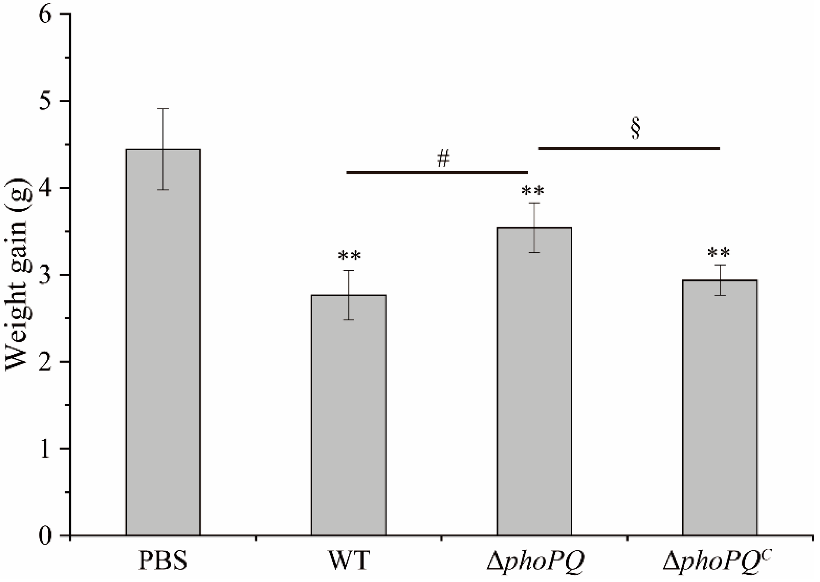
3.1. Deletion of phoPQ Gene Reduced the Count of C. sakazaki in Ileum

No C. sakazakii was detected in the control group (Figure 1). The counts of C. sakazakii colonies in the ileum of the mice in the WT group and ΔphoPQC group were 58 ± 4 CFU/mg and 36 ± 4 CFU/mg, respectively, whereas the count significantly decreased to only 3 ± 0.57 CFU/mg in the ΔphoPQ group. This indicates that the phoPQ deletion reduced the colonization ability of C. sakazakii in the mice.

3.2. PhoP/PhoQ System Affected the Histopathological Damage

Compared with the PBS group, the mice infected with C. sakazakii had slower growth from day 3 to day 10 after birth, with no mortality observed. However, the weight gain of the mice infected with the WT and ΔphoPQC strains was significantly lower (p < 0.05) compared to that of the ΔphoPQ group (Figure 2).
The H&E staining results of the jejunum, ileum, and colon of the newborn mice in the different groups are shown in Figure 3A. The intestinal morphology of the PBS group appeared normal, with no evident inflammation and damage. The jejunal injury in the mice infected with C. sakazakii was mild, while ileal and colonic lesions were severe. Due to the smaller size of the intestines in neonatal mice and the intestinal contents in the colon being relatively large, which may have interfered with the subsequent experiments, the ileum tissue was selected for the follow-up studies. The ileum tissues were observed under a magnification of 200× (Figure 3B). The ileum morphology of the mice infected with the WT strain showed severe damage, including epithelial shedding, villi destruction, and goblet cell reduction. In contrast, the ileal structure in the ΔphoPQ group remained relatively intact with reduced lesions. The ileal lesions in the ΔphoPQC group were more serious than those in the ΔphoPQ group. The histopathological scores of the three groups infected with C. sakazakii exhibited significantly higher values compared to those in the PBS group. Although no significant difference was observed between the WT group and ΔphoPQC group, both groups exhibited significantly higher scores compared to the ΔphoPQ group (p < 0.05) (Figure 3C).

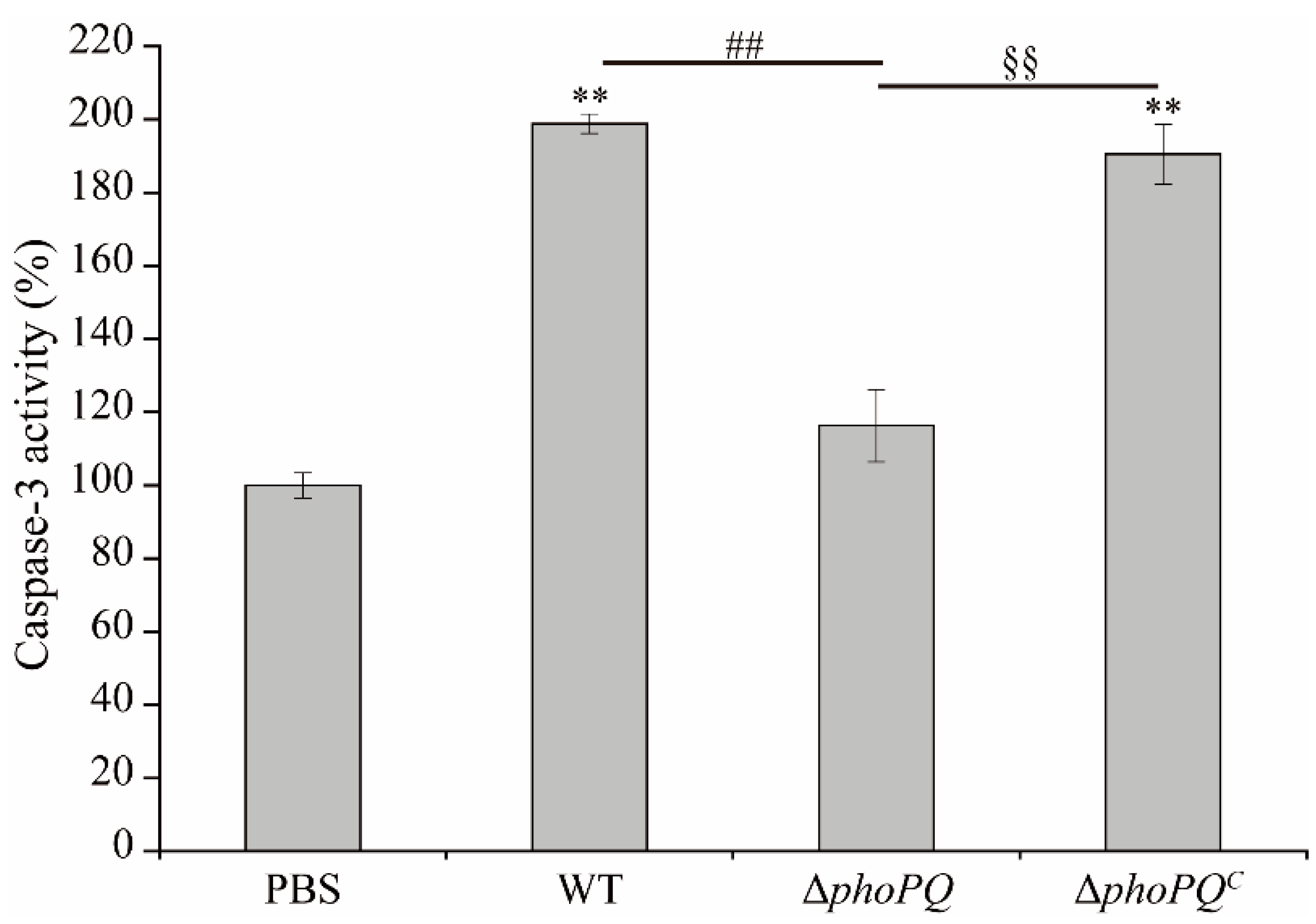
3.3. Deletion of phoPQ Gene in C. sakazakii Affected Caspase-3 Activity

The caspase-3 activity in the ileum of both the WT and ΔphoPQC groups exhibited a significant increase compared to that in the PBS group, with respective fold changes of 1.99 and 1.90. The ΔphoPQ and PBS groups exhibited comparable levels of capase-3 activity, indicating no significant difference between them (Figure 4).

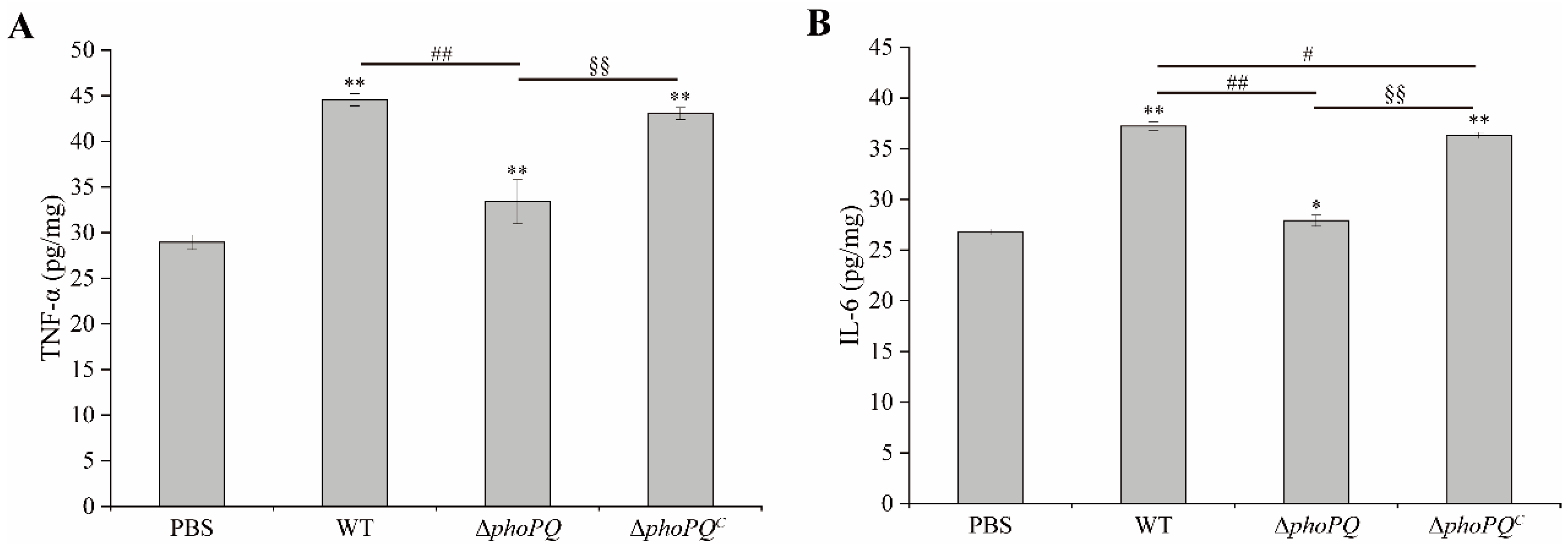
3.4. Deletion of phoPQ Gene in C. sakazakii Affected TNF-α and IL-6 Levels in Ileum

The levels of TNF-α and IL-6 in the ileum of the mice infected with the WT strain and ΔphoPQC strain were significantly elevated compared to those in the PBS group and ΔphoPQ group (p < 0.05). The TNF-α levels of the WT, ΔphoPQ, and ΔphoPQC groups increased by 53.97%, 15.38%, and 48.72%, respectively, compared with the PBS group (Figure 5A). Similarly, the IL-6 levels in the WT, ΔphoPQ, and ΔphoPQC groups increased by 38.93%, 4.14%, and 35.54%, respectively (Figure 5B). The results showed that phoPQ gene deletion significantly reduced the TNF-α and IL-6 levels within the ileum of the C. sakazakii-infected neonatal mice.

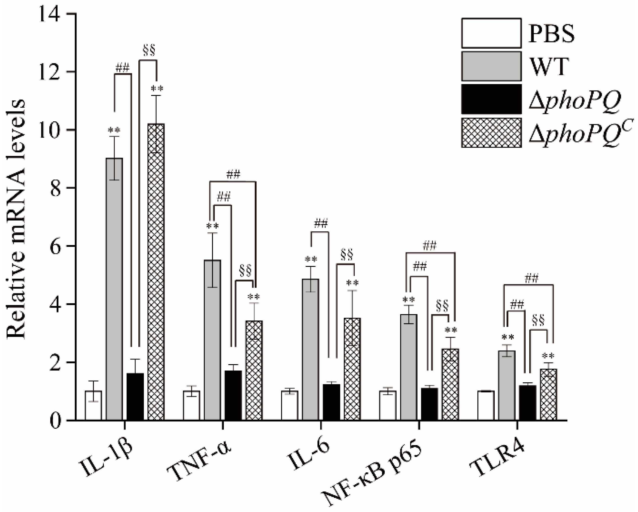
3.5. Deletion of phoPQ Gene Affected the Expression of Inflammation-Related Genes

The levels of target genes in the ileum of the neonatal mice in the four groups was detected by qRT-PCR (Figure 6). Compared to the normal mice, the mRNA transcription levels of IL-1β, TNF-α, IL-6, NF-κB p65, and TLR4 were significantly up-regulated in the ileum of the WT- and ΔphoPQC-infected mice (p < 0.05). However, the expression levels of the target genes in the ileum of the ΔphoPQ-strain-infected mice did not exhibit significant differences compared to those in the PBS group.

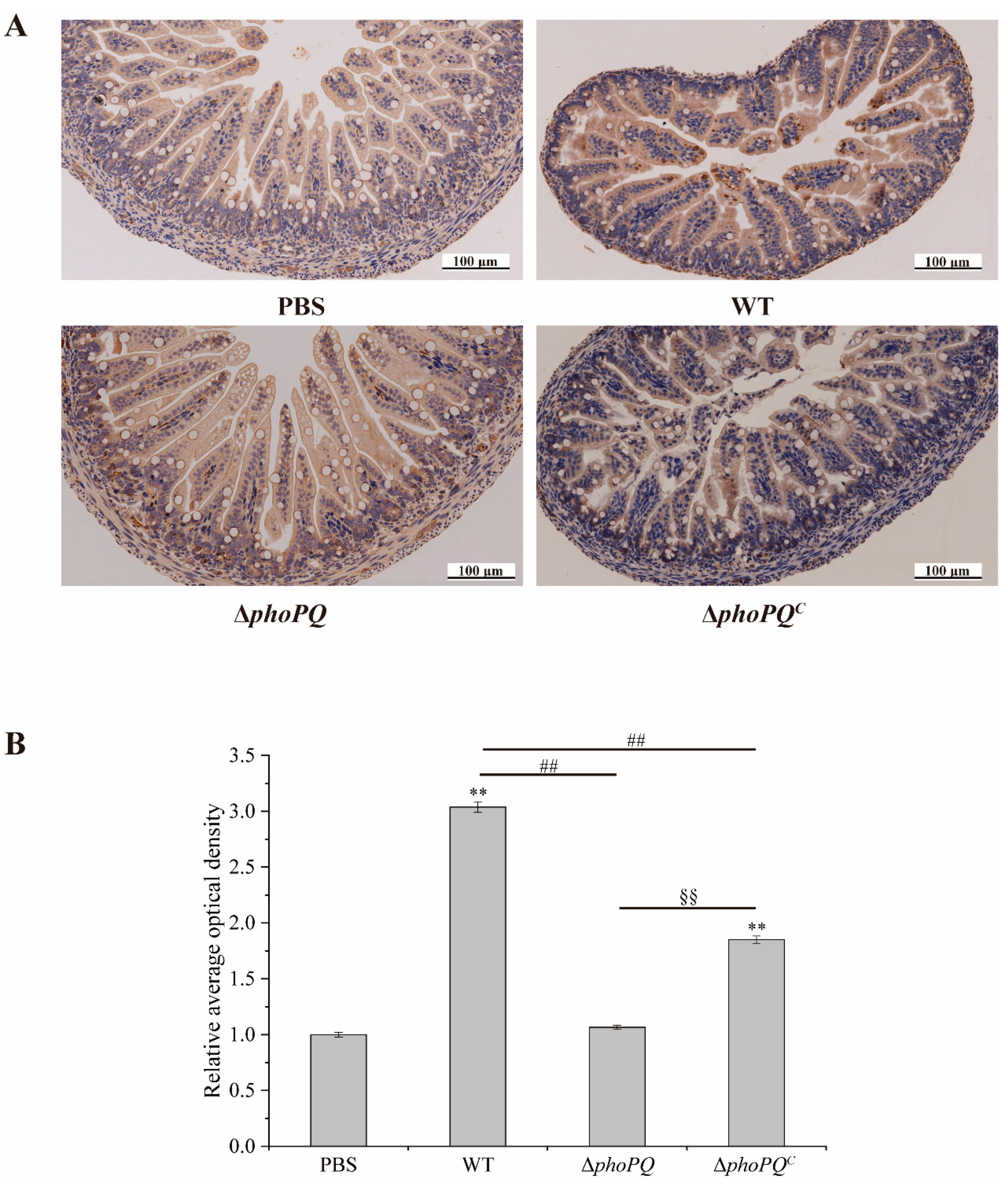
3.6. Deletion of phoPQ Gene Affected the Expression of NF-κB p65

The expression of NF-κB p65 in the ileum was measured by immunohistochemical staining (Figure 7A,B). The level of NF-κB p65 protein in the mice infected with the WT and ΔphoPQC strains exhibited a significantly higher level compared to that of the PBS group (p < 0.05). However, no significant difference was observed between the mice infected with the ΔphoPQ strains and the PBS group.

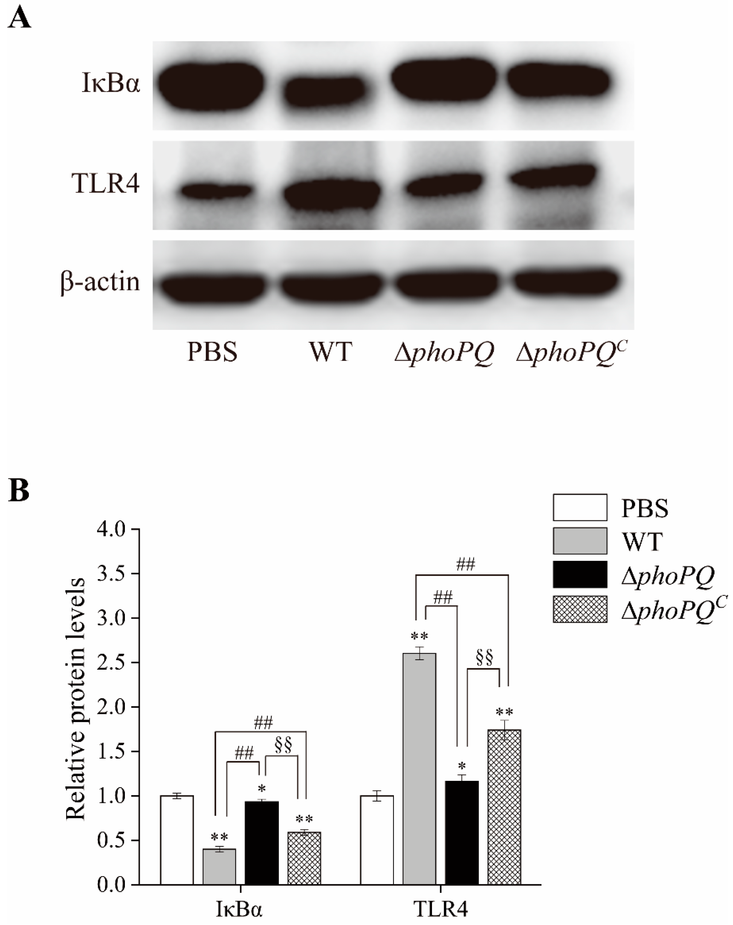
3.7. Deletion of phoPQ Gene in C. sakazakii Affected the Protein Expression of IκBα and TLR4 in the Ileum

Western blot analysis showed that TLR4 (toll-like receptor 4) protein expression was significantly up-regulated in the ileum of the C. sakazakii-infected neonatal mice compared to the control group. Moreover, the expression levels in the mice infected with the WT and ΔphoPQC strains were significantly elevated compared to those in the ΔphoPQ-infected mice. In contrast, IκBα (NF-κB inhibitor protein) protein expression was down-regulated in the C. sakazakii-infected mice, with significant decreases observed in the WT and ΔphoPQC groups compared to the ΔphoPQ group (Figure 8A,B).

4. Discussion

C. sakazakii can cause NEC in infants, with a fatality rate reaching up to 80% [30]. NEC is a high-disability, high-mortality, and devastating disease that poses a serious threat to the health of newborns [1]. Compared with BALB/c and C57BL/6 mice, CD-1 mice exhibited the highest susceptibility to C. sakazakii, having the lowest infecting and lethal doses. This suggests that CD-1 mice are suitable models for studying C. sakazakii infection in neonates [21]. Shi et al. [31] established a neonatal mouse NEC model by gavaging 3-day-old mice with 1 × 10⁷ CFU C. sakazakii ATCC29544, demonstrating that C. sakazakii infection could induce severe intestinal inflammation in mice. Weng et al. [24] found that C. sakazakii infection caused damage throughout the intestines of newborn mice, with the jejunum being slightly damaged and the ileum being severely damaged. In this study, CD-1 mice infected with C. sakazakii at 3 days old were selected to construct the NEC model, and examined the regulatory effect of PhoP/PhoQ system on intestinal inflammation. The results showed that C. sakazakii infection caused growth retardation and severe intestinal inflammation in the neonatal mice (Figure 2). The H&E staining and pathology scores indicated that the neonatal mice infected with the WT strain and ΔphoPQC strain exhibited more severe lesions than those infected with the ΔphoPQ strain, with the ileum showing the most serious damage, including intestinal epithelial shedding, villus structure destruction, and inflammatory cell infiltration (Figure 3A–C). These findings suggest that C. sakazakii causes severe intestinal injury in neonatal mice, and the deletion of the phoPQ gene reduces its pathogenicity (Figure 1, Figure 2 and Figure 3).
The main causes of NEC include prematurity, apoptosis, inflammation, and oxidative stress [32]. Apoptosis is a significant reason for the breakdown of intestinal mucosal barrier function [33]. The apoptosis process can be activated through various pathways, with caspase-3 playing a key role as an apoptosis executor [34]. Yang et al. [35] showed that C. sakazakii ATCC 29544-infected mice induced apoptosis of ileal cells by activating caspase-3 activity. Similarly, in this study, the C. sakazakii BAA-894 strain-infected neonatal mice activated caspase-3 activity. However, the caspase-3 activity in the ΔphoPQ group exhibited a significant decrease (p < 0.05) (Figure 4).
Inflammation is a protective response to cell infection or injury, mediated by the activation of various immune cells [36]. The symptoms of NEC encompass increased intestinal inflammation and elevated levels of cytokines [37]. Fan et al. demonstrated that B. fragilis inhibited C. sakazakii-induced NEC by regulating inflammation, cell apoptosis, and pyroptosis [38]. The secretion of TNF-α by various cells serves as a trigger and inducer for an inflammatory cascade [39]. IL-6 is an important immunomodulatory cytokine that promotes the migration, recruitment, and activation of inflammatory cells [40]. It has been reported that bacterial entry into tissues can cause severe inflammatory responses triggered by cytokines [41]. In this study, the ELISA results showed that the expression of TNF-α and IL-6 was significantly up-regulated in the neonatal mice infected with C. sakazakii compared to the control mice (p < 0.05). Compared to the WT and ΔphoPQC groups, the levels of TNF-α and IL-6 in the ileum of the ΔphoPQ group exhibited a significant reduction (p < 0.05) (Figure 5A,B). Similarly, the qRT-PCR results showed that, compared with the PBS group, TNF-α expression in the WT, ΔphoPQ, and ΔphoPQC groups was up-regulated by 5.51-, 1.70-, and 3.42-fold, respectively, and IL-6 expression was up-regulated by 4.85-, 1.22-, and 3.52-fold, respectively (Figure 6). Similar to TNF-α, IL-1β plays a crucial part in the host’s response to inflammation [42]. In this study, IL-1β transcription levels were significantly (p < 0.05) up-regulated in the ileum of the WT and ΔphoPQC groups compared to the PBS and ΔphoPQ groups. However, the ΔphoPQ group did not exhibit a significant difference compared to the PBS group (Figure 6). These results suggest that the PhoP/PhoQ system affects the ability of C. sakazakii to infect the intestinal tract of newborn mice by regulating the gene expression of inflammatory factors.
Toll-like receptor 4 (TLR4) mediates inflammation by recognizing lipopolysaccharides in Gram-negative bacteria [43]. The NF-κB pathway represents a pivotal downstream pathway for all LPS-mediated signal transduction pathways, suggesting that the TLR4/NF-κB pathway may serve as a crucial target for inflammatory responses and organ damage [44,45]. The TLR4/NF-κB pathway is critical in the pathogenesis of NEC [46,47]. Activation of TLR4 induces NF-κB nuclear translocation and overexpression of proinflammatory cytokines, thereby participating in the pathogenesis of NEC [48,49,50]. Zhang et al. [48] demonstrated that β-glucan may exert inhibitory effects on intestinal inflammation by modulating the TLR4/NF-κB pathway, thereby preventing NEC in newborn mice. Liu et al. [51] demonstrated that Lactobacillus reuteri mitigates the incidence and severity of NEC through modulation of the TLR4/NF-κB pathway. In this study, both the mRNA transcription levels and protein levels of NF-κB p65 in the ileum of the mice infected by the WT and ΔphoPQC strains exhibited a significant increase compared to those in the PBS group, while the expression levels of the ΔphoPQ group were similar to those in the control group (Figure 6 and Figure 7). Similarly, TLR4 mRNA and protein levels were significantly elevated in the WT- and ΔphoPQC-infected mice compared to those in the ΔphoPQ group (Figure 7 and Figure 8). However, the protein levels of IκBα in the ileum of the C. sakazakii-infected mice were significantly decreased compared to the PBS group, with lower expression levels in the WT and ΔphoPQC groups than in the ΔphoPQ group (Figure 8). Overall, the results suggest that C. sakazakii infection activates the TLR4/NF-κB pathway in the mouse ileum and may regulate inflammation mediated by this pathway through the PhoP/PhoQ system.
The PhoP/PhoQ system is critical in bacterial pathogenicity and antibiotic resistance, rendering it attractive for the development of novel antimicrobial agents. It has been reported that the PhoP/PhoQ systems in Salmonella have been targeted for screening quinazoline-based compounds with anti-virulence effects [52]. Carlos et al. [53] demonstrated that N′-(Thiophen-2-methyl-benzoylhydrazide) (A16B1) can effectively target the PhoP/PhoQ system by studying the antibacterial mechanism of A16B1 and a gentamicin protection assay, thereby mitigating the toxicity and drug resistance of Salmonella. Cai et al. [54] reported that targeting the PhoQ histidine kinase could serve as a promising therapeutic strategy against Shigella, effectively attenuating its pathogenicity. Although drugs targeting the PhoP/PhoQ system are still in the research phase, continued exploration of inhibitors against this system holds significant potential as a method for developing novel antimicrobial strategies, particularly for combating multidrug-resistant G-bacteria.

5. Conclusions

In summary, deletion of the phoPQ gene significantly reduced the pathogenicity of C. sakazakii in neonatal mice, including a reduced colonization ability in the ileum, reduced intestinal damage, decreased caspase-3 activity, and down-regulated levels of IL-1β, TNF-α, IL-6, NF-κB p65, and TLR4. In addition, C. sakazakii activates the TLR4/NF-κB pathway in the mouse ileum, and the deletion of the phoPQ gene may mitigate the inflammatory response mediated by this pathway. These findings suggest that the PhoP/PhoQ system plays a pivotal role in regulating the pathogenicity of C. sakazakii within the neonatal mouse intestinal tract.

Author Contributions

Conceptualization, X.X. and Y.M.; methodology, Y.M. and Y.Z.; software, Y.Z., Y.W., Z.Q. and Y.M.; validation, Y.Z., Y.W. and Y.L.; formal analysis, Y.M., Z.Q., Y.L. and X.X.; investigation, X.X. and Y.M.; resources, X.X., Y.W. and Z.Q.; data curation, Y.L. and Y.W.; writing—original draft preparation, Y.M.; writing—review and editing, X.X. and Y.M.; visualization, Y.M. and Y.Z.; supervision, X.X.; project administration, X.X.; funding acquisition, X.X. and Y.M. All authors have read and agreed to the published version of the manuscript.

Funding

This research was funded in part by the National Key Research and Development Program of China (2022YFD2100104), the Science and Technology Development Plan Project of Henan Province, China (242102110136; 242102110117), Key scientific research project of Henan Higher Education Institutions (24B550006), and the National Scientific Research Project Cultivation Fund project of Huanghuai University (XKPY-2023001).

Institutional Review Board Statement

The study was approved by the Animal Ethics Committee of Dalian Polytechnic University (DLPU2022082) (Approval date: 25 October 2022).

Informed Consent Statement

Not applicable.

Data Availability Statement

The original contributions presented in the study are included in the article, further inquiries can be directed to the corresponding author.

Conflicts of Interest

The authors declare that there are no conflicts of interest.

References

  1. Yang, Y.; Zhang, T.; Zhou, G.; Jiang, X.; Tao, M.; Zhang, J.; Zeng, X.; Wu, Z.; Pan, D.; Guo, Y. Prevention of necrotizing enterocolitis through milk polar lipids reducing intestinal epithelial apoptosis. J. Agric. Food Chem. 2020, 68, 7014–7023. [Google Scholar] [CrossRef]
  2. Hackam, D.J.; Sodhi, C.P. Bench to bedside—New insights into the pathogenesis of necrotizing enterocolitis. Nat. Rev. Gastroenterol. Hepatol. 2022, 19, 468–479. [Google Scholar] [PubMed]
  3. Henry, M.; Moss, R. Necrotizing enterocolitis. Annu. Rev. Med. 2009, 60, 111–124. [Google Scholar] [CrossRef]
  4. Niño, D.F.; Sodhi, C.P.; Hackam, D.J. Necrotizing enterocolitis: New insights into pathogenesis and mechanisms. Nat. Rev. Gastroenterol. Hepatol. 2016, 13, 590–600. [Google Scholar] [PubMed]
  5. Wang, L.; Gao, M.; Kang, G.; Huang, H. The potential role of phytonutrients flavonoids influencing gut microbiota in the prophylaxis and treatment of inflammatory bowel disease. Front. Nutr. 2021, 8, 798038. [Google Scholar] [CrossRef]
  6. Patel, R.M.; Denning, P.W. Intestinal microbiota and its relationship with necrotizing enterocolitis. Pediatr. Res. 2015, 78, 232–238. [Google Scholar]
  7. Jin, T.; Zhan, X.; Pang, L.; Peng, B.; Zhang, X.; Zhu, W.; Yang, B.; Xia, X. CpxAR two-component system contributes to virulence properties of Cronobacter sakazakii. Food Microbiol. 2024, 117, 104393. [Google Scholar] [CrossRef]
  8. Blackwood, B.; Hunter, C. Cronobacter spp. Microbiol. Spectr. 2016, 4, 10–1128. [Google Scholar] [CrossRef] [PubMed]
  9. Fei, P.; Jiang, Y.; Feng, J.; Forsythe, S.J.; Li, R.; Zhou, Y.; Man, C. Antibiotic and desiccation resistance of Cronobacter sakazakii and C. malonaticus isolates from powdered infant formula and processing environments. Front. Microbiol. 2017, 8, 316. [Google Scholar] [CrossRef]
  10. Guo, X.; Yu, H.; Xiong, J.; Dai, Q.; Li, Y.; Zhang, W.; Liao, X.; He, X.; Zhou, H.; Zhang, K. Pseudomonas aeruginosa two-component system LadS/PA0034 regulates macrophage phagocytosis via fimbrial protein cupA1. mBio 2024, 15, e00616-24. [Google Scholar] [CrossRef]
  11. Monteagudo-Cascales, E.; Santero, E.; Canosa, I. The regulatory hierarchy following signal integration by the CbrAB two-component system: Diversity of responses and functions. Genes 2022, 13, 375. [Google Scholar] [CrossRef] [PubMed]
  12. Gooderham, W.J.; Hancock, R.E.W. Regulation of virulence and antibiotic resistance by two-component regulatory systems in Pseudomonas aeruginosa. FEMS Microbiol. Rev. 2009, 33, 279–294. [Google Scholar] [CrossRef]
  13. Zhu, Y.; Dou, Q.; Du, L.; Wang, Y. QseB/QseC: A two-component system globally regulating bacterial behaviors. Trends Microbiol. 2023, 31, 749–762. [Google Scholar] [PubMed]
  14. Singh, P.; Goar, H.; Paul, P.; Mehta, K.; Bamniya, B.; Vijjamarri, A.; Bansal, R.; Khan, H.; Karthikeyan, S.; Sarkar, D. Dual functioning by the PhoR sensor is a key determinant to Mycobacterium tuberculosis virulence. PLoS Genet. 2023, 19, e1011070. [Google Scholar] [CrossRef]
  15. Janssen, A.; de Bakker, V.; Aprianto, R.; Trebosc, V.; Kemmer, C.; Pieren, M.; Veening, J.W. Klebsiella pneumoniae OmpR facilitates lung infection through transcriptional regulation of key virulence factors. Microbiol. Spectr. 2023, 12, e0396623. [Google Scholar] [CrossRef]
  16. Fernandez-Ciruelos, B.; Potmis, T.; Solomin, V.; Wells, J. Cross-talk between QseBC and PmrAB two-component systems is crucial for regulation of motility and colistin resistance in Enteropathogenic Escherichia coli. PLoS Pathog. 2023, 19, e1011345. [Google Scholar] [CrossRef] [PubMed]
  17. Groisman, E.A.; Duprey, A.; Choi, J. How the PhoP/PhoQ system controls virulence and Mg2+ homeostasis: Lessons in signal transduction, pathogenesis, physiology, and evolution. Microbiol. Mol. Biol. Rev. 2021, 85, e0017620. [Google Scholar]
  18. Han, J.; Gao, X.; Luo, X.; Zhu, L.; Zhang, Y.; Dong, P. The role of PhoP/PhoQ system in regulating stress adaptation response in Escherichia coli O157:H7. Food Microbiol. 2023, 112, 104244. [Google Scholar] [CrossRef]
  19. Disney-McKeethen, S.; Seo, S.; Mehta, H.; Ghosh, K.; Shamoo, Y. Experimental evolution of Pseudomonas aeruginosa to colistin in spatially confined microdroplets identifies evolutionary trajectories consistent with adaptation in microaerobic lung environments. mBio 2023, 14, e0150623. [Google Scholar] [CrossRef]
  20. Cabezudo, I.; Lobertti, C.; Véscovi, E.; Furlan, R. Effect-directed synthesis of PhoP/PhoQ inhibitors to regulate Salmonella virulence. J. Agric. Food Chem. 2022, 70, 6755–6763. [Google Scholar] [CrossRef]
  21. Richardson, A.; Lambert, S.; Smith, M. Neonatal mice as models for Cronobacter sakazakii infection in infants. J. Food Prot. 2009, 72, 2363–2367. [Google Scholar] [CrossRef] [PubMed]
  22. Ma, Y.; Zhang, Y.; Chen, K.; Zhang, L.; Zhang, Y.; Wang, X.; Xia, X. The role of PhoP/PhoQ two component system in regulating stress adaptation in Cronobacter sakazakii. Food Microbiol. 2021, 100, 103851. [Google Scholar] [CrossRef]
  23. Emami, C.; Mittal, R.; Wang, L.; Ford, H.; Prasadarao, N. Recruitment of dendritic cells is responsible for intestinal epithelial damage in the pathogenesis of necrotizing enterocolitis by Cronobacter sakazakii. J. Immunol. 2011, 186, 7067–7079. [Google Scholar] [CrossRef]
  24. Weng, M.Q.; Ganguli, K.; Zhu, W.S.; Shi, H.N.; Walker, W.A. Conditioned medium from Bifidobacteria infantis protects against Cronobacter sakazakii-induced intestinal inflammation in newborn mice. Am. J. Physiol. Gastrointest. Liver Physiol. 2014, 306, G779–G787. [Google Scholar] [CrossRef] [PubMed]
  25. Eissa, N.; Hussein, H.; Wang, H.; Rabbi, M.F.; Bernstein, C.N.; Ghia, J.E.; Sly, L.M. Stability of reference genes for messenger RNA quantification by real-time PCR in mouse dextran sodium sulfate experimental colitis. PLoS ONE 2016, 11, e0156289. [Google Scholar] [CrossRef]
  26. He, Y.; Yao, X.; Taylor, N.; Bai, Y.; Lovenberg, T.; Bhattacharya, A. RNA sequencing analysis reveals quiescent microglia isolation methods from postnatal mouse brains and limitations of BV2 cells. J. Neuroinflammation 2018, 15, 153. [Google Scholar] [CrossRef]
  27. Zhang, N.; Huan, Y.; Huang, H.; Song, G.M.; Sun, S.J.; Shen, Z.F. Atorvastatin improves insulin sensitivity in mice with obesity induced by monosodium glutamate. Acta Pharmacol. Sin. 2010, 31, 35–42. [Google Scholar] [CrossRef] [PubMed]
  28. Boytard, L.; Hadi, T.; Silvestro, M.; Qu, H.; Kumpfbeck, A.; Sleiman, R.; Fils, K.; Alebrahim, D.; Boccalatte, F.; Kugler, M.; et al. Lung-derived HMGB1 is detrimental for vascular remodeling of metabolically imbalanced arterial macrophages. Nat. Commun. 2020, 11, 4311. [Google Scholar] [CrossRef]
  29. Jin, C.J.; Liu, J.Y.; Jin, R.Y.; Yao, Y.P.; He, S.; Lei, M.; Peng, X.L. Linarin ameliorates dextran sulfate sodium-induced colitis in C57BL/6J mice via the improvement of intestinal barrier, suppression of inflammatory responses and modulation of gut microbiota. Food Funct. 2022, 13, 10574–10586. [Google Scholar] [CrossRef]
  30. Gao, J.; Han, Z.; Li, P.; Zhang, H.; Du, X.; Wang, S. Outer membrane protein F is involved in biofilm formation, virulence and antibiotic resistance in Cronobacter sakazakii. Microorganisms 2021, 9, 2338. [Google Scholar] [CrossRef]
  31. Shi, C.; Jin, T.; Guo, D.; Zhang, W.; Yang, B.; Su, D.; Xia, X. Citral attenuated intestinal inflammation induced by Cronobacter sakazakii in newborn mice. Foodborne Pathog. Dis. 2020, 17, 243–252. [Google Scholar] [CrossRef]
  32. Ufuk, C.; Cuneyt, T.; Utku, S.; Halil Ibrahim, Y.; Esra, C.; Ufuk, A.; Ismail, K.; Eyyup, K. Ginger (Zingiber officinale Roscoe) for the treatment and prevention of necrotizing enterocolitis. J. Ethnopharmacol. 2018, 225, 297–308. [Google Scholar]
  33. Janina, M.; Lilith, R.; Christoph, H.; Mats Ingmar, F.; Kirstin, F.; Delfina, M.; Jürgen, H.; Michael, Z.; Mercedes, G.d.A.; Katja, M.; et al. Antimicrobial peptides (AMPs) and the microbiome in preterm infants: Consequences and opportunities for future therapeutics. Int. J. Mol. Sci. 2024, 25, 6684. [Google Scholar] [CrossRef] [PubMed]
  34. Manabu, K.; Sally, K. Caspases and kinases in a death grip. Cell 2009, 138, 838–854. [Google Scholar]
  35. Yang, G.; Jin, T.; Yin, S.; Guo, D.; Zhang, C.; Xia, X.; Shi, C. trans-Cinnamaldehyde mitigated intestinal inflammation induced by Cronobacter sakazakii in newborn mice. Food Funct. 2019, 10, 2986–2996. [Google Scholar] [CrossRef]
  36. Hejna, M.; Kovanda, L.; Rossi, L.; Liu, Y. Mint oils: In vitro ability to perform anti-inflammatory, antioxidant, and antimicrobial activities and to enhance intestinal barrier integrity. Antioxidants 2021, 10, 1004. [Google Scholar] [CrossRef]
  37. Bu, C.; Hu, M.; Su, Y.; Yuan, F.; Zhang, Y.; Xia, J.; Jia, Z.; Zhang, L. Cell-permeable JNK-inhibitory peptide regulates intestinal barrier function and inflammation to ameliorate necrotizing enterocolitis. J. Cell Mol. Med. 2024, 28, e18534. [Google Scholar] [CrossRef] [PubMed]
  38. Fan, H.; Chen, Z.; Lin, R.; Liu, Y.; Zhi, F. Bacteroides fragilis strain ZY-312 defense against Cronobacter sakazakii-induced necrotizing enterocolitis in vitro and in a neonatal rat model. mSystems 2019, 4, e00305-19. [Google Scholar] [CrossRef]
  39. Rajendra, K.; Bhesh Raj, S.; Shraddha, T.; Evan Peter, W.; Lillian, Z.; Parimal, S.; Min, Z.; Balamurugan, S.; Balaji, B.; Subbarao Malireddi, R.K.; et al. Synergism of TNF-α and IFN-γ triggers inflammatory cell death, tissue damage, and mortality in SARS-CoV-2 infection and cytokine shock syndromes. Cell 2021, 184, 149–168.e17. [Google Scholar]
  40. Rose-John, S.; Jenkins, B.J.; Garbers, C.; Moll, J.M.; Scheller, J. Targeting IL-6 trans-signalling: Past, present and future prospects. Nat. Rev. Immunol. 2023, 23, 666–681. [Google Scholar] [CrossRef]
  41. Kaplina, A.; Kononova, S.; Zaikova, E.; Pervunina, T.; Petrova, N.; Sitkin, S. Necrotizing enterocolitis: The role of hypoxia, gut microbiome, and microbial metabolites. Int. J. Mol. Sci. 2023, 24, 2471. [Google Scholar] [CrossRef] [PubMed]
  42. Wang, Y.; Che, M.; Xin, J.; Zheng, Z.; Li, J.; Zhang, S. The role of IL-1β and TNF-α in intervertebral disc degeneration. Biomed. Pharmacother. 2020, 131, 110660. [Google Scholar] [CrossRef]
  43. Zhang, Y.; Liang, X.; Bao, X.; Xiao, W.; Chen, G. Toll-like receptor 4 (TLR4) inhibitors: Current research and prospective. Eur. J. Med. Chem. 2022, 235, 114291. [Google Scholar] [CrossRef] [PubMed]
  44. Yang, J.; Chen, Y.; Jiang, K.; Yang, Y.; Zhao, G.; Guo, S.; Guo, G. MicroRNA-106a provides negative feedback regulation in lipopolysaccharide-induced inflammation by targeting TLR4. Int. J. Biol. Sci. 2019, 15, 2308–2319. [Google Scholar] [CrossRef]
  45. Yang, R.; Hu, X.; Xie, X.; Chen, H.; Fang, H.; Zhu, L.; Li, Z. Propionic acid targets the TLR4/NF-κB signaling pathway and inhibits LPS-induced intestinal barrier dysfunction: In vitro and in vivo studies. Front. Pharmacol. 2020, 11, 573475. [Google Scholar] [CrossRef]
  46. Chhinder, P.S.; Matthew, D.N.; Richard, S.; Shonan, S.; Congrong, M.; Maria, F.B.; Thomas, P., Jr.; Anthony, M.R.; Amin, A.; Misty, G.; et al. Intestinal epithelial Toll-like receptor 4 regulates goblet cell development and is required for necrotizing enterocolitis in mice. Gastroenterology 2012, 143, 708–718.e5. [Google Scholar]
  47. Yu, R.; Jiang, S.; Tao, Y.; Li, P.; Yin, J.; Zhou, Q. Inhibition of HMGB1 improves necrotizing enterocolitis by inhibiting NLRP3 via TLR4 and NF-κB signaling pathways. J. Cell Physiol. 2019, 234, 13431–13438. [Google Scholar] [CrossRef] [PubMed]
  48. Zhang, X.; Zhang, Y.; He, Y.; Zhu, X.; Ai, Q.; Shi, Y. β-glucan protects against necrotizing enterocolitis in mice by inhibiting intestinal inflammation, improving the gut barrier, and modulating gut microbiota. J. Transl. Med. 2023, 21, 14. [Google Scholar] [CrossRef]
  49. Sun, Q.; Ji, Y.C.; Wang, Z.L.; She, X.; He, Y.; Ai, Q.; Li, L.Q. Sodium butyrate alleviates intestinal inflammation in mice with necrotizing enterocolitis. Mediat. Inflamm. 2021, 2021, 6259381. [Google Scholar] [CrossRef]
  50. De Plaen, I.G.; Liu, S.X.L.; Tian, R.; Neequaye, I.; May, M.J.; Han, X.-B.; Hsueh, W.; Jilling, T.; Lu, J.; Caplan, M.S. Inhibition of nuclear factor-kappaB ameliorates bowel injury and prolongs survival in a neonatal rat model of necrotizing enterocolitis. Pediatr. Res. 2007, 61, 716–721. [Google Scholar] [CrossRef]
  51. Liu, Y.; Fatheree, N.Y.; Mangalat, N.; Rhoads, J.M. Lactobacillus reuteri strains reduce incidence and severity of experimental necrotizing enterocolitis via modulation of TLR4 and NF-κB signaling in the intestine. Am. J. Physiol. Gastrointest. Liver Physiol. 2012, 302, G608–G617. [Google Scholar] [CrossRef] [PubMed]
  52. Carabajal, M.A.; Asquith, C.R.M.; Laitinen, T.; Tizzard, G.J.; Yim, L.; Rial, A.; Chabalgoity, J.A.; Zuercher, W.J.; Véscovi, E.G. Quinazoline-based antivirulence compounds selectively target Salmonella PhoP/PhoQ signal transduction system. Antimicrob. Agents Chemother. 2019, 64, e01744-19. [Google Scholar] [CrossRef] [PubMed]
  53. Carlos, A.L.; Ignacio, C.; Fernán, O.G.; Víctor, B.; Christian, M.; Ricard, L.E.F.; Eleonora, G.V. An allosteric inhibitor of the PhoQ histidine kinase with therapeutic potential against Salmonella infection. J. Antimicrob. Chemother. 2024, 79, 1820–1830. [Google Scholar]
  54. Cai, X.; Zhang, J.; Chen, M.; Wu, Y.; Wang, X.; Chen, J.; Zhang, J.; Shen, X.; Qu, D.; Jiang, H. The effect of the potential PhoQ histidine kinase inhibitors on Shigella flexneri virulence. PLoS ONE 2011, 6, e23100. [Google Scholar] [CrossRef] [PubMed]
Figure 1. The count of C. sakazakii in ileum. *, #, and § represent comparisons with PBS, WT, and ΔphoPQ groups, respectively. ** p, ## p, and §§ p mean p < 0.01 (n = 5/group).
Foods 13 02808 g001
Figure 2. Effect of phoPQ gene deletion on weight gain. *, #, and § represent comparisons with PBS, WT, and ΔphoPQ groups, respectively. # p, and § p mean p < 0.05, and ** p mean p < 0.01 (n = 30/group).
Foods 13 02808 g002
Figure 3. H&E staining and pathology score of neonatal mice intestinal tissues. Arrows indicate epithelial shedding, villi destruction, and goblet cell reduction. (A): H&E staining of jejunum, ileum, and colon tissue of neonatal mice (100×); (B): H&E staining of ileum tissues (200×); (C): histopathological score of ileum tissues. *, #, and § represent comparisons with PBS, WT, and ΔphoPQ groups, respectively. * p mean p < 0.05, and ** p, ## p, and §§ p mean p < 0.01.
Foods 13 02808 g003
Figure 4. Caspase-3 activity in ileum of newborn mice in PBS, WT, ΔphoPQ, and ΔphoPQC groups. *, #, and § represent comparisons with PBS, WT, and ΔphoPQ groups, respectively. ** p, ## p, and §§ p mean p < 0.01 (n = 5–6/group).
Foods 13 02808 g004
Figure 5. The levels of TNF-α (A) and IL-6 (B) in ileum of newborn mice in PBS, WT, ΔphoPQ, and ΔphoPQC groups. *, #, and § represent comparisons with PBS, WT, and ΔphoPQ groups, respectively. * p and # p mean p < 0.05, and ** p, ## p, and §§ p mean p < 0.01 (n = 5/group).
Foods 13 02808 g005
Figure 6. Relative mRNA levels of target genes in the ileum tissue of newborn mice by qRT-PCR. *, #, and § represent comparisons with PBS, WT, and ΔphoPQ groups, respectively. ** p, ## p, and §§ p mean p < 0.01 (n = 5/group).
Foods 13 02808 g006
Figure 7. Immunohistochemical staining of NF-κB p65 protein in the ileum of newborn mice. (A): Representative images of immunohistochemistry; (B): the level of NF-κB p65 protein analyzed by relative average optical density. *, #, and § represent comparisons with PBS, WT, and ΔphoPQ groups, respectively. ** p, ## p, and §§ p mean p < 0.01.
Foods 13 02808 g007
Figure 8. The protein levels of IκBα and TLR4 in the ileum. (A): Representative images; (B): relative protein levels of IκBα and TLR4. *, #, and § represent comparisons with PBS, WT, and ΔphoPQ groups, respectively. * p mean p < 0.05, and ** p, ## p, and §§ p mean p < 0.01 (n = 5/group).
Foods 13 02808 g008
Table 1. Primers used for qRT-PCR.
GeneSequence (5′-3′)References
IL-1βF: GCAACTGTTCCTGAACTCAACT
R: ATCTTTTGGGGTCCGTCAACT
[25]
TNF-αF: CCCTCACACTCAGATCATCTTCT
R: GCTACGACGTGGGCTACAG
[26]
IL-6F: TAGTCCTTCCTACCCCAATTTCC
R: TTGGTCCTTAGCCACTCCTTC
[26]
NF-κB p65F: AGGCTTCTGGGCCTTATGTG
R: TGCTTCTCTCGCCAGGAATAC
[27]
TLR4F: ATGGCATGGCTTACACCACC
R: GAGGCCAATTTTGTCTCCACA
[28]
GAPDHF: AGGTCGGTGTGAACGGATTTG
R: TGTAGACCATGTAGTTGAGGTCA
Disclaimer/Publisher’s Note: The statements, opinions and data contained in all publications are solely those of the individual author(s) and contributor(s) and not of MDPI and/or the editor(s). MDPI and/or the editor(s) disclaim responsibility for any injury to people or property resulting from any ideas, methods, instructions or products referred to in the content.

