Review

Non-Thermal Treatment Mediated by Curcumin for Enhancing Food Product Quality

1
Key Laboratory of Geriatric Nutrition and Health, Beijing Technology and Business University, Ministry of Education, Beijing 100048, China
2
National Center of Technology Innovation for Grain Industry, Comprehensive Utilization of Edible By-Products, Beijing Technology and Business University, Beijing 100048, China
3
School of Food Science and Technology, Jiangnan University, Wuxi 214122, China
*
Author to whom correspondence should be addressed.
Foods 2024, 13(23), 3980; https://doi.org/10.3390/foods13233980
Submission received: 31 October 2024 / Revised: 30 November 2024 / Accepted: 6 December 2024 / Published: 9 December 2024

Abstract

:
Increasing antibiotic resistance is one of the world’s greatest health problems, and biocide use in food disinfection, alongside other application fields, could increase antibiotic resistance. Effective and eco-friendly food decontamination treatment with minimal chemical intervention in food production is urgently needed. Synergistic antimicrobial interaction of photoactive compounds and blue-light-emitting diodes have recently been proven effective in agricultural and environmental applications. Curcumin-based non-thermal treatment has been reviewed in this work for the development of a safe and effective decontamination tool that could be adapted to the food industry. The antimicrobial mechanism of the synergistic interaction and the inhibitory efficacy against foodborne pathogens (bacteria in both vegetative form and spore, as well as in biofilms) are discussed. Further studies on curcumin and its derivative, as well as light illumination patterns, were compared for enhanced bactericidal efficacy. Moreover, studies relating to photodynamic inactivation treatment for food sanitation and food quality enhancement (cereal grains and other food products) were summarized, as well as the impact on food organoleptic and nutritional quality.

1. Introduction

Nowadays, the outbreak of foodborne diseases has become a crucial issue that threatens food safety and public health [1,2]. Due to incomplete sterilization or improper preservation, microorganisms in food could multiply quickly, resulting in food spoilage and deterioration, along with serious economic losses [3]. Therefore, exploring safe, environmentally friendly, and efficient means for controlling and eradicating harmful microorganisms has always been a concern for both researchers and consumers [4]. However, long-term use of chemical preservatives has induced antibiotic-resistant microorganisms in the environment, creating new challenges for the fresh-produce market, while the existing disinfection strategies are not efficient [5]. Non-thermal food decontamination technologies, such as ultrasound, pulsed electric field (PEF), ionizing radiation, and cold plasma (CP), have received increasing attention recently [6]. However, there are still concerns about the practical application of these technologies due to the low sterilization efficiency and the safety hazards they pose [4,7,8,9]. For instance, PEF has a limited spore inactivation efficiency and may cause structural changes [10]. And CP-treated food might induce the formation of toxic compounds [6]. Furthermore, microbial resistance to PEF and ultrasonication have been reported [11], reinforcing the urgent need for an alternative disinfection technique that is safe, efficient, and cost-effective.
Photodynamic inactivation (PDI) is a new and promising strategy for eradicating microorganisms. It involves using a photosensitizer, light, and oxygen to damage and kill cells, especially harmful ones like microorganisms [12]. PDI sterilizes food products through the synergistic interaction between photoactive compounds (photosensitizers, PS) and visible-light illumination, and it is a safe, effective, and environment- and human-friendly method [13]. Photosensitization, which does not adversely affect food texture and avoids the thermal effects associated with PEF, is currently becoming a focal point in the field of light-based food processing [2]. As a non-thermal disinfection technique, the bactericidal efficacy is attributed to the generated reactive oxygen species (ROS) by non-toxic photosensitizers (PS) upon light excitation in the presence of oxygen. The production of ROS could attack cellular components simultaneously, including nucleic acids, lipids, and proteins, causing cell death ultimately [13]. Existing non-thermal treatment methods still have some challenges, such as being time consuming, expensive, stimulating microbial resistance, and having adverse effects on food quality (e.g., color, texture, and bioactive compounds), thus limiting their commercial application in food processing [14]. Compared to other antimicrobial methods, photosensitized sterilization is economical, controllable, stable, and more efficient without affecting food texture [2,9,13]. One of the most important advantages of this multi-targeted strategy over other antibacterial approaches is no microbial resistance, making it gain increasing attention [15].
For the application in food processing, the ideal photosensitizer should have the characteristics of being natural, cost-effective, having good efficacy in low concentration, and being safe for human consumption [12]. Curcumin (CUR), as a yellow phenol compound extracted from the rhizome of turmeric, has been used as a food additive since ancient times, with low-cost, safe, non-toxic, and non-polluting properties [12,16]. Presently, numerous studies have illuminated the biological features and medicinal properties of CUR, such as antioxidant, anti-cancer, anti-inflammatory, anti-diabetic, hypolipidemic, and other pharmacological benefits [17,18,19]. CUR is a natural photosensitizer that absorbs light in the wavelength range of 400–475 nm [16]. Curcumin-mediated photosensitization targets a range of bacteria and fungi, such as Staphylococcus aureus, Enterococcus faecalis, Streptococcus mutans, Salmonella typhimurium, Streptococcus pyogenes, Lactobacillus spp., and Escherichia coli [20]. Its combination with LED light sources has been shown to be useful in the purification of fresh foods, such as aquatic products, vegetables and fruits, dairy products, and poultry [2]. Moreover, the conjugation of CUR with edible biopolymers has yielded good results in the development of anti-microbial food packaging [14]. Its application in food ingredient production, product packaging, and other fields can help to enhance food microbiological control. In turn, it extends the shelf life of food, maintains the sensory and nutritional properties of food, and reduces foodborne diseases [13].
Therefore, the aim of this review is to present the synergistic potential of curcumin and light-emitting diodes for food sanitizing and cleaning labeling solutions. Starting from the bactericidal mechanism and mode of action of photosensitization, curcumin-mediated photodynamic inactivation in food packaging, processed surfaces, and food model systems is presented. This innovative approach opens a new and prospective avenue for the development of efficient, affordable, non-genotoxic, non-hazardous, and eco-friendly non-thermal antimicrobial techniques that may substitute for chemical interventions in food processing and agriculture.

2. Mechanisms of Antimicrobial Action

In general, photosensitization is based on the activation of photosensitizers by light at an appropriate wavelength, along with the presence of oxygen molecules. The schematic diagram of the photosensitization processes is illustrated in Figure 1. The ground state photosensitizers are able to absorb the quantum of light and convert it to an excited singlet state, which is unstable and, further, can return to the ground state via releasing energy (emitting fluorescence) or transform to an excited triplet state via intersystem crossing [2]. Then, the excited triplet state PS could decay to the ground state by emitting phosphorescence or reacting with an acceptor substrate (e.g., organic molecules) to release the extra energy, resulting in the production of radicals.
These free radicals can target oxygen molecules and generate reactive oxygen species (ROS), such as hydrogen peroxide (H2O2), hydroxyl radicals (HO·), and superoxide anions (·O2) (Type I mechanism). The ROS formed by the above mechanism can attack hundreds of cells per minute and react with the sensitive groups of biological molecules, causing multi-targeted cell damage [2,21,22]. Alternatively, the energy of the excited triplet state PS can be transferred to an oxygen molecule producing highly reactive singlet oxygen (1O2), which is cytotoxic and is predominantly involved in antibacterial photosensitization (Type II mechanism) [22,23]. The 1O2 can target different biomolecules, including aromatic side chains of amino acids, sulfur-containing amino acids, nucleic acids, and unsaturated lipids [24]. Both mechanisms induced oxidative damage to the cell membrane and cellular integrity, which could cause cytoplasm leakage, ending up in cell death. This mode of multi-targeted action has been proven to be efficient against a variety of microorganisms (e.g., parasitic protozoa, bacteriophages, fungi, Gram-positive and Gram-negative bacteria in a vegetative form, spores, and biofilms) with no emerging of mutagenic risks, namely no existence of antibiotic resistance [25].
In addition, the antimicrobial effect of photosensitization might be ascribed to the photolysis products of curcumin (Figure 2). Photolysis products, such as benzaldehyde and 2-methoxy-4-vinylphenol, could interfere with the normal expression of bacterial genes [20]. Cinnamaldehyde and ferulic acid derived from curcumin were found to disrupt cellular integrity and alter cellular morphology [26]. Vanillin was found to induce the dissipation of ion gradients and the inhibition of respiration. Flavanone was capable of inactivating bacteria by inhibiting their dehydrogenase activity, disturbing the normal physiological and biochemical reactions of the organism [27].

3. Light Sources and Irradiation Mode for Photosensitization

The light sources for the activation of photosensitizers include metal halide lamps, short-arc xenon, narrow-band UV lamps, and light-emitting diodes (LEDs). LED technology is based on semiconductors that emit selected ranges of wavelengths within the visible-light spectrum. Compared with UV illumination and conventional sources of light, LEDs, in terms of spectral properties and radiation pattern, show numerous advantages, including being environmentally friendly, energy-saving, safe, having a long lifespan, and being effective at causing a lethal photobiological response [2,24], without the concerns of harmful impacts on human health and deleterious effect on treated foods [28,29,30,31].
In general, the antimicrobial activity of LEDs is dependent on variable factors, such as the wavelength and irradiation mode. The present LED spectrum covers a wide range (250 nm–7 μm) and can be subdivided based on different wavelengths, among which blue LED (400–470 nm) was the most effective for microbial decontamination. Light irradiation can be performed in different manners, among them fractional or continuous. The continuous mode is traditionally used in PDT treatments, while the fractionation of the light dose is described in the literature as an advantageous protocol. This is because it allows for tissue reoxygenation during the period when it is not being illuminated, thus potentializing the PDT effect.

4. Bacteriostatic Action of Photosensitization

The effectiveness of synergistic interaction by CUR and LED on planktonic microorganisms has been summarized in Table 1. In this paper, the reduction of the number of bacterial colonies is evaluated by the logarithmic value of the total number of bacterial colonies per milliliter of sample (log CFU/mL). Both Gram-positive bacteria and Gram-negative bacteria were effectively inactivated by the combined treatment of CUR and blue-light (400–500 nm) illumination [32]. Aided by CUR (75 μM), a 3.57 log CFU/mL reduction of Staphylococcus aureus was achieved after 10 min of light exposure (139 J/cm2). Meanwhile, significant antimicrobial action against Aeromonas hydrophila, Salmonella typhimurium, Pseudomonas aeruginosa, and Escherichia coli was observed, with a corresponding reduction of 3.33, 1.26, 0.24, and 1.29 log CFU/mL, respectively. Similarly, Jiang et al. found that CUR (0–2.5 μM) and blue LED (3 J/cm2) reduced the growth of S. aureus by around 2.5 log CFU/mL [33]. Using CUR (5 μM) and blue light (3.6 J/cm2), Gao et al. found that the viability of E. coli DH5α decreased by 3.5 log CFU/mL in a dose-dependent manner [34]. Wu et al. [35] reported that the combined treatment of CUR (10 and 20 μM) and LED irradiation (470 nm, 3.6 J/cm2) resulted in a >6.5 log CFU/mL reduction of Vibrio parahaemolyticus. The bacterial death was partially induced by the ROS generated during PDI treatment, which disrupted the structure of the cell membrane, attacked intracellular amino acids, and broke down nucleotides. Najafi et al. [36] reported that Aggregatibacter actinomycetemcomitans was susceptible (inhibition of ~3 log) to CUR (2.5 mg/mL) and LED (420–480 nm, 120 J/cm2). Using CUR (3.7 mg/L) as PSs, a complete inactivation (6.1 log CFU/mL) of Listeria innocua was achieved after LED (400–500 nm) irradiation for 30 min [37]. It has been shown that treatment with 60 μM curcumin and blue light (430 nm, 33.01 J/cm2) could reduce 0.31 log CFU/mL and 1.39 log CFU/mL of Pseudomonas lundensis and Brochothrix thermosphacta, respectively [38]. Additionally, Saraiva et al. [39] found that curcumin combined with 450 nm light (0.81 J/cm2) in an aqueous solution inhibited 7 log CFU/mL of Pseudomonas fluorescens. Pang et al. [40] found that Penicillium expansum was completely inhibited by CUR (400 μM) and LED treatment in a liquid medium.
In addition to vegetative cells, the survival of spores is supposed to gain special attention in the food industry due to their high resistance to conventional sterilization approaches. Photodynamic inactivation has been reported to reduce >3 log CFU/mL of Aspergillus flavus spores by using 15–50 μM CUR and 84 J/cm2 illumination dose [41]. Al-Asmari, Mereddy, and Sultanbaw [42] also reported that CUR (600–1000 μM) and illumination (dose of 96 J/cm2) could reduce spores of Aspergillus niger, Aspergillus. flavus, Penicillium griseofulvin, Penicillium chrysogenum, and Zygosaccharomyces bailii by 78.1–99%. Furthermore, the inhibition ratio achieved 100% for Fusarium oxysporum and Candida albicans after CUR (800 μM) combined illumination treatment (dose of 360 J/cm2). The effect of photoinactivation partially relied upon the uptake and accumulation of CUR on the spore surface, which may be impacted by the chemical properties of the spore membranes. It has also been pointed out that PDI-induced ROS production and biochemical reactions could result in obvious abnormalities in spore morphology and ultrastructure, such as cell wall atrophy, cytoplasmic exocytosis, and nuclear membrane blurring [43]. These results indicated that CUR-mediated PDI could be applied as a valuable alternative sanitation technology against planktonic bacteria and even fungal spores.
Table 1. The parameters and effect of CUR-mediated PDI against foodborne pathogens.
MicroorganismConcentration
of CUR
WavelengthEnergy DensityIrradiation Time/DoseReductionReferences
S. aureus ATCC 25923
A. hydrophila ATCC 7966
S. Typhimurium ATCC 14028
P. aeruginosa ATCC 27853
E. coli ATCC 25922
75 μM470 nm678 mW/cm210–30 min
139, 278, 417 J/cm2
~3–4 log CFU/mL[32]
S. aureus0–2.5 μM470 nm60 mW/cm23 J/cm2~2.5 log CFU/mL[33]
E. coli DH5α5, 10, 20 μM470 nm0.06 W/cm23.6 J/cm23.5 log CFU/mL [34]
V. parahaemolyticus
ATCC 17802
5, 10, 20 μM470 nm0.06 W/cm23.6 J/cm2>6.5 log CFU/mL[35]
A. actinomycetemcomitans
ATCC 33384
2.5 mg/mL420~480 nm400 mW/cm2120 J/cm2~3 log CFU/mL[36]
L. innocua NCTC 112883.7 mg/L400~500 nm150 mW/cm230 min6.1 log CFU/mL[37]
P. Lundensis
B. thermosphacta
60 μM430 nm34 W30 min
33.01 J/cm2
0.31 log CFU/mL
1.39 log CFU/mL
[38]
P. fluorescens ATCC 1352562.5 mg/mL450 nm2.7 mW/cm25 min
0.81 J/cm2
7 log CFU/mL[39]
P. expansum400 μM420 nm50 W20 min
330 J/cm2
100%[40]
A. flavus ATCC 288935–100 μM 420 nmNS12–84 J/cm2>3 log CFU/mL[41]
A. niger ATCC 6275
A. flavus ATCC 9643
P. griseofulvum ATCC 48927
P. chrysogenum ATCC 10106
F. oxysporum ATCC 62606
C. albicans ATCC 10231
Z. bailii ATCC 42476
100–1000 μM 370–680 nmNS24–96 J/cm20.66–2.49 log CFU/mL[42]
P. italicum75 μmol/L460 nm200 mW/cm220 min
4.61 log CFU/mL[44]
NS: not specified.

5. Photosensitization for Decontamination of Biofilms

The formation of biofilms adhering to food and various food-processing contact surfaces is an important cause of product contamination. Biofilm is a membranous multicellular complex, formed by self-encapsulation, attached to the surface of the host cavity or biological material. It can secrete polysaccharide substrates, lipoproteins, etc., which enhance resistance to sanitizers and sterilization procedures during food processing [22]. CUR-mediated PDI has been proven to be effective in controlling cross-contamination by biofilms in the food industry (Table 2).
Sanitá et al. [45] reported the inhibitory effect of CUR-mediated PDI against Candida dubliniensis and showed an 82.05% reduction for biofilm cultures. Similar results have been reported for biofilms of Candida albicans, Candida Glabrata, and Streptococcus mutans after CUR and blue-light treatment [46]. The authors found that 48 h biofilm was more resistant than 24 h biofilm, which was probably related to the increased amount of extracellular substance during the longer mature period [47]. The viability of Candida albicans biofilm decreased with the increase in the CUR concentration, and the highest PDI efficacy (87% reduction) was achieved when treated with a longer incubation time (20 min) before irradiation [48]. For Enterococcus faecalis biofilm, CUR (5.0 mg/mL) combined with LED (450 nm, 300–420 J/cm2) could result in a reduction of 68.5% [49]. Furthermore, with the irradiation of 14.4 J/cm2, a decrease of 80% for P. aeruginosa biofilm was reached when a CUR microemulsion (5 ppm) was combined with EDTA (125 ppm) [50]. Abdulrahman et al. [51] stated that extracellular polymeric substances (EPS), an essential component of P. aeruginosa biofilm, were reduced by CUR-mediated PDI. This, in turn, affected the stability of the biofilm, which led to cell death in the end. Meanwhile, PDI treatment was able to inhibit the expression of population-sensing genes (QS), disrupting the normal expression of the biofilm formation-related gene. It was also found that 75 μmol/L CUR combined with 250 mg/L Vc reduced Penicillium italicum biofilm by 3.23 log CFU/mL after blue-light (460 nm, 200 mW/cm2) irradiation for 20 min [44]. The study indicated that the reduction of biofilm was caused by the significant structural damage and morphological changes made by PDI.
Table 2. The parameters and effects of CUR-mediated PDI against biofilms.
MicroorganismConcentration
of CUR
WavelengthEnergy DensityIrradiation Time/
Dose
ReductionReferences
C. dubliniensis20, 30, 40 μM455 nm22 mW/cm25.28 J/cm282.05%[45]
C. albicans ATCC 90028
C. glabrata ATCC 2001
S. mutans ATCC 25175
80, 100, 120 μM440–460 nm22 mW/cm229 min
37.5 J/cm2
lower livability[46]
C. albicans ATCC 900285, 10, 20 μM440–460 nm22 mW/cm21.32–26.4 J/cm2lower livability[48]
E. faecalis
ATCC 29212
5.0 mg/mL450 nmNS300–420 J/cm268.5%[49]
P. aeruginosa
BCRC 12154
5 ppm CUR
125 ppm EDTA
455 nm16 mW/cm215 min
14.4 J/cm2
80%[50]
P. Italicum75 μmol/L460 nm200 mW/cm220 min~2.3Log CFU/mL[44]
NS: not specified.

6. Efficiency of Synergistic Interaction in Food Model

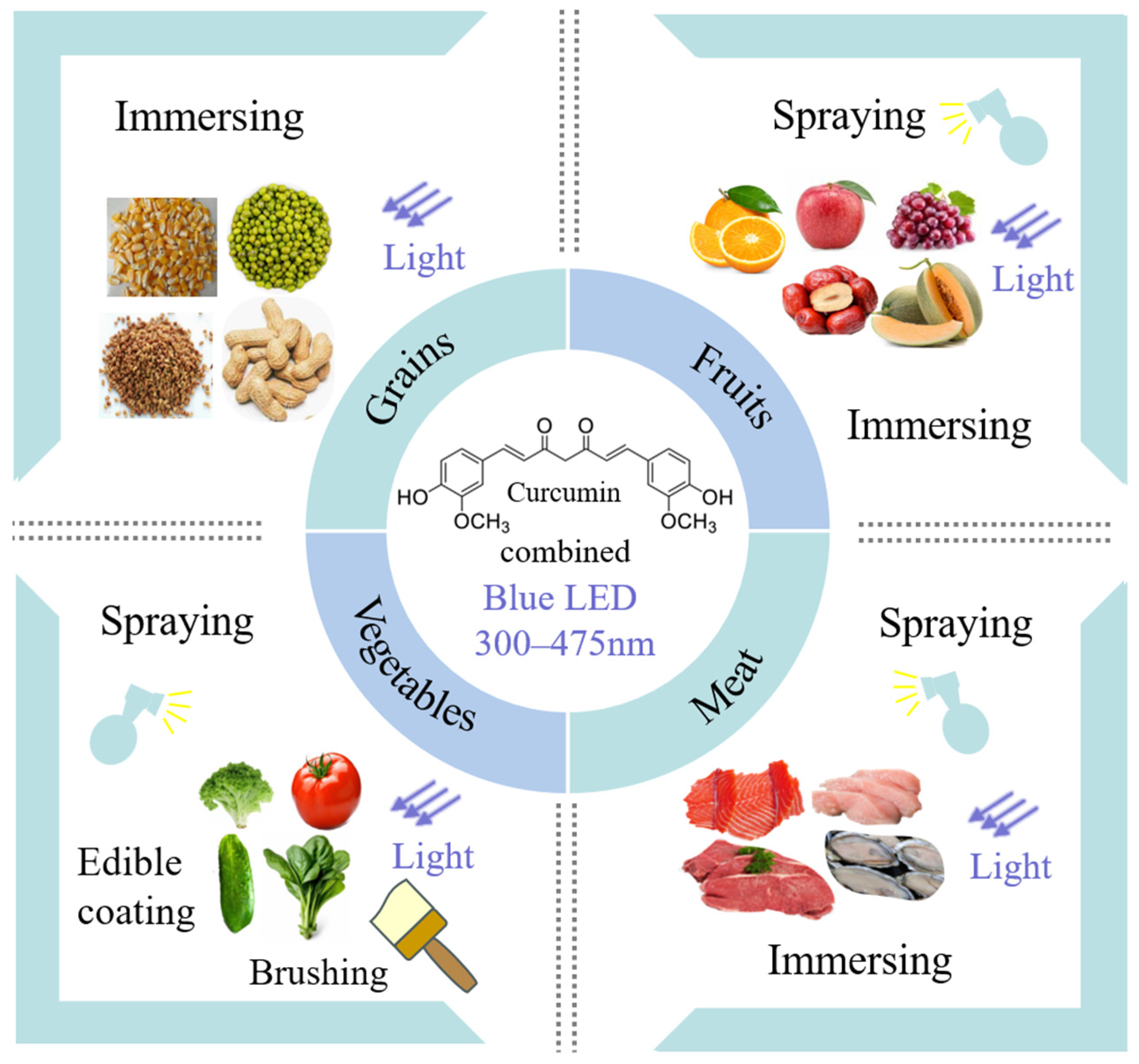
Due to the increasing demands for a healthy diet, the tendency of consumption of aquatic products, grains, and fresh fruit and vegetables has increased. However, raw food materials have been known to be vehicles for the transmission of human diseases and have become the second leading cause of food-borne illnesses. Obviously, there is a need to develop novel processing technologies that are more effective and do not diminish the organoleptic properties and nutritional value of the treated foodstuffs. The practical application of CUR and LED in different food model systems has been listed in Table 3 and shown in Figure 3.

6.1. Fruits and Vegetables

Fresh fruits have high moisture content and sugar, which provides favorable conditions for the growth and reproduction of microorganisms. The current technology for fruit surface sanitizing involves chlorine water disinfection, high hydrostatic pressure treatment, electrolyzed oxidized water, ozonation, and X-ray irradiation [52]. Nevertheless, these methods are not fully applied to commercialization due to their limitations, while CUR-mediated PDI has been shown to be a promising alternative for the disinfection of fresh fruit without notable negative effects.
The fungal growth on the date surface was inhibited by spraying CUR solution on the date surface combined with blue-light illumination [52]. The shelf-life was prolonged from 7 days and 28 days to 14 days and 98 days for dates stored at 30 °C and at 4 °C, respectively, without quality loss, such as moisture content, total sugar, and total phenol content. Spoilage of fresh-cut Hami melons could be prevented by spraying curcumin solution (10–50 µM) and exposure to blue LED [53]. The combined treatment effectively reduced microbial growth by 1.8 log CFU/g, approximatively.
The antibacterial efficiency of CUR-mediated PDI against E. coli was verified on the surface of a fresh-cut “Fuji” apple, which was positively related to CUR concentration and irradiation time [54]. Additionally, the sensory and nutritional qualities of the fresh-cut apple were maintained during storage, including color, weight, ascorbic acid content, total phenolic content, and total antioxidant capacity. The enzyme activities of polyphenol oxidase and peroxidase were inhibited, which further prevented the unwanted browning of the fresh-cut apple. Aurum and Nguyen [55] showed that PDI could effectively inactivate ~2.4 log E. coli on the surface of a grape with exposure to blue light (36.3 J/cm2). Moreover, the photoactivation treatment largely preserved the original quality of the stored grape, including vitamin C, color, and firmness.
For the decontamination of orange juice, a combination of CUR and blue LED could inactivate E. coli and S. aureus, respectively [56]. Meanwhile, there was no significant change in the total phenolic, flavonoid, and hesperidin contents after the treatments. Additionally, Wang et al. [44] found that CUR-mediated PDI was able to reduce Penicillium italicum by 2.5 log CFU/mL in fresh-cut oranges. Photodynamic treatment with curcumin (0–75 μmol/L) had little effect on the color of the orange slices and also impeded the decrease in pH of the oranges during storage.
Fresh vegetables are highly susceptible to microbial contamination during processing and storage. The effect of CUR-mediated PDI on preventing microbial cross-contamination of fresh vegetables (spinach leaves and cherry tomato) has been verified [57]. Polyvinylpyrrolidone-CUR (PVP-C) has also been reported to cause a reduction of 2.6 log CFU/mL S. aureus in cucumber [58].

6.2. Cereal Grains

Grains are often contaminated with microorganisms and molds during harvesting, transportation, and storage, which seriously affects the quality and functional properties of grains [60]. Molds not only reduce the nutritional value of grains but also form mycotoxins and allergenic spores, which are the biggest spoilage organisms for grain quality [71]. Although new technologies such as cold plasma, irradiation, ozone, and biological methods have been developed for microbial safety, they still have limited applications [72,73]. Recently, non-thermal photosensitization has been proven to have good potential in grain storage and preservation.
It has been reported that the high-intensity light pulses generated by the LED can be used to reduce pathogenic bacteria in low-aw foods [74]. For example, the maximum reduction of the Salmonella cocktail was 2.91 log after wheat flour was treated with a 395 nm pulsed LED for 60 min in a semi-closed system [75]. Similarly, the number of Salmonella in wheat flour was reduced by 1.07, 2.42, 3.67, and 2.64 CFU/g after treatments with 275, 365, 395, and 455 nm LEDs for 60 min at 25 °C and 75% relative humidity, respectively [76]. Not coincidentally, another study found that 5 h of LED blue-light treatment (430–470 nm, 36 W) on wheat flour with a high bacterial load could reduce the number of Escherichia coli colonies by 4.9 (lg (CFU/g)), while reducing the number of Bacillus cereus colonies by 4.7 (lg (CFU/g)), and the sterilizing effect was positively correlated with the duration of light exposure [77].
Additionally, the use of a photosensitizer-synergistic LED antimicrobial has a better sterilizing effect and a wider range of application scenarios. The photosensitizer riboflavin has potential antimicrobial, and antibiotic periplasmic activity against the rice leaf blight fungus (Xanthomonas oryzae pv. oryzae) [78]. In addition, CUR (25 μM) at a light dose of 4.32 J/cm2 produced significant photoinactivation of S. saprophyticus in vitro (approximately 5 log reduction), and there was a photo-antimicrobial effect on fresh millet dough (initial bacterial loads of 5 log CFU/g and 3 log CFU/g) of 1.04 log CFU/g and 0.81 log CFU/g [64]. Curcumin-based photosensitization has the advantages of being natural, green, economical, and effective in removing the aflatoxins that are commonly present in grain [79]. The effect of CUR-mediated PDI has also been evaluated for preventing the contamination of grains and seeds. It was found that CUR (1000 μM) and LED (430 nm, 104.2 J/cm2) photoinactivated Aspergillus flavus spores on maize kernels by 2.3 log CFU/mL and inhibited aflatoxin B1 (AFB1) production without affecting carotenoids [65]. A significant reduction (1.8–2.2 log CFU/g) of A. flavus spores on corn kernels was achieved after CUR (45 μM) and irradiation (420 nm) treatment [41]. Furthermore, the production of aflatoxin B1 in maize kernel was notably reduced from 305.9 μg/kg to 82.4 μg/kg after the CUR-mediated PDI treatment of A. flavus [66]. Nguenha et al. [67] reported that the photosensitization of ethanol and propylene glycol solubilized curcumin effectively and reduced the number of Aspergillus flavus on yellow and white corn kernel surfaces by 2.04 log CFU/mL and 3.33 log CFU/mL, respectively. Fungal growth in photosensitized maize kernels and flour was delayed by 14 and 7 days under 25 °C storage conditions. After 20 days of storage, AFB1 was not detected in maize kernels, and the amount of AFB1 in maize flour was reduced by 91%. Moreover, CUR (50 μΜ) and a 43.0 J/cm2 treatment inhibited the production of zearalenone (ZEN) by F. graminearum, contributing to a reduction from 1.15 mg/kg to 0.58 mg/kg. When the CUR concentration was ≥70 μM and the light energy was 43.0 J/cm2, F. graminearum. was completely inactivated [68].
Reductions of E. coli (~3 log CFU/mL) on fenugreek and mung beans were achieved when treated with the cationic CUR derivatives (SACUR-3) combined with blue LED (432 nm) and illumination (33.8 J/cm2) [69]. Additionally, CUR-mediated PDI was effective in mitigating the contamination of peanut seeds by Aspergillus flavus [70]. These results indicated that non-thermal treatment was a potential way to control the contamination of grains and seeds.

6.3. Animal-Based Food

Seafood, rich in nutritive value and biologically active ingredients, has aroused wide concern, as it is perishable at ordinary temperatures. Gao et al. reported that a 2.5 log reduction of E. coli DH5α in oysters was observed by using CUR combined with blue light [34]. And the PDI-treated oysters have been proven to be safe and nontoxic through cytotoxicity tests. Similarly, CUR solution sprayed on the surfaces of oysters following blue-light (470 nm) irradiation was found to extend the storage period from 8 days to 12 days stored at 5 ± 1 °C [80]. The PDI treatment improved oyster quality, lowered the oxidation degree, and slowed down the decomposition of nutrients. Chen et al. reported that CUR-mediated PDI was able to completely inhibit Vibrio parahaemolyticus on oysters when irradiated with blue light (455–460 nm, 9.36 J/cm2) [61]. PDI treatment inhibited the decrease in the pH value of oysters, reduced the production of TVB-N, and inhibited the lipid oxidation of oysters, prolonging the shelf-life of oysters.
CUR-mediated PDI has also been reported to be effective against the norovirus, which is one of the most crucial seafood-borne viruses and the leading cause of human acute gastroenteritis outbreaks [62]. A CUR solution (5, 10, 20 µM) combined with LED light (470 nm, 3.6 J/cm2) could cause a reduction of 1.15 log PFU/mL of norovirus in oysters by destroying the viral nucleic acids and capsid protein structure. Curcumin combined with 18.72 J/cm2 blue light inactivated ~3.0 log CFU/g of bacteria on salmon, achieving 99.9% bacterial inhibition. In addition, CUR-mediated PDI effectively inhibited salmon color and pH change and maintained muscle fiber integrity and SOD activity, preventing salmon protein and lipid degradation [61].
An investigation of PDI mediated by CUR was also conducted in meat products. In a beef model, PDI treatment decreased Pseudomonas lundensis and Brochothrix thermosphacta by 0.15 log CFU/g and 0.08 log CFU/g, respectively [38]. PDI treatment reduces the breakdown of proteins and lipids, reduces the production of undesirable odors, and maintains the bright-red color of beef.
Antimicrobial photodynamic protocols applying curcumin as a PS use a wide variety of parameters, and their efficiency in antimicrobial photodynamic therapy is related to the concentration they use and the species of microorganisms they target. The PDI of curcumin was effective in reducing the bacterial population, and its bactericidal effect was positively correlated with the duration of light exposure [2]. Considering the complexity of food ingredients, growth patterns of microorganisms, and practical use scenarios, it is difficult to compare the results of these descriptions using universal operational parameters [20].

7. Antimicrobial Effect of Curcumin Derivatives

In addition to the free form of curcumin, researchers have focused on developing new strategies to improve the physical–chemical properties of CUR by using micro-/nano-encapsulation and its derivatives [81]. These novel strategies have gained wide acceptance due to their advantages and application prospects over traditional materials. The anti-microbial activity of CUR derivatives is summarized in Table 4.
UNCPs-CUR synthesized by combining upconversion nanoparticles with CUR by using polyethyleneimine showed a complete inhibition (6 log CFU/mL) on methicillin-resistant S. aureus (MRAS) [82]. The combination of CUR-loaded cationic nanoparticles (CUR.NP) and LED illumination resulted in a decrease of >6.2 log of Staphylococcus saprophyticus and 2.9 log of E. coli [83]. A CUR/polyurethane nanocomposite was also found to inhibit E. coli and S. aureus (~6 log CFU/mL) completely after irradiation at 470 nm due to the phototoxicity and singlet oxygen produced [84]. Compared with the CUR solution, the synthesis of an antibacterial coating by using CUR in combination with blue-light illumination offered another new thought for preventing bacterial infection on food surfaces. Condat et al. [85] found that a CUR-derivative coating composed of epoxidized oil could achieve an inhibition rate of 99% and 95% against S. aureus and E. coli, respectively.
Microencapsulated CUR combined with blue LED achieved a good inhibitory effect against P. aeruginosa [50]. A complete photodynamic inhibition of P. aeruginosa was observed when a CUR microemulsion was combined with EDTA. Similarly, Hu et al. reported that the addition of EDTA significantly increased the antibacterial effect of CUR-mediated PDI against Burkholderia cepaci [86]. The bacterial inhibition rate was up to approximately 100% when treated with 50 μw CUR, including 0.4% EDTA following an irradiation dosage of 28 J/cm2. The reason might be due to the destruction of lipopolysaccharides in the outer membrane of Gram-negative bacteria caused by EDTA. In addition, NovaSol®–CUR was reported to sterilize Staphylococcus aureus in chicken meat, resulting in a reduction of 1.7 log [58].
Chen et al. prepared a 2,3-dialdehyde cellulose (DAC) antimicrobial membrane by adding a β-cyclodextrin/curcumin (β-CD/Cur) complex [87]. The film combined with PDI treatment was able to completely inhibit L. monocytogenes (>99.9%), Vibrio parahaemolyticus (>99.0%), and Shewanella putrefaciens (>99.9%). It is mainly the action of ROS (1O2, H2O2, etc.) produced by CUR after being excited by light and attacking intracellular proteins, nucleic acids, and lipids that kill bacteria.

8. Conclusions and Future Trends

The antimicrobial and antifungal activity of curcumin-mediated photodynamic inhabitation was reviewed, focusing on the inhibitory efficacy in vitro and in various food systems. Additionally, CUR in its free form and the derivatives with improved water solubility and chemical stability, including nano-/micro-formulations, polymeric micelles, and hydrogels, were evaluated. CUR-mediated PDI can also be used in combination with other physical sterilization methods to enhance the bactericidal effect, such as microwaves, ultrasound, pulsed light, and electron-beam irradiation. The large differences between different food matrices and compositions greatly increase the complexity of food sterilization and storage and preservation. Further research should focus on how PDI treatment affects the physicochemical properties and nutritional value of foods under actual food-processing conditions. In order to achieve the commercialized application of PDI for food sanitation and preservation, interdisciplinary research involving food science, microbiology, nutriology, and mathematical modeling is needed.

Author Contributions

Conceptualization, Z.W.; software, H.Y.; validation, H.Y.; formal analysis, Z.L.; investigation, H.Y.; resources, Z.W.; writing—original draft preparation, Z.W.; writing—review and editing, J.L.; supervision, J.L.; funding acquisition, Z.W. and J.L. All authors have read and agreed to the published version of the manuscript.

Funding

This work was supported by the National Key Research and Development Program of China under Grant number 2022YFF1100605 and the Beijing Nova Program under grant number 20230484307.

Data Availability Statement

No new data were created or analyzed in this study.

Conflicts of Interest

The authors declare no conflicts of interest.

References

  1. Settanni, L.; Corsetti, A. Application of bacteriocins in vegetable food biopreservation. Int. J. Food Microbiol. 2008, 121, 123–138. [Google Scholar] [CrossRef] [PubMed]
  2. Luksiene, Z.; Brovko, L. Antibacterial Photosensitization-Based Treatment for Food Safety. Food Eng. Rev. 2013, 5, 185–199. [Google Scholar] [CrossRef]
  3. Chalak, A.; Abou-Daher, C.; Chaaban, J.; Abiad, M.G. The global economic and regulatory determinants of household food waste generation: A cross-country analysis. Waste Manag. 2016, 48, 418–422. [Google Scholar] [CrossRef] [PubMed]
  4. Pereira, R.N.; Vicente, A.A. Environmental impact of novel thermal and non-thermal technologies in food processing. Food Res. Int. 2010, 43, 1936–1943. [Google Scholar] [CrossRef]
  5. Li, X.; Farid, M. A review on recent development in non-conventional food sterilization technologies. J. Food Eng. 2016, 182, 33–45. [Google Scholar] [CrossRef]
  6. Pan, Y.Y.; Sun, D.W.; Han, Z. Applications of electromagnetic fields for nonthermal inactivation of microorganisms in foods: An overview. Trends Food Sci. Technol. 2017, 64, 13–22. [Google Scholar] [CrossRef]
  7. Zhao, W.; Tang, Y.L.; Lu, L.X.; Chen, X.; Li, C.Y. Review: Pulsed Electric Fields Processing of Protein-Based Foods. Food Bioprocess Technol. 2014, 7, 114–125. [Google Scholar] [CrossRef]
  8. Bisht, B.; Bhatnagar, P.; Gururani, P.; Kumar, V.; Tomar, M.S.; Sinhmar, R.; Rathi, N.; Kumar, S. Food irradiation: Effect of ionizing and non-ionizing radiations on preservation of fruits and vegetables—A review. Trends Food Sci. Technol. 2021, 114, 372–385. [Google Scholar] [CrossRef]
  9. Patil, S.; Moiseev, T.; Misra, N.N.; Cullen, P.J.; Mosnier, J.P.; Keener, K.M.; Bourke, P. Influence of high voltage atmospheric cold plasma process parameters and role of relative humidity on inactivation of Bacillus atrophaeus spores inside a sealed package. J. Hosp. Infect. 2014, 88, 162–169. [Google Scholar] [CrossRef]
  10. Ghoshal, G. Comprehensive review on pulsed electric field in food preservation: Gaps in current studies for potential future research. Heliyon 2023, 9, e17532. [Google Scholar] [CrossRef]
  11. Cebrián, G.; Condón, S.; Mañas, P. Influence of growth and treatment temperature on Staphylococcus aureus resistance to pulsed electric fields: Relationship with membrane fluidity. Innov. Food Sci. Emerg. Technol. 2016, 37, 161–169. [Google Scholar] [CrossRef]
  12. Sobotta, L.; Skupin-Mrugalska, P.; Piskorz, J.; Mielcarek, J. Non-porphyrinoid photosensitizers mediated photodynamic inactivation against bacteria. Dye. Pigment. 2019, 163, 337–355. [Google Scholar] [CrossRef]
  13. Luksiene, Z.; Zukauskas, A. Prospects of photosensitization in control of pathogenic and harmful micro-organisms. J. Appl. Microbiol. 2009, 107, 1415–1424. [Google Scholar] [CrossRef] [PubMed]
  14. Damyeh, M.S.; Mereddy, R.; Netzel, M.E.; Sultanbawa, Y. An insight into curcumin-based photosensitization as a promising and green food preservation technology. Compr. Rev. Food Sci. Food Saf. 2020, 19, 1727–1759. [Google Scholar] [CrossRef] [PubMed]
  15. Nitzan, Y.; Ashkenazi, H. Photoinactivation of Acinetobacter baumannii and Escherichia coli B by a cationic hydrophilic porphyrin at various light wavelengths. Curr. Microbiol. 2001, 42, 408–414. [Google Scholar] [CrossRef] [PubMed]
  16. Stanic, Z. Curcumin, a Compound from Natural Sources, a True Scientific Challenge—A Review. Plant Food Hum. Nutr. 2017, 72, 1–12. [Google Scholar] [CrossRef]
  17. Martelli, G.; Giacomini, D. Antibacterial and antioxidant activities for natural and synthetic dual-active compounds. Eur. J. Med. Chem. 2018, 158, 91–105. [Google Scholar] [CrossRef]
  18. Suliman, M.; Kzar, M.H.; Juma, A.S.M.; Ali, I.A.; Yasin, Y.; Sayyid, N.H.; Alkhafaji, A.T.; Kadhim, A.J.; Abid, M.M.; Alawadi, A.H.; et al. B40 and SiB39 fullerenes enhance the physicochemical features of curcumin and effectively improve its anti-inflammatory and anti-cancer activities. J. Mol. Liq. 2024, 395, 123816. [Google Scholar] [CrossRef]
  19. Legaba, B.C.; Galinari, C.B.; dos Santos, R.S.; Bruschi, M.L.; Gremiao, I.D.F.; Boechat, J.S.; Pereira, S.A.; Malacarne, L.C.; Caetano, W.; Bonfim-Mendonça, P.S.; et al. In vitro antifungal activity of curcumin mediated by photodynamic therapy on Sporothrix brasiliensis. Photodiagnosis Photodyn. Ther. 2023, 43, 103659. [Google Scholar] [CrossRef]
  20. Dias, L.D.; Blanco, K.C.; Mfouo-Tynga, I.S.; Inada, N.M.; Bagnato, V.S. Curcumin as a photosensitizer: From molecular structure to recent advances in antimicrobial photodynamic therapy. J. Photochem. Photobiol. C 2020, 45, 100384. [Google Scholar] [CrossRef]
  21. Cieplik, F.; Deng, D.M.; Crielaard, W.; Buchalla, W.; Hellwig, E.; Al-Ahmad, A.; Maisch, T. Antimicrobial photodynamic therapy—What we know and what we don’t. Crit. Rev. Microbiol. 2018, 44, 571–589. [Google Scholar] [CrossRef] [PubMed]
  22. Silva, A.F.; Borges, A.; Giaouris, E.; Mikcha, J.M.G.; Simoes, M. Photodynamic inactivation as an emergent strategy against foodborne pathogenic bacteria in planktonic and sessile states. Crit. Rev. Microbiol. 2018, 44, 667–684. [Google Scholar] [CrossRef] [PubMed]
  23. Jori, G.; Fabris, C.; Soncin, M.; Ferro, S.; Coppellotti, O.; Dei, D.; Fantetti, L.; Chiti, G.; Roncucci, G. Photodynamic therapy in the treatment of microbial infections: Basic principles and perspective applications. Lasers Surg. Med. 2006, 38, 468–481. [Google Scholar] [CrossRef] [PubMed]
  24. Costa, L.; Faustino, M.A.F.; Neves, M.; Cunha, A.; Almeida, A. Photodynamic Inactivation of Mammalian Viruses and Bacteriophages. Viruses 2012, 4, 1034–1074. [Google Scholar] [CrossRef]
  25. Lubart, R.; Lipovski, A.; Nitzan, Y.; Friedmann, H. A possible mechanism for the bactericidal effect of visible light. Laser Ther. 2011, 20, 17–22. [Google Scholar] [CrossRef]
  26. Zhang, S.; He, Z.; Xu, F.; Cheng, Y.; Waterhouse, G.I.N.; Sun-Waterhouse, D.; Wu, P. Enhancing the performance of konjac glucomannan films through incorporating zein–pectin nanoparticle-stabilized oregano essential oil Pickering emulsions. Food Hydrocolloid. 2022, 124, 107222. [Google Scholar] [CrossRef]
  27. Chen, B.; Huang, J.; Li, H.; Zeng, Q.-H.; Wang, J.J.; Liu, H.; Pan, Y.; Zhao, Y. Eradication of planktonic Vibrio parahaemolyticus and its sessile biofilm by curcumin-mediated photodynamic inactivation. Food Control 2020, 113, 107181. [Google Scholar] [CrossRef]
  28. Kumar, A.; Ghate, V.; Kim, M.J.; Zhou, W.B.; Khoo, G.H.; Yuk, H.G. Inactivation and changes in metabolic profile of selected foodborne bacteria by 460 nm LED illumination. Food Microbiol. 2017, 63, 12–21. [Google Scholar] [CrossRef]
  29. Kleinpenning, M.M.; Smits, T.; Frunt, M.H.A.; van Erp, P.E.J.; van de Kerkhof, P.C.M.; Gerritsen, R. Clinical and histological effects of blue light on normal skin. Photodermatol. Photoimmunol. Photomed. 2010, 26, 16–21. [Google Scholar] [CrossRef]
  30. Matsumura, Y.; Ananthaswamy, H.N. Toxic effects of ultraviolet radiation on the skin. Toxicol. Appl. Pharmacol. 2004, 195, 298–308. [Google Scholar] [CrossRef]
  31. Maclean, M.; MacGregor, S.J.; Anderson, J.G.; Woolsey, G. Inactivation of Bacterial Pathogens following Exposure to Light from a 405-Nanometer Light-Emitting Diode Array. Appl. Environ. Microbiol. 2009, 75, 1932–1937. [Google Scholar] [CrossRef] [PubMed]
  32. Penha, C.B.; Bonin, E.; da Silva, A.F.; Hioka, N.; Zanqueta, É.; Nakamura, T.U.; de Abreu, B.A.; Campanerut-Sá, P.A.Z.; Mikcha, J.M.G. Photodynamic inactivation of foodborne and food spoilage bacteria by curcumin. LWT-Food Sci. Technol. 2017, 76, 198–202. [Google Scholar] [CrossRef]
  33. Jiang, Y.; Leung, A.W.; Hua, H.; Rao, X.; Xu, C. Photodynamic Action of LED-Activated Curcumin against Staphylococcus aureus Involving Intracellular ROS Increase and Membrane Damage. Int. J. Photoenergy 2014, 2014, 637601. [Google Scholar] [CrossRef]
  34. Gao, Y.; We, J.; Li, Z.J.; Zhang, X.; Lu, N.; Xu, C.H.; Leung, A.W.; Xu, C.S.; Tang, Q.J. Curcumin-mediated photodynamic inactivation (PDI) against DH5α contaminated in oysters and cellular toxicological evaluation of PDI-treated oysters. Photodiagnosis Photodyn. Ther. 2019, 26, 244–251. [Google Scholar] [CrossRef]
  35. Wu, J.; Mou, H.; Xue, C.; Leung, A.W.; Xu, C.; Tang, Q.J. Photodynamic effect of curcumin on Vibrio parahaemolyticus. Photodiagnosis Photodyn. Ther. 2016, 15, 34–39. [Google Scholar] [CrossRef]
  36. Najafi, S.; Khayamzadeh, M.; Paknejad, M.; Poursepanj, G.; Kharazi Fard, M.J.; Bahador, A. An In Vitro Comparison of Antimicrobial Effects of Curcumin-Based Photodynamic Therapy and Chlorhexidine, on Aggregatibacter actinomycetemcomitans. J. Lasers Med. Sci. 2016, 7, 21–25. [Google Scholar] [CrossRef]
  37. Bonifacio, D.; Martins, C.; David, B.; Lemos, C.; Neves, M.; Almeida, A.; Pinto, D.; Faustino, M.A.F.; Cunha, A. Photodynamic inactivation of Listeria innocua biofilms with food-grade photosensitizers: A curcumin-rich extract of Curcuma longa vs commercial curcumin. J. Appl. Microbiol. 2018, 125, 282–294. [Google Scholar] [CrossRef] [PubMed]
  38. Lu, H.; Zheng, S.; Fang, J.; Zhu, J. Photodynamic inactivation of spoilers Pseudomonas lundensis and Brochothrix thermosphacta by food-grade curcumin and its application on ground beef. Innov. Food Sci. Emerg. Technol. 2023, 87, 103410. [Google Scholar] [CrossRef]
  39. Saraiva, B.B.; Campanholi, K.D.S.; Machado, R.R.B.; Nakamura, C.V.; Silva, A.A.; Caetano, W.; Pozza, M.S.D. Reducing Pseudomonas fluorescens in milk through photodynamic inactivation using riboflavin and curcumin with 450 nm blue light-emitting diode. Int. Dairy J. 2024, 148, 105787. [Google Scholar] [CrossRef]
  40. Pang, J.; Zhang, F.; Wang, Z.; Wu, Q.; Liu, B.; Meng, X. Inhibitory effect and mechanism of curcumin-based photodynamic inactivation on patulin secretion by Penicillium expansum. Innov. Food Sci. Emerg. Technol. 2022, 80, 103078. [Google Scholar] [CrossRef]
  41. Temba, B.A.; Fletcher, M.T.; Fox, G.P.; Harvey, J.J.W.; Sultanbawa, Y. Inactivation of Aspergillus flavus spores by curcumin-mediated photosensitization. Food Control 2016, 59, 708–713. [Google Scholar] [CrossRef]
  42. Al-Asmari, F.; Mereddy, R.; Sultanbawa, Y. A novel photosensitization treatment for the inactivation of fungal spores and cells mediated by curcumin. J. Photochem. Photobiol. B 2017, 173, 301–306. [Google Scholar] [CrossRef] [PubMed]
  43. Song, L.L.; Zhang, F.; Yu, J.S.; Wei, C.L.; Han, Q.M.; Meng, X.H. Antifungal effect and possible mechanism of curcumin mediated photodynamic technology against Penicillium expansum. Postharvest Biol. Technol. 2020, 167, 111234. [Google Scholar] [CrossRef]
  44. Wang, Y.; Zhao, Y.; Wu, R.; Gao, J.; Chen, M.; Cui, Y.; Hao, J.; Han, J.; Matthews, K. Photodynamic inactivation of curcumin combined with ascorbic acid against Penicillium italicum in vitro and on fresh-cut orange. LWT 2023, 182, 114900. [Google Scholar] [CrossRef]
  45. Sanitá, P.V.; Pavarina, A.C.; Dovigo, L.N.; Ribeiro, A.P.D.; Andrade, M.C.; Mima, E.G.D. Curcumin-mediated anti-microbial photodynamic therapy against Candida dubliniensis biofilms. Lasers Med. Sci. 2018, 33, 709–717. [Google Scholar] [CrossRef]
  46. Quishida, C.C.C.; Mima, E.G.D.; Jorge, J.H.; Vergani, C.E.; Bagnato, V.S.; Pavarina, A.C. Photodynamic inactivation of a multispecies biofilm using curcumin and LED light. Lasers Med. Sci. 2016, 31, 997–1009. [Google Scholar] [CrossRef] [PubMed]
  47. Chandra, J.; Kuhn, D.M.; Mukherjee, P.K.; Hoyer, L.L.; McCormick, T.; Ghannoum, M.A. Biofilm formation by the fungal pathogen Candida albicans:: Development, architecture, and drug resistance. J. Bacteriol. 2001, 183, 5385–5394. [Google Scholar] [CrossRef]
  48. Dovigo, L.N.; Pavarina, A.C.; Ribeiro, A.P.D.; Brunetti, I.L.; Costa, C.A.D.; Jacomassi, D.P.; Bagnato, V.S.; Kurachi, C. Investigation of the Photodynamic Effects of Curcumin Against Candida albicans. Photochem. Photobiol. 2011, 87, 895–903. [Google Scholar] [CrossRef]
  49. Pourhajibagher, M.; Kazemian, H.; Chiniforush, N.; Hosseini, N.; Pourakbari, B.; Azizollahi, A.; Rezaei, F.; Bahador, A. Exploring different photosensitizers to optimize elimination of planktonic and biofilm forms of Enterococcus faecalis from infected root canal during antimicrobial photodynamic therapy. Photodiagnosis Photodyn. Ther. 2018, 24, 206–211. [Google Scholar] [CrossRef]
  50. Liu, C.H.; Lee, W.S.; Wu, W.C. Photodynamic inactivation against Pseudomonas aeruginosa by curcumin microemulsions. RSC Adv. 2016, 6, 63013–63022. [Google Scholar] [CrossRef]
  51. Abdulrahman, H.; Misba, L.; Ahmad, S.; Khan, A.U. Curcumin induced photodynamic therapy mediated suppression of quorum sensing pathway of Pseudomonas aeruginosa: An approach to inhibit biofilm in vitro. Photodiagnosis Photodyn. Ther. 2020, 30, 101645. [Google Scholar] [CrossRef] [PubMed]
  52. Al-Asmari, F.; Mereddy, R.; Sultanbawa, Y. The effect of photosensitization mediated by curcumin on storage life of fresh date (Phoenix dactylifera L.) fruit. Food Control 2018, 93, 305–309. [Google Scholar] [CrossRef]
  53. Lin, Y.L.; Hu, J.M.; Li, S.Y.; Hamzah, S.S.; Jiang, H.Q.; Zhou, A.R.; Zeng, S.X.; Lin, S.L. Curcumin-Based Photodynamic Sterilization for Preservation of Fresh-Cut Hami Melon. Molecules 2019, 24, 2374. [Google Scholar] [CrossRef] [PubMed]
  54. Tao, R.; Zhang, F.; Tang, Q.J.; Xu, C.S.; Ni, Z.J.; Meng, X.H. Effects of curcumin-based photodynamic treatment on the storage quality of fresh-cut apples. Food Chem. 2019, 274, 415–421. [Google Scholar] [CrossRef]
  55. Aurum, F.S.; Nguyen, L.T. Efficacy of photoactivated curcumin to decontaminate food surfaces under blue light emitting diode. J. Food Process. Eng. 2019, 42, e12988. [Google Scholar] [CrossRef]
  56. Bhavya, M.L.; Hebbar, H.U. Sono-photodynamic inactivation of Escherichia coli and Staphylococcus aureus in orange juice. Ultrason. Sonochemistry 2019, 57, 108–115. [Google Scholar] [CrossRef]
  57. de Oliveira, E.F.; Tosati, J.V.; Tikekar, R.V.; Monteiro, A.R.; Nitin, N. Antimicrobial activity of curcumin in combination with light against Escherichia coli O157:H7 and Listeria innocua: Applications for fresh produce sanitation. Postharvest Biol. Technol. 2018, 137, 86–94. [Google Scholar] [CrossRef]
  58. Tortik, N.; Spaeth, A.; Plaetzer, K. Photodynamic decontamination of foodstuff from Staphylococcus aureus based on novel formulations of curcumin. Photochem. Photobiol. Sci. 2014, 13, 1402–1409. [Google Scholar] [CrossRef]
  59. de Oliveira, E.F.; Tikekar, R.; Nitin, N. Combination of aerosolized curcumin and UV-A light for the inactivation of bacteria on fresh produce surfaces. Food Res. Int. 2018, 114, 133–139. [Google Scholar] [CrossRef]
  60. Los, A.; Ziuzina, D.; Akkermans, S.; Boehm, D.; Cullen, P.J.; Van Impe, J.; Bourke, P. Improving microbiological safety and quality characteristics of wheat and barley by high voltage atmospheric cold plasma closed processing. Food Res. Int. 2018, 106, 509–521. [Google Scholar] [CrossRef]
  61. Chen, B.; Huang, J.; Liu, Y.; Liu, H.; Zhao, Y.; Wang, J.J. Effects of the curcumin-mediated photodynamic inactivation on the quality of cooked oysters with Vibrio parahaemolyticus during storage at different temperature. Int. J. Food Microbiol. 2021, 345, 109152. [Google Scholar]
  62. Wu, J.; Hou, W.; Cao, B.; Zuo, T.; Xue, C.; Leung, A.W.; Xu, C.; Tang, Q.J. Virucidal efficacy of treatment with photodynamically activated curcumin on murine norovirus bio-accumulated in oysters. Photodiagnosis Photodyn. Ther. 2015, 12, 385–392. [Google Scholar] [CrossRef]
  63. Tosati, J.V.; de Oliveira, E.F.; Oliveira, J.V.; Nitin, N.; Monteiro, A.R. Light-activated antimicrobial activity of turmeric residue edible coatings against cross-contamination of Listeria innocua on sausages. Food Control 2018, 84, 177–185. [Google Scholar] [CrossRef]
  64. Wang, Z.Y.; Jia, Y.T.; Li, W.Y.; Zhang, M. Antimicrobial photodynamic inactivation with curcumin against Staphylococcus saprophyticus, in vitro and on fresh dough sheet. LWT 2021, 147, 111567. [Google Scholar] [CrossRef]
  65. Nguenha, R.; Damyeh, M.S.; Phan, A.D.T.; Hong, H.T.; Chaliha, M.; O’Hare, T.J.; Netzel, M.E.; Sultanbawa, Y. Effect of Photosensitization Mediated by Curcumin on Carotenoid and Aflatoxin Content in Different Maize Varieties. Appl. Sci. 2021, 11, 5902. [Google Scholar] [CrossRef]
  66. Temba, B.A.; Fletcher, M.T.; Fox, G.P.; Harvey, J.; Okoth, S.A.; Sultanbawa, Y. Curcumin-based photosensitization inactivates Aspergillus flavus and reduces aflatoxin B1 in maize kernels. Food Microbiol. 2019, 82, 82–88. [Google Scholar] [CrossRef] [PubMed]
  67. Nguenha, R.J.; Damyeh, M.S.; Hong, H.T.; Chaliha, M.; Sultanbawa, Y. Effect of solvents on curcumin as a photosensitizer and its ability to inactivate Aspergillus flavus and reduce aflatoxin B1 in maize kernels and flour. J. Food Process. Preserv. 2022, 46, e16169. [Google Scholar] [CrossRef]
  68. Jingjing, W.; Jieer, Q.I.U.; Tiantian, H.E.; Aiyao, Y.; Ming, X.U.; Yang, L.I.U. Curcumin-mediated Photodynamic Inhibition of ZEN Mycotoxin Production by Fusarium graminearum. Mod. Food Sci. Technol. 2023, 39, 305–311. [Google Scholar]
  69. Glueck, M.; Schamberger, B.; Eckl, P.; Plaetzer, K. New horizons in microbiological food safety: Photodynamic Decontamination based on a curcumin derivative. Photochem. Photobiol. Sci. 2017, 16, 1784–1791. [Google Scholar] [CrossRef]
  70. Chen, L.; Li, X.; Chen, J.; Lin, R.; Mai, Y.; Lin, Y.; Wang, G.; Chen, Z.; Zhang, W.; Wang, J.; et al. Formulation with zinc acetate enhances curcumin’s inherent and photodynamic antimicrobial effects for food preservation. Food Control 2024, 157, 110220. [Google Scholar] [CrossRef]
  71. Hu, Y.M.; Nie, W.; Hu, X.Z.; Li, Z.G. Microbial decontamination of wheat grain with superheated steam. Food Control 2016, 62, 264–269. [Google Scholar] [CrossRef]
  72. Mir, S.A.; Dar, B.N.; Shah, M.A.; Sofi, S.A.; Hamdani, A.M.; Oliveira, C.A.F.; Moosavi, M.H.; Khaneghah, A.M.; Sant’Ana, A.S. Application of new technologies in decontamination of mycotoxins in cereal grains: Challenges, and perspectives. Food Chem. Toxicol. 2021, 148, 111976. [Google Scholar] [CrossRef] [PubMed]
  73. Los, A.; Ziuzina, D.; Bourke, P. Current and Future Technologies for Microbiological Decontamination of Cereal Grains. J. Food Sci. 2018, 83, 1484–1493. [Google Scholar] [CrossRef] [PubMed]
  74. Song, K.; Taghipour, F.; Mohseni, M. Microorganisms inactivation by continuous and pulsed irradiation of ultraviolet light-emitting diodes (UV-LEDs). Chem. Eng. J. 2018, 343, 362–370. [Google Scholar] [CrossRef]
  75. Du, L.H.; Prasad, A.J.; Gänzle, M.; Roopesh, M.S. Inactivation of Salmonella spp. in wheat flour by 395 nm pulsed light emitting diode (LED) treatment and the related functional and structural changes of gluten. Food Res. Int. 2020, 127, 108716. [Google Scholar] [CrossRef]
  76. Subedi, S.; Du, L.H.; Prasad, A.; Yadav, B.; Roopesh, M.S. Inactivation of Salmonella and quality changes in wheat flour after pulsed light-emitting diode (LED) treatments. Food Bioprod. Process. 2020, 121, 166–177. [Google Scholar] [CrossRef]
  77. Ziyuan, W.; Wanyi, L.; Zichu, M.; Meng, Z.; Jinghan, W.; Min, Z. Efficacy of light emitting diode (LED) blue light treatment in bacterial decontamination of wheat flour and its influence on wheat flour quality. Food Sci. 2022, 43, 117–124. [Google Scholar]
  78. Vishakha, K.; Das, S.; Ganguli, A. Photodynamic antibacterial and antibiofilm activity of riboflavin against Xanthomonas oryzae pv oryzae: An ecofriendly strategy to combat bacterial leaf blight (BLB) rice disease. Arch. Microbiol. 2022, 204, 566. [Google Scholar] [CrossRef]
  79. Lan, X.; Liu, Y.Y.; Wang, L.; Wang, H.Y.; Hu, Z.; Dong, H.; Yu, Z.W.; Yuan, Y.K. A review of curcumin in food preservation: Delivery system and photosensitization. Food Chem. 2023, 424, 136464. [Google Scholar] [CrossRef]
  80. Liu, F.; Li, Z.J.; Cao, B.B.; Wu, J.; Wang, Y.M.; Xue, Y.; Xu, J.; Xue, C.H.; Tang, Q.J. The effect of a novel photodynamic activation method mediated by curcumin on oyster shelf life and quality. Food Res. Int. 2016, 87, 204–210. [Google Scholar] [CrossRef]
  81. Hussain, Z.; Thu, H.E.; Amjad, M.W.; Hussain, F.; Ahmed, T.A.; Khan, S. Exploring recent developments to improve antioxidant, anti-inflammatory and antimicrobial efficacy of curcumin: A review of new trends and future perspectives. Mater. Sci. Eng. C 2017, 77, 1316–1326. [Google Scholar] [CrossRef] [PubMed]
  82. Ye, Y.; Li, Y.; Fang, F. Upconversion nanoparticles conjugated with curcumin as a photosensitizer to inhibit methicillin-resistant Staphylococcus aureus in lung under near infrared light. Int. J. Nanomed. 2014, 9, 5157–5165. [Google Scholar] [CrossRef] [PubMed]
  83. Agel, M.R.; Baghdan, E.; Pinnapireddy, S.R.; Lehmann, J.; Schäfer, J.; Bakowsky, U. Curcumin loaded nanoparticles as efficient photoactive formulations against gram-positive and gram-negative bacteria. Colloids Surf. B Biointerfaces 2019, 178, 460–468. [Google Scholar] [CrossRef]
  84. Markovic, Z.; Kovácová, M.; Micusík, M.; Danko, M.; Svajdlenková, H.; Kleinová, A.; Humpolícek, P.; Lehocky, M.; Markovic, B.T.; Spitalsky, Z. Structural, mechanical, and antibacterial features of curcumin/polyurethane nanocomposites. J. Appl. Polym. Sci. 2019, 136, 47283. [Google Scholar] [CrossRef]
  85. Condat, M.; Mazeran, P.E.; Malval, J.P.; Lalevée, J.; Morlet-Savary, F.; Renard, E.; Langlois, V.; Andalloussi, S.A.; Versace, D.L. Photoinduced curcumin derivative-coatings with antibacterial properties. RSC Adv. 2015, 5, 85214–85224. [Google Scholar] [CrossRef]
  86. Hu, J.M.; Lin, S.L.; Tan, B.K.; Hamzah, S.S.; Lin, Y.; Kong, Z.H.; Zhang, Y.; Zheng, B.D.; Zeng, S.X. Photodynamic inactivation of Burkholderia cepacia by curcumin in combination with EDTA. Food Res. Int. 2018, 111, 265–271. [Google Scholar] [CrossRef]
  87. Chen, L.; Dong, Q.; Shi, Q.; Du, Y.; Zeng, Q.; Zhao, Y.; Wang, J.J. Novel 2,3-Dialdehyde Cellulose-Based Films with Photodynamic Inactivation Potency by Incorporating the β-Cyclodextrin/Curcumin Inclusion Complex. Biomacromolecules 2021, 22, 2790–2801. [Google Scholar] [CrossRef]
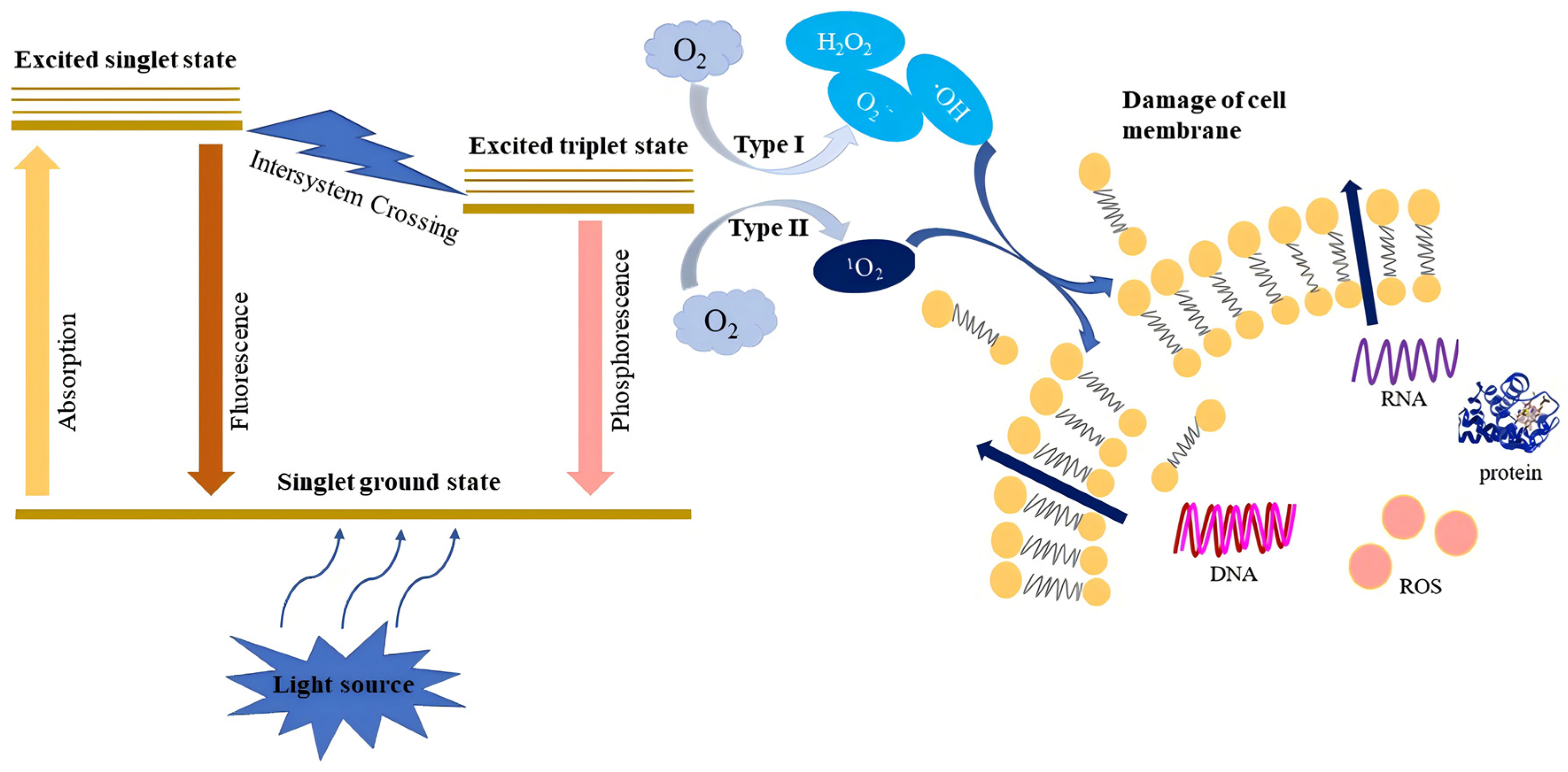
Figure 1. Schematic diagram of photosensitization processes.
Foods 13 03980 g001
Figure 2. Photolysis products of curcumin and antibacterial mechanism.
Foods 13 03980 g002
Figure 3. The practical application of CUR-mediated PDI in different food model systems.
Foods 13 03980 g003
Table 3. Antimicrobial activity of CUR-mediated PDI applied in food products.
FoodstuffTest StrainsCUR
Concentration
Light SourceIrradiation Time
/Light Dose
ReductionReferences
FruitFresh date fruitNS1000, 1400, 1800 μM420 nm10, 15 min
180, 270 J/cm2
shelf life extended by
7 days at 30 °C
[52]
Hami melonNS10, 20, 40, 50 μM460 nm5, 30, 60, 90 min~1.8 log CFU/g at storage[53]
Fresh-cut applesE. coli
ATCC 25922
0.5, 2, 10, 50 μM298 mW/cm2150–510 s~0.95 log CFU/g[54]
GrapeE. coli1600 μM465–470 nm
4.5–30.2 mW/cm2
9.1, 18.1, 27.2, 36.3 J/cm20.95, 1.26, 2.18, 2.40 log CFU/g[55]
Orange juiceE. coli
ATCC 11775
S. aureus
ATCC 12600
50, 100 μM462 ± 3 nm70 J/cm21.06 log CFU/mL
2.34 log CFU/mL
[56]
Orange slicesP. italicum75 μmol/L460 nm
100 mW/cm2
20 min~2.5 log CFU/g[38]
VegetableSpinach leaves
Cherry tomato
E. coli O157:H7
ATCC 700728
L. innocua
ATCC 33090
5 mg/mL320–400 nm
32 W/m2
5 min4 log CFU/mL in wash water
>100 CFU/cm2 on surface of product
[57]
CucumberS. aureus
ATCC 25923
50, 100 μM435 nm
9.4 mW/cm2
33.8 J/cm22.6 log CFUs[58]
Spinach
Lettuce
Tomato
E. coli O157:H7
ATCC 700728
L. innocua
ATCC 33090
10 mg/mL320–400 nm
6.8 W/m2
5 min
20.4 kJ/m2
3 log CFU/mL
1.7–3.3 log CFU/mL
[59]
SeafoodOystersNS10 μM470 nm
0.06 W/cm2
90 s
5.4 J/cm2
storage time extended by 7 days at 4 °C[60]
E. coli DH5α5, 10, 20 μM470 nm
0.06 W/cm2
3.6 J/cm22.5 log CFU/g[34]
V. parahaemolyticus100 μM10 W,
455–460 nm
30 min
9.36 J/cm2
no visible colony was detectable[61]
Norovirus5, 10, 20 μM470 nm3.6 J/cm21.15 log PFU/mL[62]
MeatChicken meatS. aureus
ATCC 25923
50, 100 μM435 nm
9.4 mW/cm2
33.8 J/cm21.7 log CFUs[58]
SausagesL. innocua
ATCC 33090
150 μg/mL320–400 nm
32 ± 0.2 W/m2
30 min>3 log CFU/mL[63]
BeefP. lundensis
B. thermosphacta
60 μM34 W, 430 nm33.01 J/cm20.15 log CFU/g
0.08 log CFU/g
[38]
Grains and SeedsMillet doughS. saprophyticus25 μM420–470 nm4.32 J/cm2~1 log CFU/g[64]
Corn kernelsA. flavus spores
aflatoxin B1
1000 μM430 nm104.2 J/cm22.3 log CFU/mL
inhibited aflatoxin B1 production
[65]
A. flavus spores0, 25, 45 μM420 nm60 J/cm2~2 log CFU/g[41]
aflatoxin B150 μM420 nm60 J/cm2production of aflatoxin B1 decreased significantly[66]
A. flavus spores
aflatoxin B1
1000, 1250, 1500 μM420 nm60 J/cm2storage time extended by 14 days at 25 °C
inhibited aflatoxin B1 production
[67]
zearalenone50 μΜ455~460 nm43.0 J/cm2ZEN decreased from 1.15 mg/kg to 0.58 mg/kg[68]
Corn flourA. flavus spores
aflatoxin B1
1000, 1500 μM420 nm60 J/cm2storage time extended by 7 days at 25 °C
inhibited aflatoxin B1 production
[67]
Fenugreek
Mung beans
E. coli
ATCC 25922
10, 50, 100 μM432 nm
9.4 mW/cm2
33.8 J/cm2>3 log CFUs[69]
Peanut seedsA. flavus20 mg/mL450 nmNS0.3 log CFU/g[70]
NS: not specified.
Table 4. Investigations on photodynamic inactivation of CUR derivatives.
CUR DerivativesBacteriaConcentrationLight SourceIrradiation Time/DoseReductionReferences
UNCPs-CURS. aureus0, 10, 20, 40, 80, 320 µg/mL 980 nm
0.5 W/cm2
30 min0–100%[82]
CUR.NPS. saprophyticus
DSM No. 18669
E. coli
DSM No. 6897
NS457 nm105.6 J/cm2>6.2 log CFU/mL
~2.9 log CFU/mL
[83]
CUR/polyurethane nanocompositesE. coli
CCM 4517
S. aureus
CCM 4516
NS470 nm1 h
6 h
100%[84]
CUR derivative coatingsS. aureus
E. coli
NS420 nm2, 6, 24, 48 h99%
95%
[85]
Microemulsion combined with EDTAP. aeruginosa5 ppm CUR
15.4 ppm EDTA
455 nm
16 mW/cm2
15 min
14.4 J/cm2
90%[59]
CUR combined EDTAB. cep aci50 μM CUR
0.4% EDTA
NS28 J/cm2100%[86]
CUR-ZnP. aeruginosa (ATCC27853)
E. coli O157:H7 (BNCC192101)
P. Mirabilis (BNCC337267)
E. coli (ATCC 25922)
1 mg/mL425 nm
6.25 mW/cm2
50 J/cm21.3 log CFU/mL
1.3 log CFU/mL
1.0 log CFU/mL
1.7 log CFU/mL
[70]
CUR-β-CDL. monocytogenes
V. parahaemolyticus
S. putrefaciens
0.16 μM10 W
455−460 nm
60 min
13.68 J/cm2
3.79 log CFU/g
2.81 log CFU/g
3.27 log CFU/g
[87]
NS: Not specified.
Disclaimer/Publisher’s Note: The statements, opinions and data contained in all publications are solely those of the individual author(s) and contributor(s) and not of MDPI and/or the editor(s). MDPI and/or the editor(s) disclaim responsibility for any injury to people or property resulting from any ideas, methods, instructions or products referred to in the content.