Article

Efficacy of Hovenia dulcis Fruit Extract in Hangover Mitigation: Double-Blind Randomized Clinical Evaluation

1
Natural Products Convergence R&D Division, Kwangdong Pharmaceutical Co., Ltd., Gwacheon 13840, Republic of Korea
2
Department of Food and Bioproduct Sciences, University of Saskatchewan, Saskatoon, SK S7N 5A8, Canada
3
Prairie Tide Diversified Inc., Saskatoon, SK S7J 0R1, Canada
4
Department of Family Medicine, Soonchunhyang University Bucheon Hospital, Bucheon 22972, Republic of Korea
5
Global Medical Research Center, Seoul 06526, Republic of Korea
6
Department of Food and Biotechnology, Korea University, Sejong 30019, Republic of Korea
7
Department of Food Regulatory Science, Korea University, Sejong 30019, Republic of Korea
*
Authors to whom correspondence should be addressed.
These authors contributed equally to this work.
Foods 2024, 13(24), 4084; https://doi.org/10.3390/foods13244084
Submission received: 8 November 2024 / Revised: 9 December 2024 / Accepted: 16 December 2024 / Published: 17 December 2024

Abstract

:
The fruit of Hovenia dulcis Thunb. (HD) is renowned for its medicinal properties and is rich in bioactive compounds, traditionally used in East Asian medicine as a natural antidote for alcohol intoxication. A randomized, double-blind, crossover, placebo (PLA)-controlled clinical trial was conducted to compare the effects of beverages containing 0.475% HD (HDB), HD combined with 0.1% Pueraria lobata extract (HDPB), and HD combined with 0.02% glutathione yeast extract (HDGB) with the PLA on the symptoms of a hangover. Subjects (n = 30) were randomized into six test groups consuming three beverages, including the PLA. After ingestion, blood alcohol and blood acetaldehyde concentrations were measured at 0, 0.25, 0.5, 1, 2, 4, 6, and 15 h post-alcohol consumption. No significant differences in hematology tests and vital signs were observed amongst the treatment groups; however, at 0.5 and 6 h, the blood alcohol concentrations of the HDB and HDPB groups were significantly lower compared to the PLA group (p < 0.05). Additionally, the blood acetaldehyde concentrations in the HDPB group showed significantly lower values than the PLA after 6 h (p < 0.05). These findings suggest that HD may aid in alcohol detoxification and limit acetaldehyde accumulation. This indicates the potential for HD as a functional food ingredient for alleviating hangover symptoms.

1. Introduction

A hangover from alcohol is characterized by a collection of symptoms that appear the day after heavy drinking, usually when the blood alcohol level is nearly zero [1]. Physiological symptoms of hangovers include thirst, drowsiness, headache, fatigue, and sweating, while mental symptoms may involve dizziness, depression, anxiety, and irritability [2]. These symptoms not only cause discomfort but also impair cognitive and physical functions, leading to significant social and economic impacts [3]. The pathophysiology of hangovers is complex and multifactorial, involving dehydration, electrolyte imbalance, intestinal disturbances, and accumulation of toxic metabolites such as acetaldehyde [4,5]. Therefore, many hangover remedies are marketed every year but there is currently a lack of clinical trials to evaluate their solid effectiveness.
Alcohol metabolism begins primarily with its absorption and transport through the bloodstream to the liver. In the liver, alcohol is converted to acetaldehyde by alcohol dehydrogenase (ADH) [6]. Acetaldehyde is then further oxidized to acetate by aldehyde dehydrogenase (ALDH). The acetate is finally broken down into carbon dioxide and water, which are eliminated from the body [7]. Excessive alcohol consumption can lead to liver damage, typically indicated by elevated levels of liver enzymes such as alanine aminotransferase (ALT) and aspartate aminotransferase (AST) [8,9]. These enzymes are markers of liver cell injury, and their elevated levels can signal liver inflammation or damage. In addition, alcohol metabolism produces reactive oxygen species, which can cause oxidative damage to various tissues, including the liver, brain, and gastrointestinal tract, and are a significant cause of hangover symptoms.
In recent years, natural remedies for the prevention and treatment of hangovers have garnered increasing attention [10]. Compared to conventional medications—which can cause serious side effects such as ataxia, impaired attention, and confusion—natural bioactive compound products are considered safer alternatives [11]. For example, Mesembryanthemum crystallinum (ice plant) [12], Pueraria lobata (kudzu flower) [13], and Artemisia herbs [14], plant extracts, and glutathione yeast extract (GY) [15], which is a fermented extract, have been shown to relieve hangover symptoms or promote alcohol metabolism.
This study aims to evaluate the efficacy of Hovenia dulcis Thunb. extract (HD), P. lobata root extract (PL), and GY as natural remedies for hangover relief. HD exhibits strong antioxidant activity due to its high flavonoid content [16,17,18], lowering liver enzyme levels—including ALT and AST—improving liver function, and preventing pathological changes in liver tissue [19]. Polysaccharides derived from HD were reported to regulate hepatic fatty acid metabolism, reduce inflammation [20], and support the liver’s protection and xenobiotic metabolism [21]. HD also enhances alcohol metabolism by increasing ADH and ALDH activity [22] while reducing fatigue [23]. These activities highlight its potential as a functional food for hangover prevention.
PL exhibits strong antioxidant activity due to its polyphenols, including puerarin [24]. The antioxidant property of this substance helps to neutralize free radicals, thereby protecting cells from oxidative stress. Additionally, PL has been demonstrated to enhance blood circulation and forestall the development of cardiovascular diseases [25], which is of paramount importance for maintaining optimal heart health. It also safeguards nerves [26], which can help in preventing neurodegenerative diseases, and manages metabolic disorders [27], such as diabetes and obesity. Furthermore, it alleviates alcohol-induced liver damage [28], supporting liver health and function.
Similarly, GY has been reported to have strong antioxidant activity [29], enhancing the body’s ability to combat oxidative stress. This extract has been demonstrated to enhance the immune system [30], thereby providing a robust defense against infections and diseases. It protects nerves [31], similar to PL, which suggests a potential synergistic effect in neuroprotection. Additionally, GY prevents metabolic disorders such as diabetes [32], contributing to overall metabolic health. It also supports liver detoxification [33], aiding in the removal of toxins and improving liver function. The combined use of PL and GY leverages their shared antioxidant properties and complementary health benefits. When combined, they can provide enhanced protection against oxidative stress, support cardiovascular and liver health, improve metabolic function, and offer neuroprotective effects. This synergistic approach highlights their potential as effective natural remedies for promoting overall human health and well-being.
The objective of this study is to conduct clinical trials to evaluate the efficacy of HD, PL, and GY, both individually and in combination, in preventing hangover symptoms. By examining their potential as natural remedies, this study is designed to contribute to the development of safer and more effective treatments for hangovers, potentially alleviating their significant social and economic impacts.

2. Materials and Methods

2.1. Study Participants and Inclusion/Exclusion Criteria

The Global Medical Research Center located in Seoul (Republic of Korea) enrolled participants in this clinical trial. The inclusion criteria were based on Chang and Kim [34] but modified as follows: (1) aged between 19 and 40 years; (2) body mass index (BMI) between 18.5 and 25 kg/m2; (3) a history of experiencing hangovers. Subsequently, participants were required to meet the following criteria: (1) consumption of alcohol within the previous 30 days; (2) exhaled alcohol concentration of 0.00% at the initial visit on Day 2; and (3) consent to participate in a human clinical trial, along with a signed consent form. The criterion related to the experience of hangovers meant that participants must have previously experienced hangover symptoms, such as headaches, nausea, fatigue, and sensitivity to light and sound following alcohol consumption. This ensured that the study population was relevant to the research on hangover alleviation.
Individuals who did not meet the following criteria were excluded from participation: (1) individuals undergoing treatment for a serious medical condition, including but not limited to cardiovascular (hypertension), immune, respiratory, endocrine (diabetes), renal and urinary, neurologic and musculoskeletal, psychiatric, infectious disease, and malignancy; (2) individuals with a history of peptic ulcer disease, gastroesophageal reflux disease, or a history of serious gastrointestinal disease (e.g., Crohn’s disease); (3) individuals who are pregnant or lactating or who intend to become pregnant during the course of this human application study; (4) individuals with an alcohol use disorder or recent heavy alcohol consumption that may affect the study results; (5) individuals taking medications that affect alcohol metabolism (e.g., antidepressants) or pose the risk of gastrointestinal bleeding (e.g., warfarin, clopidogrel, aspirin, non-steroidal anti-inflammatory drugs); (6) individuals who have used medications that enhance liver function (disulfiram class); (7) individuals who have used drugs, dietary supplements, or medications that affect drug-metabolizing enzymes within a specified time period prior to the screening visit; (8) individuals with abnormal laboratory results (e.g., AST, ALT, GPT), including creatinine, thyroid-stimulating hormone, fasting blood glucose levels; (9) individuals who have participated in another interventional clinical trial within the past month or intend to participate in another clinical trial during this study; (10) individuals with known sensitivities or allergies to any of the food trial ingredients; and (11) other reasons for exclusion at the discretion of the investigator.

2.2. Study Design

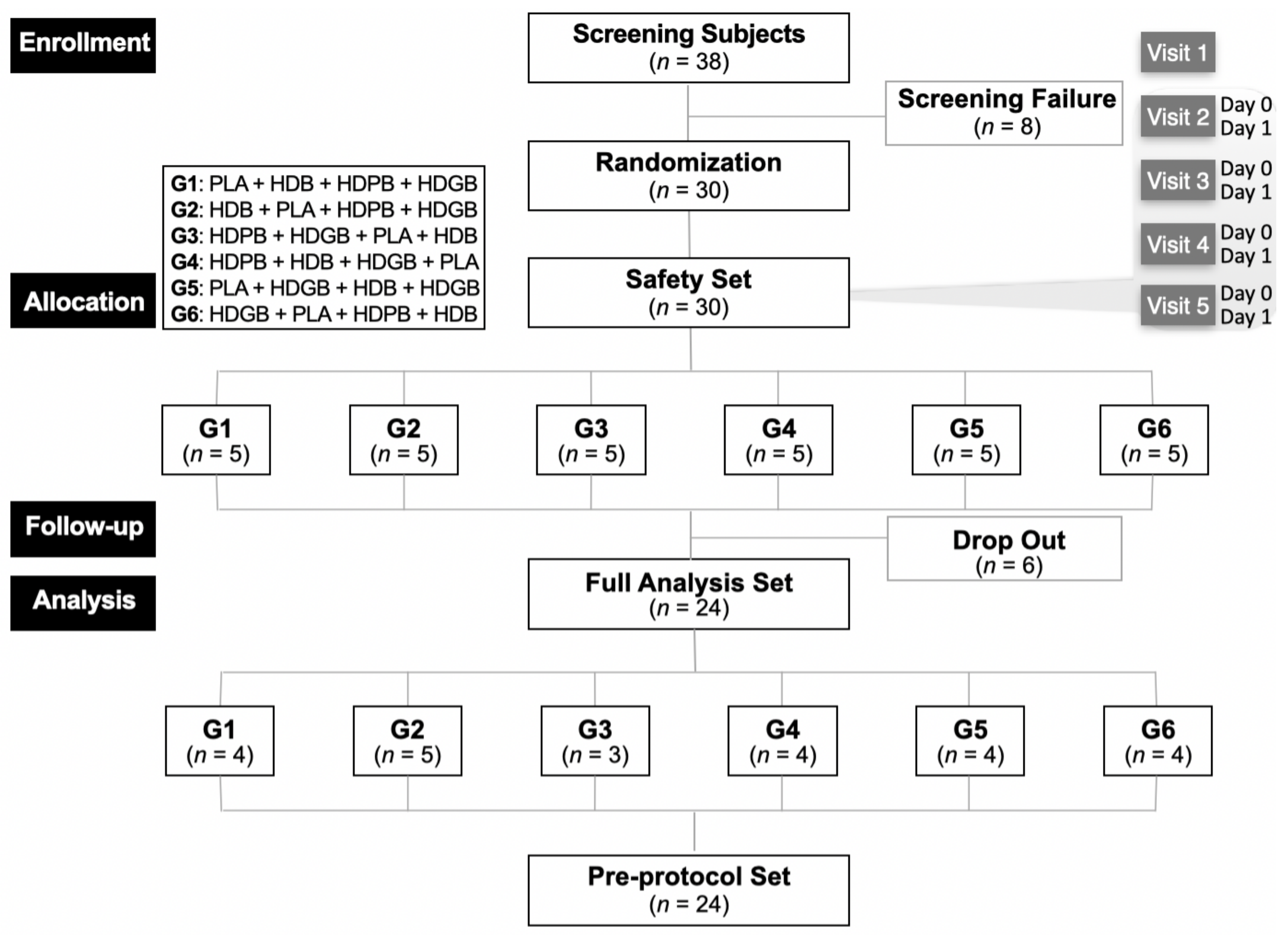
This study was approved by the Institutional Review Board of the Global Medical Research Center (IRB No. GIRB-24216-ZL) and registered with the Centers for Disease Control and Prevention Clinical Research Information Service (CRIS No. KCT0009899). It was conducted from April 29 to June 16, 2024. This study was designed as a randomized, double-blind, parallel-group, placebo (PLA)-controlled trial (Figure 1). On the day of screening, participants were randomly assigned to different groups, including one PLA group and three test groups, using a double-blind method. A statistician prepared the randomization list in collaboration with the sponsor. Both participants and researchers were unaware of group allocation until the end of the study. Power analyses were performed to estimate the sample size required to test for superiority [35].
The human clinical trial was a crossover design trial with randomization to one of six arms. The required number of human clinical trial subjects was 30 to account for the dropout rate (20%) (Table 1). Participants were sent for the human clinical trial at Visits 1 and 2 (Day 0). Participants were screened to determine if they met the inclusion/exclusion criteria by completing questionnaires on demographics, lifestyle, medical history, and medication history. They also underwent physical examination, vital signs (blood pressure, pulse), anthropometry (height, BMI, weight), clinical pathology tests, and pregnancy tests (for women of childbearing age) [36]. The drinking behavior questionnaire included questions about the type of alcohol, amount consumed, number of times per week, and hangovers experienced in the past month. At Visit 2 (Day 0), all human subjects meeting the inclusion/exclusion criteria were assigned to each of the following arms by assignment code using blocked randomization [37]. To ensure balanced randomization between the intake arms, the ratio of the number of human subjects in each arm was equalized to 1:1:1:1:1:1. Randomized human subjects received a single, alternating dose of the investigational food (Test Food I or Test Food II or Test Food III or control food) at Visits 2, 3, 4, and 5, respectively, with a 7-day washout period between each pair of consecutive visits (Figure 1).

2.3. Interventions

The concentrated aqueous samples were kindly provided by Kwangdong Pharmaceutical Co., Ltd. (Gwacheon, Republic of Korea). Four samples, including the control (PLA), were administered orally as 500 mL beverages, the specific compositions of which are shown in Table 2: 0.475% (w/v) HD beverage (HDB); HD combined with 0.1% (w/v) PL (HDPB); and HD combined with 0.02% GY (HDGB). The PLA sample consisted of 0.012% caramel color powder, and 0.070% flavor dissolved in purified water. In addition, three samples contained vitamin C (0.020%), sodium bicarbonate (0.023%), glycine (0.010%), and flavorings (0.105%) as minor ingredients, all ingredients are expressed in weight per volume. Each sample was identical in appearance, shape, color, taste, sweetness, packaging, and additives. All participants received an identical meal before the test and consumed either the PLA or the intervention beverage 2 h after the meal. Thirty minutes after ingesting the test substance, the participants consumed whiskey (40% alcohol, 0.9 g/kg body weight) in two 30-minute increments. To control for variables and ensure the accuracy and reliability of experimental results, subjects were instructed to fast after alcohol ingestion. At each visit, subjects were instructed to abstain from alcohol for 24 h prior to the next visit. Upon waking the next morning, subjects completed a hangover symptom questionnaire 15 h after alcohol ingestion.

2.4. Outcome Measures

2.4.1. Acute Hangover Scale (AHS)

The AHS is used to assess hangover symptoms and their severity [38]. This scale consists of 9 items that rate the severity of each symptom and provide an overall hangover severity score. Each item is scored from 0 to 7, with the total AHS score being the average of all items. The symptom severity is categorized as ‘none’ (0 points), ‘mild’ (1 point), ‘moderate’ (4 points), or ‘incapacitating’ (7 points).

2.4.2. Alcohol and Acetaldehyde Analysis During Blood Sample Handling and Collection

Blood was taken at 0, 0.25, 0.5, 1, 2, 4, 6, and 15 h after alcohol consumption at Visits 2, 3, 4, and 5 (Days 0 and 1). A catheter filled with saline for the injection was placed in a vein in the participant’s arm to obtain 5 mL of blood at each time point. Blood samples were placed in BD Vacutainer® NaF tubes (BD, Milan, Italy), stored at 4 °C, and analyzed immediately according to the guidelines of the external laboratory [39]. Samples were discarded immediately after analysis and not used for any other purpose.

2.4.3. Analysis of Alcohol and Acetaldehyde in Blood

All clinical blood samples were stored as described in Section 2.4.2 and processed for ethanol and acetaldehyde concentration analyses. Blood samples treated with anticoagulants were centrifuged, and the supernatant plasma was collected. For analysis, 200 µL of human plasma was mixed with 500 µL of saturated NaCl solution in a headspace vial and 100 µL of 0.005% n-butanol was added. Samples were analyzed on an Agilent 5977 series GC system (Agilent Technologies Inc., Palo Alto, CA, USA) with a CTC headspace GC/MS detector (CTC Analytics AG, Zwingen, Switzerland). Ethanol, acetaldehyde, and n-butanol were chromatographically separated on a Discovery HP-INNOWAX column (0.32 mm × 30 m, 0.5 µm, Sigma-Aldrich, St. Louis, MO, USA). Helium was used as the carrier gas at a constant flow at 3 mL/min and the interfacial temperature was set at 200 °C. An electron ionization system with 70 eV ionization energy was used for GC-MS detection. The initial temperature of the GC oven was maintained at 35 °C for 3 min, after which it was increased to 85 °C at a rate of 40 °C per minute and held for a further 2 min. The equilibration temperature and time for the headspace sampler were 70 °C and 10 min, respectively. The injection volume was 250 µL, and the split ratio was 100:1. The mass spectrometer was operated in single-ion monitoring mode with ethanol set to m/z 45, 46, and 31 and acetaldehyde set to m/z 43, 41, and 29. Quantification was based on m/z values of 45 for ethanol and 43 for acetaldehyde [11].

2.5. Safety Assessments

Vital signs, including systolic and diastolic blood pressure, temperature, pulse rate, and clinical laboratory tests (hematology, biochemistry, and urinalysis) were performed on participants at screening (Visit 1) and Visit 2 (Day 1) [40]. In addition, adverse events were identified during the study through interviews or questionnaires administered to the participants.

2.6. ADH and ALDH Enzyme Activity Assay

To measure markers of alcohol hangover, ADH and ALDH activity were determined in plasma after blood separation. The activities of ADH (K787; BioVision Inc., Milpitas, CA, USA) and ALDH (K731; BioVision Inc., Milpitas, CA, USA) were determined using commercial kits. All assay procedures were performed according to the manufacturer’s instructions [11].

2.7. Statistical Analysis

Statistical analyses were performed using SAS version 9.4 from the SAS Institute (Cary, NC, USA). Data were analyzed by calculating the mean ± standard deviation (SD) with appropriate descriptive statistics, and the significance of differences was determined by two-tailed t-tests at * p < 0.05 and ** p < 0.01.

2.7.1. Validation Variables

The degree of change in blood alcohol and acetaldehyde concentrations (hourly concentrations, Cmax, Tmax, and AUC) of each group were analyzed using repeated measures analysis of variance (RM ANOVA) [41]. ANOVA was performed to evaluate whether there was a statistically significant difference in the intake effect between each group under the assumption of no residual effect. RM ANOVA was used to analyze the difference between the intake effects of each group at each time of AHS to evaluate whether there was a statistically significant difference.

2.7.2. Safety Evaluation Variables

All adverse events that occurred after the ingestion of the study food were tabulated and evaluated through the calculation of an incidence rate. The proportion of human subjects with adverse events in each group was calculated and compared using the Chi-square test or Fisher’s exact test [42]. The hematological and blood chemistry test values between the two groups (Test Food I and control food, Test Food II and control food, Test Food III and control food) were evaluated for statistically significant differences by ANOVA or Kruskal–Wallis test according to normality, and post hoc tests were performed if necessary. The frequency and proportion of normal and abnormal urinalysis were calculated and compared using the Chi-square test or Fisher’s exact test. Intra-group comparisons of pre- and post-consumption changes in vital signs (blood pressure, pulse) were analyzed using paired t-tests. The magnitude of change between groups (Test Food I and control food, Test Food II and control food, Test Food III and control food) was assessed for statistically significant differences using ANOVA or Kruskal–Wallis test depending on normality, and post hoc tests were performed if necessary.

3. Results and Discussion

3.1. Enrollment

A total of 30 participants were enrolled, with 8 excluded during the screening process, resulting in 24 participants included in the experimental analysis from 29 April 2024 to 16 June 2024. During the experimental period, six participants were excluded from the final analysis. Regarding efficacy data, the per-protocol (PP) set analysis represents the primary analytical approach, complemented by the full analysis (FA) set analysis. The PP analysis method provides the maximum potential efficacy of a treatment under strict adherence to the protocol, making it suitable for assessing the effectiveness of the protocol itself. In contrast, the FA analysis method maximizes the reflection of the real conditions of the study population, thereby enhancing the external validity of the results [43]. For demographic data, the PP set analysis is the primary method, while for safety data, the safety set analysis is used. None of the participants in the FA set were excluded from the PP set analysis set among those in the FA set; therefore, both the FA set and the PP set were analyzed with the same number of cases (n = 24) (Figure 2).

3.2. General Participant Characteristics

Participant demographics and pre-intake characteristics are compared in Table 3. All pre-intake characteristics, including demographics, were compared between the intake groups to identify factors that differed; however, the experimental group comprised 12 (50%) males and 12 (50%) females. There was no statistically significant difference in age, with a mean age of 29.19 ± 3.86 years for the experimental group. In addition, the intake groups were matched to ensure that there were no significant differences in exercise status, smoking status and amount, sleep duration, height, and weight between the two groups, thus ensuring comparability between the groups.

3.3. Clinical Pathology Assessment for Hangover Relief

A total of 30 subjects were randomized into the test and control groups for a human application trial to obtain more realistic safety data and conduct a comprehensive risk assessment. The primary method used was a safety set analysis [44]. No statistically significant differences were observed between each group and the PLA group for any hematological test at 15 h post-drinking (Table 4) and no serious adverse events occurred. The hematological test of monocyte analysis showed that the HDGB group had the lowest monocyte count of 6.6 ± 1.2% (p = 0.2959), while the other groups, including the PLA, had similar counts of 7.1–7.3%. In the blood chemistry test, ALT analysis showed that HDB and HDPB were 19 ± 15 and 17 ± 12 U/L, respectively, compared to the lower values (both 15 ± 11 U/L) for the PLA and HDGB. However, these changes were within the normal range and were not found to be clinically significant. In addition, there was no statistically significant difference between the groups in any of the urine test parameters, including glucose, ketones, and bilirubin at 15 h after drinking. Therefore, no safety concerns were reported.
Table 5 presents the data on vital signs, including blood pressure and pulse, and body measurements such as weight. According to the research results of Seppä et al. [45] and Kawano et al. [46], vital sign changes in pulse and blood pressure after drinking increased the pulse rate immediately after drinking, and blood pressure decreased due to the acute depression effect. Blood pressure began to rise after 8 h (subacute pressure effect), and blood pressure returned to its previous level 24 h after abstinence. In this study, vital signs of blood pressure and pulse were measured 15 h after drinking and no statistically significant differences were observed between the groups in any of the measured parameters; however, after 15 h, significant differences were observed between the PLA and HDB (p = 0.0060) or HDPB (p = 0.0309) in the systolic blood pressure analysis of the change from baseline using the paired samples t-test. In addition, in the diastolic blood pressure analysis using the paired samples t-test, significant differences were observed between the PLA (p = 0.0462) and samples (t-test). However, for pulse rate, the analysis showed no statistically significant differences between the groups (t-test).

3.4. Survey of the Symptoms of a Hangover

AHS scores are utilized to assess various physical and cognitive issues, including headache, nausea, fatigue, and concentration difficulties, [47,48] which are commonly associated with the severity of hangover symptoms. In this study, AHS scores following alcohol consumption were compared between the experimental groups (HD, HDPB, and HDGB) and the control group (PLA) to determine any statistically significant differences. Table 6 presents the AHS results measured 15 h after alcohol consumption, with a total score ranging from 3.0 ± 2.7 to 3.8 ± 3.3. There were no significant differences between the experimental and control groups (PLA). The experimental group exhibited similar scores to the PLA group across various symptoms, including thirst, hangovers, headaches, dizziness/fainting, anorexia, gastrointestinal disorders, nausea, and heart palpitations. In addition, there was no significant difference in alcohol consumption patterns between different GG genotypes, which is consistent with previous research suggesting that genetic factors may not significantly influence hangover symptoms in this context [49].
Previous studies have suggested that blood ethanol levels, rather than acetaldehyde, play a critical role in hangover severity [50]; however, recent evidence suggests that both systemic alcohol consumption and acetaldehyde can lead to increased acetaldehyde accumulation in both blood and brain tissue in mice [51]. In addition, a recent clinical trial also found an association between acetaldehyde levels and hangover symptoms [52,53]. Accordingly, the present study sought to ascertain the efficacy of the experimental treatment in alleviating the symptoms of alcohol withdrawal by measuring the concentrations of blood alcohol and acetaldehyde.
The peak blood alcohol concentration (Cmax), time to peak blood concentration (Tmax), and area under the concentration–time curve (AUC) from 0 to 15 h after drinking are shown in Table 7. Compared to the PLA, HDPB sample ingestion slightly reduced blood alcohol concentrations in AUC and Cmax but there were no statistically significant differences between groups. Blood alcohol and acetaldehyde concentrations by group after alcohol ingestion are shown in Table 7. Although the AUC levels of HDPB and HDB in blood alcohol tended to be lower than those of the PLA, these differences did not achieve statistical significance (p = 0.0800). Additionally, no significant differences were observed in Cmax and Tmax among the samples. In the analysis using the paired samples t-test, no significant differences were observed between the PLA and HDB or HDGB. Additionally, comparative analysis of AUC, Cmax, and Tmax for blood acetaldehyde showed no significant differences among the test groups.

3.5. Changes in Blood Alcohol and Acetaldehyde Levels

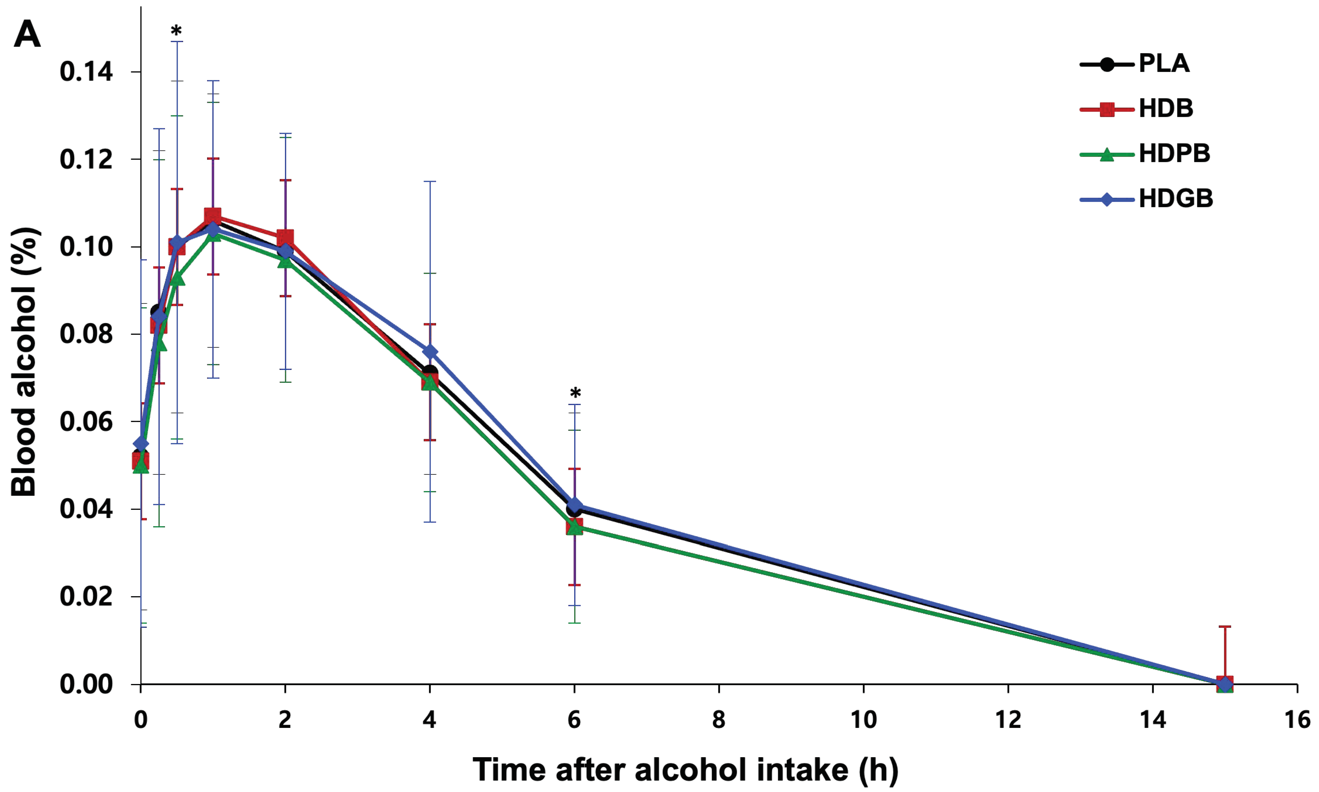
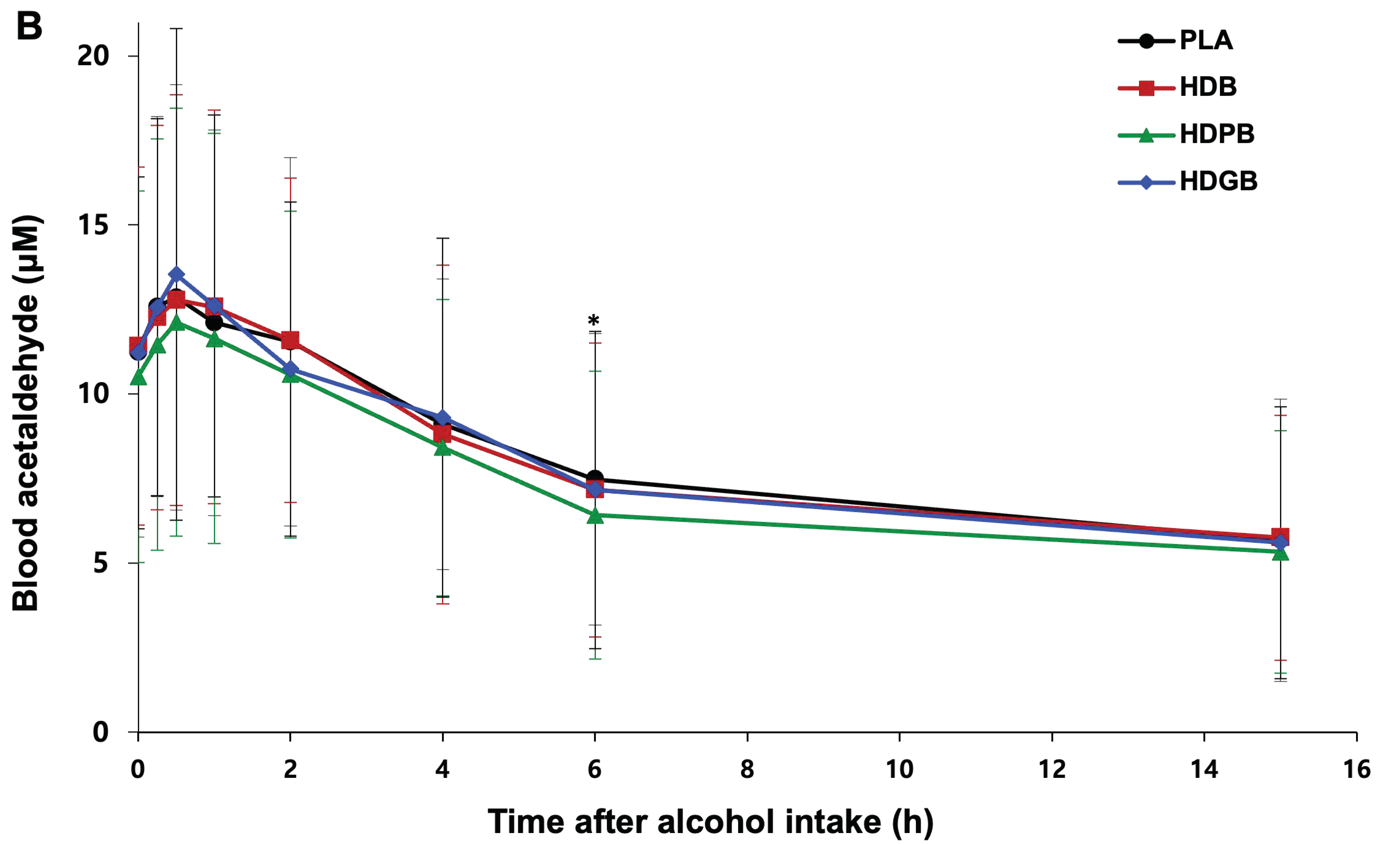
Figure 3 shows the changes in blood alcohol and acetaldehyde concentrations over a 15-h period following consumption. Previous research suggests that blood alcohol concentrations typically peak at approximately 1 h after drinking and then decline [54,55]. Our study also displayed this, and our comparisons between each treatment group and the PLA using the paired samples t-test showed no significant differences at most times (0.0, 0.25, 1.0, 2.0, 4.0, 15 h), except at 0.5 and 6 h. Interestingly, the HDPB group (0.093 ± 0.037%) had lower blood alcohol levels compared to the PLA group (0.100 ± 0.038%) at 0.5 h post-consumption (p = 0.0396). Although statistically significant, the observed difference is minimal and may not hold biological or practical significance in a broader context [56].
This finding is consistent with Thomes et al. [57], who observed similar trends in blood alcohol levels after alcohol consumption, suggesting that the potential of HD and PL extracts might influence alcohol metabolism. In addition, both the HDB group (0.036 ± 0.018%) and the HDPB group (0.036 ± 0.022%) had lower blood alcohol concentrations after 6 h than the PLA group (0.040 ± 0.022%) (p = 0.0368), further supporting the hypothesis that certain extracts may play a role in effectively modulating alcohol metabolism. In addition, the study by Park et al. [58] highlights the importance of understanding changes in blood alcohol and acetaldehyde concentrations, which are critical factors in the severity of hangover symptoms. Analysis of blood acetaldehyde concentrations showed that subjects receiving HDPB had consistently lower levels than those of the other treatment groups at all times; however, significant differences were only observed at 6 h, where the HDPB group (6.419 ± 4.257 μM) had significantly lower acetaldehyde levels compared to the PLA group (7.477 ± 4.313 μM) (p = 0.0251). This is consistent with findings from medical studies highlighting the role of acetaldehyde in alcohol metabolism and its potential impact on hangover severity [59]. These findings reinforce the potential benefits of using a combination of HD and PL, a natural plant-derived extract, in managing alcohol-related adverse effects.

4. Conclusions

This study demonstrated the potential hangover-relieving effects of a group of extracts (HDPB and HDGB) when added to HD in individuals aged 19 to 40 years who had previously experienced hangovers. A double-blind, randomized, PLA-controlled crossover trial showed that blood alcohol levels in the HDPB group were significantly lower than those in the PLA group at 0.5 h after ingestion (p < 0.05). At 6 h after ingestion, both the HDB and HDPB groups had significantly lower blood alcohol concentrations than the PLA group (p < 0.05). Notably, although statistically significant differences were not observed at all time points, the HDPB group consistently had lower blood alcohol levels than the PLA group throughout this study. Similarly, the results for blood acetaldehyde showed that the HDPB group had significantly lower levels than the PLA group at 6 h after ingestion and maintained consistently lower levels than the PLA group at all time points. These results confirm that the consumption of HDB and HDPB foods can significantly improve hangover symptoms by reducing blood acetaldehyde concentrations and blood alcohol concentrations.

Author Contributions

Conceptualization, D.H.P. and K.W.L.; investigation, D.H.P.; validation, K.W.L. and Y.Y.S.; formal analysis, D.H.P.; data curation, Y.Y.S., M.J.T.R. and G.X.; writing—original draft preparation, D.H.P., K.W.L. and Y.Y.S.; writing—review and editing, Y.Y.S., M.J.T.R., I.P., S.-H.L., J.-Y.P., E.P., S.-B.L., I.A.K., G.X., J.Y.H. and Y.J.K.; visualization, Y.Y.S.; supervision, Y.J.K.; project administration, K.W.L.; funding acquisition, Y.J.K. All authors have read and agreed to the published version of the manuscript.

Funding

This research was supported by a grant from the Graduate School Education Program of Regulatory Sciences for Functional Food under grant No. 21153MFDS604 funded by the Ministry of Food and Drug Safety (MFDS) of the Republic of Korea.

Institutional Review Board Statement

This study was approved by the Institutional Review Board of the Global Medical Research Center (IRB No. GIRB-24216-ZL, 23 February 2024).

Informed Consent Statement

All participants received detailed explanations about the purpose and content of this study from the researchers and voluntarily provided written consent.

Data Availability Statement

The original contributions presented in this study are included in the article; further inquiries can be directed to the corresponding author.

Acknowledgments

The authors acknowledge the kind contribution of the concentrated aqueous samples from Kwangdong Pharmaceutical Co., Ltd. (Gwacheon, Republic of Korea). Academic support for the research project was obtained from the Bio-medicinal Technology group at Korea University (Sejong, Republic of Korea). The authors also wish to thank Suk Nam Kang of the Department of Animal Resources, Daegu University (Daegu, Republic of Korea), for his assistance with the statistical analyses and Su Bin Lee of the Department of Food Regulatory Science, Korea University (Sejong, Republic of Korea), for her assistance with this research.

Conflicts of Interest

D.H.P., K.W.L., I.P., S.-H.L., J.-Y.P. and E.P. are employees of Kwangdong Pharmaceutical Co., Ltd., and their role in this study reflects their role as researchers and does not represent the position of their current or former employers. M.J.T.R. is the founder of, and has an equity interest in, Prairie Tide Diversified Inc. (PTD, Saskatoon, SK, Canada). Y.Y.S. is the Korean Branch Representative for PTD in the Republic of Korea. The terms of this arrangement have been reviewed and approved by the University of Saskatchewan by their Conflicts of Interest policy. No other authors declare any conflicts of interest.

References

  1. van Schrojenstein Lantman, M.; van de Loo, J.A.E.; Mackus, M.; Verster, J.C. Development of a definition for the alcohol hangover: Consumer descriptions and expert consensus. Curr. Drug Abuse Rev. 2016, 9, 148–154. [Google Scholar] [CrossRef] [PubMed]
  2. Verster, J.C.; Scholey, A.; van de Loo, A.J.A.E.; Benson, S.; Stock, A.-K. Updating the definition of the alcohol hangover. J. Clin. Med. 2020, 9, 823–827. [Google Scholar] [CrossRef] [PubMed]
  3. Nutt, P.D.; Liu, Y.-C.; Chen, M.-S.; Wang, D.-L.; Palmer, E.; Tyacke, R. Alcohol hangover: The health impact with a historic and Chinese perspective. Drug Sci. Policy Law 2020, 6, 1–5. [Google Scholar] [CrossRef]
  4. Bereda, G. Mental and physical symptoms of alcohol hangover. Neuropsychopharmacol. Disord. 2022, 1, 1–3. [Google Scholar] [CrossRef]
  5. Penning, R.; van Nuland, M.; Fliervoet, L.A.; Olivier, B.; Verster, J.C. The pathology of alcohol hangover. Curr. Drug Abuse Rev. 2010, 3, 68–75. [Google Scholar] [CrossRef]
  6. Sin, H.J.; Choung, S.Y.; Kang, S.; Kwon, H.T.; Kim, B.H. Anti-alcohol and anti-aldehyde hangover effect of aldehyde dehydrogenase related compounds in rat. J. Environ. Health Sci. 2023, 49, 99–107. [Google Scholar] [CrossRef]
  7. Yun, M.-K.; Jeong, H.C.; Lee, S.-J.; Lee, S.J. Effect of Lactobacillus fermented garlic extract powder on alcohol and acetaldehyde metabolism. J. Korean Soc. Food Sci. Nutr. 2023, 52, 357–362. [Google Scholar] [CrossRef]
  8. Tsai, J.; Ford, E.S.; Li, C.; Zhao, G. Past and current alcohol consumption patterns and elevations in serum hepatic enzymes among US adults. Alcohol. Behav. 2012, 37, 78–84. [Google Scholar] [CrossRef]
  9. Nivukoski, U.; Bloigu, A.; Bloigu, R.; Aalto, M.; Laatikainen, T.; Niemelä, O. Liver enzymes in alcohol consumers with or without binge drinking. Alcohol 2019, 78, 13–19. [Google Scholar] [CrossRef]
  10. Choi, E.J.; Kim, H.; Hong, K.-B.; Suh, H.J.; Ahn, Y. Hangover-relieving effect of ginseng berry kombucha fermented by Saccharomyces cerevisiae and Gluconobacter oxydans in ethanol-treated cells and mice model. Antioxidants 2023, 12, 774. [Google Scholar] [CrossRef]
  11. Jung, S.H.; Lee, Y.H.; Lee, E.K.; Park, S.-D.; Shim, J.-J.; Lee, J.-L.; Yoo, H.H. Effects of plant-based extract mixture on alcohol metabolism and hangover improvement in humans: A randomized, double-blind, paralleled, placebo-controlled clinical trial. J. Clin. Med. 2023, 12, 5244–5258. [Google Scholar] [CrossRef] [PubMed]
  12. Lee, S.Y.; Choi, H.D.; Yu, S.N.; Kim, S.H.; Park, S.K.; Ahn, S.C. Biological activities of Mesembryanthemum crystallinum (ice plant) extract. J. Life Sci. 2015, 25, 638–645. [Google Scholar] [CrossRef]
  13. Yamazaki, T.; Hosono, T.; Matsushita, Y.; Kawashima, K.; Someya, M.; Nakajima, Y.; Narui, K.; Hibi, Y.; Ishizaki, M.; Kinjo, J. Pharmacological studies on Puerariae Flos. IV: Effects of Pueraria thomsonii dried flower extracts on blood ethanol and acetaldehyde levels in humans. Int. J. Clin. Pharmacol. Res. 2002, 22, 23–28. [Google Scholar] [PubMed]
  14. Song, G.; Han, H.; Park, S.; Sa, S.; Chung, W.; Lee, B.Y. Effects of GSH on alcohol metabolism and hangover improvement in humans: A randomized double-blind placebo-controlled crossover clinical trial. Foods 2024, 16, 3262–3275. [Google Scholar] [CrossRef] [PubMed]
  15. Lee, K.-S.; Kim, G.-H.; Seong, B.-J.; Kim, H.-H.; Kim, M.-Y.; Kim, M.-R. Effects of aqueous medicinal herb extracts and aqueous fermented extracts on alcohol-metabolizing enzyme activities. Food Sci. Preserv. 2009, 16, 259–265. [Google Scholar]
  16. Sung-Hee, K.; Jin-Yeul, M. A study on the extraction and efficacy of bioactive compound from Hovenia dulcis. Korean J. Pharm. 2006, 21, 11–15. [Google Scholar] [CrossRef]
  17. Lee, S.; Kim, H.; Park, Y.; Ryu, S.; Yeon, S.; Turk, A.; Lee, H.; Hwang, B.; Lee, M. Quantitation of antioxidant and α-glucosidase inhibitory flavonoids in the fruits, fruit stalks and seeds of Hovenia dulcis. J. Pharm. 2023, 54, 242–248. [Google Scholar] [CrossRef]
  18. Kong, L.; Li, J.; Zhang, X. Prevention and cure of acute alcohol intoxication in mice by administration of compound of Japanese raisintree fruit, lobed kudzuvine flower bud and lightyellow sophora root. Adv. Sci. Technol. 2014, 15, 874–880. [Google Scholar]
  19. Fang, H.-L.; Lin, H.-Y.; Chan, M.-C.; Lin, W.-L.; Lin, W.-C. Treatment of chronic liver injuries in mice by oral administration of ethanolic extract of the fruit of Hovenia dulcis. Asian J. Clin. Med. 2007, 35, 693–703. [Google Scholar] [CrossRef]
  20. Choi, R.-Y.; Woo, M.-J.; Ham, J.R.; Lee, M.-K. Anti-steatotic and anti-inflammatory effects of Hovenia dulcis Thunb. extracts in chronic alcohol-fed rats. Biochem. Pharmacol. 2017, 90, 393–401. [Google Scholar] [CrossRef]
  21. Kim, H.; Kim, Y.J.; Jeong, H.Y.; Kim, J.Y.; Choi, E.-K.; Chae, S.W.; Kwon, O. A standardized extract of the fruit of Hovenia dulcis alleviated alcohol-induced hangover in healthy subjects with heterozygous ALDH2: A randomized, controlled, crossover trial. J. Ethnopharmacol. 2017, 209, 167–174. [Google Scholar] [CrossRef] [PubMed]
  22. Chen, S.; Zhong, G.; Li, A.; Li, S.; Wu, L. Influence of Hovenia dulcis on alcohol concentration in blood and activity of alcohol dehydrogenase (ADH) of animals after drinking. Zhongguo Zhong Yao Za Zhi 2006, 31, 1094–1096. [Google Scholar] [PubMed]
  23. Na, C.-S.; Yoon, S.Y.; Kim, J.B.; Na, D.-S.; Dong, M.-S.; Lee, M.-Y.; Hong, C.Y. Anti-fatigue activity of Hovenia dulcis on a swimming mouse model through the inhibition of stress hormone expression and antioxidation. Asian J. Clin. Med. 2013, 41, 945–955. [Google Scholar] [CrossRef] [PubMed]
  24. Xu, X.; Guo, Y.; Chen, S.; Ma, W.; Xu, X.; Hu, S.; Jin, L.; Sun, J.; Mao, J.; Shen, C. The positive influence of polyphenols extracted from Pueraria lobata root on the gut microbiota and its antioxidant capability. Front. Nutr. 2022, 9, 868188–868200. [Google Scholar] [CrossRef]
  25. Zhou, Y.X.; Zhang, H.; Peng, C. Effects of puerarin on the prevention and treatment of cardiovascular diseases. Front. Pharmacol. 2021, 12, 771793–771800. [Google Scholar] [CrossRef]
  26. Gao, E.; Wang, W.; Huang, Y.; Luo, Z.; Chen, B.; Xiao, S.; Li, D. Puerariae lobatae Radix: Progress in extraction, separation methods and pharmacological activities research. Separations 2024, 11, 195–210. [Google Scholar] [CrossRef]
  27. Li, Z.; Cao, W.; Zhang, Y.; Lai, S.; Ye, Y.; Bao, J.; Fu, A. Puerarin ameliorates non-alcoholic fatty liver disease by inhibiting lipid metabolism through FMO5. Front. Pharmacol. 2024, 15, 1423634–1423645. [Google Scholar] [CrossRef]
  28. Gao, R.; Huang, Q.; Zeng, Y.; Chen, D.; Jia, Z.; Han, B.; Huang, X.; Wang, Q.; Hu, X.; Liao, M.; et al. Pueraria lobataPrunus mume complex alleviates alcoholic liver disease by regulating lipid metabolism and inhibiting inflammation: A transcriptome and gut microbiota analysis. Foods 2024, 13, 2431–2440. [Google Scholar] [CrossRef]
  29. Wang, Y.; Zhang, H.; Li, X.; Liu, Y.; Chen, F. Antioxidant properties of glutathione-enriched yeast extract in vitro and in vivo. Food Chem. 2022, 370, 130978–130985. [Google Scholar] [CrossRef]
  30. Kim, J.; Lee, S.; Park, J.; Choi, H.; Kim, S. Immunomodulatory effects of glutathione-enriched yeast extract on macrophage activation. J. Funct. Foods 2023, 98, 105295. [Google Scholar] [CrossRef]
  31. Chen, Y.; Wang, X.; Zhang, L.; Li, Y.; Zhao, Y. Neuroprotective effects of glutathione-enriched yeast extract against oxidative stress-induced neuronal damage. Neurochem. Int. 2024, 152, 105276–105285. [Google Scholar]
  32. Liu, L.; Zhu, S.; Zhang, Y.; Zhu, Z.; Xue, Y.; Liu, X. Hovenia dulcis fruit peduncle polysaccharides reduce intestinal dysbiosis and hepatic fatty acid metabolism disorders in alcohol-exposed mice. Foods 2024, 13, 1145–1155. [Google Scholar] [CrossRef] [PubMed]
  33. Zhao, X.; Li, W.; Zhang, H.; Chen, F.; Liu, Y. Protective effects of glutathione-enriched yeast extract on alcohol-induced liver injury in rats. Food Funct. 2023, 14, 4567–4578. [Google Scholar]
  34. Chang, A.-K.; Kim, S.-H. Predictors of weight-control behavior in healthy weight and overweight Korean middle-aged women. Int. J. Environ. Res. Public Health 2022, 19, 7546–7555. [Google Scholar] [CrossRef]
  35. Kinoshita, T.; Shinoda, M.; Nishizaki, Y.; Shiraki, K.; Hirai, Y.; Kichikawa, Y.; Tsushima, K.; Shinkai, M.; Komura, N.; Yoshida, K.; et al. A multicenter, double-blind, randomized, parallel-group, placebo-controlled study to evaluate the efficacy and safety of camostat mesilate in patients with COVID-19 (CANDLE study). BMC Med. 2022, 20, 342–350. [Google Scholar] [CrossRef]
  36. Patterson, E.; Tan, H.T.T.; Groeger, D.; Andrews, M.; Buckley, M.; Murphy, E.F.; Groeger, J.A. Bifidobacterium longum 1714 improves sleep quality and aspects of well-being in healthy adults: A randomized, double-blind, placebo-controlled clinical trial. Sci. Rep. 2024, 14, 3725–3735. [Google Scholar] [CrossRef]
  37. Efird, J. Blocked randomization with randomly selected block sizes. Int. J. Environ. Res. Public Health 2011, 8, 15–20. [Google Scholar] [CrossRef]
  38. Rohsenow, D.J.; Howland, J.; Minsky, S.J.; Greece, J.; Almeida, A.; Roehrs, T.A. The acute hangover scale: A new measure of immediate hangover symptoms. Addict. Behav. 2007, 32, 1314–1320. [Google Scholar] [CrossRef]
  39. Thiel, A.; Rümbeli, R.; Mair, P.; Yeman, H.; Beilstein, P. 3-NOP: ADME studies in rats and ruminating animals. Food Chem. Toxicol. 2019, 125, 528–539. [Google Scholar] [CrossRef]
  40. Wankhede, S.; Mohan, V.; Thakurdesai, P. Beneficial effects of fenugreek glycoside supplementation in male subjects during resistance training: A randomized controlled pilot study. J. Sport Health Sci. 2016, 5, 176–182. [Google Scholar] [CrossRef]
  41. Lee, S.H.; Choi, S.P.; Park, E.O.; Jung, S.J.; Chae, S.W.; Park, Y.S. Alleviation of hangover effects by DA-5521: A randomized, double-blind, placebo-controlled, crossover trial. Food Eng. Prog. 2024, 28, 20–30. [Google Scholar] [CrossRef]
  42. Lu, C.L.; Yang, L.Q.; Liu, X.H.; Jin, X.Y.; Wang, F.X.; Friedemann, T.; Robinson, N.; Schröder, S.; Lu, H.Z.; Liu, J.P. Chinese herbal medicine Shufeng Jiedu Capsule for patients with mild to moderate coronavirus disease 2019 (COVID-19): Protocol for a randomized, blinded, placebo control trial. Eur. J. Integr. Med. 2023, 62, 102286–102295. [Google Scholar] [CrossRef]
  43. Tripepi, G.; Chesnaye, N.C.; Dekker, F.W.; Zoccali, C.; Jager, K.J. Intention to treat and per protocol analysis in clinical trials. Nephrology 2020, 25, 513–517. [Google Scholar] [CrossRef] [PubMed]
  44. Moon, D.U.; Lee, J.H.; Song, Y.J.; Kim, K.R.; Kang, M.J.; Choi, S.H.; Lee, J.S.; Lee, Y.J.; Kim, Y.I.; Jung, Y.C. A randomized, multicenter, treatment-as-usual controlled clinical trial to evaluate the safety and efficacy of digital cognitive behavioral therapy for eating disorders in South Korea: Study protocol. Res. Sq. 2024, 1, 1–10. [Google Scholar] [CrossRef]
  45. Seppä, K.; Sillanaukee, P. Binge drinking and ambulatory blood pressure. Hypertens 1999, 33, 79–82. [Google Scholar] [CrossRef]
  46. Kawano, Y.; Abe, H.; Kojima, S.; Ashida, T.; Yoshida, K.; Imanishi, M.; Yoshimi, H.; Kimura, G.; Kuramochi, M.; Omae, T. Acute depressor effect of alcohol in patients with essential hypertension. Hypertens 1992, 20, 219–226. [Google Scholar] [CrossRef]
  47. Penning, R.; McKinney, A.; Verster, J.C. Alcohol hangover symptoms and their contribution to the overall hangover severity. Alcohol Alcohol. 2012, 47, 248–252. [Google Scholar] [CrossRef]
  48. Verster, J.C.; Severeijns, N.R.; Sips, A.S.; Saeed, H.M.; Benson, S.; Scholey, A.; Bruce, G. Alcohol hangover across the lifespan: Impact of sex and age. Alcohol Alcohol. 2021, 50, 589–598. [Google Scholar] [CrossRef]
  49. Lin, Z.; Wang, H.; Jones, A.W.; Wang, F.; Zhang, Y.; Rao, Y. Evaluation and review of ways to differentiate sources of ethanol in postmortem blood. Int. J. Legal Med. 2020, 134, 2081–2093. [Google Scholar] [CrossRef]
  50. Mackus, M.; Van de Loo, A.J.A.E.; Garssen, J.; Kraneveld, A.D.; Verster, J.C. The association between ethanol elimination rate and hangover severity. Int. J. Environ. Res. Public Health 2020, 17, 4324–4332. [Google Scholar] [CrossRef]
  51. Jamal, M.; Ameno, K.; Tanaka, N.; Ito, A.; Takakura, A.; Kumihashi, M.; Kinoshita, H. Ethanol and acetaldehyde after intraperitoneal administration to Aldh2-knockout mice-reflection in blood and brain levels. Neurochem. Res. 2016, 41, 1029–1034. [Google Scholar] [CrossRef] [PubMed]
  52. Lee, K.W.; Xu, G.; Paik, D.H.; Shim, Y.Y.; Reaney, M.J.T.; Park, I.; Lee, S.-H.; Park, J.-Y.; Park, E.; Lee, S.-B.; et al. Clinical evaluation of Hovenia dulcis extract combinations for effective hangover relief in humans. Foods 2024, 13, 4021. [Google Scholar] [CrossRef]
  53. Peana, A.T.; Sánchez-Catalán, M.J.; Hipólito, L.; Rosas, M.; Porru, S.; Bennardini, F.; Romualdi, P.; Caputi, F.F.; Candeletti, S.; Polache, A.; et al. Mystic acetaldehyde: The never-ending story on alcoholism. Front. Behav. Neurosci. 2017, 11, 81–90. [Google Scholar] [CrossRef]
  54. Chen, L.; Smith, G.D.; Harbord, R.M.; Lewis, S.J. Alcohol intake and blood pressure: A systematic review implementing a Mendelian randomization approach. PLoS Med. 2008, 5, e52. [Google Scholar] [CrossRef]
  55. Chen, X.; Dong, X.; Zhu, R.; Xue, Q.; Zhang, D.; Liu, X.; Zheng, L.; Jiang, Y. Abnormally high blood acetaldehyde concentrations suggest potential postmortem ethanol generation. J. Anal. Toxicol. 2021, 45, 748–755. [Google Scholar] [CrossRef]
  56. Ialongo, C. Blood alcohol concentration in the clinical laboratory: A narrative review of the preanalytical phase in diagnostic and forensic testing. Biochem. Med. 2024, 34, 10501. [Google Scholar] [CrossRef]
  57. Thomes, P.G.; Rasineni, K.; Saraswathi, V.; Kharbanda, K.K.; Clemens, D.L.; Sweeney, S.A.; Kubik, J.L.; Donohue, T.M., Jr.; Casey, C.A. Natural recovery by the liver and other organs after chronic alcohol use. Alcohol Res. Curr. Rev. 2021, 41, 5. [Google Scholar] [CrossRef]
  58. Park, S.H.; Lee, Y.S.; Sim, J.; Seo, S.; Seo, W. Alcoholic liver disease: A new insight into the pathogenesis of liver disease. Arch. Pharm. Res. 2022, 45, 447–459. [Google Scholar] [CrossRef]
  59. Guo, R.; Ren, J. Alcohol and acetaldehyde in public health: From marvel to menace. Int. J. Environ. Res. Public Health 2010, 7, 1285–1301. [Google Scholar] [CrossRef]
Figure 1. Overview of human clinical trial.
Foods 13 04084 g001
Figure 2. A flow chart illustrating the selection and allocation of participants in this study. Thirty eligible subjects were randomized to one of the six groups, followed by a one-week washout period, and then crossed over to the other group. All subjects completed the study.
Foods 13 04084 g002
Figure 3. Effect of samples and PLA on (A) blood alcohol and (B) acetaldehyde concentrations after alcohol consumption at different time points (0, 0.25, 0.5, 1, 2, 4, 6, and 15 h). Group comparisons were conducted using p-values derived from either the two-sample t-test (T) or Wilcoxon rank sum test (W). * p < 0.05.
Foods 13 04084 g003aFoods 13 04084 g003b
Table 1. Timeline for human clinical trials.
PeriodScreening 1Active Treatment 2
Visit12345
Day−1401010101
Window Period (Day) 3 +7 +7 +7
Written consent
Demographics 4
Lifestyle research 5
Medical and surgical history 6
Medication history, non-medication history 7
Physical examination
Vital signs (blood pressure, pulse) 8
Body instrumentation 9Height and BMI
Weight
Clinical pathology 10
Pregnancy reaction test 10
Alcohol degradation genetic testing 11
Breath alcohol test 12
Drinking habits survey
Validity evaluationBlood Alcohol, acetaldehyde concentrations 13
Alcohol Hangover Scale (AHS) 14
Evaluating human subject suitability
Randomization
Consumption of human investigational foods/alcohol 15
Checking for adverse events
Human subject training 16
1 Visit 2 (Day 0) occurred within 14 days of Visit 1. Visit 1 and Visit 2 (Day 0) occurred on the same day. If some tests were missing from Visit 1, they could be performed before randomization at Visit 2 (Day 0). 2 Visits 2, 3, 4, and 5 took place overnight at the site and included the same diet 2 h before ingestion of the investigational food. 3 Visits 3, 4, and 5 (Day 0) were performed after a rest period of 7 days after the fallow period. 4 At Visit 1, gender, date of birth, and age were asked about. 5 At Visit 1, smoking, exercise, and total sleep duration were asked about. 6 Medical history, including surgical procedures within 1 month (30 days) of Visit 1 [however, in the case of gastrointestinal diseases (Crohn’s disease) or gastrointestinal surgery (but not simple appendectomy or hernia surgery), a full medical history was taken with no time limit]. 7 A review of the medication and non-medication history within one month (30 days) preceding Visit 1 was conducted. Subsequently, at each visit, an examination of alterations in the medication and non-medication history in comparison to those identified at Visit 1 was undertaken, and the findings were duly recorded. 8 At Visits 2, 3, 4, and 5 (Days 0 and 1), vital signs (blood pressure and pulse) were measured 2 h before ingestion of the investigational food and 15 h after completion of drinking. 9 Height was measured to the nearest 0.1 cm, and weight was rounded to the nearest 0.1 kg. 10 Human clinical trial subjects fasted for 8 h before blood draws and were screened for the following items: Clinical pathology tests at Visit 1 were applicable if results were available within 4 weeks before Visit 1 (excluding pregnancy reactivity tests) and may be re-tested for abnormal results at the discretion of the human clinical trial investigator. Clinicopathologic examinations at Visits 2, 3, 4, and 5 (Day 1) were performed 15 h after completion of drinking. Specimens were stored and analyzed by the external laboratory manual and discarded immediately after analysis without further use. Hematologic tests: WBC, RBC, Hb, Hct, platelet, neutrophil, lymphocyte, monocyte, eosinophil, basophil. Blood chemistry tests: AST (GOT), ALT (GPT), γ-GTP, total protein, blood urea nitrogen (BUN), creatinine, uric acid, alkaline phosphatase (ALP), bilirubin, glucose, total cholesterol, HDL-cholesterol, LDL-cholesterol, triglyceride. Urinalysis: glucose, ketone, bilirubin, RBC (erythrocyte), urobilinogen, Nitrite, WBC (leukocyte). 11 Specimens were stored and analyzed by the Exotic Petrographic Institution Manual, discarded immediately after completion of the analysis, and not used for any secondary purposes. 12 Before randomization at Visit 2 (Day 0) and before the start of the human clinical trial at Visits 3, 4, and 5 (Day 0) to determine alcohol consumption. 13 Samples were collected at Visits 2, 3, 4, and 5 (Days 0 and 1) before drinking and at 0, 0.25, 0.5, 1, 2, 4, 6, and 15 h after completion of drinking. Specimens were stored and analyzed according to the external laboratory manual and discarded immediately after analysis without further use for secondary purposes. 14 Visits 2, 3, 4, and 5 (Days 0 and 1) were conducted 1, 2, 4, and 15 h after completion of drinking. 15 The human food was consumed in divided portions at 30-minute intervals before alcohol consumption, followed by human food and alcohol (0.9 g/kg body weight) together in divided portions at 30-minute intervals. Fasting was observed after alcohol consumption. 16 Human subjects were instructed to abstain from alcohol within 24 h of the next visit at Visit 1 and Visits 2, 3, and 4 (Day 1).
Table 2. Ingredients of the PLA group and three experimental beverage groups.
Ingredients (g)Beverage Group Contents (%)
HDBHDPBHDGBPLA
HD 10.4750.4750.4750
PL 200.100
GY 3000.020
Sodium bicarbonate0.0230.0230.0230
Vitamin C0.0200.0200.0200
Glycine0.0100.0100.0100
Flavors0.1050.1050.1050.070
Caramel pigment powder0000.012
Purified water99.36799.26799.34799.918
Abbreviations: HDB—HD beverage; HDPB—HD beverage combined with 0.1% PL; HDGB—HD beverage combined with 0.02% GY; PLA—placebo; HD—H. dulcis extract; PL—P. lobata extract; GY—glutathione yeast extract. 1 Concentrated aqueous HD: 60 Brix, 50% solid on drying. 2 Concentrated aqueous PL: 50 Brix, 40% solid on drying. 3 Powder GY: yeast extract 100% (L-glutathione > 10%).
Table 3. Demographic information and pre-intake characteristics of human subjects (PP set).
VariablesG1 (n = 4)G2 (n = 5)G3 (n = 3)G4 (n = 4)G5 (n = 4)G6 (n = 4)p-Value 1
Gender
n (%)
Male3 (75.00)3 (60.00)1 (33.33)2 (50.00)2 (50.00)1 (25.00)0.8946 (F)
Female1 (25.00)2 (40.00)2 (66.67)2 (50.00)2 (50.00)3 (75.00)
AgeMean ± SD30.50 ± 5.2632.80 ± 4.3826.33 ± 1.5328.25 ± 3.3028.25 ± 6.5529.00 ± 2.160.2075 (K)
Min, Max26.00, 38.0028.00, 40.0025.00, 28.0024.00, 32.0024.00, 38.0027.00, 32.00
Whether you smoke, n (%)Yes1 (25.00)2 (40.00)1 (33.33)1 (25.00)1 (25.00)0 (0.00)0.9525 (F)
No3 (75.00)3 (60.00)2 (66.67)3 (75.00)3 (75.00)4 (100.00)
Smoking amount (cigarette/day)Mean ± SD10.006.50 ± 4.957.001.005.00NS 20.5252(K)
Min, Max10.003.00, 10.007.001.005.00NS
Exercise or not
n (%)
No1 (25.00)2 (40.00)0 (0.00)1 (25.00)1 (25.00)0 (0.00)0.7085 (F)
1–2 times/week3 (75.00)1 (20.00)1 (33.33)1 (25.00)1 (25.00)3 (75.00)
3–4 times/week0 (0.00)2 (40.00)2 (66.67)2 (50.00)1 (25.00)1 (25.00)
5–6 times/week0 (0.00)0 (0.00)0 (0.00)0 (0.00)1 (25.00)0 (0.00)
Daily0 (0.00)0 (0.00)0 (0.00)0 (0.00)0 (0.00)0 (0.00)
Total sleep time
(h/day)
Mean ± SD7.75 ± 0.507.60 ± 0.896.67 ± 0.587.25 ± 0.967.50 ± 0.587.50 ± 1.000.4311(K)
Min, Max7.00, 8.006.00, 8.006.00, 7.006.00, 8.007.00, 8.006.00, 8.00
Height
(cm)
Mean ± SD174.03 ± 11.11169.46 ± 10.83167.43 ± 1.59172.33 ± 9.24175.33 ± 10.56166.63 ± 3.200.8130 (K)
Min, Max158.30, 184.30157.30, 183.80165.60, 168.50164.20, 182.50164.20, 184.60164.70, 171.40
Weight
(kg)
Mean ± SD
Min, Max
66.48 ± 13.7964.26 ± 11.2855.77 ± 1.6668.13 ± 12.1870.38 ± 12.5363.10 ± 6.250.6024 (A)
48.00, 78.6050.30, 76.6054.20, 57.5057.60, 81.5057.60, 83.9057.20, 69.50
1 p-value for Chi-square test (C) or Fisher’s exact test (F) in categorical variables, p-value for ANOVA (A) or Kruskal–Wallis test (K) in continuous variables. 2 NS, non-smoker.
Table 4. Hematology and blood biochemical tests at 15 h post-consumption for safety evaluation (safety set).
Parameters 1HDB (n = 27)HDPB (n = 28)HDGB (n = 27)PLA (n = 27)p-Value 2
WBC (103/μL)6.2 ± 1.76.0 ± 1.46.6 ± 1.86.3 ± 1.70.7388 (K)
RBC (106/μL)4.63 ± 0.464.6 ± 0.484.61 ± 0.494.62 ± 0.470.9941 (A)
Hb (g/dL)14.0 ± 1.414.0 ± 1.514.0 ± 1.514.0 ± 1.41.0000 (A)
Hct (%)43.5 ± 4.143 ± 4.143.5 ± 4.343.2 ± 4.00.9962 (A)
Platelet (103/μL)277 ± 64262 ± 54270 ± 63274 ± 560.8224 (A)
Neutrophil (%)45.7 ± 8.448 ± 8.347.9 ± 9.445.3 ± 8.40.6103 (A)
Lymphocyte (%)42.8 ± 8.741.1 ± 9.141.3 ± 9.542.9 ± 9.00.8278 (A)
Monocyte (%)7.1 ± 1.77.2 ± 1.76.6 ± 1.27.3 ± 1.30.2959 (K)
Eosinophil (%)3.8 ± 1.93.5 ± 1.63.5 ± 2.23.8 ± 2.30.9384 (K)
Basophil (%)0.64 ± 0.340.73 ± 0.300.68 ± 0.340.63 ± 0.310.6845 (A)
AST (GOT) (U/L)19.4 ± 7.318.8 ± 5.919.2 ± 7.217.3 ± 4.80.6886 (K)
ALT (GPT) (U/L)19 ± 1517 ± 1215 ± 1115 ± 110.8195 (K)
γ-GTP (U/L)23 ± 2321 ± 1821 ± 1723 ± 240.9917 (K)
Total protein (g/dL)7.4 ± 0.397.16 ± 0.297.22 ± 0.337.22 ± 0.230.9486 (K)
BUN (mg/dL)12.9 ± 2.712.8 ± 3.113.4 ± 3.313.2 ± 2.40.9703 (K)
Creatinine (mg/dL)0.81 ± 0.150.80 ± 0.160.81 ± 0.140.78 ± 0.160.8200 (A)
Uric acid (mg/dL)5.8 ± 1.16.0 ± 1.35.9 ± 1.35.7 ± 1.30.7620 (A)
ALP (U/L)62 ± 2261 ± 2562 ± 2261 ± 220.9848 (K)
Bilirubin (mg/dL)0.77 ± 0.320.72 ± 0.240.78 ± 0.290.77 ± 0.290.9394 (K)
Glucose (mg/dL)74 ± 5.476.2 ± 6.674.6 ± 5.074.2 ± 5.70.4531 (A)
Total cholesterol (mg/dL)201 ± 38197 ± 44193 ± 46201 ± 420.5444 (K)
HDL cholesterol (mg/dL)65 ± 1464 ± 1265 ± 1467 ± 140.8874 (A)
LDL cholesterol (mg/dL)117 ± 34113 ± 42111 ± 43117 ± 410.6057 (K)
Triglyceride (mg/dL)104 ± 37113 ± 46104 ± 41106 ± 360.8643 (K)
Values are presented as mean ± SD. 1 Hematologic tests: white blood cell (WBC), red blood cell (RBC), hemoglobin (Hb), hematocrit (Hct), platelet, neutrophil, lymphocyte, monocyte, eosinophil, basophil. Blood chemistry tests: AST (GOT), ALT (GPT), γ-GTP, total protein, blood urea nitrogen (BUN), creatinine, uric acid, alkaline phosphatase (ALP), bilirubin, glucose, total cholesterol, HDL-cholesterol, LDL-cholesterol, triglyceride. 2 p-value for ANOVA (A) or Kruskal–Wallis test (K).
Table 5. Vital signs (blood pressure and pulse) (safety set).
ParametersHDB (n = 27)HDPB (n = 28)HDGB (n = 27)PLA (n = 27)p-Value 1
Systolic blood pressure
(mmHg)
Baseline (before ingestion)116 ± 13115 ± 11116 ± 13117 ± 130.9602 (A)
15 h after drinking112 ± 12111 ± 12114 ± 11114 ± 12
Change from baseline−4.0 ± 6.9−4.0 ± 9.3−1.7 ± 8.7−2.8 ± 100.4753 (K)
p-value 20.0060 **0.0309 *0.34160.1709
Diastolic blood pressure
(mmHg)
Baseline (before ingestion)71 ± 1471 ± 1370 ± 1173 ± 130.8774 (K)
15 h after drinking69 ± 1266 ± 1269 ± 1169 ± 11
Change from baseline−2.6 ± 12.4−5.4 ± 11.7−0.15 ± 9.12−4.0 ± 10.00.3476 (A)
p-value 20.28070.0227 *0.93210.0462 *
Pulse
(Times/min)
Baseline (before ingestion)82 ± 1283 ± 1283 ± 1182 ± 100.9688 (A)
15 h after drinking83 ± 1480 ± 1283 ± 1585 ± 13
Change from baseline1.3 ± 13.4−2.0 ± 12.8−0.04 ± 10.843.2 ± 9.30.4186 (A)
p-value 20.62990.40820.98570.0888
Values are presented as mean ± SD. 1 p-value for ANOVA (A) or Kruskal–Wallis test (K). 2 p-value for the paired t-test as * p < 0.05 and ** p < 0.01 versus the PLA.
Table 6. Hourly AHS (points) from the FA and PP sets.
SymptomsHDB (n = 24)HDPB (n = 24)HDGB (n = 24)PLA (n = 24)p-Value 1
Total score3.8 ± 3.33.6 ± 3.33.2 ± 3.03.4 ± 2.70.3951
Thirst2.1 ± 2.02.2 ± 2.02.3 ± 2.02.0 ± 1.90.7680
Hangover0.50 ± 0.720.54 ± 0.930.25 ± 0.610.25 ± 0.610.1147
Fatigue0.92 ± 1.060.63 ± 0.920.63 ± 1.170.75 ± 1.030.5307
Headache0.08 ± 0.280.17 ± 0.640.04 ± 0.200.00 ± 0.000.4491
Dizziness/Fainting0.04 ± 0.200.00 ± 0.000.00 ± 0.000.00 ± 0.000.3982
Anorexia0.00 ± 0.000.00 ± 0.000.00 ± 0.000.00 ± 0.00NA 2
Gastrointestinal disorder0.08 ± 0.280.04 ± 0.200.00 ± 0.000.04 ± 0.200.2645
Nausea0.04 ± 0.200.00 ± 0.000.00 ± 0.000.00 ± 0.000.3982
Heart palpitations0.04 ± 0.200.00 ± 0.000.00 ± 0.000.00 ± 0.000.3982
Values are presented as mean ± SD. 1 Compared within groups; p-value for RM ANOVA. 2 NA, not applicable.
Table 7. Variations in blood alcohol and acetaldehyde concentrations by group from 0 to 15 h after drinking (PP set).
VariablesConcentration 1HDB (n = 24)HDPB (n = 24)HDGB (n = 24)PLA (n = 24)p-Value 2
Blood Alcohol (%)AUC0.64 ± 0.220.62 ± 0.250.67 ± 0.260.66 ± 0.240.0800
Cmax0.113 ± 0.0340.109 ± 0.0360.122 ± 0.0500.112 ± 0.0350.2263
Tmax1.09 ± 0.581.01 ± 0.571.10 ± 0.871.01 ± 0.510.7620
Blood Acetaldehyde (µM)AUC119 ± 63109 ± 60118 ± 66121 ± 630.1000
Cmax14.0 ± 6.013.1 ± 6.214.8 ± 7.414.0 ± 6.50.4342
Tmax0.69 ± 0.511.0 ± 1.41.1 ± 1.21.3 ± 3.00.5489
Values are presented as mean ± SD. 1 AUC, area under the concentration–time curve; Cmax, peak blood alcohol concentration; Tmax, time to reach Cmax. 2 p-value for the paired t-test (compared between groups).
Disclaimer/Publisher’s Note: The statements, opinions and data contained in all publications are solely those of the individual author(s) and contributor(s) and not of MDPI and/or the editor(s). MDPI and/or the editor(s) disclaim responsibility for any injury to people or property resulting from any ideas, methods, instructions or products referred to in the content.