Abstract
Active chitosan-based films, blended with fibrous chestnut (Castanea sativa Mill.) tannin-rich extract were used to pack Gouda cheese that has been contaminated with spoilage microflora Pseudomonas fluorescens, Escherichia coli, and fungi Penicillium commune. A comprehensive experimental plan including active chitosan-based films with (i) chestnut extract (CE), (ii) tannic acid (TA), and (iii) without additives was applied to evaluate the film′s effect on induced microbiological spoilage reduction and chemical indices of commercial Gouda cheese during 37 days while stored at 4 °C and 25 °C, respectively. The cheese underwent microbiology analysis and chemical assessments of ultra-high-performance liquid chromatography (UHPLC) (cyclopiazonic acid), pH, and moisture content. The biopackaging used for packing cheese was characterized by mechanical properties before food packaging and analyzed with the same chemical analysis. The cheese microbiology showed that the bacterial counts were most efficiently decreased by the film without additives. However, active films with CE and TA were more effective as they did not break down around the cheese and showed protective properties against mycotoxin, moisture loss, and pH changes. Films themselves, when next to high-fat content food, changed their pH to less acidic, acted as absorbers, and degraded without plant-derived additives.
1. Introduction
In chemical attributes, the main macromolecules in Gouda cheese are lipids (24%) followed by proteins. With the nutritious media, this semihard cheese can be highly susceptible to microbial hazard in ambient conditions (room temperature, high O2), including the mold’s ability to stay vital at refrigeration temperature, but also at low values of O2, pH, and water activity [1]. The water activity of the Gouda cheese itself has been reported as an average of 0.972 [2]. When contaminated with pathogenic bacteria or toxigenic fungi, a rapid growth fosters deterioration of the texture and mycotoxin production [3,4]. There is a list of cheese contaminators, but a few concerning ones that can be named are bacteria Pseudomonas fluorescens and Escherichia coli and fungi Penicillium commune [5,6]. The latter is known for producing mycotoxin cyclopiazonic acid (CPA), strongly implicated as a causative agent in mycotoxicosis for animals and is potentially harmful to humans [7,8,9]. For these reasons, various types of cheese require a different kind of packaging concepts that meet the demand for prolonged shelf life.
Within current technologies to protect cheese from spoilage, it is either paraffined or packed in flexible film, including conventional polyethylene (PE) or similar polymer materials such as polyethylene terephthalate (PET) or polypropylene (PP) [10]. Both materials represent good barrier properties, although PE (or similar) material act as a single-use packaging and thus in most cases ends up as a non-degradable waste. The wax layer favors bacterial growth, causing off-flavors and gas formation when not applied to the cheese surface properly [2]. On the other hand, films using chitosan constitute thin layers of materials that have been successfully tested to substitute synthetic packaging and prolong the shelf life of cheese in regard to safety [11,12,13]. Numerous natural substances have been tested and interpreted through modeling to design applicable biomaterials [14,15]. However, to our knowledge, none of the created compositions take into account adjacent food macromolecular composition and its characteristic microbial population effect on the films themselves [16]. Therefore, additionally to food analysis in the biopackaging, it is necessary to monitor how the films are changed in times when they are in contact with certain food macromolecules/microbes. The simultaneous analysis of two matrices is required input to the ongoing engineering of the films, which makes choosing suitable storage material for food more efficient.
Engineering of the bio-based film starts with the matrix-forming biomaterial. Chitosan is a long-chain polymer with reactive OH− and β-(1–4) positioned NH2+ groups, mainly chemically converted biomaterial from chitin, and used as one among others [17]. Due to the conversion, it receives higher solubility in a mildly acetic aqueous solution, which is vital for the film-forming solution (FFS) homogeneity. By being able to aggregate with negatively charged molecules of fats (oleic, linoleic, palmitic, stearic, linolenic), it performs antihyperlipidemic action through ionic complexes (between NH2+ and O−) [18]. In the form of film, it bestows antimicrobial properties through either electrostatic interaction with the cell wall by changing the cell permeability, or metal chelation with the outcome of collapsing/distorting the outer membrane. In the end, the DNA of bacteria will be damaged and depleted [19]. The biopolymer deacetylation (DA) level, molecular weight, and pH are essential factors to the antimicrobial activity [20,21]. In fact, values of DA and molecular weight (Mw) are in correlation. The high numeric value of these two parameters gives chitosan enhanced binding affinity and uptake capacity; employs chitosan into a non-degradable, less penetrable film matrix formation, which is essential when preserving food and plays a role when applying against certain spoilage bacteria. Furthermore, since chitosan amine groups are becoming ionized at pH < 6, antimicrobial activity improves at low pH [19].
The above-mentioned properties of chitosan are well-aligned with properties of plant derivative extracts, frequently used as active additives to engineered biofilms [22]. In this regard, the chestnut extract is one of the widespread derivatives that is also known for enhancing the film’s permeability properties [23,24]. Various plant segments (fruits, leaves, galls, bark, and wood) are used for the extract. It is a multifunctional component mainly consisting of starch (40–60%), followed by condensed tannins with flavonoid core and, most importantly, hydrolyzable tannins (HTs). HTs are a mix of simple phenols, which also have the coloring effect. Their chemistry is broad, covering interactions not only with proteins but several other organic nitrogen compounds, including arginine, chitin, and chitosan. The higher reactivity is related to sufficient amine groups and higher Mw [25]. Studies report chestnut extract tannins interactions with abdominal cholesterol and thus lowering adiposity in mice [26], a decrease in Cladosporium cladosporioides on sheep cheese rind [7], L. monocytogenes in Emmental cheese [27], E. coli, P. fluorescens in mozzarella cheese [28], and reduction in mycotoxins [29]. Yet even though the chestnut extract is widely spread, there seems to be a gap in knowledge of how the component combines with chitosan to prevent food spoilage.
This work aimed to prepare new food packaging material, as in chitosan-based films with chestnut extract, to enhance the chitosan-based film’s antimicrobial activity by incorporating active HTs from chestnut extract (CE). For better realization of the activity, the films were coupled with reference (tannic acid:chitosan (TA:CH) and CH) films. Enhanced film′s antimicrobial activity was tested on induced Gouda cheese spoilage, chosen to study the impact on high lipid food. To do so, comparative analysis of microbiology, ultra-high-performance liquid chromatography (UHPLC), pH and moisture mobility were applied in two conditions (4 °C and 25 °C) for 37 days. To our knowledge, there are no reports on CPA measurements in biopackaging, which was one of the objectives of this study. Biopackaging changes and food changes are described and presented as a two-way system.
2. Materials and Methods
2.1. Materials
High molecular weight chitosan (CH) (acetylation degree ≥ 75%, 310–375 kDa), lactic acid (LA) (purity ≥ 85%), and tannic acid (TA) were purchased from Sigma-Aldrich (Steinheim, Germany), while methanol and acetonitrile were purchased from Avantor Performance Materials (Gliwice, Poland) and Honeywell (Hannover, Germany), respectively. Sodium dihydrogen phosphate dihydrate and ortho-phosphoric acid (purity ≥ 85%) were obtained from Merck (Darmstadt, Germany), ammonium acetate from Kemika (Zagreb, Croatia), and glycerol (GLY) from Pharmachem Sušnik (Ljubljana, Slovenia). Commercially available CE (≥75% tannins; <4% of ash) was provided by the company Tanin Sevnica (Sevnica, Slovenia). All chemicals except LA were of analytical grade. According to EU legislations (EU) 2017/2470 (chitosan) 2017, (EU) 2017/66 (tannic acid) 2016, (EU) no 231/2012 (lactic acid and glycerol) 2012 [30] chestnut extract specification (provided by producer company), all the substances can be considered as food additives. Milli-Q® water was used throughout all the experiments.
2.2. Film-Forming Solutions and Chitosan-Based Films
2.2.1. Film-Forming Solutions
All FFSs formulations were prepared at ambient conditions by adding predetermined amounts of CH (% w/v) and GLY (% w/w, calculated per mass of CH) in the solvent (1% (v/v) aqueous solution of lactic acid) followed by continuous stirring (1000 rpm; 12 h; room temperature, 24 °C) on RCT magnetic stirrer (IKA, Staufen, Germany) [31]. The predetermined amounts of CE or TA were added subsequently after the mixing step, and the mixtures were homogenized (6000 rpm; 2 min) on Ultra-Turrax® T50 (IKA) and left overnight to get rid of the air bubbles formed during this process. A small amount of stable foam that was formed on the top of the mixtures was eventually removed by using a laboratory spatula.
2.2.2. Chitosan-Based Films
Prepared FFSs were cast in polyurethane Petri dishes (approximately 0.32 mL/cm) and left under constant airflow box (Microbium d.o.o, Ljubljana, Slovenia) at room temperature 24 ± 2 °C for the next 24 h. Obtained films were peeled off from rectangular Petri dishes (12 cm × 12 cm), and stored in an airtight container (24 °C, no exposure to light) until further analysis.
2.3. Gouda Cheese Preparation
The Gouda cheese was purchased from a local supermarket in Slovenia, Ljubljana. Before repacking, the cheese blocks were cut under the sterile constant airflow box into uniform pieces with an average weight of 22 g.
2.4. Experimental Design
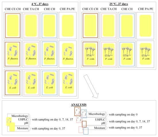
A 4:4:2 factorial experiment design (4 different packaging sets × 4 time points × 2 temperatures) was implemented during this study (Scheme 1). Accordingly, three sets of chitosan-based films, namely chestnut extract:chitosan (CE:CH), tannic acid:chitosan (TA:CH), and blank chitosan (CH) with extra layer of polyamide:polyethylene (PA:PE) vacuum bags (Status d.o.o Metlika, Slovenia) were prepared. A reference set with PA:PE was prepared next to biopackaging sets.
Scheme 1.
Experimental design. The cheese (■—CHE) was packed in three different sachets (□—chestnut extract (CE), □— tannic acid (TA), □— chitosan (CH)) and additionally into vacuum packaging (□—polyamide:polyethylene (PA:PE)).
The precut cheese was placed onto a Petri dish, inoculated with chosen spoilage microorganisms (P. fluorescens, E. coli, P. commune), and packed into a sachet of one 12 cm × 12 cm sheet of chitosan film. Each sachet was prepared by heat sealing (165 °C, 700 Pa, 7 s) on HST-H6 heat seal tester PARAM® (Labthink, Jinan, China) along the long and then short edge. After the insertion of the spoiled food, the sachet was heat-sealed once more from a short open edge. The same process was completed with the unspoiled food and all the procedures were conducted under the sterile conditions of a constant airflow box. All the cheese:biofilms sample sets were additionally packed into an extra layer of PA:PE vacuum bags and vacuum sealed. This was achieved using a vacuum sealer (Status d.o.o, Metlika, Slovenia) with a purpose of preventing environmental effects and to study the ultimate mutual impact of the matrices inside the sets. The named sample sets with spoilage bacteria were stored in 4 °C conditions while samples with spoilage fungi in environmental conditions of 4 °C and 25 °C for 37 days (day 0, 7, 14 and 37). The full name of the samples and their abbreviations are presented in Table 1.

Table 1.
Abbreviations used in the manuscript.
2.4.1. Bacterial Inoculation
E. coli K12 and P. fluorescens NRRL B-253 were grown overnight at 30 °C in 2x YT medium. The medium was discarded, and the culture was resuspended in sterile saline solution. Cheese bricks were aseptically cut to a rectangular dimension of 50 mm × 25 mm × 8 mm (~22 g). Then, 100 µL of bacterial inoculum (E. coli 6.5 log10 CFU/g and P. fluorescens 8.3 log10 CFU/g) was spread onto the surface of the cheese slice and covered with biofilm or in case of control, no biofilm was used. The sachet was put into PA:PE bag and vacuum-sealed (Status d.o.o, Metlika, Slovenia) and stored at 4 °C.
2.4.2. Fungal Inoculation
P. commune NRRL 894 was grown on malt extract agar plates for 10 days to obtain spores. Spores from the plate were collected in sterile saline solution and homogenized with vortexing. Then, 100 µL (4.3 log10 CFU/g) of fungal inoculum was spread onto the surface of the cheese slice and covered with chitosan film, or in case of control, only a vacuum bag was used. The packet was put into PA:PE bag and vacuum-sealed (Status d.o.o, Metlika, Slovenia) and stored at 4 °C or 25 °C.
2.5. Microbiological Analysis
On days 0, 7, 14, and 37, each cheese sample was opened aseptically and cut in half. One half of the sample (approximately 10 g) was mixed with 90 mL of sterile saline solution and homogenized with a stomacher (Lawson Scientific, Ningbo, China). The suspensions were appropriately diluted in sterile saline solution and plated on selective medium. The 2x YT medium with incubation at 37 °C for 24 h was used for E. coli count, Pseudomonas selective agar and incubation at 30 °C for 48 h was used for P. fluorescens count, and malt extract agar with incubation at 25 °C for 5 days was used for P. commune enumeration.
2.6. Chemical and Physical Analysis
2.6.1. UHPLC
Analysis of the liquid samples was performed by using ultra-high-performance liquid chromatography—UHPLC (Thermo-Fisher Scientific UltiMate™ 3000, Waltham, MA USA)—equipped with a 5.0 μm; 4.6 × 150 mm Hypersil GOLDTM Amino column (ThermoFisher Scientific, Waltham, MA USA), heated to 30 °C, and equipped with a DAD detector. Cyclopiazonic acid was identified by retention time and UV-Vis spectra comparison to reference standards using UV–VIS spectra between 282–283 nm. The compound was quantified by external calibration standards. The mobile phase with a flow rate of 0.6 mL/min consisted of a 20:80 aqueous phase (50 mM ammonium acetate buffer solution with pH 5) and organic phase (acetonitrile), respectively. Then, 20 μL of the sample was injected, and a stationary method of a single mobile phase was applied. All the peaks shown in the chromatogram (Supplementary Materials) were identified and quantified. The retention time of cyclopiazonic acid was 3.68 min.
For the determination of the CPA concentrations in the samples, a calibration curve with seven dilution levels was prepared (concentrations range in between 50 ng/mL–0.025 mg/mL). The linear calibration curve was created by plotting the ratio of the peak area of CPA versus the CPA concentrations in the standards. To reduce the measurement errors, the standard series was measured at least in duplicate.
The chitosan-based film sample preparation for UHPLC took place accordingly: the sample (2 × 2 cm) was placed next to a wall of a 1 mL plastic vial, covered with 1.5 mL of methanol, and the received solution was shaken on a thermoshaker TS-100C (Biosan, Riga, Latvia) for 1 h. Afterward, the film was removed from the methanol, and the excess solvent was evaporated under a stream of N2. Received solid matter was restored with 1.5 mL of phosphate buffer (5 mM, pH 2.8). After 10 min of the diffusion process, the solvent sample was subjected to UHPLC. Spiking of film samples (400 µL) with 100 µL of CPA standard solution (0.1 mg/mL) was carried out to ensure the concentration to be within the concentration range used in the calibration curve. The Gouda cheese sample preparation took place according to Zambonin et al. with a slight modification [32] as follows: the cheese sample (0.5 g) was previously cut into small pieces with a knife and weighted into a vial, 1.5 mL of methanol was added and sonicated for 10 min. Reactive methanol was separated from the cheese sample by filtering through the PTFE-20/13 UHPLC filter (Sartorius, Goettingen, Germany) and then evaporated under a stream of N2 at ambient conditions (25 °C). Received solid matter was restored with 1.5 mL of phosphate buffer (5 mM, pH 2.8) and then subjected to UHPLC. Recovery calculations were done by spiking cheese samples (400 µL) with 100 µL of CPA standard solution (0.1 mg/mL).
2.6.2. Moisture Content
The moisture analyzer HE 53 (Mettler Toledo, Wien, Austria) at room temperature was used to measure the moisture content (MC) of cheese and films. Cheese samples were analyzed by receiving a cheese piece from cold storage, cutting it into smaller particles, and immediately placing it onto a measuring plate for analysis. The film samples were handled similarly, using scissors for cutting.
2.6.3. pH Value
The pH value of the cheese and film samples were measured at room temperature using a benchtop 781 pH/ion meter (Metrohm AG, Ionenstrasse, Switzerland). The cheese samples were aseptically homogenized in sterile saline solution with a stomacher (Lawson Scientific, Ningbo, China) and analyzed directly. The film pH values were received by measuring the pH of water solvent. For the exact results, the film samples (2 × 2 cm) were immersed into water for two hours and then removed from the water to finalize film activity in the solvent.
2.6.4. Mechanical Properties
Mechanical characterization of chitosan-based films was performed by following the guidelines from the American Society for Testing and Materials (ASTM) D 882 standard method [33]. Rectangular film samples (8 cm × 2 cm) were tested on the Multitest 2.5-i universal testing machine (Mecmesin, Slinfold, UK) equipped with a 100 N load cell, at a crosshead speed of 5mm min−1. Tensile strength (TS) was calculated by dividing the load with the average original cross-sectional area in the gage length segment (6 cm) of the sample, while elongation at break (EB) was calculated as the ratio between increased length after breakage and the initial gage length.
2.6.5. Active Properties
The total phenolic content (TPC) of active sachets (~5 mg) was determined by Folin-Ciocalteu’s (FC) phenol reagent according to the protocol outlined in our previous study [34]. Briefly, the small rectangular samples were added into water, followed by the successive addition of FC phenol reagent and aqueous solution of Na2CO3 (10% w/v) in the amount of 10 vol% and 20 vol% based on the volume of water, respectively. After the incubation of samples (2 h in dark, 24 °C), the absorbance was measured at 765 nm using the Synergy TM 2 Multi-Detection Microplate Reader (BioTek, Winooski, VT, USA). Gallic acid was used as the standard, and the results were expressed as the mass of gallic acid equivalent (GAE) per mass of the films.
2.7. Statistical Analysis
The data were subjected to one-way analysis of variance (ANOVA) with a confidence level of 95% (p ≤ 0.05). All the results in triplicate are expressed as the mean ± standard deviation.
3. Results and Discussion
3.1. Characterization of the Chitosan-Based Films
3.1.1. Mechanical Properties
The mechanical properties of the packaging material used in this study were measured prior to their use as follows: TS (CH) = 6.7 MPa, TS (TA:CH) = 15.0 MPa, TS (CE:CH) = 15.6 MPa, TS (PA:PE) = 27.5 MPa, and EB (CH) = 75.9%, EB (TA:CH) = 28.5%, EB (CE:CH) = 22.9%, EB (PA:PE) = 40.0% (Figure S1). This correlates with what has been shown previously in a comprehensive modeling study by Bajić et al. [14]. Overall statistical difference according to the analysis of variance showed that the films TS property can be considered as different (p < 0.05) when active components are added into films, wherein TA and CE showed similar results. In regard to EB, all the films were considered different (p < 0.05). Thus, the TS and EB of the biopolymer films were deemed significantly lower (p < 0.05) than conventional PA:PE packaging. The values of the tensile strength (TS) and elongation at break (EB) should be adequate when acceptable integrity of good packaging material is requested. The materials produced show strong integrity, considering that heavy food, e.g., fresh pasta, has been packed into similar material and the material withstood the load during the 2 month shelf life [34].
3.1.2. Activity
Along with mechanical properties, the activity of the films was determined and expressed through the total phenolic content (TPC) value. Films used for packing Gouda cheese in this study received TPC values of CH = 0.5 mgGAE gfilm−1, TA:CH = 3.2 mgGAE gfilm−1, CE:CH = 17.2 mgGAE gfilm−1 (Figure S2). Almost six-fold higher (p < 0.05) activity by CE was seen and thus indicates the pure compound’s (TA) lack of efficiency as an active component. The higher activity of the CE in the film was expected due to its abundant, diverse phenolic content. Its adverse effect has shown to be even higher than of similar extracts such as oak (9.0 mgGAE gfilm−1) and hop (12.7 mgGAE gfilm−1) [31,35].
3.2. Gouda Cheese Spoilage Microbiota Reduction with the CE:CH Film
3.2.1. Bacteria Reduction with Chitosan-Based Films
The cheese was inoculated with approximately 6.5 log10 CFU/g of E. coli and wrapped in different types of chitosan films, as seen in Figure 1a. The E. coli count on inoculated cheese (iCHE PA:PE), stored at 4 °C, remained throughout the 37 days incubation period at the same value (±0.6 log10 CFU/g) as it was at the beginning of the experiment. Between the different tested chitosan films, the E. coli count dropped most dramatically with the inoculated chitosan (iCH) film (p < 0.05). Accordingly, without the additives (CE or TA), the reduction in E. coli count was approximately 2 log10 CFU/g. The primary reduction in bacterial count in these samples happened during the first 14 days, while from 14 to 37 days, the E. coli count stagnated. In the cheese samples wrapped with inoculated chestnut extract:chitosan (iCE:CH) and inoculated tannic acid:chitosan (iTA:CH) films, the reduction in E. coli count was 1 log10 CFU/g, and it happened in the first 7 days of the cheese storage.
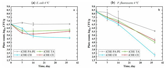
Figure 1.
The effect of biopolymer films (CH, TA:CH, CE:CH) on bacterial count on cheese during storage at 4 °C inoculated with: (a) E. coli and (b) P. fluorescens. Values are means (n = 2 × 3) with standard errors that are significantly different within columns (p < 0.05; Table S1). tannic acid (TA), chitosan (CH), chestnut extract (CE), polyamide (PA), polyethylene (PE), inoculated cheese (iCHE).
According to the evidence of Blaiotta et al. [36], Gram-positive lactic acid bacteria (LAB) cell protection could be retrieved when immobilized in CE fiber in an acidic environment. Considering that E. coli is Gram-negative, and positively charged chitosan-based materials disrupt the cell membrane [37], it could be considered that CE fiber has the protective effect on the intact cells. Although, it is believed that the effect is not merely related to the fiber. The CE is demonstrated to contain reducing sugars, provides adenosine triphosphate (ATP), and improves the bacteria’s survival [36]. Accordingly, TA:CH films have a similar effect to CE:CH films (Figure 1a), disputing the sugars or fiber effect and indicating the effect of hydrolyzable tannins, and indicating there were destructive effects on the spoilage bacteria, E. coli, but only to some extent (until day seven). The lysing result of the neat iCH film stands out in terms of less complexity and its ability to form hydrogen bonds with a higher amount of active cites [37].
The second spoilage bacteria, P. fluorescens, was inoculated on cheese in approximately 8.2 log10 CFU/g (Figure 1b). The cheese was stored at 4 °C. Conformably, the drop of P. fluorescens count was most dramatic with the iCH film, where the count dropped for 6 log10 CFU/g, and samples with the iCE:CH and iTA:CH films showed bacteria count reduction for approximately 4 log10 CFU/g (p < 0.05). Non-inoculated cheese samples were also included in the analysis to exclude the contamination (not shown on graph) and the samples did not show any E. coli or Pseudomonas spp. presence. Prior the experiment initiation, the count of P. fluorescens at inoculation was slightly higher than the E. coli count, but it seems that the latter bacteria species is more susceptible to long term storage at low temperatures or the LAB, naturally present in the cheese, was a habitual defense system. The count of P. fluorescens dropped even in the samples that were not wrapped in biopolymer films. Herein, one of the main differences between the two bacteria is in their survival conditions. Specifically, P. fluorescens is unable to grow under anaerobic conditions, hence the drop. E. coli, on the other hand, withstands anaerobic conditions to some extent.
3.2.2. Fungi Reduction with Chitosan-Based Films
For cheese inoculation with fungi, approximately 4 log10 CFU/g of P. commune spores were used and incubated at two different temperatures. At refrigeration conditions (4 °C), the drop of P. commune count was minimal (less than 1 log10 CFU/g) in all the samples regardless of packaging (Figure 2a), which confirms the good survival of the mold spores over time at low temperatures. This shows that the P. commune spores are susceptible to components of the chitosan films, temperature conditions, and naturally present LAB only until day 14.
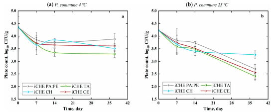
Figure 2.
The effect of biopolymer films (CH, TA:CH, CE:CH) on P. commune count on cheese during storage at: (a) 4 °C and (b) 25 °C. Values are means (n = 2 × 2) with standard errors that are significantly different within columns (p < 0.05; Table S1).
On the other hand, the room temperature conditions (25 °C) affected P. commune survival on the cheese samples over time (Figure 2b). After 37 days, the count of P. commune had dropped for approximately 2 log10 CFU/g on iCHE TA:CH, iCHE CE:CH and iCHE PA:PE samples, while on iCHE CH samples, the P. commune count had dropped for only 1 log10 CFU/g (p < 0.05). Contrary to our study, the study by Duan et al. [28] revealed that the mold count increased (highest at 5.12 log10 CFU/g) during 30 day incubation at 10 °C in control (untreated) samples of the cheese, while the count reduction (highest at 1.90 log10 CFU/g) was seen when the mozzarella cheese was wrapped in chitosan-based films or coatings. Different species of mold and cheese were used in the study. Evidently, the higher temperature contributes to the film’s active components diffusion process, which has also been shown by Ouattara et al. [38]. Additionally, in a study by Esposito et al. [23], CE, with its polyphenols, was shown to have an inhibiting effect on fungus, both in mycelial and spore form. We also expected that the mold count would increase on cheese wrapped in iCHE PA:PE at room temperature conditions, which are ideal for mold growth. On the contrary, the P. commune count dropped during the 37 days long incubation period. Being strictly aerobic mold, most likely, the reason for the reduction is the activity of the LAB. The study of Cheong et al. [39] confirms our theory, where the antifungal effect of LAB was shown on several mold species, P. commune being one of them.
3.2.3. Influence of Chitosan-Based Films on Mycotoxin CPA from Cheese
The cheese was inoculated with P. commune and repacked into chitosan-based antimicrobial films to retain induced mold growth and secondary metabolite cyclopiazonic acid (CPA) formation. The presence of tannins in the packaging material at a higher temperature (25 °C) hindered the CPA production in cheese samples and had an endorsing effect while held at low temperature (4 °C).
Based on the analysis of variance, which was employed on spiked and non-spiked sample results prior to the subtraction to observe the difference in CPA production, the sample groups were identified as different (p < 0.05) while stored in two temperatures (Table S2).
As seen in Figure 3b, mycotoxin CPA concentration in cheese samples at 25 °C significantly decreased from 6400 (day seven) to 533 µg/kg (day 37) when packed in iCE:CH film, and from 3600 to 1300 µg/kg in iTA:CH film. Furthermore, the values presented are the subtraction of inoculated cheese with the P.commune and non-inoculated samples, which gave the direct comparison of the CE and TA effect on growth, eliminating the need for the partitioning between phases.
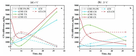
Figure 3.
The effect of biopolymer films (CH, TA:CH, CE:CH) on cyclopiazonic acid (CPA) production in cheese (0.5 g), compared with mycotoxin accumulation in films (2 × 2 cm) at: (a) 4 °C and (b) 25 °C. Dashed lines represent packaging material results. Values are gained by subtraction of spiked non-inoculated sample mean from the spiked inoculated sample mean values, which are significantly different within rows (p < 0.05; Table S2).
The cheese packed with iCH film remained constant (200 µg/kg), and the CHE PA:PE sample (without protective film) showed an increase (800 to 1400 µg/kg) in CPA concentration. Likewise, packaging materials themselves depicted opposite outcomes pointing to interaction with CPA and thus possible protective features in regard to food safety (Figure 3).
The mycotoxin concentrations measured from films were low but kept appearing. In the presence of the CE in the film, CPA concentration increased up to 3200 µg/kg (day 37) while with TA, the used UHPLC method enabled the identification of any CPA due to shadowing complex formation with TA (Figure S3). Accordingly, the film without any additives (CH) tended to continue accumulating the mycotoxin, having a CPA concentration of 2200 µg/kg on day 37.
The CPA concentration levels measured in refrigerated cheese (4 °C) elevated from 5900 (day seven) to 8367 µg/kg (day 37), 1867 to 7600 µg/kg, 700 to 5800 µg/kg, and demoted from 1400 to 267 µg/kg when packed in CE:CH, TA:CH, CH and PA:PE packaging, respectively (Figure 3a).
Higher CPA results measured from cheese could be considered as a consequence of tannin chemistry that allows conjugation with CPA (a tetradic indole acid/N-compound) to form stable linkages between carboxylic groups and amines, similarly to the protein–tannin complex reaction [40]. Additionally, the high temperature is advantageous for tannins chemical reactivity [25] and optimal for lactic acid bacteria (LAB) from cheese to be dominant in the microbial competition [41]. Accumulation in CH film at 25 °C may be attributed to the chemical properties of chitosan, possessing more free hydroxyl radicals [42] to continually interact with (Figure 3b). Undetectable CPA in the iTA:CH films is most likely the result of the higher chemical reactivity of tannic acid, but only until a certain limit (being an unvaried chemical) compared to the iCE:CH film, which contains a source of various, more abundant tannin-rich chestnut extract [43]. Furthermore, chitosan-based materials have been observed to have a strong absorption mechanism for various chemical components [44,45], macromolecules [46,47], and also for mycotoxins [48,49], which is used as a safety precaution to conjugate the unwanted particles. This explanation appears to correlate with the specific film activity of this study in terms of mycotoxin. Rapid CPA forming in cheese samples (at 4 °C) is an irrefutable indication of the film’s low activity after packaging, seemingly related to mechanical properties. Cooling storage has a uniforming effect on biopolymers allocation and molecular configuration in materials [50,51]. This is confirmed by the results of inexistent CPA concentration within films on days zero to seven and 37. In time, the chitosan-based chestnut extract film reaches its moisture equilibrium [34], depending on the food product that it is in contact with.
Furthermore, the films potentially act as an absorber in low-temperature conditions, as on day 14 the iCE:CH and iCH film samples depicted higher CPA presence—most certainly influenced by moisture mobility.
Lately, only a few attempts to quantify CPA in cheese have been reported [40]. Comparatively, in white mold cheese, CPA was reported within a range of 1.83–3610 µg/kg measured by HPLC-MS/MS [50,52], and in inoculated cheddar cheese under modified atmosphere (CO2 + O2) within range of 4–280 µg/kg measured by HPLC [53]. To our knowledge, there are no reports covering CPA quantification in biofilm matrices that have been in contact with food. Although, theoretical multiple mycotoxin absorption by cross-linked chitosan polymer has been reported to be 5.67 g/kg [48]. The results of inoculated gouda cheese (<8367 µg/kg) and chitosan-based films (<3200 µg/kg 2 × 2 cm2) remain in the same range reported by Zhao et al. [48] and therefore report non-toxicity in protective films. This is concluded based on the 50% (LD50) lethal dose in rats by oral ingestion (36,000 µg/kg) [54].
3.3. pH Value
The overall pH deviation between stored Gouda cheese and acidic chitosan-based films was assessed at every time point to determine the flux direction of acidity known to influence microbial growth. In comparison to the CHE PA:PE/iCHE PA:PE samples, a significant conversion of cheese to become more acidic, while the films altered towards a neutral pH, was observed. All the cheese packed in different chitosan films, conditioned at 25 °C, had a pH decrease from 5.6 to 5.2. The pH of cheese from the iCH film started to decrease right away compared to the results of cheese packed in the iCE:CH and iTA:CH films (Figure 4b,c). The incorporation of CE into chitosan-based film showed not to have an additional pH lowering effect on the cheese compared to the TA effect (p = 0.05). A delayed change of pH was determined in the latter packaging’s until day 14 when it started to stabilize. In regard to the iCHE PA:PE cheese samples, a wave-like pH change was seen with a sharp pH increase from 5.6 to 5.8, down to 5.7 by day 14 and up again to 5.9 on day 37.
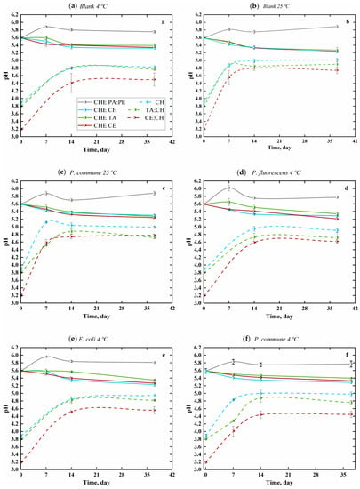
Figure 4.
Effect of biopolymer films (CH, TA:CH, CE:CH) on pH in cheese, compared to simultaneous pH change in films (2 × 2 cm) at 4 °C in (a) Blank (non-inoculated), (d) P. fluorescens (e) E. coli, (f) P. commune and at 25 °C in (b) Blank (non-inoculated), (c) P. commune sample sets. Dashed lines represent packaging material results. Values are means (n = 2 × 1) with standard errors that are significantly different within columns (p < 0.05; Table S3).
The cheese pH changed slightly differently when stored at 4 °C. While packed in the TA:CH/iTA:CH film, the pH values always stayed above the values of other biofilm-packed cheese, and did not go lower than pH 5.3, regardless of the spoilage microbes (p < 0.05). The influence of the spoilage microbes on the cheese pH could be observed. To be more exact, without spoilage bacteria, it took 7 days for the pH of cheese in the TA:CH film to drop (Figure 4a). With Gram-negative bacteria, a pH drop started to happen after 7 and 14 days, respectively, with E. coli and P. fluorescens (Figure 4d,e). P. commune influenced pH decrease uniformly in each biopackaging throughout the time points (Figure 4f).The iCHE PA:PE sample results were diverse with a higher temperature, although after a similar pH jump, they were strongest with P. fluorescens presence up to pH 6.0, and the iCHE PA:PE samples reached a plateau in pH value from day 14.
The pH trend in films (25 °C) took a similar course in every sample set, with a difference in the films containing tannin-additives, being both lower and identical in values (acidic) (p < 0.05) than the CH film. The pH of the CH film rose to 5.0 when it stabilized. In the TA:CH and CE:CH films, the pH reached a plateau at 4.8 on average, which was similar in the presence of P. commune (Figure 4b,c). Simultaneously, lower temperature (4 °C) seemed to prevent pH elevation and kept it at the levels of pH 4.4 (blank, P. fluorescens, E. coli) and 4.6 (P. commune) in the CE:CH films. The preventing effect did not apply to the TA:CH film and the pH was recognized as statistically different from the other tannin-rich films (p < 0.05).
Based on the CHE PA:PE/iCHE PA:PE results, moisture mobility influence could be considered, and the samples could experience the so-called buffering effect. This reflects matrix degradation, which leads to free water mobility with a result of elevated pH values. In our previous work, we have seen that active components diffusion in between the food matrix and active CH:CE film reaches an equilibrium and thus explains the plateau after day 14. The observation correlates to the studies of other authors, confirming that the chitosan application has the pH altering effect in a different direction and only to a small extent. For example, when the cheese is packed with gelatin:chitosan film, the pH changes from 4.6 to 4.4 [55]. The pH is reported to increase from 5.3 to 5.9 while incorporated in cheese mixture and packed in alginate coating plus a modified atmosphere environment [56]. Saloio cheese’s pH ranged from 4.72 to 5.12 (day 37) when coated with chitosan-based natamycin edible film [57].
The changes in the pH of films are mostly attributed to the electrostatic and hydrophobic interactions between chitosan (cationic) and lipids (anionic) [18,58]. Furthermore, it can be presumed that the warmer environment affects the chitosan network for higher diffusion of active components, while lower temperature keeps the integrity of the material network. Additionally, CE contains fiber, which contributes to the material integrity and thus to a lower diffusion.
3.4. Moisture Mobility
The moisture mobility between chitosan-based films and stored Gouda cheese was evaluated for its possible impact on the growth of pathogenic microbes on cheese. Analysis using an automated moisture analyzer was applied to samples initially and at the end of the experiment. Expectantly, the films acted as moisture absorbers, while cheese yielded moisture. The MC in cheese samples, contaminated with two different Gram-negative bacteria, in an anaerobic surrounding at 4 °C, depicts no distinct species that influence associated differences. Temperature’s influence on fungi inoculated cheese, packed in different packaging, stands out with similarity when conditioned at 4 °C and diversity at 25 °C. The CH film without additives performs superior absorbing properties compared to the cheese, while the CE:CH and the TA:CH films, receive results with uniform outcomes, most potentially due to high degradation and intact matrices, respectively (Table 2).

Table 2.
The effect of biopolymer films (CH, TA:CH, CE:CH) on cheese′s moisture content (MC) compared to moisture accumulation in films (both in % per 0.5 g). Values are means (n = 2 × 2) with standard errors, which within a row are significantly different (p < 0.05).
All the samples were packed in additional conventional PA:PE packaging to avoid environmental effects on the MC. However, the CHE PA:PE cheese MC results refer to temperature affected disparities and diversity aspects of biological material after repacking. A slightly lower MC was measured compared to the starting point MC of 33.5 ± 2.0%. Moisture contents on day 37 were the lowest in the CH/iCH packed cheese samples (20.1 ± 2.2%), with results being on average 24.6 ± 1.8% higher in the TA:CH/iTA:CH packed cheese and even higher in the CE:CH/iCE:CH packed cheese (25.6 ± 2.0%) (p < 0.05). Likewise, by the same time, the CH/iCH films gained moisture on average up to 62.4 ± 1.5%, the TA:CH/iTA:CH films 51.0 ± 1.6%, and the CE:CH/iCE:CH films 52.2 ± 2.3% (day 37) showing matrix binding similarities in additive-films. Enhanced moisture absorption is most likely attributed to (i) CH: protein interactions and thus lowering the cheese’s protective lipid layer [18], (ii) CE being a starch (good free water binder) incorporator into chitosan-based films [24], and (iii) TA, via its cross-linking mechanism, being able to strengthen the film matrix to be resilient to moisture absorption [59]. Controlling moisture during cheese processing has a technical connotation for final cheese quality [3], and these results show the benefits of CE in regard to moisture absorption, performing absorption to a lower extent and preserving cheese moisture levels time-wise.
3.5. Effect of the Film on a Food Safety
In addition to the mechanical integrity, it is mandatory to determine the safety of a new material when it is applied on food. All the ingredients used in films were of natural origin and declared as food safe [30]. A comprehensive study by Hu and Gänzle [60] shows chitosan’s bactericidal effect towards several pathogenic microbes on artificially contaminated intermediate moisture foods, and states that the lethality is limited up to 5 log10 CFU/g. Films incorporated with CE in this study are able to inhibit induced spoilage up to 4 log10 CFU/g, indicating the efficiency to ensure food safety. One of the goals of this study was get more insight into the fungi development and its mycotoxin production. With an interesting outcome and a positive outcome in regard to food safety, the mycotoxin is absorbed into the film’s matrix, but does not migrate back to the food surface when the storing temperature is 25 °C (Figure 3). This outcome broadens the storage possibilities surrounding temperature range; however, it should be emphasized that this was not the case at 4 °C and further studies should be conducted for the nethermost temperature. Furthermore, food safety is in balance when several food processes (lipolysis, acidity change) are induced. According to our results of moisture content and pH change that correlates to acidity, the films could be considered as promoting food safety, as the changes were minimal.
4. Conclusions
Based on the results, it can be concluded that chitosan film enriched with chestnut extract reduces extreme bacterial (up to 6 log10 CFU/g) and fungal (up to 4 log10 CFU/g) contamination more actively at 25 °C than at 4 °C. The primary decrease in contaminators happened at 14 days and towards P. fluorescens most efficiently. The results indicate that the addition of commercial chestnut extract has the equivalent effect of chemical grade tannic acid and chitosan film solely lacks protection—first because of the high degradation. The chestnut extract enriched chitosan film seems to operate as an absorbent of mycotoxin cyclopiazonic acid at 25 °C while lowering the toxicity level in cheese. Although, even in airtight conditions, the cheese yielded moisture to the chestnut extract chitosan film throughout all the samples, on average up to 8 ± 2% of its initial moisture content. This alters the authentic Gouda cheese color to a dark brown (Figures S4 and S5). Consequently, packing a high lipid food product—Gouda cheese—into chestnut extract enriched chitosan film could be a good strategy as it influences its pH for only 0.2 units and ensures food safety with active compounds, certainly even longer than 37 days. For further studies, the selected antioxidant and antimicrobial biomarkers extracted from the CE can be used to provide cheese with no visual effects. Additionally, the components chosen for in vivo protection of packed food are entirely safe for use in the food.
Supplementary Materials
The following are available online at https://www.mdpi.com/2304-8158/9/11/1645/s1, Supplementary Materials: Supplementary data; Table S1: The effect of biopolymer film (CH, TA:CH, CE:CH) on bacterial counts (log10 CFU/g) on cheese during storage at 4 °C and 25 °C when inoculated with E. coli, P. fluorescens and P. commune, Table S2: The effect of biopolymer film (CH, TA:CH, CE:CH) on CPA production in cheese (0.5 g), compared with mycotoxin accumulation in films (2 × 2 cm) during storage at 4 °C and 25 °C. Slash-marked spaces depict missing parallels for CH and CH:CE samples and inability to record results for TA:CH and iTA:CH. Calculations in µg/kg are expressed per 1 g of the sample, Table S3: The effect of biopolymer film (CH, TA:CH, CE:CH) on pH in cheese, compared to simultaneous pH change in films (2 × 2 cm) at 4 °C and 25 °C, Figure S1: Mechanical properties of films used for packing Gouda cheese, Figure S2: Total phenolic content of films used for packing Gouda cheese. Gallic acid was used as a standard, and the results were expressed as the mass of gallic acid equivalent (GAE) per mass of the film, Figure S3: UHPLC chromatograms of iTA:CH (film) inoculated with P. commune (■) and 0.025 mg/L CPA standard (■). Spectra depict possible shadowing complex formation between the CPA- and TA- containing samples, Figure S4: Gouda cheese packed in CH, TA:CH and CE:CH biofilms (from left to right), Figure S5: The appearance of the cheese at the beginning (day 0) and the end of the storage (after 37 days, for all the other pictures).
Author Contributions
Conceptualization, K.K., H.Š. and U.N.; methodology, K.K., H.Š. and U.N.; software, K.K; validation, K.K., H.Š., M.B. and U.N.; formal analysis, K.K. and H.Š.; investigation, K.K. and H.Š.; resources, U.N. and B.L.; data curation, K.K. and H.Š.; writing—original draft preparation, K.K. and H.Š.; writing—review and editing, K.K., H.Š., M.B. and U.N.; visualization, K.K. and H.Š.; supervision, U.N.; project administration, U.N.; funding acquisition, U.N. and B.L. All authors have read and agreed to the published version of the manuscript.
Funding
This work was facilitated with the financial support provided by the BioApp project (Interreg V-A Italy- Slovenia 2014–2020) and the Slovenian Research Agency (research core funding no. P2-0152).
Conflicts of Interest
The authors declare no conflict of interest.
References
- Kapoor, R.; Metzger, L.E. Process Cheese: Scientific and Technological Aspects—A Review. Compr. Rev. Food Sci. Food Saf. 2008, 7, 194–214. [Google Scholar] [CrossRef]
- Walstra, P.; Noomen, A.; Geurts, T.J. Dutch-Type Varieties. In Cheese: Chemistry, Physics and Microbiology, 2nd ed.; Fox, P.F., Ed.; Springer: Boston, MA, USA, 1993; p. 56. [Google Scholar]
- Lei, T.; Sun, D. Developments of nondestructive techniques for evaluating quality attributes of cheeses: A review. Trends Food Sci. Tech. 2019, 88, 527–542. [Google Scholar] [CrossRef]
- Sengun, I.Y.; Yaman, D.B.; Gonul, S.A. Mycotoxins and mould contamination in cheese: A review. World Mycotoxin J. 2008, 1, 291–298. [Google Scholar] [CrossRef]
- Yang, Y.; Li, G.; Wu, D.; Liu, J.; Li, X.; Luo, P.; Hu, N.; Wang, H.; Wu, Y. Recent advances on toxicity and determination methods of mycotoxins in foodstuffs. Trends Food Sci. Tech. 2020, 96, 233–252. [Google Scholar] [CrossRef]
- Saravani, M.; Ehsani, A.; Aliakbarlu, J.; Ghasempour, Z. Gouda cheese spoilage prevention: Biodegradable coating induced by Bunium persicum essential oil and lactoperoxidase system. Food Sci. Nutr. 2018, 7, 959–968. [Google Scholar] [CrossRef]
- Dorner, J.W.; Sobolev, V.S.; Yu, W.; Chu, F.S. Immunochemical Method for Cyclopiazonic Acid. In Mycotoxin Protocols. Methods in Molecular Biology; Trucksess, M.W., Pohland, A.E., Eds.; Humana Press Inc.: Totowa, NJ, USA, 2001; p. 72. [Google Scholar]
- Messini, A.; Buccioni, A.; Minieri, S.; Mannelli, F.; Mugnai, L.; Comparini, C.; Venturi, M.; Viti, C.; Pezzati, A.; Rapaccini, S. Effect of chestnut tannin extract (Castanea sativa Miller) on the proliferation of Cladosporium cladosporioides on sheep cheese rind during the ripening. Int. Dairy J. 2017, 66, 6–12. [Google Scholar] [CrossRef]
- Agriopoulou, S.; Stamatelopoulou, E.; Varzakas, T. Advances in Analysis and Detection of Major Mycotoxins in Foods. Foods 2020, 9, 518. [Google Scholar] [CrossRef]
- Kõrge, K.; Laos, K. The influence of different packaging materials and atmospheric conditions on the properties of pork rinds. J. Appl. Packag. Res. 2019, 11, 1–8. [Google Scholar]
- Wang, H.; Qian, J.; Ding, F. Emerging chitosan-based films for food packaging applications. J. Agric. Food Chem. 2018, 66, 395–413. [Google Scholar] [CrossRef]
- Cazón, P.; Velazquez, G.; Ramírez, J.A.; Vázquez, M. Polysaccharide-based films and coatings for food packaging: A review. Food Hydrocoll. 2017, 68, 136–148. [Google Scholar] [CrossRef]
- Mujtaba, M.; Morsi, R.E.; Kerch, G.; Elsabee, M.Z.; Kaya, M.; Labidi, J.; Khawar, K.M. Current advancements in chitosan-based film production for food technology; A review. Int. J. Biol. Macromol. 2019, 121, 889–904. [Google Scholar] [CrossRef] [PubMed]
- Bajić, M.; Oberlintner, A.; Kõrge, K.; Likozar, B.; Novak, U. Formulation of active food packaging by design: Linking composition of the film-forming solution to properties of the chitosan-based film by response surface methodology (RSM) modelling. Int. J. Biol. Macromol. 2020, 160, 971–978. [Google Scholar] [CrossRef] [PubMed]
- Thakur, R.; Saberi, B.; Pristijono, P.; Stathopoulos, C.E.; Golding, J.B.; Scarlett, C.J.; Bowyer, M.; Vuong, Q.V. Use of response surface methodology (RSM) to optimize pea starch–chitosan novel edible film formulation. J. Food Sci. Technol. 2017, 54, 2270–2278. [Google Scholar] [CrossRef] [PubMed]
- Costa, M.J.; Maciel, L.C.; Teixeira, J.A.; Vicente, A.A.; Cerqueira, M.A. Use of edible films and coatings in cheese preservation: Opportunities and challenges. Food Res. Int. 2018, 107, 84–92. [Google Scholar] [CrossRef] [PubMed]
- Vicente, F.A.; Bradic, B.; Novak, U.; Likozar, B. α-Chitin dissolution, N-deacetylation and valorization in deep eutectic solvents. Biopolymers 2020, 1–9. [Google Scholar] [CrossRef] [PubMed]
- Wydro, P.; Krajewska, B.; Hac-Wydro, K. Chitosan as a Lipid Binder: A Langmuir Monolayer Study of Chitosan-Lipid Interactions. Biomacromolecules 2007, 8, 2611–2617. [Google Scholar] [CrossRef] [PubMed]
- Hosseinnejad, M.; Jafari, S.M. Evaluation of different factors affecting antimicrobial properties of chitosan. Int. J. Biol.Macromol. 2016, 85, 467–475. [Google Scholar] [CrossRef]
- Rajoka, M.S.R.; Zhao, L.; Mehwish, H.M.; Wu, Y.; Mahmood, S. Chitosan and its derivatives: Synthesis, biotechnological applications, and future challenges. Appl. Microbiol. Biotechnol. 2019, 103, 1557–1571. [Google Scholar] [CrossRef]
- Benbettaïeb, N.; O′Connell, C.; Viaux, A.; Bou-Maroun, E.; Seuvre, A.; Brachais, C.; Debeaufort, F. Sorption kinetic of aroma compounds by edible bio-based films from marine-by product macromolecules: Effect of relative humidity conditions. Food Chem. 2019, 298, 125064. [Google Scholar] [CrossRef]
- Novak, U.; Bajić, M.; Kõrge, K.; Oberlintner, A.; Murn, J.; Lokar, K.; KTriler, V.; Likozar, B. From waste/residual marine biomass to active biopolymer-based packaging film materials for food industry applications—A review. Phys. Sci. Rev. 2019, 5, 20190099. [Google Scholar] [CrossRef]
- Esposito, T.; Celano, R.; Pane, C.; Piccinelli, A.L.; Sansone, F.; Picerno, P.; Zaccardelli, M.; Aquino, R.P.; Mencherini, T. Chestnut (Castanea sativa Miller.) Burs Extracts and Functional Compounds: UHPLC-UV-HRMS Profiling, Antioxidant Activity, and Inhibitory Effects on Phytopathogenic Fungi. Molecules 2019, 24, 302. [Google Scholar] [CrossRef] [PubMed]
- Torres, M.D.; Moreira, R. Production of hydrogels with different mechanical properties by starch roasting: A valorization of industrial chestnut by-products. Ind. Crops Prod. 2019, 128, 377–384. [Google Scholar] [CrossRef]
- Adamczyk, B.; Simon, J.; Kitunen, V.; Adamczyk, S.; Smolander, A. Tannins and Their Complex Interaction with Different Organic Nitrogen Compounds and Enzymes: Old Paradigms versus Recent Advances. ChemistryOpen 2017, 6, 610–614. [Google Scholar] [CrossRef] [PubMed]
- Rodrigues, P.; Ferreira, T.; Nascimento-Gonçalves, E.; Seixas, F.; Gil da Costa, R.M.; Martins, T.; Neuparth, M.J.; Pires, M.J.; Lanzarin, G.; Félix, L.; et al. Dietary Supplementation with Chestnut (Castanea sativa) Reduces Abdominal Adiposity in FVB/n Mice: A Preliminary Study. Biomedicines 2020, 8, 75. [Google Scholar] [CrossRef]
- Coma, V.; Martial-Gros, A.; Garreau, S.; Copinet, A.; Salin, F.; Deschamps, A. Edible Antimicrobial Films Based on Chitosan Matrix. J. Food Sci. 2002, 67, 1–8. [Google Scholar] [CrossRef]
- Duan, J.; Park, S.I.; Daeschel, M.A.; Zhao, Y. Antimicrobial chitosan-lysozyme (CL) films and coatings for enhancing microbial safety of mozzarella cheese. J. Food Sci. 2007, 72, 355–362. [Google Scholar] [CrossRef] [PubMed]
- Bargiacchi, E.; Bellotti, P.; Pinelli, P.; Costa, G.; Miele, S.; Romani, A.; Zambelli, P.; Scardigli, A. Use of Chestnut Tannins Extract as Anti-Oxidant, Anti-Microbial Additve and to Reduce Nitrosamines and Mycotoxins. US Patent Application Publication, 2015. Available online: https://patents.google.com/patent/US20150223512A1/en (accessed on 19 October 2020).
- EUR-lex (European Union Law). Official Journal of the European Union. Available online: https://eur-lex.europa.eu/homepage.html (accessed on 19 October 2020).
- Bajić, M.; Ročnik, T.; Oberlintner, A.; Scognamiglio, F.; Novak, U.; Likozar, B. Natural plant extracts as active components in chitosan-based films: A comparative study. Food Packag. Shelf Life 2019, 21, 100365. [Google Scholar] [CrossRef]
- Zambonin, C.G.; Monaci, L.; Aresta, A. Determination of cyclopiazonic acid in cheese samples using solidphase microextraction and high performance liquid chromatography. Food Chem. 2001, 75, 249–254. [Google Scholar] [CrossRef]
- ASTM International (American Society for Testing and Materials). ASTM Volume 08.01 Plastics (I): C1147-D3159. Designation D 882: Standard Test Method for Tensile Properties of Thin Plastic Sheeting. Available online: https://www.astm.org/Standards/D882 (accessed on 19 October 2020).
- Kõrge, K.; Bajić, M.; Likozar, B.; Novak, U. Active chitosan–chestnut extract films used for packaging and storage of fresh pasta. Int. J. Food Sci. 2020, 1–10. [Google Scholar] [CrossRef]
- Bajić, M.; Jalšovec, H.; Travan, A.; Novak, U.; Likozar, B. Chitosan-based films with incorporated supercritical CO2 hop extract: Structural, physicochemical, and antibacterial properties. Carbohydr. Polym. 2019, 219, 261–268. [Google Scholar] [CrossRef]
- Blaiotta, G.; La Gatta, B.; Di Capua, M.; Di Luccia, A.; Coppola, R.; Aponte, M. Effect of chestnut extract and chestnut fiber on viability of potential probiotic Lactobacillus strains under gastrointestinal tract conditions. Food Microbiol. 2013, 36, 161–169. [Google Scholar] [CrossRef] [PubMed]
- Liu, H.; Du, Y.; Wang, X.; Sun, L. Chitosan kills bacteria through cell membrane damage. Int. J. Food Microbiol. 2004, 95, 147–155. [Google Scholar] [CrossRef] [PubMed]
- Ouattara, B.; Simard, R.E.; Piette, G.; Bégin, A.; Holley, R.A. Diffusion of Acetic and Propionic Acids from Chitosan-based Antimicrobial Packaging Films. J. Food Sci. 2000, 65, 768–773. [Google Scholar] [CrossRef]
- Cheong, E.Y.; Sandhu, A.; Jayabalan, J.; Le, T.T.K.; Nhiep, N.T.; Ho, H.T.M.; Zwielehner, J.; Bansal, N.; Turner, M.S. Isolation of lactic acid bacteria with antifungal activity against the common cheese spoilage mould Penicillium commune and their potential as biopreservatives in cheese. Food Control 2014, 46, 91–97. [Google Scholar] [CrossRef]
- Ostry, V.; Toman, J.; Grosse, Y.; Malir, F. Cyclopiazonic acid: 50th anniversary of its discovery. World Mycotoxin J. 2018, 11, 135–148. [Google Scholar] [CrossRef]
- Barr, J.G. Effects of Volatile Bacterial Metabolites on the Growth, Sporulation and Mycotoxin Production of Fungi. J. Sci. Food Agric. 1976, 27, 324330. [Google Scholar] [CrossRef]
- Arslan, B.; Soyer, A. Effects of chitosan as a surface fungus inhibitor on microbiological, physicochemical, oxidative and sensory characteristics of dry fermented sausages. Meat Sci. 2018, 145, 107–113. [Google Scholar] [CrossRef]
- Comandini, P.; Lerma-García, M.J.; Simó-Alfonso, E.F.; Toschi, T.G. Tannin analysis of chestnut bark samples (Castanea sativa Mill.) by HPLC-DAD–MS. Food Chem. 2014, 157, 290–295. [Google Scholar] [CrossRef]
- Vakili, M.; Rafatullah, M.; Salamatinia, B.; Abdullah, A.Z.; Ibrahim, M.H.; Tan, K.B.; Gholami, Z.; Amouzgar, P. Application of chitosan and its derivatives as adsorbents for dye removal from water and wastewater: A review. Carbohydr. Polym. 2014, 113, 115–130. [Google Scholar] [CrossRef]
- Crini, G.; Torri, G.; Lichtfouse, E.; Kyzas, G.Z.; Wilson, L.D. Morin-Crini, N. Cross-Linked Chitosan-Based Hydrogels for Dye Removal. In Sustainable Agriculture Reviews; Crini, G., Lichtfouse, E., Eds.; Springer: Cham, Switzerland, 2019; Volume 36, pp. 381–425. [Google Scholar]
- Ylitalo, R.; Lehtinenc, S.; Wuolijoki, E.; Ylitalo, P.; Lehtimäkid, T. Cholesterol-lowering Properties and Safety of Chitosan. Arzneimittel-Forsch. 2002, 52, 1–7. [Google Scholar] [CrossRef]
- Chiu, C.-Y.; Yen, T.-E.; Liu, S.-H.; Chiang, M.-T. Comparative Effects and Mechanisms of Chitosan and Its Derivatives on Hypercholesterolemia in High-Fat Diet-Fed Rats. Int. J. Mol. Sci. 2020, 21, 92. [Google Scholar] [CrossRef] [PubMed]
- Zhao, Z.; Liu, N.; Yang, L.; Wang, J.; Song, S.; Nie, D.; Yang, X.; Hou, J.; Wu, A. Cross-linked chitosan polymers as generic adsorbents for simultaneous adsorption of multiple mycotoxins. Food Control 2015, 57, 362–369. [Google Scholar] [CrossRef]
- Thipe, V.C.; Bloebaum, P.; Khoobchandani, M.; Karikachery, A.R.; Katti, K.K.; Katti, K.V. Green nanotechnology: Nanoformulations against toxigenic fungi to limit mycotoxin production. In Nanomycotoxicology; Rai, M., Abd-Elsalam, K.A., Eds.; Elsevier Inc.: Columbia, MO, USA, 2020; pp. 155–188. [Google Scholar]
- Qiao, C.; Ma, X.; Zhang, J.; Yao, J. Effect of hydration on water state, glass transition dynamics and crystalline structure in chitosan films. Carbohydr. Polym. 2019, 206, 602–608. [Google Scholar] [CrossRef] [PubMed]
- Hamdi, M.; Nasri, R.; Hajji, S.; Nigen, M.; Li, S.; Nasri, M. Acetylation degree, a key parameter modulating chitosan rheological, thermal and film-forming properties. Food Hydrocoll. 2019, 87, 48–60. [Google Scholar] [CrossRef]
- Ansari, P.; Häubl, G. Determination of cyclopiazonic acid in white mould cheese by liquid chromatography–tandem mass spectrometry (HPLC–MS/MS) using a novel internal standard. Food Chem. 2016, 211, 978–982. [Google Scholar] [CrossRef] [PubMed]
- Taniwaki, M.H.; Hocking, A.D.; Pitt, J.I.; Fleet, G.H. Growth of fungi and mycotoxin production on cheese under modified atmospheres. Int. J. Food Microbiol. 2001, 68, 125–133. [Google Scholar] [CrossRef]
- Purchase, I.F.H. The Acute Toxicity of the Mycotoxin Cyclopiazonic Acid to Rats. Toxicol. Appl. Pharmacol. 1971, 18, 114–123. [Google Scholar] [CrossRef]
- Amjadi, S.; Emaminia, S.; Nazari, M.; Davudian, S.H.; Roufegarinejad, L.; Hamishehkar, H. Application of Reinforced ZnO Nanoparticle-Incorporated Gelatin Bionanocomposite Film with Chitosan Nanofiber for Packaging of Chicken Fillet and Cheese as Food Models. Food Bioprocess Tech. 2019, 12, 1205–1219. [Google Scholar] [CrossRef]
- Del Nobile, M.A.; Gammariello, D.; Conte, A.; Attanasio, M. A combination of chitosan, coating and modified atmosphere packaging for prolonging Fior di latte cheese shelf life. Carbohydr. Polym. 2009, 78, 151–156. [Google Scholar] [CrossRef]
- Fajardo, P.; Martins, J.T.; Fuciños, C.; Pastrana, L.; Teixeira, J.A.; Vicente, A.A. Evaluation of a chitosan-based edible film as carrier of natamycin to improve the storability of Saloio cheese. J. Food Eng. 2010, 101, 349–356. [Google Scholar] [CrossRef]
- Ham-Pichavant, F.; Sebe, G.; Pardon, P.; Coma, V. Fat resistance properties of chitosan-based paper packaging for food applications. Carbohydr. Polym. 2005, 61, 259–265. [Google Scholar] [CrossRef]
- Rivero, S.; García, M.A.; Pinotti, A. Crosslinking capacity of tannic acid in plasticized chitosan films. Carbohydr. Polym. 2010, 82, 270–276. [Google Scholar] [CrossRef]
- Hu, Z.; Gänzle, M.G. Challenges and opportunities related to the use of chitosan as a food preservative. J. Appl. Microbiol. 2019, 126, 1318–1331. [Google Scholar] [CrossRef] [PubMed]
Publisher’s Note: MDPI stays neutral with regard to jurisdictional claims in published maps and institutional affiliations. |
© 2020 by the authors. Licensee MDPI, Basel, Switzerland. This article is an open access article distributed under the terms and conditions of the Creative Commons Attribution (CC BY) license (http://creativecommons.org/licenses/by/4.0/).