Article

A Novel Pseudomonas geniculata AGE Family Epimerase/Isomerase and Its Application in d-Mannose Synthesis

1
State Key Laboratory of Food Science and Technology, Jiangnan University, 1800 Lihu Avenue, Wuxi 214122, China
2
School of Biotechnology and Key Laboratory of Industrial Biotechnology Ministry of Education, Jiangnan University, 1800 Lihu Avenue, Wuxi 214122, China
*
Author to whom correspondence should be addressed.
Foods 2020, 9(12), 1809; https://doi.org/10.3390/foods9121809
Submission received: 29 September 2020 / Revised: 3 December 2020 / Accepted: 3 December 2020 / Published: 6 December 2020
(This article belongs to the Section Food Physics and (Bio)Chemistry)

Abstract

:
d-mannose has exhibited excellent physiological properties in the food, pharmaceutical, and feed industries. Therefore, emerging attention has been applied to enzymatic production of d-mannose due to its advantage over chemical synthesis. The gene age of N-acetyl-d-glucosamine 2-epimerase family epimerase/isomerase (AGEase) derived from Pseudomonas geniculata was amplified, and the recombinant P. geniculata AGEase was characterized. The optimal temperature and pH of P. geniculata AGEase were 60 °C and 7.5, respectively. The Km, kcat, and kcat/Km of P. geniculata AGEase for d-mannose were 49.2 ± 8.5 mM, 476.3 ± 4.0 s−1, and 9.7 ± 0.5 s−1·mM−1, respectively. The recombinant P. geniculata AGEase was classified into the YihS enzyme subfamily in the AGE enzyme family by analyzing its substrate specificity and active center of the three-dimensional (3D) structure. Further studies on the kinetics of different substrates showed that the P. geniculata AGEase belongs to the d-mannose isomerase of the YihS enzyme. The P. geniculata AGEase catalyzed the synthesis of d-mannose with d-fructose as a substrate, and the conversion rate was as high as 39.3% with the d-mannose yield of 78.6 g·L−1 under optimal reaction conditions of 200 g·L−1 d-fructose and 2.5 U·mL−1 P. geniculata AGEase. This novel P. geniculata AGEase has potential applications in the industrial production of d-mannose.

1. Introduction

d-mannose is an epimer of d-glucose with the isomerization at the C-2 position [1]. It is in a free state in various fruit peels including orange, peach, and apple. It does not cause significant increases in blood sugar because it cannot be well metabolized in the body, and it has been extensively employed in the food, pharmaceutical, and feed industries [2,3]. Recently, researchers described the application of d-mannose as a good alternative in the prevention and treatment of urinary tract infections (UTIs) [4,5]. d-mannose can be extracted from jujube, apple pulp, and orange peel [6,7,8]. However, this method is complicated and generates low yield (jujube 12.9% [6], apple 2.3–4.0% [7], and orange peel 6.1% [8]) and high production costs. Through chemical methods, d-mannose can be produced from d-glucose in 1% molybdate under acidic conditions. The conversion rate of this method is about 32.0% [9]. However, this method produces a large amount of acidic waste liquid that pollutes the environment, and the final separation and purification is also challenging.
Enzymatic synthesis is an alternative method that uses d-fructose or d-glucose as the substrate to produce d-mannose via enzyme catalysis. This method has advantages such as mild reaction conditions, fewer byproducts, easy separation and purification processes, no environmental pollutants, and low cost, and plays an increasingly important role in the preparation of d-mannose [10]. The N-acetyl-d-glucosamine 2-epimerase family epimerase/isomerase (AGEase) includes N-acetyl-d-glucosamine 2-epimerase, Cellobiose 2-epimerase, and YihS enzyme subfamilies that all catalyze isomerization at the C-2 position [11,12,13]. The catalytic substrate of N-acetyl-d-glucosamine 2-epimerase is the modified sugar N-acetyl-d-mannosamine, which can be isomerized into N-acetyl-d-glucosamine [11]. Cellobiose 2-epimerase can catalyze oligosaccharides containing β-1,4 glycoside bonds, such as cellobiose, lactose, 4-O-beta-d-mannose-d-glucose, etc. [12]. YihS enzyme exhibits aldose-ketose isomerase activity, catalyzing isomerization of unmodified oligosaccharides such as d-mannose, d-fructose, and d-glucose, as well as threose and xylulose [13]. Several enzymes have been reported to be useful for producing d-mannose, including d-mannose isomerase (d-MIase, EC 5.3.1.7), d-lyxose isomerase (d-LIase, EC 5.3.1.15), and cellobiose 2-epimerase (CEase, EC 5.1.3.11), which belongs to AGEase and produces d-mannose with d-fructose or d-glucose as substrate (Table 1) [14,15,16,17,18]. Identification of novel d-mannose producing enzymes is essential for the enhancement of d-mannose production.
In this study, a gene age encoding for AGEase was cloned from Pseudomonas geniculata. The recombinant P. geniculata AGEase was investigated and identified as d-MIase. The property of P. geniculata AGEase for producing d-mannose was also characterized.

2. Materials and Methods

2.1. Strains, Chemicals, and Enzymes

P. geniculata containing the AGEase gene was stored in our laboratory. The cloning host Escherichia coli JM109 and the expression host E. coli BL21 (DE3) were purchased from Bao Bioengineering Co., Ltd. (Dalian, China). The vector pET-24a was purchased from Novagen Co., Ltd. (Shanghai, China). The Plasmid pET-24a-age was constructed by this study. The restriction enzymes (Nde I and Hind III), DNA polymerase Primer STAR® HS, T4 DNA ligase, rTaq enzyme, and alkaline phosphatase CIAP were purchased from Bao Bioengineering Co., Ltd. (Dalian, China). The DNA recovery kits, PCR product purification kits, and Plasmid extraction kits were purchased from Tiangen Biochemical Technology Co., Ltd. (Shanghai, China). All chemicals and reagents with analytical grade were purchased from Sinopharm Chemical Reagent Co., Ltd. (Shanghai, China).

2.2. Construction of Recombinant Plasmid

Two oligonucleotide primers (F: 5′-TATACATATGAGCACCTCGCCCGATTTC-3′, R: 5′-GCCGCAAGCTTCAACGCACCACGTTCAAC-3′), with restriction sites Nde I and Hind III, were utilized to amplify the gene age by PCR using the whole P. geniculata genome as a template. After purification and digestion, the PCR product was digested and ligated into the vector pET-24a. The recombinant plasmid pET-24a-age was transformed into E. coli BL21 (DE3) for further expression.

2.3. Expression of Recombinant P. geniculata AGEase

A single colony of recombinant E. coli BL21 (DE3) containing pET-24a-age plasmid was inoculated in 10 mL LB medium, and seed culture medium was obtained after being cultured for 8 h at 37 °C and 200 r·min−1. The seed culture was inoculated to 50 mL TB fermentation medium at a vaccination rate of 5%. The recombinant E. coli BL21 (DE3)/pET-24a-age was cultured at 37 °C for 1.5–2 h until the OD600 reached 0.6. Inducer Isopropyl β-d-Thiogalactoside (IPTG) at a 0.1 mM final concentration was added, then the culture was induced at 25 °C for 24 h. All of the cultures mentioned above contained 100 μg·mL−1 Kanamycin. The expression of P. geniculata AGEase was checked by 12% SDS-PAGE.

2.4. Purification of Recombinant P. geniculata AGEase

After flask fermentation for 24 h, 400 mL fermentation culture was centrifuged at 7741 g for 20 min. The cell pellet was resuspended in 200 mL sodium phosphate buffer (50 mM, pH 7.5). Crude enzyme solution was obtained by crushing harvested cells with a high-pressure homogenate crusher. Recombinant P. geniculata AGEase was precipitated by addition of 60% (NH4)2SO4, isolated, and purified using the AKTA protein purification system with the Superdex 200 column (column size: 10 mm × 30 cm, flow rate: 0.5 mL·min−1). Enzyme purity was checked by 12% SDS-PAGE. Bradford assay was used for the determination of protein concentration. The disodium hydrogen phosphate-sodium dihydrogen phosphate buffer (20 mM, pH 7.5) was used for purification. All the above solutions needed to be filtered before they could be used. All purification steps were conducted at 4 °C.

2.5. Recombinant P. geniculata AGEase Activity Assay

P. geniculata AGEase activity was determined at 60 °C by the cysteine-carbazole method [19]. A total of 900 μL 0.1 M d-mannose solution in 50 mM sodium phosphate buffer (pH 7.5) was mixed with 0.1 mL enzyme, and the reaction was performed at 60 °C for 1 min, then was terminated by adding 5 mL perchloric acid. Then, 1 mL of the above solution was mixed with 6 mL 75% (v/v) H2SO4 solution and 0.2 mL 1.5% (w/v) cysteine hydrochloride solution. Finally, 0.2 mL 0.12% carbazole ethanol solution was added and the final solution was incubated at 60 °C for 10 min, then cooled in ice water for 5 min. The absorbance at 560 nm was measured, and the concentration of d-fructose was calculated according to the d-fructose standard curve. One unit of P. geniculata AGEase activity is defined as the amount of enzyme required to convert d-mannose to 1 μmol of d-fructose per minute with 0.1 M of d-mannose as substrate.

2.6. Characterization of Purified P. geniculata AGEase

The optimal pH of P. geniculata AGEase was determined by measuring the activity of recombinant P. geniculata AGEase at different pH values using sodium phosphate buffers with different pH values (50 mM, pH 5.0–9.0), and the highest enzyme activity was defined as 100%. The P. geniculata AGEase was placed in sodium phosphate buffer with different pH values (50 mM, pH 5.0–10.0) and stored at 4 °C for 24 h; the enzyme activity of recombinant P. geniculata AGEase was measured to determine the pH stability of P. geniculata AGEase, and the highest activity was defined as 100%. The optimal temperature was determined by assaying the activity of P. geniculata AGEase at 30–70 °C, and the highest enzyme activity was defined as 100%. The P. geniculata AGEase was incubated at 50 °C, and samples were taken periodically to determine the residual activity of the P. geniculata AGEase to determine its stability at 50 °C, and the initial enzyme activity was defined as 100%. d-glucose, d-fructose, d-mannose, d-xylose, d-xylose, N-acetyl-d-glucosamine, and N-acetyl-d-mannosamine were used as substrates to determine the substrate specificity of P. geniculata AGEase.

2.7. Structure Modeling

The theoretical structure of P. geniculata AGEase was obtained by homology modeling utilizing the Swiss Model Protein Modeling Server [20]. These homology models were constructed by employing the crystal structure of Marinomonas mediterranea d-mannose isomerase (PDB ID: 5 × 32) with 57.6% similarity as the template. PyMol was utilized to visualize the generated model structures [21].

2.8. Determination of the Kinetic Parameters

Activity assays were conducted for different substrates at 10, 20, 30, 50, 100, 150, 200, 250, 300, 400, 500, 700, and 1000 mM. Km (mM) and kcat (s−1) of purified P. geniculata AGEase for substrates were calculated from Michaelis–Menten equation after fitting non-linear regression curves. Catalytic efficiency (kcat/Km) of P. geniculata AGEase was calculated.

2.9. Production of d-Mannose from d-Fructose by P. geniculata AGEase

Synthesis of d-mannose was carried out in flasks at 150 r·min−1 in a water bath shaker in 10 mL volume. In order to investigate the optimal temperature of d-mannose production, reactions were carried out at 40–60 °C in sodium phosphate buffer (50 mM, pH 7.5) containing 20% (w/v) d-fructose and 2.5 U·mL−1 P. geniculata AGEase. To investigate the optimal pH of d-mannose production, reactions were carried out at the optimal temperature over the pH range of 6.0–9.0 (disodium hydrogen phosphate-potassium dihydrogen phosphate buffer, 50 mM) in mixtures containing 20% (w/v) (1.1 M) d-fructose and 2.5 U·mL−1 (0.18 μM) P. geniculata AGEase for 2 h. Different amounts of P. geniculata AGEase were added under the same conditions of temperature, pH, substrate concentration, and time to investigate the effect of enzyme dosage on the production of d-mannose. The concentration of d-fructose was varied to investigate the optimal substrate concentration on d-mannose production under the same conditions of 50 °C, pH 7.5, and 2.5 U·mL−1 P. geniculata AGEase.

2.10. High-Performance Liquid Chromatography (HPLC) Analysis of Products

An HPLC (Agilent Technologies, Santa Clara, CA, USA, NH2 column, 250 × 4.6 mm; Cosmosil, Japan; G1362A RID detector) was used to determine the quantities of products. After reaction, the samples were boiled for 10 min then centrifuged at 7741 g for 10 min to obtain the supernatant to measure the yield of d-mannose. All samples were filtered through 0.22 μm cellulose acetate membranes and analyzed by HPLC. The mobile phase was 75% (v/v) acetonitrile with 0.8 mL·min1 flow rate. The yield of d-mannose in the enzyme conversion product was calculated by the external standard method.

3. Results and Discussion

3.1. Cloning and Expression of P. geniculata AGEase

The gene age was cloned and P. geniculata AGEase was expressed in E. coli BL21 (DE3). The crude enzyme solutions of P. geniculata AGEase were obtained through cell disruption, and exhibited a 43 kDa protein band on SDS-PAGE (Figure 1a). This result demonstrated the successful expression of P. geniculata AGEase in E. coli BL21 (DE3).

3.2. Purification of P. geniculata AGEase and Identification of Biochemical Properties

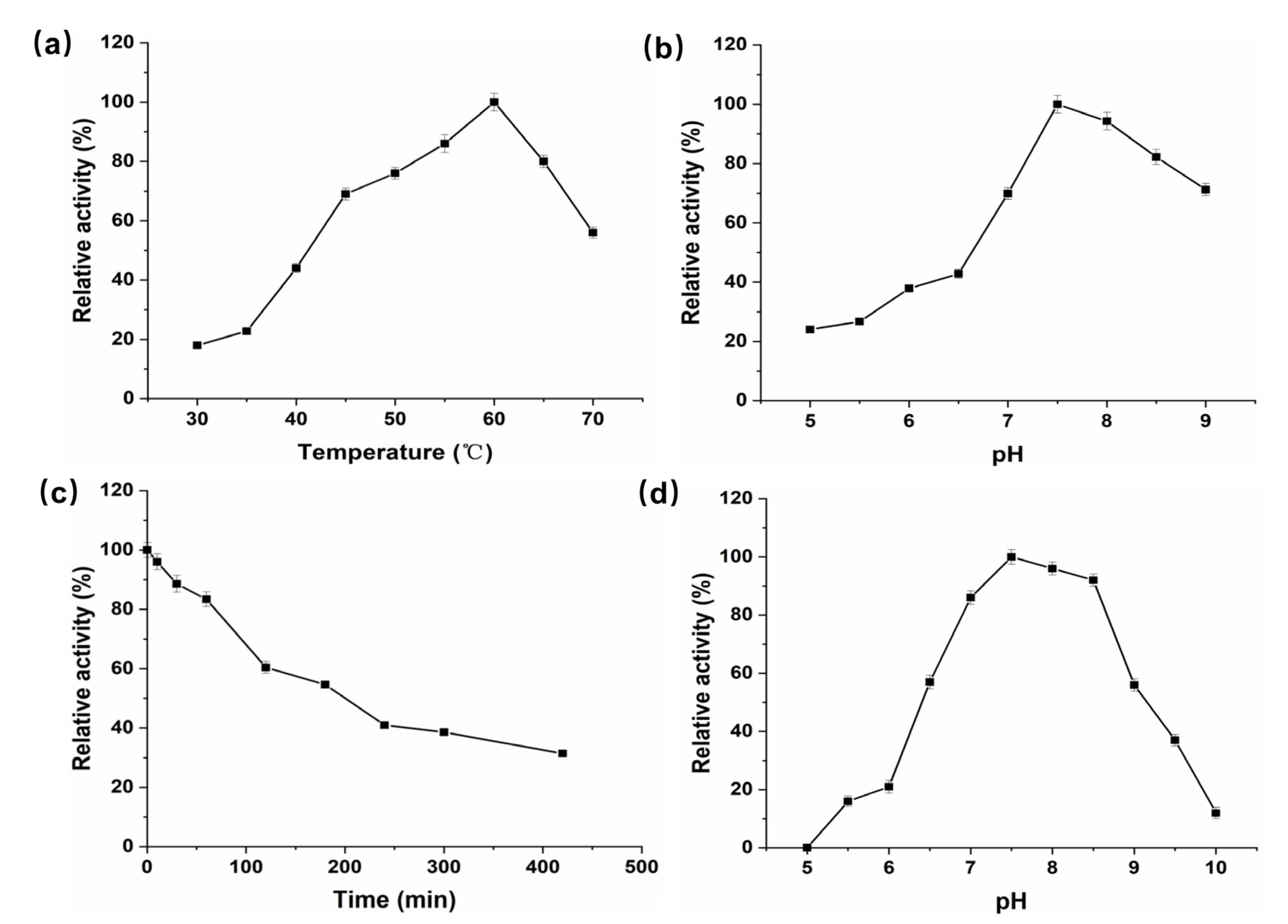
The crude enzyme was purified by gel column Superdex 200 chromatography. Purified product was subjected to SDS-PAGE and showed a single band at 43 kDa (Figure 1b). P. geniculata AGEase specific activity was 295.3 U·mg−1. The optimal temperature for P. geniculata AGEase was 60 °C (Figure 2a), and optimal pH was pH 7.5 (Figure 2b). Purified enzyme solution was incubated at 50 °C and sampled regularly to analyze residual enzyme activity to determine thermostability; the half-life of the enzyme at 50 °C was about 3 h (Figure 2c). Purified enzyme solution was incubated in 50 mM sodium phosphate buffer solution with different pH values at 4 °C for 24 h to determine pH stability. P. geniculata AGEase was stable at pH 7.0–8.5 (Figure 2d).

3.3. P. geniculata AGEase Substrate Specificity

In this study, d-glucose, d-fructose, d-mannose, d-lyxose, d-xylulose, d-xylose, N-acetyl-d-glucosamine, and N-acetyl-d-mannosamine were used as substrates for enzymatic conversion by P. geniculata AGEase at 50 °C and pH 7.5 (Table 2). P. geniculate AGEase could catalyze the isomerization between d-glucose and d-fructose; d-fructose and d-mannose; and d-lyxose and d-xylulose. The substrate specificity of P. geniculata AGEase was consistent with that of YihS reported by Itoh et al. [13].
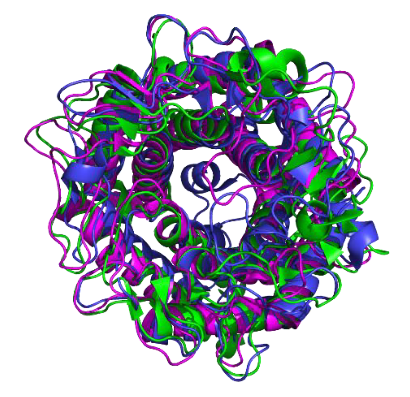
Although the amino acid sequence homology in the AGE family is low (20–40%), tertiary structures of family members have high homology, and all have an obvious (α/α)6-barrel motif (Figure 3) [11]. The tertiary structure study of the AGE enzyme family shows that the active centers of the N-acetyl-d-glucosamine 2-epimerase and YihS enzymes are significantly different. Since N-acetyl-d-glucosamine 2-epimerase catalyzes a modified sugar, the active center consists of two histidines, while the YihS enzyme active center is composed of three histidines. The three-dimensional structure of P. geniculata AGEase was simulated by using SWISS-MODEL. The three-dimensional structure of P. geniculata AGEase showed a catalytic center of three histidines: His179, His251, and His395, consistent with the active center of Salmonella enterica YihS enzyme (Figure S1) [13]. The results suggested that P. geniculata AGEase belonged to the YihS enzyme subfamily of the AGE enzyme family.
YihS enzyme is a kind of aldose-ketose isomerase that catalyzes interconversion between d-glucose and d-fructose; d-fructose and d-mannose; and d-lyxose and d-xylulose. To further determine the categorization of P. geniculata AGEase, the kinetics for different substrates were investigated (Table 3). P. geniculata AGEase had the lowest Km for d-mannose among these substrates, consistent with the reported substrate specificity of mannose isomerase [14]. Therefore, P. geniculata AGEase can be identified as d-MIase in the YihS enzyme of the AGE enzyme family.

3.4. Preparation of D-Mannose Using P. geniculata AGEase with d-Fructose as Substrate

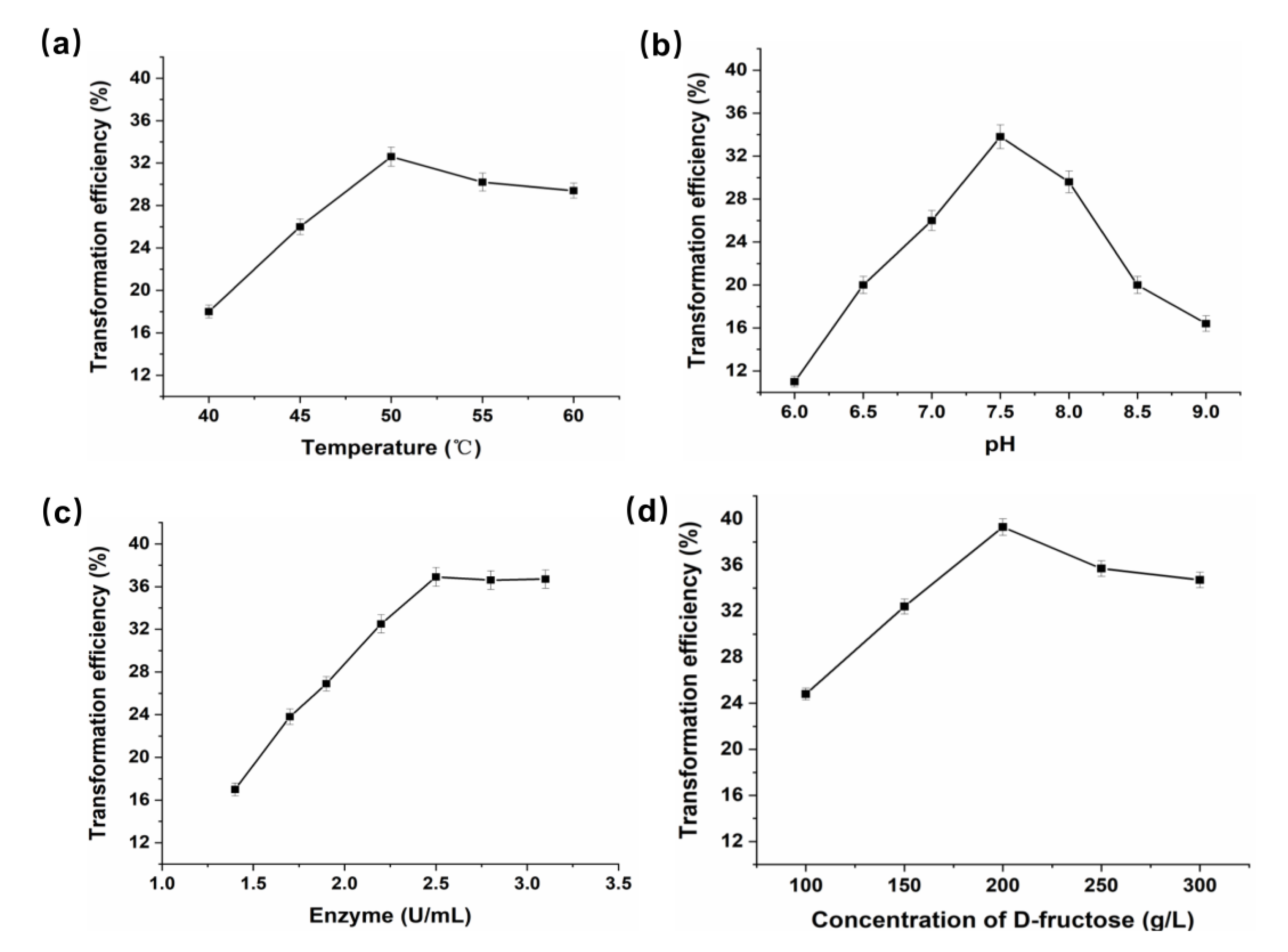
d-fructose was used as the substrate to generate d-mannose via catalysis by P. geniculata AGEase. The effect of temperature on the reaction was investigated at 40–60 °C (Figure 4a). The optimal temperature for generating d-mannose from d-fructose by P. geniculata AGEase was 50 °C. The effect of pH on the reaction was investigated at pH 6.0–9.0 (Figure 4b). The optimal pH of the reaction was pH 7.5. Between pH 6.0 and 7.5, conversion increased with pH increase; conversion decreased rapidly with increasing pH between pH 7.5 and 9.0. The impact of the amount of P. geniculata AGEase was investigated by adding different enzyme amounts at 50 °C and pH 7.5 with 250 g·L−1 of d-fructose as substrate. Increasing the amount of enzyme increased the conversion rate, with a highest rate of 36.9% within 2.5 U·mL−1 enzyme (Figure 4c). Further increasing the amount of enzyme did not increase the conversion rate.
The effect of d-fructose concentration on the reaction was investigated at optimal reaction conditions (50 °C, pH 7.5, and 2.5 U·mL−1 enzyme) with 100–300 g·L−1 d-fructose (Figure 4d). A total of 200 g·L−1 d-fructose yielded a maximum of 78.6 g·L−1 d-mannose after 2 h of reaction with a highest rate of 39.3%. The previously reported highest conversion rate of d-mannose from d-fructose catalyzed by Pseudomonas sp. No 2120 d-MIase was 36.7% with 200 g·L−1 substrate after a 32 h reaction [16]. The reaction time of P. geniculata AGEase was much shorter than that of Pseudomonas sp. No. 2120 d-MIase, which is suitable for d-mannose production.

4. Conclusions

The age gene encoding AGEase from P. geniculata was obtained and recombinant P. geniculata AGEase was successfully expressed in E. coli BL21 (DE3). P. geniculata AGEase was confirmed to be the d-MIase belonging to the YihS enzyme of the AGE enzyme family. P. geniculata AGEase exhibited a high conversion rate of 39.3% for d-mannose production from d-fructose. This identified P. geniculata AGEase has great value for d-mannose production.

Supplementary Materials

The following are available online at https://www.mdpi.com/2304-8158/9/12/1809/s1, Figure S1: Active centers of AGE family epimerase/isomerase, Figure S2: High-performance liquid chromatography (HPLC) of d-fructose and d-mannose. The age gene encoding P. geniculata AGEase.

Author Contributions

Conceptualization, Z.L. and Y.L.; Methodology, Z.L. and Y.L.; Validation, Z.L.; Formal Analysis, Z.L. and Y.L.; Investigation, J.W. and S.C.; Data Curation, Z.L. and Y.L.; Writing-Original Draft Preparation, Z.L. and Y.L.; Writing-Review and Editing, Z.L., J.W., and S.C.; Supervision, S.C.; Project Administration, S.C. All authors have read and agreed to the final version of the manuscript.

Funding

This research received no external funding.

Acknowledgments

This work was financially supported by the National Natural Science Foundation of China (31972032) and (31730067); the Natural Science Foundation of Jiangsu Province (BK20190586); the High-level Innovation and Entrepreneurship Talents Introduction Program of Jiangsu Province (5816010241200090); and the National Science Foundation for Distinguished Young Scholars (31425020).

Conflicts of Interest

The authors declare no conflict interest.

References

  1. Hu, X.; Shi, Y.; Zhang, P.; Miao, M.; Zhang, T.; Jiang, B. d-Mannose: Properties, production, and applications: An overview. Compr. Rev. Food Sci. Food Saf. 2016, 15, 773–785. [Google Scholar] [CrossRef] [Green Version]
  2. Ichikawa, M.; Scott, D.A.; Losfeld, M.E.; Freeze, H.H. The metabolic origins of mannose in glycoproteins. J. Biol. Chem. 2014, 289, 6751–6761. [Google Scholar] [CrossRef] [PubMed] [Green Version]
  3. Wu, H.; Zhang, W.; Mu, W. Recent studies on the biological production of d-mannose. Appl. Microbiol. Biotechnol. 2019, 103, 8753–8761. [Google Scholar] [CrossRef] [PubMed]
  4. Scribano, D.; Sarshar, M.; Prezioso, C.; Lucarelli, M.; Angeloni, A.; Zagaglia, C.; Palamara, A.T.; Ambrosi, C. d-Mannose treatment neither affects uropathogenic Escherichia coli properties nor induces stable FimH modifications. Molecules 2020, 25, 316. [Google Scholar] [CrossRef] [PubMed] [Green Version]
  5. Sarshar, M.; Behzadi, P.; Ambrosi, C.; Zagaglia, C.; Palamara, A.T.; Scribano, D. FimH and anti-adhesive therapeutics: A disarming strategy against uropathogens. Antibiotics 2020, 9, 397. [Google Scholar] [CrossRef] [PubMed]
  6. Wang, B. Chemical characterization and ameliorating effect of polysaccharide from Chinese jujube on intestine oxidative injury by ischemia and reperfusion. Int. J. Biol. Macromol. 2011, 48, 386–391. [Google Scholar] [CrossRef] [PubMed]
  7. Gheyas, F.; Blankenship, S.M.; Young, E.; McFeeters, R. Dietary fibre content of thirteen apple cultivars. J. Sci. Food Agric. 1997, 75, 333–340. [Google Scholar] [CrossRef]
  8. Aravantinos-Zafiris, G.; Oreopoulou, V.; Tzia, C.; Thomopoulos, C. Fibre fraction from orange peel residues after Pectin extraction. LWT 1994, 27, 468–471. [Google Scholar] [CrossRef]
  9. Zhao, G.H.; Wang, G.B.; Li, J.P.; Cao, Y.H. The orthogonal experiment analysis on the producing mannose from dextrose by catalyzig effect of ammonium molybdate. Contemp. Chem. Ind. 2005, 34, 39–41. [Google Scholar]
  10. Bicas, J.L.; Silva, J.C.; Dionísio, A.P.; Pastore, G.M. Biotechnological production of bioflavors and functional sugars. Food Sci. Technol. 2010, 30, 7–18. [Google Scholar] [CrossRef] [Green Version]
  11. Lee, Y.C.; Wu, H.M.; Chang, Y.N.; Wang, W.C.; Hsu, W.H. The central cavity from the (alpha/alpha)6 barrel structure of Anabaena sp. CH1 N-Acetyl-d-glucosamine 2-epimerase contains two key histidine residues for reversible conversion. J. Mol. Biol. 2007, 367, 895–908. [Google Scholar] [CrossRef] [PubMed]
  12. Fujiwara, T.; Saburi, W.; Matsui, H.; Mori, H.; Yao, M. Structural insights into the epimerization of β-1,4-linked oligosaccharides catalyzed by cellobiose 2-epimerase, the sole enzyme epimerizing non-anomeric hydroxyl groups of unmodified sugars. J. Biol. Chem. 2014, 289, 3405–3415. [Google Scholar] [CrossRef] [PubMed] [Green Version]
  13. Itoh, T.; Mikami, B.; Hashimoto, W.; Murata, K. Crystal structure of YihS in complex with d-Mannose: Structural annotation of Escherichia coli and Salmonella enterica yihS-encoded proteins to an aldose–ketose isomerase. J. Mol. Biol. 2008, 377, 1443–1459. [Google Scholar] [CrossRef] [PubMed]
  14. Palleroni, N.J.; Doudoroff, M. Mannose isomerase of Pseudomonas saccharophila. J. Biol. Chem. 1956, 218, 535–548. [Google Scholar] [PubMed]
  15. Liu, Q. The construction of recombinant Escherichia coli of expression d-mannose ketol isomerase and study of enzymic characterization. Master’s thesis, Beijing University of Chemical Technology, Beijing, China, 2010. [Google Scholar]
  16. Zhang, J. Study on d-mannose isomerase produced by Pseudomonas sp. No. 2120. Master’s thesis, Jiangnan University, Jiangsu, China, 2009. [Google Scholar]
  17. Wu, H.; Chen, M.; Guang, C.; Zhang, W.; Mu, W. Characterization of a recombinant d-mannose-producing d-lyxose isomerase from Caldanaerobius polysaccharolyticus. Enzym. Microb. Technol. 2020, 138, 109553. [Google Scholar] [CrossRef] [PubMed]
  18. Park, C.S.; Kim, J.E.; Choi, J.G.; Oh, D.K. Characterization of a recombinant cellobiose 2-epimerase from Caldicellulosiruptor saccharolyticus and its application in the production of mannose from glucose. Appl. Microbiol. Biotechnol. 2011, 92, 1187–1196. [Google Scholar] [CrossRef] [PubMed]
  19. Dische, Z.; Borenfreund, E. A new spectrophotometric method for the detection and determination of keto sugars and trioses. J. Biol. Chem. 1951, 192, 583–587. [Google Scholar] [PubMed]
  20. Waterhouse, A.; Bertoni, M.; Bienert, S.; Studer, G.; Tauriello, G.; Gumienny, R.; Heer, F.T.; de Beer, T.A.P.; Rempfer, C.; Bordoli, L.; et al. SWISS-MODEL: Homology modelling of protein structures and complexes. Nucleic Acids Res. 2018, 46, W296–W303. [Google Scholar] [CrossRef] [PubMed] [Green Version]
  21. The PyMOL Molecular Graphics System, Version 2.0 Schrodinger, LLC. Available online: https://pymol.org/2/ (accessed on 29 September 2020).
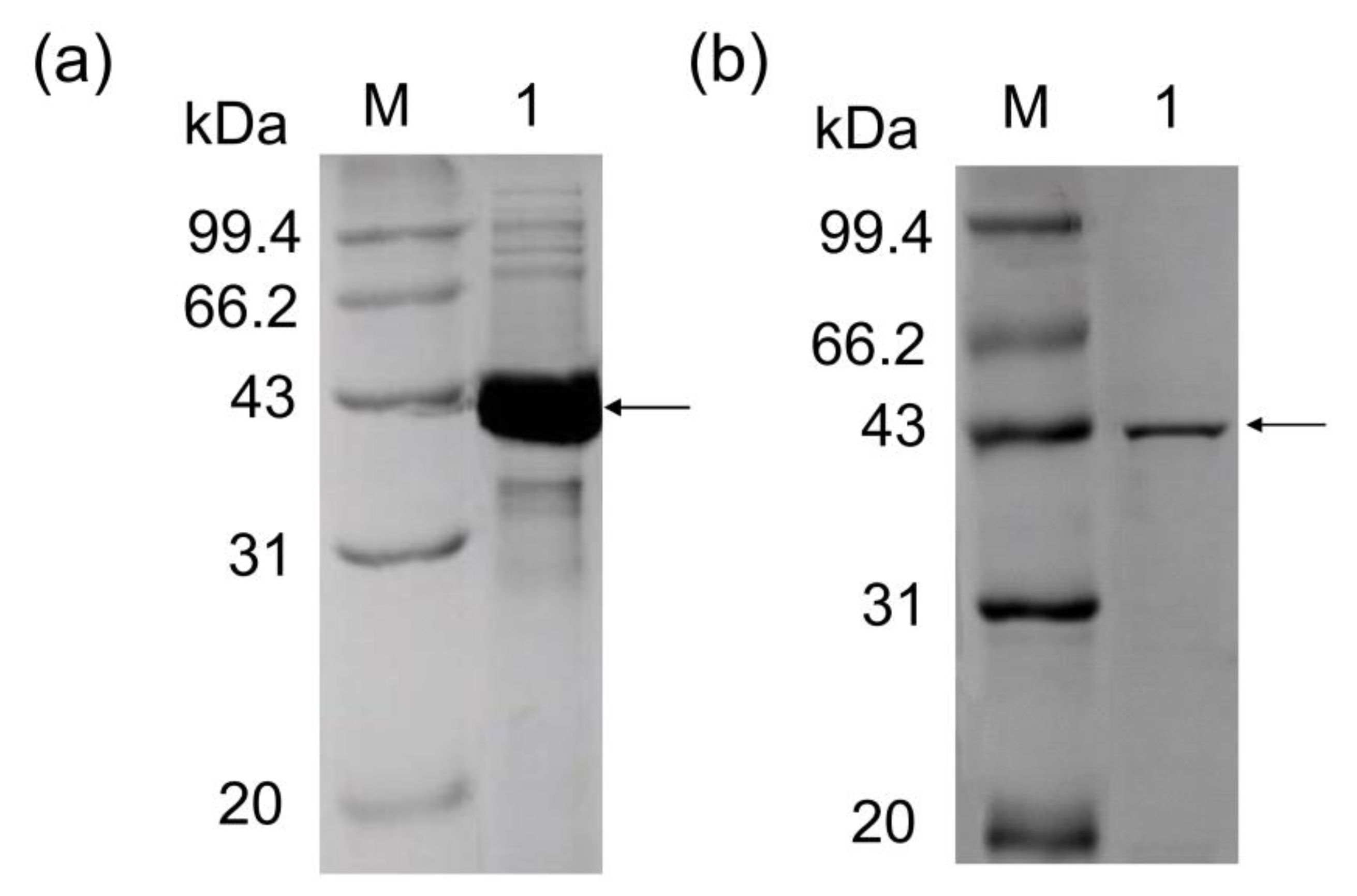
Figure 1. SDS-PAGE analysis of recombinant P. geniculata AGEase. (a) Lanes: M, molecular mass markers (97.4, 66.2, 43, 31, and 20 kDa). 1, lysates of whole-cell expressing recombinant P. geniculata AGEase after IPTG induction for 24 h. (b) Lanes: M, molecular mass markers (97.4, 66.2, 43, 31, and 20 kDa). 1, P. geniculata AGEase purified by gel column Superdex 200.
Foods 09 01809 g001
Figure 2. Enzymatic properties of recombinant P. geniculata AGEase. Optimal temperature for P. geniculata AGEase (a), the reactions were performed in 50 mM sodium phosphate buffer (pH 7.5) at 30–70 °C. Optimal pH for P. geniculata AGEase (b), the reactions were performed in 50 mM sodium phosphate buffer (pH 5.0–9.0) at 60 °C. Thermostability of P. geniculata AGEase at 50 °C (c), the reactions were performed in 50 mM sodium phosphate buffer (pH 7.5). The pH stability of P. geniculata AGEase at pH 5.0–10.0 (d), the reactions were performed in 50 mM sodium phosphate buffer (pH 5.0–10.0) at 60 °C.
Foods 09 01809 g002
Figure 3. Three-dimensional structure of AGE enzyme family. N-acetyl-d-glucosamine 2-epimerase (PDB code: 1FP3) is highlighted in green; YihS enzyme (PDB code: 2AFA) is highlighted in blue; cellobiose 2-epimerase (PDB code: 3WKG) is highlighted in purple. The central region of this structure is the active site of the AGE family enzyme.
Foods 09 01809 g003
Figure 4. Effects of temperature, pH, enzyme concentration, and substrate concentration on D-mannose production. (a) Optimal temperature. The reactions were performed in 50 mM sodium phosphate buffer (pH 7.5) at 40–60 °C with 20% d-fructose and 2.5 U·mL−1 P. geniculata AGEase; (b) Optimal pH. The reactions were performed in 50 mM sodium phosphate buffer (pH 6.0–9.0) at 50 °C with 20% D-fructose and 2.5 U·mL−1 P. geniculata AGEase; (c) Optimal enzyme concentration. The reactions with different enzyme concentrations were performed in 50 mM sodium phosphate buffer (pH 7.5) at 50 °C with 20% D-fructose; (d) Optimal substrate concentration. The reactions with different substrate concentrations were performed in 50 mM sodium phosphate buffer (pH 7.5) at 50 °C with 2.5 U·mL−1 P. geniculata AGEase.
Foods 09 01809 g004
Table 1. Conversion efficiency of enzymes from different strains catalyzing substrate to d-mannose.
SourceSubstrateConditionsTransformation Efficiency (%)
Pseudomonas saccharophilad-MIase1.1 g/L d-fructose30 °C, pH 7.4 29.0 [14]
Agrobacterium radiobactord-MIase250 g/L d-fructose45 °C, pH 7.5, 2 h29.2 [15]
Pseudomonas sp. d-MIase200 g/L d-fructose55 °C, pH 7.5, 32 h36.7 [16]
Caldanaerobius polysaccharolyticusd-LIase100 g/L d-fructose 65 °C, pH 6.5, 4 h25.6 [17]
Caldicellulosiruptor saccharolyticus CEase500 g/L d-glucose75 °C, pH 7.5, 3 h15.0 [18]
Table 2. Substrate specificity of recombinant P. geniculata AGEase.
SubstratesProductsTransformation Efficiency (%)
d-glucosed-fructose5.4
d-fructosed-mannose33.8
d-mannosed-fructose67.9
d-xylulosed-lyxose23.1
d-lyxosed-xylulose12.4
cellobioseN.D.N.D.
d-xyloseN.D.N.D.
N-acetyl-d-mannosamineN.D.N.D.
N-acetyl-d-glucosamineN.D.N.D.
N.D. Not detected by assay conditions used.
Table 3. Kinetic parameters for recombinant P. geniculata AGEase.
Substrateskcat (s−1)Km (mM)kcat/Km (s−1·mM−1)
d-mannose476.3 ± 4.049.2 ± 8.59.7 ± 0.5
d-fructose176.5 ± 1.2117.0 ± 22.51.5 ± 0.1
d-glucose67.2 ± 0.9382.0 ± 88.10.2 ± 0.01
Publisher’s Note: MDPI stays neutral with regard to jurisdictional claims in published maps and institutional affiliations.