Article

Toxicokinetics, Percutaneous Absorption and Tissue Distribution of Benzophenone-3, an UV Filtering Agent, in Rats

1
College of Pharmacy, Daegu Catholic University, Gyeongsan 38430, Gyeongbuk, Korea
2
School of Pharmacy, Sungkyunkwan University, Suwon 16419, Gyeonggi, Korea
3
College of Pharmacy, Wonkwang University, Iksan 54538, Jeonbuk, Korea
4
College of Pharmacy, Dankook University, Cheonan 31116, Chungnam, Korea
5
R&D Center, GL Pharm Tech Corp., Seongnam-si 13202, Gyeonggi, Korea
6
Center for Human Risk Assessment, Dankook University, Cheonan 31116, Chungnam, Korea
*
Authors to whom correspondence should be addressed.
These authors contributed equally to this work.
Toxics 2022, 10(11), 672; https://doi.org/10.3390/toxics10110672
Submission received: 14 October 2022 / Revised: 2 November 2022 / Accepted: 4 November 2022 / Published: 7 November 2022
(This article belongs to the Special Issue Toxicokinetics of Chemicals in Consumer Products)

Abstract

:
The aim of this study was to evaluate in vitro skin permeation and deposition, in vivo toxicokinetics, percutaneous absorption and tissue distribution of benzophenone-3 (BP-3) in rats. Four transdermal formulations containing BP-3 were prepared and evaluated for in vitro skin permeation and deposition of BP-3 using Franz diffusion cells. A gel formulation was used in subsequent in vivo percutaneous absorption due to its high in vitro skin permeation and deposition. Compared to intravenous (i.v.) injection, the prolonged terminal t1/2 (3.1 ± 1.6 h for i.v. injection and 18.3 ± 5.8 h for topical application) was observed indicating occurrence of flip-flop kinetics after topical application. The bioavailability of BP-3 after topical application was 6.9 ± 1.8%. The tissue-to-plasma partition coefficient (kp) for testis, considered a toxic target for BP-3, was less than 1. Overall, findings of this study may be useful for risk assessment of BP-3.

1. Introduction

Benzophenone-3 (2-hydroxy-4-methoxybenzophenone, BP-3), a UV filtering agent in cosmetic products and an organic ultraviolet (UV) filter in sunscreen that absorbs both UVa and UVb radiation at wavelengths from 200–350 nm, is widely used to protect the skin from UV rays [1,2,3,4]. Recently, as the use of sunscreens increases, the possibility of systemic exposure of BP-3 through the skin penetration is also increasing [5,6,7].
In many countries, the maximum allowable content of BP-3 in a product is regulated at 5–6% in the United States, Japan and Korea, and 10% in Europe [8,9,10,11].
To date, various studies about the toxicity of BP-3 including its endocrine disrupting potential have been reported [12,13,14,15,16]. By using MCF-7 and MDA-kb2-cells, BP-3 showed estrogenic and anti-androgenic effects. The pS2, estrogen-regulated protein was significantly up-regulated by BP-3 [17]. It is reported that BP-3 has both in vitro anti-estrogenic and anti-androgenic activities [18,19]. In addition, the mutagenic effect of BP-3 was observed in a salmonella assay [20], and the BP-3 induced sister chromatid exchange and chromosomal aberrations in Chinese hamster ovary cells [21].
Several reports about the in vivo endocrine disrupting activity of BP-3 have been published. Following oral and intra-peritoneal administrations, the increase of uterine weight was observed in rats [17,22,23]. In male rats, an antiestrogenic effect was also observed [24]. Furthermore, dietary contained 5% BP-3 decreased sperm concentration in caudal epididymis in F344/N rat and B6C3F mice [25].
The effect of formulation on in vitro skin permeation of BP-3 has been reported. Using a static diffusion cell, the permeability of BP-3 contained in emulsion and petroleum jelly was evaluated. The O/W emulsion maintained a high BP-3 concentration in the epidermis, dermis, stratum corneum, and receptor fluid, and showed a significant difference in increased permeability compared to the petroleum jelly [26]. Another study reported a significant difference in permeability and flux of BP-3 among light liquid paraffin, coconut oil, 50:50 ethanol:coconut oil, aqueous cream, and oily cream formulation. The highest flux was observed following the administration of light liquid paraffin [27].
Following a topical application of a cosmetic product containing BP-3, the systemic exposure of BP-3 was observed in human volunteers. The lotion containing 6% of BP-3 was applied and urine was collected for 48 h. Up to 48 h, BP-3 corresponding to 1-2% of the dose was detected in urine, which indicated a transdermal absorption of BP-3 [28]. BP-3 was also detected in urine after transdermal administration of sunscreen containing 4% BP-3 at 2 mg/cm2 in 11 volunteers. It is reported that the average total amount excreted was 11 mg, which is about 0.4% of the applied dose [29]. In a follow-up study, BP-3 was detected in urine after repeated transdermal administration of sunscreen containing 4% BP-3 for 5 days on the whole bodies of 25 volunteers. The excreted BP-3 via urine was 1.2–8.7% of the dosed amount, suggesting the potential for accumulation of BP-3 by daily exposure [5]. After repeated topical applications of 2 mg/cm2 sunscreen containing 10% BP-3, plasma and urine concentrations were determined. BP-3 was detected in plasma 1 h after the first administration [30]. Following lotion and aerosol spray containing BP-3 that were repeatedly applied to the skin for 4 days, BP-3 was detected in plasma. BP-3 was detected in the stratum corneum even on days 7 and 14 following the first administration [31,32]. To date, toxicokinetic parameters including absolute bioavailability in humans have not been reported.
In animal studies, BP-3 has been reported to be rapidly absorbed following oral and transdermal administration in rats and piglets [33,34,35,36]. Following oral administration, BP-3 concentration in the liver was highest among tissues [34]. Although various animal experiments have been reported, the absolute bioavailability and tissue distribution at the steady state of BP-3 have been limitedly identified.
Therefore, the present study was carried out to investigate the effect of the formulation on the skin permeability, the toxicokinetics including absolute bioavailability, and the tissue distribution at a steady state of BP-3.

2. Materials and Methods

2.1. Materials

The following materials were used as received, without further purification: petroleum jelly (Unilever, London, UK), medium-chain triglycerides (Sasol Germany GmbH, Hamburg, Germany), marketed body emulsion containing “free” BP-3 (Neutrogena Co., Los Angeles, CA, USA), carbomer 940 (Lubrizol Corp., Wickliffe, OH, USA), poloxamer 188 (BASF, Ludwigshafen, Germany). BP-3, nonivamide, acetic acid, formic acid, ethylenediamine tetraacetic acid (EDTA) sodium, methyl paraben, mineral oil, sodium hydroxide, and beeswax were purchased from Sigma Aldrich Co. (St. Louis, MO, USA). Acetonitrile, water, and methanol (all HPLC grade) were purchased from Avantor (Radnor, PA, USA).

2.2. HPLC-UV and LC-MS/MS Analysis

BP-3 concentrations in samples obtained from in vitro skin permeation studies were determined by a HPLC-UV. A Waters 2690 separation module with a Waters 2487 dual λ absorbance detector (Waters, Milford, MA, USA) was applied. The UV detection wavelength was set at 290 nm. The mobile phase was a mixture of acetonitrile and 0.1% acetic acid (70:30 v/v%), and the flow rate of the mobile phase was 1.2 mL/min. The lower limit of quantification (LLOQ) was 50 ng/mL. The receptor fluid samples were analyzed without extraction steps, and stripped tapes and harvested skin layers were extracted with methanol for 12 h. Separately, BP-3 concentrations in plasma, skin and tissue samples obtained from the in vivo toxicokinetic studies were determined by a validated liquid chromatography mass spectroscopy (LC-MS/MS) method. An API 2000 LC-MS/MS system (Applied Biosystems/MDS Sciex, Toronto, ON, Canada) coupled with a Waters 2690 HPLC (Waters Corporation, Milford, MA, USA) was applied. Rat biological samples were prepared by protein precipitation with acetonitrile. The 150 μL of acetonitrile was added to 50 μL of plasma sample. The mixture was vortex-mixed for 1 min and then centrifugated for 10 min at 4000× g. The 100 μL of supernatant was diluted with an identical volume of distilled water and a portion (10 μL) was injected into LC-MS/MS. For preparation of tissue homogenates samples, 350 μL of acetonitrile was added to 50 μL of biological samples. The subsequent procedure was the same as the plasma preparation. Washed swabs and stripped tapes were extracted with methanol with a 12-h agitation in a shaking bath (Lab Companion, Daejeon, Korea). Separations were achieved on a Kinetex C18 column (50 × 2.1 mm ID, 2.6 μm, Phenomenex, Torrance, CA, USA). The mobile phase was a mixture of methanol and 0.05% formic acid (85:15 v/v%), and the flow rate of the mobile phase was 0.3 mL/min. The multiple reaction monitoring was based on the transition of m/z of 229.0 → 150.9 for BP-3 and 294.3 → 137.2 for nonivamide (internal standard). The LLOQ of plasma and tissue homogenates were 1 and 5 ng/mL, respectively. The assay was validated using the matrix-matched quality control (QC) samples (including QC samples at LLOQ). The intra- and inter-day accuracy and precision ranged from 97.5% to 109.1% and from 1.3% to 8.5%, respectively.

2.3. In Vitro Skin Permeability

Rat skins were mounted on Franz diffusion cells (area: 1.77 cm2). The receptor cell (volume: 10 mL) was filled with phosphate buffered saline (PBS, pH 7.4) containing 5% Tween 80 to maintain sink conditions. After equilibration, approximately 200 mg of transdermal formulations (petroleum jelly, oil, lotion, and gel) with incorporated BP-3 were applied to the donor cell. The protocol for incorporation of BP-3 in transdermal formulations have been described previously [37]. The composition of BP-3 formulations was listed in Table 1. The receptor medium was stirred at 600 rpm and maintained at 37 ± 0.5 °C throughout the experiment for 48 h. At 0, 2, 4, 6, 8, 12, 24, 36, and 48 h following applications, receptor media were collected and replaced with an equal volume of fresh media. Immediately after the last receptor medium collection, the stratum corneum (30 times of tape strips), viable epidermis, and dermis were separated to determine amounts of BP-3 in the skin. The collected samples were kept at −20 °C until analysis.

2.4. In Vivo Toxicokinetics

To characterize the dermal absorption kinetics, BP-3 was intravenously and topically administered in male Sprague-Dawley rats (8–10 weeks of age) (Samtako Co., Osan, Gyeonggi-do, Korea). Prior to the study, the rats were acclimatized for at least 1 week at a temperature of 23 ± 2 °C with a 12-h light/dark cycle and a relative humidity of 50%. BP-3 was intravenously injected at a dose of 1 mg/kg. The dosing solutions were prepared by dissolving BP-3 in a mixture of ethanol:PEG 400:Tween 80:saline (2:2.5:0.5:5, v/v) at concentrations of 0.5 mg/mL. Blood samples were collected from the jugular vein at 0, 2, 5, 15, 30 min, 1, 2, 4, 8, and 12 h following injection. Plasma samples were harvested by centrifugation at 4000× g for 10 min and stored at −20 °C until analysis.
In vivo transdermal absorption kinetics of BP-3 were characterized after topical application of gel formulation. Prior to administration, the back skin of the rat covering an area of 3 × 3 cm was shaved with an electric clipper (Golden A5, Oster, Rye, NY, USA). Next, the 200 mg of BP-3 containing gel was evenly applied to the shaved area. At 0, 6, 12, 24, 36, 48, 72, and 96 h following application, blood samples were collected from the jugular vein and centrifuged at 4000× g for 10 min. Immediately after the last blood sample collection, the animals were sacrificed and the stratum corneum (30 times of tape strips), viable epidermis, and dermis were separated to determine amounts of BP-3 in the skin. The viable epidermis and dermis were homogenized in saline (Tissue tearer, Biospec Co., Bartlesville, OK, USA). The collected samples were stored at −20 °C until analysis.
The toxicokinetic parameters were calculated by standard noncompartmental analysis in WinNonlin (Pharsight, Cary, NC, USA). These parameters included the initial plasma concentration (C0), area under the plasma concentration–time curve from time zero to last sampling point (AUClast), and terminal half-life (t1/2). The peak plasma concentration (Cmax) and the time to reach Cmax (Tmax) were directly obtained from the observations. The absolute bioavailability (F) of BP-3 after transdermal application was calculated as F (%) = 100 (AUCtransdermal × Dosei.v.)/(AUCi.v. × Dosetransdermal).
BP-3 tissue distribution at a steady state was evaluated following continuous i.v. infusion at a rate of 0.23 mg/h for 12 h. At 0, 2, 4, 8, 12 h following administration, blood samples were collected via the jugular vein and centrifuged at 4000× g for 10 min. Harvested plasma samples were immediately stored at −20 °C until analysis. At 12 h, kidney, lung, brain, heart, liver, spleen, testis, stomach, small intestine, and large intestine were collected. Collected samples were homogenized in saline. The tissues were stored at −20 °C until analysis. Using plasma and tissue concentration of BP-3 at steady state, the partition coefficient (kp) as the tissue-to-plasma concentration ratio for each tissue was calculated.

3. Results

3.1. In Vitro Skin Permeation of BP-3

In vitro skin permeation profiles of BP-3 through excised rat skin after application of BP-3 formulations are presented in Figure 1. The respective flux of BP-3 was 0.11 ± 0.06 μg/cm2/h for petroleum jelly, 0.11 ± 0.05 μg/cm2/h for oil, 0.14 ± 0.03 μg/cm2/h for lotion, and 0.25 ± 0.02 μg/cm2/h for gel. The flux for gel formulation was significantly higher than that for the other formulations (p < 0.05, one-way ANOVA, post hoc; Tukey’s).
In vitro skin deposition of BP-3 in the stratum corneum and the epidermis/dermis of excised rat skin after 48 h application of BP-3 formulations is summarized in Table 2. Except for the gel, the deposition of BP-3 in the stratum corneum was similar to that in the epidermis/dermis. In the case of the gel, the deposition of BP-3 in the stratum corneum, which is considered to be able to be systemically exposed, was significantly higher than that in the epidermis/dermis.
Among the formulations tested for in vitro skin permeation and deposition of BP-3, gel formulation showed the highest skin permeation and deposition. Consequently, gel formulation was used for further in vivo toxicokinetic study.

3.2. In Vivo Percutaneous Absorption

This study was performed to characterize the disposition and topical bioavailability of BP-3. The average plasma concentration-time profiles of BP-3 obtained after i.v. injection (1 mg/kg) and topical application (40 mg/kg) are presented in Figure 2. A multi-exponential decline was observed in BP-3 plasma concentration after i.v. injection. Following topical application, BP-3 was slowly absorbed and eliminated.
The mean toxicokinetic parameters of BP-3 after i.v. injection and topical application in rats are summarized in Table 3. The t1/2 was significantly different between groups, indicating the occurrence of flip-flop kinetics after the topical application of gel. The mean bioavailability of BP-3 after topical application of the gel was 6.9%.

3.3. Tissue Distribution

Since BP-3 is considered to be exposed continuously, the tissue distribution of BP-3 was evaluated under a steady state. The average plasma concentration-time profile of BP-3 during i.v. infusion is shown in Figure 3. At 12 h following i.v. infusion, the average observed plasma concentration was 180.50 ± 52.29 ng/mL. The steady-state concentrations and kp values of BP-3 in ten different tissues are summarized in Table 4. The highest kp was observed for the large intestine, followed by the lung, kidney, small intestine, stomach, heart, brain, liver, spleen, and testis.

4. Discussion

Although BP-3, a representative UV filtering agent has endocrine disrupting potential, limited toxicokinetic properties for risk assessment have been reported to date. In the present study, we investigated the in vitro skin permeability of BP-3 using four different transdermal formulations, in vivo toxicokinetics and tissue distribution.
From the in vitro diffusion study, penetration rate of BP-3 from gel formulation was significantly higher than other formulations which indicates the presence of significant effect of the formulation on BP-3 skin penetration. According to OECD guidance, chemical in the stratum corneum remaining after topical exposure is considered to be systemically absorbable [38]. Following the gel administration, about 17% of dosed amount of BP-3 remained in the stratum corneum. Therefore, it was expected that a significant amount of BP-3 contained in the gel formulation would penetrate the skin.
To assume the worst-case scenario, the gel was applied to an in vivo toxicokinetic study. In this study, the absolute bioavailability of BP-3 following topical application was calculated as 6.9 ± 1.8%. The terminal t1/2 was significantly longer in the transdermal administration group than the i.v. injected group, which is suggestive of a flip-flop. A flip-flop is observed when the absorption rate from dosing area into the blood is significantly slower than the elimination rate of the chemical in the blood. In the present study, the slow absorption rate of BP-3 through skin layers was reflected on the elimination phase of the plasma concentration-time profile, making it seem like an increase of the half-life. Considering in vitro diffusion results, this flip-flop phenomenon is expected to have occurred by the sustained transdermal absorption of BP-3 from the stratum corneum. In the previous study, a similar flip-flop was observed following a gel formulation containing homosalate, an UV filtering agent [37].
Due to the flip-flop phenomenon during transdermal absorption, the duration of systemic exposure of BP-3 was significantly prolonged. As such, as the half-life is prolonged by flip-flop, the accumulation ratio and time to reach a steady state of the chemical following repeated exposure may significantly increase. Thus, in the case of the skin permeability and overall systemic absorption rate being affected by a change of formulation, the characterization of in vivo toxicokinetics becomes more important for accurate risk assessment.
Following i.v. infusion, BP-3 was mainly distributed to the large intestine, lung, and kidney. BP-3 distribution to the testis, considered as a toxic target due to the anti-androgenic and estrogenic effect of BP-3, was lower than that of most tissues.

5. Conclusions

Through this study, we suggested that the skin permeability of BP-3 is significantly affected by the formulation, and we investigated the overall toxicokinetic properties including absorption kinetics, disposition and tissue distribution. These results are expected to be utilized for the further risk assessment of BP-3.

Author Contributions

Conceptualization, K.-B.K., B.S.S. and T.H.K.; methodology, W.J., S.H.S. and S.H.R.; formal analysis, S.S., K.-B.K., B.S.S. and T.H.K.; investigation, S.H.S. and T.H.K.; writing-original draft preparation, W.J., S.H.S. and T.H.K.; writing—review and editing, W.J., S.H.S., S.S., T.H.K. and B.S.S. All authors have read and agreed to the published version of the manuscript.

Funding

This work was supported by grants (19172MFDS221 and 22202MFDS158) from the Ministry of Food and Drug Safety, Republic of Korea.

Institutional Review Board Statement

Not applicable.

Informed Consent Statement

Not applicable.

Data Availability Statement

Not applicable.

Conflicts of Interest

The authors declare that they have no conflicts of interest.

References

  1. Christian, M. Final report on the safety assessment of benzophenones-1,-3,-4,-5,-9, and-11. J. Am. College Toxicol 1983, 2, 35–73. [Google Scholar]
  2. Clydesdale, G.J.; Dandie, G.W.; Muller, H.K. Ultraviolet light induced injury: Immunological and inflammatory effects. Immunol. Cell Biol. 2001, 79, 547–568. [Google Scholar] [CrossRef]
  3. De Gruijl, F.R. Photocarcinogenesis: UVA vs. UVB radiation. Ski. Pharmacol. Physiol. 2002, 15, 316–320. [Google Scholar]
  4. Kaidbey, K.; Gange, R.W. Comparison of methods for assessing photoprotection against ultraviolet A in vivo. J. Am. Acad. Dermatol. 1987, 16, 346–353. [Google Scholar] [CrossRef]
  5. Gonzalez, H.; Farbrot, A.; Larkö, O.; Wennberg, A.M. Percutaneous absorption of the sunscreen benzophenone-3 after repeated whole-body applications, with and without ultraviolet irradiation. Br. J. Dermatol. 2006, 154, 337–340. [Google Scholar] [CrossRef]
  6. Hayden, C.G.J.; Cross, S.E.; Anderson, C.; Saunders, N.A.; Roberts, M.S. Sunscreen penetration of human skin and related keratinocyte toxicity after topical application. Ski. Pharmacol. Physiol. 2005, 18, 170–174. [Google Scholar] [CrossRef]
  7. Sarveiya, V.; Risk, S.; Benson, H.A. Liquid chromatographic assay for common sunscreen agents: Application to in vivo assessment of skin penetration and systemic absorption in human volunteers. J. Chromatogr. B 2004, 803, 225–231. [Google Scholar] [CrossRef]
  8. The Society of Japanese Pharmacopoeia. Japanese standard of cosmetic ingredients, 2nd ed.; Yakuji Nippo Ltd.: Tokyo, Japan, 1985. [Google Scholar]
  9. U.S. FDA Department of Health and Human Services. Sunscreen Drug Products for Over-the-Counter Human Use; 21 CFR, Subchapter D, part 352; U.S. FDA Department of Health and Human Services: Spring, MD, USA, 2013.
  10. European Commission (EC). Council Directive 83/574/EEC; European Commission (EC): Brussels, Belgium, 1983. [Google Scholar]
  11. Korea Food and Drug Administration. Regulation on the Designation of Raw Materials for Cosmetics; Korea Food and Drug Administration: Cheongju, Korea, 2012; Volume 137.
  12. Fent, K.; Kunz, P.Y.; Gomez, E. UV filters in the aquatic environment induce hormonal effects and affect fertility and reproduction in fish. CHIMIA Int. J. Chem. 2008, 62, 368–375. [Google Scholar]
  13. Heneweer, M.; Muusse, M.; van den Berg, M.; Sanderson, J.T. Additive estrogenic effects of mixtures of frequently used UV filters on pS2-gene transcription in MCF-7 cells. Toxicol. Appl. Pharmacol. 2005, 208, 170–177. [Google Scholar] [CrossRef] [PubMed]
  14. Kunz, P.Y.; Fent, K. Estrogenic activity of UV filter mixtures. Toxicol. Appl. Pharmacol. 2006, 217, 86–99. [Google Scholar] [CrossRef]
  15. Schreurs, R.; Lanser, P.; Seinen, W.; van der Burg, B. Estrogenic activity of UV filters determined by an in vitro reporter gene assay and an in vivo transgenic zebrafish assay. Arch. Toxicol. 2002, 76, 257–261. [Google Scholar] [CrossRef]
  16. Sieratowicz, A.; Kaiser, D.; Behr, M.; Oetken, M.; Oehlmann, J. Acute and chronic toxicity of four frequently used UV filter substances for Desmodesmus subspicatus and Daphnia magna. J. Environ. Sci. Health A 2011, 46, 1311–1319. [Google Scholar] [CrossRef] [PubMed]
  17. Schlumpf, M.; Cotton, B.; Conscience, M.; Haller, V.; Steinmann, B.; Lichtensteiger, W. In vitro and in vivo estrogenicity of UV screens. Environ. Health Perspect. 2001, 109, 239–244. [Google Scholar] [CrossRef]
  18. Molina-Molina, J.M.; Escande, A.; Pillon, A.; Gomez, E.; Pakdel, F.; Cavaillès, V.; Balaguer, P. Profiling of benzophenone derivatives using fish and human estrogen receptor-specific in vitro bioassays. Toxicol. Appl. Pharmacol. 2008, 232, 384–395. [Google Scholar] [CrossRef] [Green Version]
  19. Schreurs, R.H.; Sonneveld, E.; Jansen, J.H.; Seinen, W.; van der Burg, B. Interaction of polycyclic musks and UV filters with the estrogen receptor (ER), androgen receptor (AR), and progesterone receptor (PR) in reporter gene bioassays. Toxicol. Sci. 2005, 83, 264–272. [Google Scholar] [CrossRef] [PubMed] [Green Version]
  20. Zeiger, E.; Anderson, B.; Haworth, S.; Lawlor, T.; Mortelmans, K.; Speck, W. Salmonella mutagenicity tests: III. Results from the testing of 255 chemicals. Environ. Mutagenesis 1987, 9, 61–109. [Google Scholar] [CrossRef]
  21. French, J.E. NTP technical report on the toxicity studies of 2-Hydroxy-4-methoxybenzophenone (CAS No. 131-57-7) Administered Topically and in Dosed Feed to F344/N Rats and B6C3F1 Mice. Toxic. Rep. Ser. 1992, 21, 1-E14. [Google Scholar]
  22. Schlecht, C.; Klammer, H.; Jarry, H.; Wuttke, W. Effects of estradiol, benzophenone-2 and benzophenone-3 on the expression pattern of the estrogen receptors (ER) alpha and beta, the estrogen receptor-related receptor 1 (ERR1) and the aryl hydrocarbon receptor (AhR) in adult ovariectomized rats. Toxicology 2004, 205, 123–130. [Google Scholar] [CrossRef]
  23. Suzuki, T.; Kitamura, S.; Khota, R.; Sugihara, K.; Fujimoto, N.; Ohta, S. Estrogenic and antiandrogenic activities of 17 benzophenone derivatives used as UV stabilizers and sunscreens. Toxicol. Appl. Pharmacol. 2005, 203, 9–17. [Google Scholar] [CrossRef] [PubMed]
  24. Blair, R.; Fang, H.; Branham, W.S.; Hass, B.; Dial, S.L.; Moland, C.L.; Tong, W.; Shi, L.; Perkins, R.; Sheehan, D.M. Estrogen receptor relative binding affinities of 188 natural and xenochemicals: Structural diversity of ligands. Toxicol. Sci. 2000, 54, 138–153. [Google Scholar] [CrossRef] [Green Version]
  25. National Toxicity Program. NTP Technical Report on the Toxicity Studies of 2-Hydroxy-4-Methoxylbenzophenone in F244/N Rats and B6C3F1 Mice (Dosed Feed and Dermal Studies); NTP. Tox. 21; NTP Research: Triangle Park, NC, USA, 1991. [Google Scholar]
  26. Treffel, P.; Gabard, B. Skin penetration and sun protection factor of ultra-violet filters from two vehicles. Pharm. Res. 1996, 13, 770–774. [Google Scholar] [CrossRef] [PubMed]
  27. Benson, H.A.; Sarveiya, V.; Risk, S.; Roberts, M.S. Influence of anatomical site and topical formulation on skin penetration of sunscreens. Ther. Clin. Risk Manag. 2005, 1, 209–218. [Google Scholar]
  28. Hayden, C.G.; Roberts, M.S. Systemic absorption of sunscreen after topical application. Lancet 1997, 350, 863–864. [Google Scholar] [CrossRef]
  29. Gustavsson Gonzalez, H.; Farbrot, A.; Larkö, O. Percutaneous absorption of benzophenone-3, a common component of topical sunscreens. Clin. Exp. Dermatol. 2002, 27, 691–694. [Google Scholar] [CrossRef]
  30. Janjua, N.R.; Mogensen, B.; Andersson, A.M.; Petersen, J.H.; Henriksen, M.; Skakkebæk, N.E.; Wulf, H.C. Systemic absorption of the sunscreens benzophenone-3, octyl-methoxycinnamate, and 3-(4-methyl-benzylidene) camphor after whole-body topical application and reproductive hormone levels in humans. J. Investig. Dermatol. 2004, 123, 57–61. [Google Scholar] [CrossRef]
  31. Matta, M.K.; Zusterzeel, R.; Pilli, N.R.; Patel, V.; Volpe, D.A.; Florian, J.; Oh, L.; Bashaw, E.; Zineh, I.; Sanabria, C.; et al. Effect of Sunscreen Application Under Maximal Use Conditions on Plasma Concentration of Sunscreen Active Ingredients: A Randomized Clinical Trial. JAMA 2019, 321, 2082–2091. [Google Scholar] [CrossRef] [Green Version]
  32. Matta, M.K.; Florian, J.; Zusterzeel, R.; Pilli, N.R.; Patel, V.; Volpe, D.A.; Yang, Y.; Oh, L.; Bashaw, E. Effect of Sunscreen Application on Plasma Concentration of Sunscreen Active Ingredients. JAMA 2020, 323, 256–267. [Google Scholar] [CrossRef]
  33. El Dareer, S.M.; Kalin, J.R.; Tillery, K.F.; Hill, D.L. Disposition of 2-hydroxy-4-methoxybenzophenone in rats dosed orally, intravenously, or topically. J. Toxicol. Environ. Health Part A Curr. Issues 1986, 19, 491–502. [Google Scholar] [CrossRef] [PubMed]
  34. Kadry, A.M.; Okereke, C.S.; Abdel-Rahman, M.S.; Friedman, M.A.; Davis, R.A. Pharmacokinetics of benzophenone-3 after oral exposure in male rats. J. Appl. Toxicol. 1995, 15, 97–102. [Google Scholar] [CrossRef]
  35. Kasichayanula, S.; House, J.D.; Wang, T.; Gu, X. Percutaneous characterization of the insect repellent DEET and the sunscreen oxybenzone from topical skin application. Toxicol. Appl. Pharmacol. 2007, 223, 187–194. [Google Scholar] [CrossRef]
  36. Okereke, C.S.; Kadry, A.M.; Abdel-Rahman, M.S.; Davis, R.A.; Friedman, M.A. Metabolism of benzophenone-3 in rats. Drug Metab. Dispos. 1993, 21, 788–791. [Google Scholar] [PubMed]
  37. Kim, T.H.; Shin, B.S.; Kim, K.-B.; Shin, S.W.; Seok, S.H.; Kim, M.K.; Kim, E.J.; Kim, D.; Kim, M.G.; Park, E.-S.; et al. Percutaneous Absorption, Disposition, and Exposure Assessment of Homosalate, a UV Filtering Agent, in Rats. J. Toxicol Environ. Health A 2014, 77, 202–213. [Google Scholar] [CrossRef] [PubMed]
  38. Organization for Economic Cooperation and Development. OECD Guideline for the Testing of Chemicals: Skin Absorption: In Vitro Method; OECD: Paris, France, 2004. [Google Scholar]
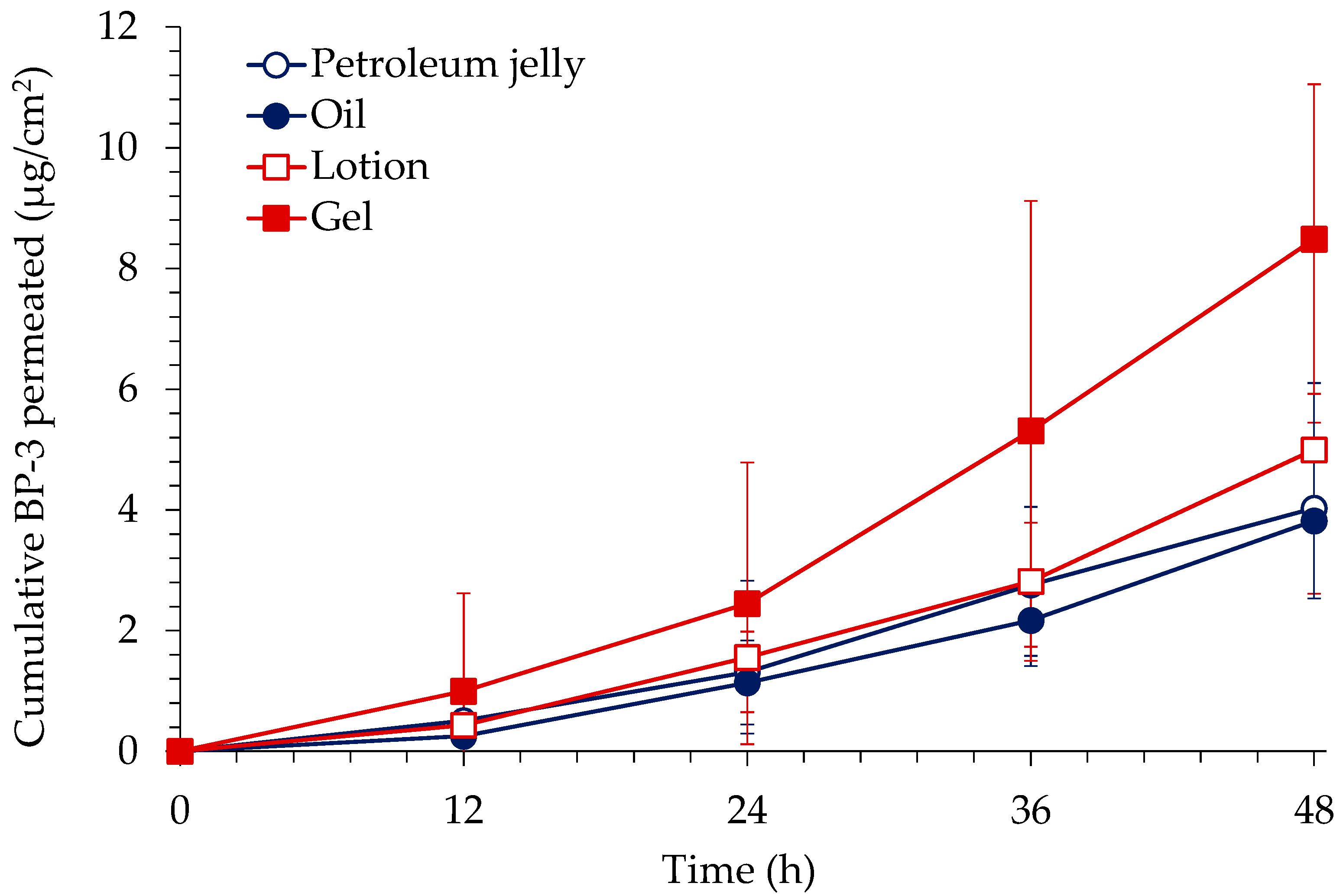
Figure 1. In vitro skin permeation profile of BP-3 through excised rat skin after application of BP-3 formulations (mean ± S.D., n = 6 each).
Toxics 10 00672 g001
Figure 2. Average plasma concentration-time profiles of BP-3 obtained after i.v. injection and transdermal administration at doses of 1 mg/kg (n = 4) and 40 mg/kg (n = 5), respectively, in rats (mean ± S.D.).
Toxics 10 00672 g002
Figure 3. Average plasma concentration-time profile of BP-3 during i.v. infusion in rats (n = 4, mean ± S.D.).
Toxics 10 00672 g003
Table 1. Composition of BP-3 formulations.
Petroleum JellyOilLotionGel
Benzophenone-3
(5%)
Benzophenone-3
(5%)
Benzophenone-3
(5%)
Benzophenone-3
(5%)
Petroleum jelly
(95%)
Miglyol
(18.7%)
Miglyol
(18.7%)
Miglyol
(18.7%)
Mineral oil
(72.3%)
Marketed body
emulsion (75.8%)
Carbomer 940
(1.1%)
Bees wax
(4.0%)
Surfactant
(0.5%)
Poloxamer 188
(0.2%)
EDTA Na
(0.1%)
Methyl paraben (0.1%)
NaOH
(10.0%)
Surfactant
(0.5%)
DW
(64.3%)
Table 2. In vitro skin deposition of BP-3 in the stratum corneum and the epidermis/dermis of excised rat skin after 48 h application of BP-3 formulations (mean ± S.D., n = 6).
FormulationEpidermis/Dermis (%)Stratum Corneum (%)
Petroleum jelly2.1 ± 0.31.0 ± 0.5
Oil2.1 ± 0.62.8 ± 1.6
Lotion3.3 ± 0.93.4 ± 1.3
Gel4.7 ± 1.017.0 ± 4.5 *
* Gel was significantly different from petroleum jelly, oil, and lotion (one-way ANOVA, post hoc; Tukey’s test, p < 0.05).
Table 3. Toxicokinetic parameters of BP-3 obtained after i.v. injection and transdermal administration at doses of 1 mg/kg and 40 mg/kg, respectively, in rats (mean ± S.D.).
Parametersi.v. (n = 4)Transdermal (n = 5)
t1/2 (h) *3.1 ± 1.618.3 ± 5.8
Tmax (h)-4.8 ± 3.5
C0 (ng/mL)577.8 ± 121.7-
Cmax (ng/mL)-26.4 ± 4.6
AUClast (ng·h/mL)208.2 ± 23.6550 ± 155.4
AUCinfinity (ng·h/mL)219.2 ± 25.9781.4 ± 311.6
F (%)-6.9 ± 1.8
* t1/2 obtained after i.v. injection was significantly different that from transdermal administration (one-way ANOVA, post hoc; Tukey’s test, p < 0.05).
Table 4. Average concentration and plasma-to-tissue partition coefficients (kp) of BP-3 in tissues determined under steady state conditions (n = 4, mean ± S.D.).
TissuesConcentration (ng/mL or ng/g)kp
Plasma180.50 ± 52.29-
Kidney620.68 ± 297.704.00 ± 2.87
Liver269.19 ± 52.141.54 ± 0.27
Spleen155.71 ± 27.630.89 ± 0.13
Lung710.00 ± 326.774.50 ± 2.87
Heart363.14 ± 132.572.11 ± 0.87
Testis136.74 ± 70.880.73 ± 0.16
Stomach476.16 ± 99.722.70 ± 0.44
Small intestine532.22 ± 296.662.92 ± 1.35
Large intestine1174.44 ± 972.836.39 ± 5.77
Brain352.75 ± 95.812.07 ± 0.81
Publisher’s Note: MDPI stays neutral with regard to jurisdictional claims in published maps and institutional affiliations.