

Potential Involvement of Oxidative Stress, Apoptosis and Proinflammation in Ipconazole-Induced Cytotoxicity in Human Endothelial-like Cells


Abstract
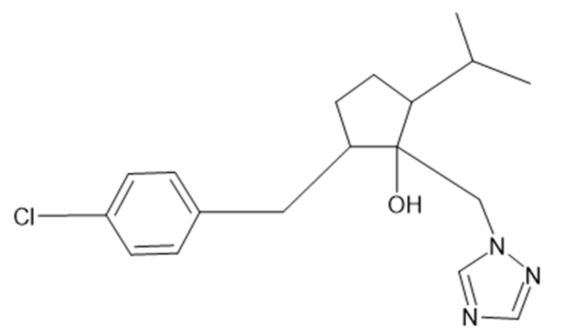
:1. Introduction
2. Materials and Methods
2.1. Reagents
2.2. Cell Culture
2.3. Cell Viability Evaluation (MTT)
2.4. ROS Production
2.5. Apoptotic Assay with Caspase 3/7 Activity
2.6. Molecular Biomarkers Assessment
2.7. Statistics
3. Results
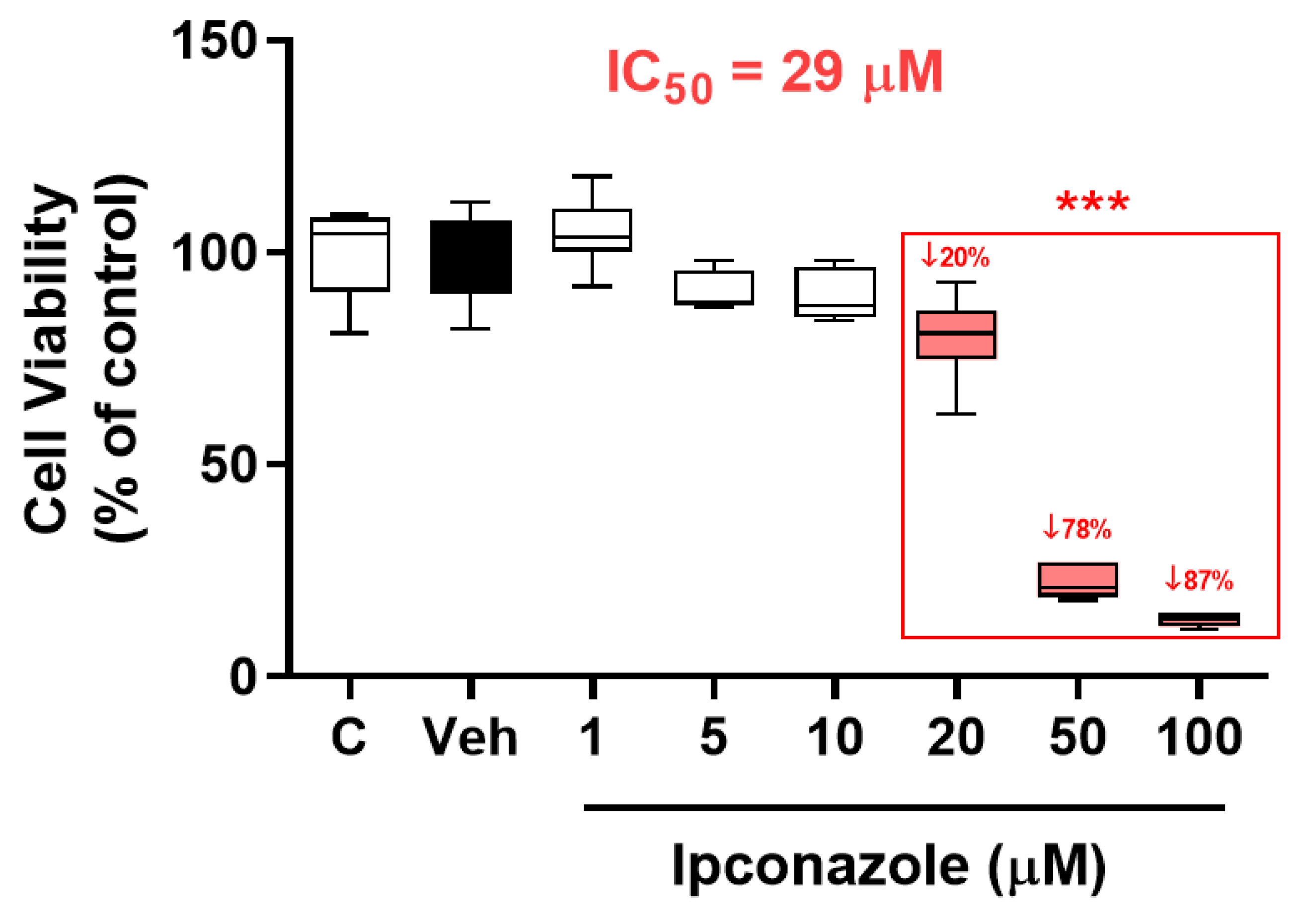
3.1. Cell Viability
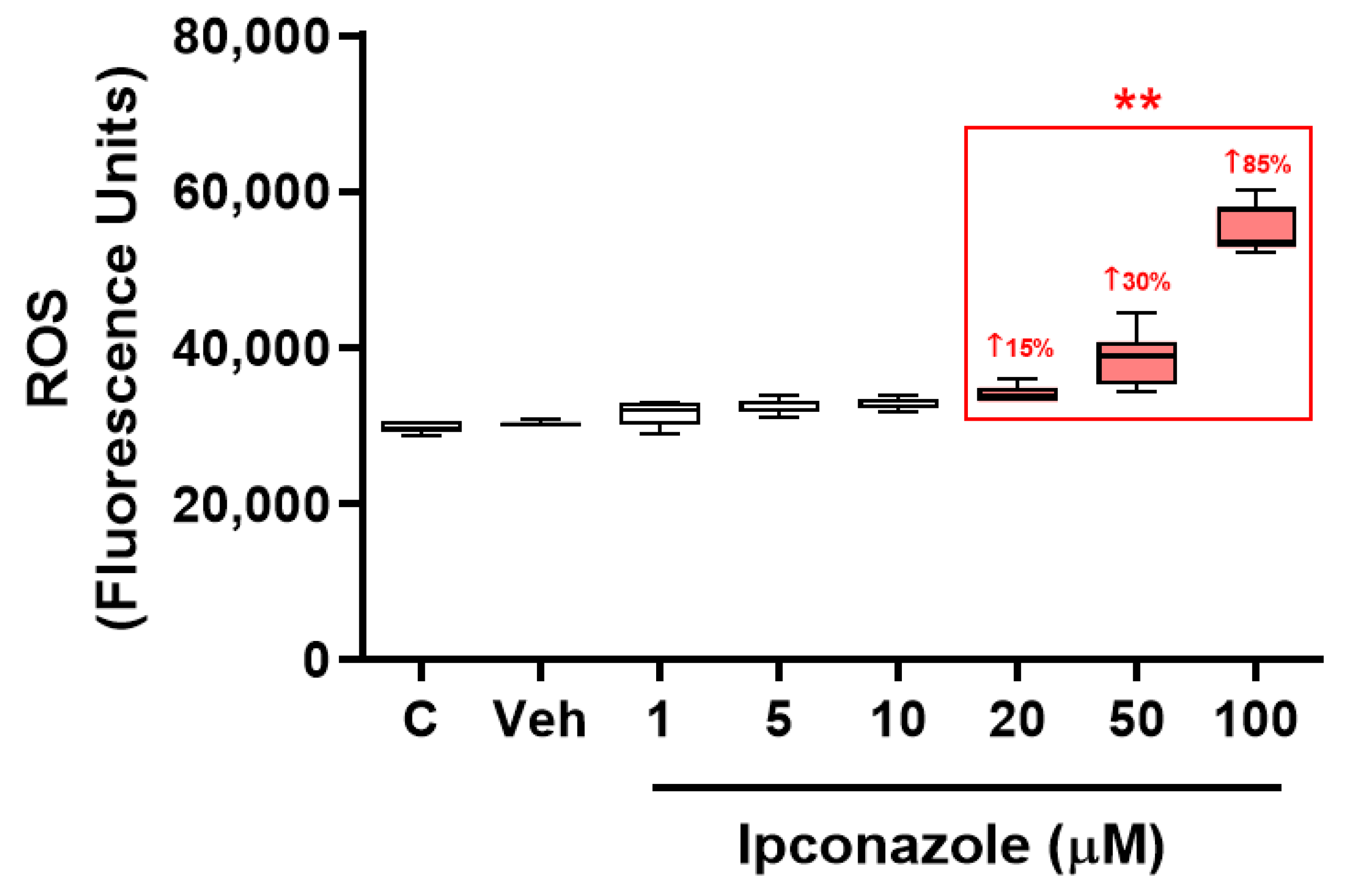
3.2. ROS Generation
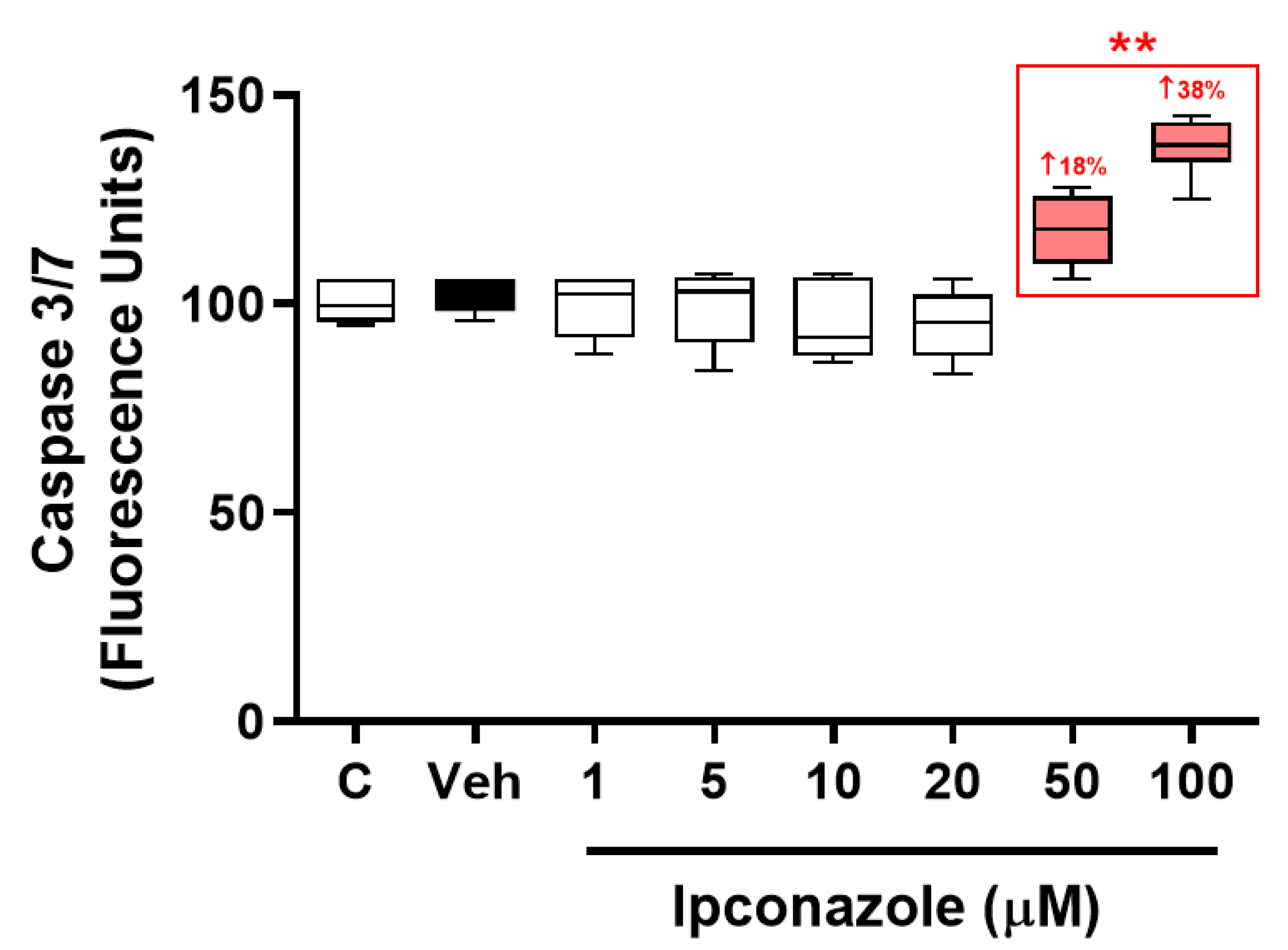
3.3. Caspase 3/7 Activity
3.4. Biomarkers of Cell Death
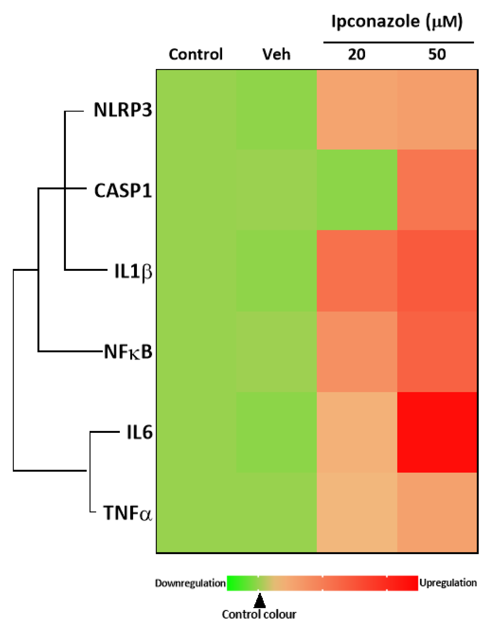
3.5. Inflammasome Complex
3.6. Antioxidant Molecular Biomarkers
4. Discussion
5. Conclusions
Author Contributions
Funding
Institutional Review Board Statement
Informed Consent Statement
Data Availability Statement
Conflicts of Interest
References
- European Food Safety Authority (EFSA). Statement concerning the review of the approval of the active substance ipconazole. EFSA J. 2022, 20, e07133. [Google Scholar]
- USEPA. Pesticides—Fact Sheet for Ipconazole; USEPA: Washington, DC, USA, 2004; pp. 1–17.
- Usman, M.; Farooq, M.; Wakeel, A.; Nawaz, A.; Cheema, S.A.; Rehman, H.U.; Ashraf, I.; Sanaullah, M. Nanotechnology in agriculture: Current status, challenges and future opportunities. Sci. Total Environ. 2020, 721, 137778. [Google Scholar] [CrossRef] [PubMed]
- Liu, T.; Fang, K.; Liu, Y.; Zhang, X.; Han, L.; Wang, X. Enantioselective residues and toxicity effects of the chiral triazole fungicide hexaconazole in earthworms (Eisenia fetida). Environ. Pollut. 2021, 270, 116269. [Google Scholar] [CrossRef] [PubMed]
- Roman, D.L.; Voiculescu, D.I.; Filip, M.; Ostafe, V.; Isvoran, A. Effects of Triazole Fungicides on Soil Microbiota and on the Activities of Enzymes Found in Soil. A Review. Agriculture 2021, 11, 893. [Google Scholar] [CrossRef]
- Teysseire, R.; Manangama, G.; Baldi, I.; Carles, C.; Brochard, P.; Bedos, C.; Delva, F. Determinants of non-dietary exposure to agricultural pesticides in populations living close to fields. A systematic review. Sci. Total Environ. 2021, 761, 143294. [Google Scholar] [CrossRef]
- Zhang, H.; Qian, M.; Wang, J.; Yang, G.; Weng, Y.; Jin, C.; Li, Y.; Jin, Y. Insights into the effects of difenoconazole on the livers in male mice at the biochemical and transcriptomic levels. J. Hazard. Mater. 2022, 422, 126933. [Google Scholar] [CrossRef] [PubMed]
- Wang, X.; Ni, H.; Xu, W.; Wu, B.; Xie, T.; Zhang, C.; Cheng, J.; Li, Z.; Tao, L.; Zhang, Y. Difenoconazole induces oxidative DNA damage and mitochondria mediated apoptosis in SH-SY5Y cells. Chemosphere 2021, 283, 131160. [Google Scholar] [CrossRef]
- Sanchez, C.L.; Souders, C.L.; Pena-Delgado, C.J.; Nguyen, K.T.; Kroyter, N.; Ahmadie, N.E.; Aristizabal-Henao, J.J.; Bowden, J.A.; Martyniuk, C.J. Neurotoxicity assessment of triazole fungicides on mitochondrial oxidative respiration and lipids in differentiated human SH-SY5Y neuroblastoma cells. Neurotoxicology 2020, 80, 76–86. [Google Scholar] [CrossRef]
- Ben Othmène, Y.; Monceaux, K.; Karoui, A.; Ben Salem, I.; Belhadef, A.; Abid-Essefi, S.; Lemaire, C. Tebuconazole induces ROS-dependent cardiac cell toxicity by activating DNA damage and mitochondrial apoptotic pathway. Ecotoxicol. Environ. Saf. 2020, 204, 111040. [Google Scholar] [CrossRef] [PubMed]
- Pan, J.S.; Hong, M.Z.; Ren, J.L. Reactive oxygen species: A double-edged sword in oncogenesis. World J. Gastroenterol. 2009, 15, 1702–1707. [Google Scholar] [CrossRef]
- Ferreira, D.; da Motta, A.C.; Kreutz, L.C.; Toni, C.; Loro, V.L.; Barcellos, L.J. Assessment of oxidative stress in Rhamdia quelen exposed to agrichemicals. Chemosphere 2010, 79, 914–921. [Google Scholar] [CrossRef] [PubMed]
- Chaâbane, M.; Koubaa, M.; Soudani, N.; Elwej, A.; Grati, M.; Jamoussi, K.; Boudawara, T.; Ellouze Chaabouni, S.; Zeghal, N. Nitraria retusa fruit prevents pen-conazole-induced kidney injury in adult rats through modulation of oxidative stress and histopathological changes. Pharm. Biol. 2017, 55, 1061–1073. [Google Scholar] [CrossRef]
- Heusinkveld, H.J.; Molendijk, J.; Van Den Berg, M.; Westerink, R.H. Azole fungicides disturb intracellular Ca2+ in an additive manner in dopaminergic PC12 cells. Toxicol. Sci. 2013, 134, 374–381. [Google Scholar] [CrossRef]
- Chen, L.; Teng, H.; Zhang, K.Y.; Skalicka-Woźniak, K.; Georgiev, M.I.; Xiao, J. Agrimonolide and Desmethylagrimonolide Induced HO-1 Expression in HepG2 Cells through Nrf2-Transduction and p38 Inactivation. Front. Pharmacol. 2017, 7, 513. [Google Scholar] [CrossRef] [PubMed]
- Soudani, N.; Ben Amara, I.; Sefi, M.; Boudawara, T.; Zeghal, N. Effects of selenium on chromium (VI)-induced hepatotoxicity in adult rats. Experimental and toxi-cologic pathology. Exp. Toxicol. Pathol. 2011, 63, 541–548. [Google Scholar] [CrossRef] [PubMed]
- Yang, J.D.; Liu, S.H.; Liao, M.H.; Chen, R.M.; Liu, P.Y.; Ueng, T.H. Effects of tebuconazole on cytochrome P450 enzymes, oxidative stress, and endocrine disruption in male rats. Environ. Toxicol. 2018, 33, 899–907. [Google Scholar] [CrossRef]
- Castillo, G.; Barrios-Arpi, L.; Ramos-Gonzalez, M.; Vidal, P.; Gonzales-Irribarren, A.; Ramos-Cevallos, N.; Rodríguez, J.L. Neurotoxicity associated with oxidative stress and inflammasome gene expression induced by allethrin in SH-SY5Y cells. Toxicol. Ind. Health 2022, 38, 777–788. [Google Scholar] [CrossRef]
- Barrios-Arpi, L.; Arias, Y.; Lopez-Torres, B.; Ramos-Gonzalez, M.; Ticli, G.; Prosperi, E.; Rodríguez, J.L. In Vitro Neurotoxicity of Flumethrin Pyrethroid on SH-SY5Y Neuroblastoma Cells: Apoptosis Associated with Oxidative Stress. Toxics 2022, 10, 131. [Google Scholar] [CrossRef] [PubMed]
- Bauernfeind, F.G.; Horvath, G.; Stutz, A.; Alnemri, E.S.; MacDonald, K.; Speert, D.; Fernandes-Alnemri, T.; Wu, J.; Monks, B.G.; Fitzgerald, K.A.; et al. Cutting edge: NF-kappa B activating pattern recognition and cytokine receptors license NLRP3 inflammasome activation by regulating NLRP3 expression. J. Immunol. 2009, 183, 787–791. [Google Scholar] [CrossRef]
- Hata, A.N.; Engelman, J.A.; Faber, A.C. The BCL2 Family: Key Mediators of the Apoptotic Response to Targeted Anticancer Therapeutics. Cancer Discov. 2015, 5, 475–487. [Google Scholar] [CrossRef]
- Tait, S.W.; Green, D.R. Mitochondria and cell death: Outer membrane permeabilization and beyond. Nat. Rev. Mol. Cell Biol. 2010, 11, 621–632. [Google Scholar] [CrossRef]
- Knebel, C.; Heise, T.; Zanger, U.M.; Lampen, A.; Marx-Stoelting, P.; Braeuning, A. The azole fungicide tebuconazole affects human CYP1A1 and CYP1A2 expression by an aryl hydrocarbon receptor-dependent pathway. Food Chem. Toxicol. 2019, 123, 481–491. [Google Scholar] [CrossRef]
- Hamdi, H.; Graiet, I.; Abid-Essefi, S.; Eyer, J. Epoxiconazole profoundly alters rat brain and properties of neural stem cells. Chemosphere 2022, 288, 132640. [Google Scholar] [CrossRef]
- Hamdi, H.; Abid-Essefi, S.; Eyer, J. Cytotoxic and genotoxic effects of epoxiconazole on F98 glioma cells. Chemosphere 2019, 229, 314–323. [Google Scholar] [CrossRef] [PubMed]
- Schwarzbacherová, V.; Wnuk, M.; Lewinska, A.; Potocki, L.; Zebrowski, J.; Koziorowski, M.; Holečková, B.; Šiviková, K.; Dianovský, J. Evaluation of cytotoxic and genotoxic activity of fungicide formulation Tango® Super in bovine lymphocytes. Environ. Pollut. 2017, 220, 255–263. [Google Scholar] [CrossRef]
- Chen, P.J.; Moore, T.; Nesnow, S. Cytotoxic effects of propiconazole and its metabolites in mouse and human hepatoma cells and primary mouse hepatocytes. Toxicol. In Vitro 2008, 22, 1476–1483. [Google Scholar] [CrossRef]
- Theunissen, P.T.; Robinson, J.F.; Pennings, J.L.; De Jong, E.; Claessen, S.M.; Kleinjans, J.C.; Piersma, A.H. Transcriptomic concentration-response evaluation of valproic acid, cyproconazole, and hexaconazole in the neural embryonic stem cell test (ESTn). Toxicol. Sci. 2012, 125, 430–438. [Google Scholar] [CrossRef] [PubMed]
- Rigoulet, M.; Yoboue, E.D.; Devin, A. Mitochondrial ROS generation and its regulation: Mechanisms involved in H(2)O(2) signaling. Antioxid. Redox Signal 2011, 14, 459–468. [Google Scholar] [CrossRef]
- Kovac, S.; Angelova, P.R.; Holmström, K.M.; Zhang, Y.; Dinkova-Kostova, A.T.; Abramov, A.Y. Nrf2 regulates ROS production by mitochondria and NADPH oxidase. Biochim. Biophys. Acta 2015, 1850, 794–801. [Google Scholar] [CrossRef]
- Fridovich, I. Superoxide anion radical (O2−), superoxide dismutases, and related matters. J. Biol. Chem. 1997, 272, 18515–18517. [Google Scholar] [CrossRef] [PubMed]
- Wang, Y.; Branicky, R.; Noë, A.; Hekimi, S. Superoxide dismutases: Dual roles in controlling ROS damage and regulating ROS signaling. J. Cell Biol. 2018, 217, 1915–1928. [Google Scholar] [CrossRef]
- Hamdi, H.; Rjiba-Touati, K.; Ayed-Boussema, I.; M’nassri, A.; Chaabani, H.; Rich, S.; Abid-Essefi, S. Epoxiconazole caused oxidative stress related DNA damage and apoptosis in PC12 rat Pheochromocytoma. Neurotoxicology 2022, 89, 184–190. [Google Scholar] [CrossRef]
- Hamdi, H.; Abid-Essefi, S.; Eyer, J. Neuroprotective effects of Myricetin on Epoxiconazole-induced toxicity in F98 cells. Free Radic. Biol. Med. 2021, 164, 154–163. [Google Scholar] [CrossRef] [PubMed]
- Hamdi, H.; Ben Salem, I.; Ben Othmène, Y.; Annabi, E.; Abid-Essefi, S. The involvement of ROS generation on Epoxiconazole-induced toxicity in HCT116 cells. Pestic. Biochem. Physiol. 2018, 148, 62–67. [Google Scholar] [CrossRef] [PubMed]
- Jiang, X.; Wang, J.; Liu, J.; Zhu, H.; Hu, J.; Sun, X.; Zhou, W. Resveratrol ameliorates penconazole-induced cardiotoxicity by inhibition of oxidative stress and apoptosis in zebrafish larvae. Ecotoxicol. Environ. Saf. 2023, 256, 114865. [Google Scholar] [CrossRef]
- Ben Othmène, Y.; Monceaux, K.; Belhadef, A.; Karoui, A.; Ben Salem, I.; Boussabbeh, M.; Abid-Essefi, S.; Lemaire, C. Triazole fungicide tebuconazole induces apoptosis through ROS-mediated endoplasmic reticulum stress pathway. Environ. Toxicol. Pharmacol. 2022, 94, 103919. [Google Scholar] [CrossRef]
- Nesnow, S.; Grindstaff, R.D.; Lambert, G.; Padgett, W.T.; Bruno, M.; Ge, Y.; Chen, P.J.; Wood, C.E.; Murphy, L. Propiconazole increases reactive oxygen species levels in mouse hepatic cells in culture and in mouse liver by a cytochrome P450 enzyme mediated process. Chem. Biol. Interact. 2011, 194, 79–89. [Google Scholar] [CrossRef]
- Roelofs, M.J.E.; Temming, A.R.; Piersma, A.H.; Van den Berg, M.; Van Duursen, M.B.M. Conazole fungicides inhibit Leydig cell testosterone secretion and androgen receptor activation in vitro. Toxicol. Rep. 2014, 1, 271–283. [Google Scholar] [CrossRef]
- Othmène, Y.B.; Salem, I.B.; Hamdi, H.; Annabi, E.; Abid-Essefi, S. Tebuconazole induced cytotoxic and genotoxic effects in HCT116 cells through ROS generation. Pestic. Biochem. Physiol. 2021, 174, 104797. [Google Scholar] [CrossRef] [PubMed]
- Mesnage, R.; Defarge, N.; Spiroux de Vendômois, J.; Séralini, G.E. Major pesticides are more toxic to human cells than their declared active principles. Biomed. Res. Int. 2014, 2014, 179691. [Google Scholar] [CrossRef] [PubMed]
- Ben Othmène, Y.; Hamdi, H.; Annabi, E.; Amara, I.; Ben Salem, I.; Neffati, F.; Najjar, M.F.; Abid-Essefi, S. Tebuconazole induced cardiotoxicity in male adult rat. Food Chem. Toxicol. 2020, 137, 111134. [Google Scholar] [CrossRef]
- Lee, W.Y.; Lee, R.; Park, H.J. Tebuconazole Induces ER-Stress-Mediated Cell Death in Bovine Mammary Epithelial Cell Lines. Toxics 2023, 11, 397. [Google Scholar] [CrossRef] [PubMed]
- Othmène, Y.B.; Hamdi, H.; Salem, I.B.; Annabi, E.; Amara, I.; Neffati, F.; Najjar, M.F.; Abid-Essefi, S. Oxidative stress, DNA damage and apoptosis induced by tebuconazole in the kidney of male Wistar rat. Chem. Biol. Interact. 2020, 330, 109114. [Google Scholar] [CrossRef]
- Kumar, N.; Awoyemi, O.; Willis, A.; Schmitt, C.; Ramalingam, L.; Moustaid-Moussa, N.; Crago, J. Comparative Lipid Peroxidation and Apoptosis in Embryo-Larval Zebrafish Exposed to 3 Azole Fungicides, Tebuconazole, Propiconazole, and Myclobutanil, at Environmentally Relevant Concentrations. Environ. Toxicol. Chem. 2019, 38, 1455–1466. [Google Scholar] [CrossRef] [PubMed]
- Morgan, A.M.; Hassanen, E.I.; Ogaly, H.A.; Al Dulmani, S.A.; Al-Zahrani, F.A.M.; Galal, M.K.; Kamel, S.; Rashad, M.M.; Ibrahim, M.A.; Hussien, A.M. The ameliorative effect of N-acetylcysteine against penconazole induced neurodegenerative and neuroinflammatory disorders in rats. J. Biochem. Mol. Toxicol. 2021, 35, e22884. [Google Scholar] [CrossRef]
- Zhu, B.; Liu, L.; Gong, Y.X.; Ling, F.; Wang, G.X. Triazole-induced toxicity in developing rare minnow (Gobiocypris rarus) embryos. Environ. Sci. Pollut. Res. Int. 2014, 21, 13625–13635. [Google Scholar] [CrossRef]
- Ma, F.; Li, Y.; Yu, Y.; Li, Z.; Lin, L.; Chen, Q.; Xu, Q.; Pan, P.; Wang, Y.; Ge, R.S. Gestational exposure to tebuconazole affects the development of rat fetal Leydig cells. Chemosphere 2021, 262, 127792. [Google Scholar] [CrossRef] [PubMed]
- Zhao, F.; Cao, F.; Li, H.; Teng, M.; Liang, Y.; Qiu, L. The effects of a short-term exposure to propiconazole in zebrafish (Danio rerio) embryos. Environ. Sci. Pollut. Res. Int. 2020, 27, 38212–38220. [Google Scholar] [CrossRef] [PubMed]
- Guo, H.; Callaway, J.B.; Ting, J.P. Inflammasomes: Mechanism of action, role in disease, and therapeutics. Nat. Med. 2015, 21, 677–687. [Google Scholar] [CrossRef] [PubMed]
- Dumas, A.; Amiable, N.; de Rivero Vaccari, J.P.; Chae, J.J.; Keane, R.W.; Lacroix, S.; Vallières, L. The inflammasome pyrin contributes to pertussis toxin-induced IL-1β synthesis, neutrophil intravascular crawling and autoimmune encephalomyelitis. PLoS Pathog. 2014, 10, e1004150. [Google Scholar] [CrossRef]
- Wen, H.; Gris, D.; Lei, Y.; Jha, S.; Zhang, L.; Huang, M.T.; Brickey, W.J.; Ting, J.P. Fatty acid-induced NLRP3-ASC inflammasome activation interferes with insulin signaling. Nat. Immunol. 2011, 12, 408–415. [Google Scholar] [CrossRef] [PubMed]
- Zhang, L.; Ai, C.; Bai, M.; Niu, J.; Zhang, Z. NLRP3 Inflammasome/Pyroptosis: A Key Driving Force in Diabetic Cardiomyopathy. Int. J. Mol. Sci. 2022, 23, 10632. [Google Scholar] [CrossRef]
- Zhang, M.; He, Q.; Chen, G.; Li, P.A. Suppression of NLRP3 Inflammasome, Pyroptosis, and Cell Death by NIM811 in Rotenone-Exposed Cells as an in vitro Model of Parkinson’s Disease. Neurodegener. Dis. 2020, 20, 73–83. [Google Scholar] [CrossRef]
- Won, J.H.; Park, S.; Hong, S.; Son, S.; Yu, J.W. Rotenone-induced Impairment of Mitochondrial Electron Transport Chain Confers a Selective Priming Signal for NLRP3 Inflammasome Activation. J. Biol. Chem. 2015, 290, 27425–27437. [Google Scholar] [CrossRef]
- Liu, Z.; Zhao, H.; Liu, W.; Li, T.; Wang, Y.; Zhao, M. NLRP3 inflammasome activation is essential for paraquat-induced acute lung injury. Inflammation 2015, 38, 433–444. [Google Scholar] [CrossRef] [PubMed]
- Jang, Y.; Lee, A.Y.; Jeong, S.H.; Park, K.H.; Paik, M.K.; Cho, N.J.; Kim, J.E.; Cho, M.H. Chlorpyrifos induces NLRP3 inflammasome and pyroptosis/apoptosis via mitochondrial oxidative stress in human keratinocyte HaCaT cells. Toxicology 2015, 338, 37–46. [Google Scholar] [CrossRef]
- Villaorduña, C.; Mendoza-Carlos, M.; Chuyma, M.; Avilés, J.; Avalos-Diaz, A.; Lozano-Reategui, R.; Garcia-Ruiz, J.; Panduro-Tenazoa, N.; Vargas, J.; Moran-Quintanilla, Y.; et al. Ipconazole Induces Oxidative Stress, Cell Death, and Proinflammation in SH-SY5Y Cells. Toxics 2023, 11, 566. [Google Scholar] [CrossRef] [PubMed]


| Gene | Forward | Reverse |
|---|---|---|
| Cell Death | ||
| BAX (BCL-2-associated X protein) | CCCCCGAGAGGTCTTTTTCC′ | CCTTGAGCACCAGTTTGCTG′ |
| CASP3 (caspase 3) | ‘GTGGAGGCCGACTTCTTGTA′ | TTTCAGCATGGCACAAAGCG′ |
| APAF1 (apoptotic protease-activating factor 1) | TCTTCCAGTGGTAAAGATTCAGTT′ | CGGAGACGGTCTTTAGCCTC′ |
| BNIP3 (BCL2-interacting protein 3) | CCTCAGCATGAGGAACACGA′ | GCCACCCCAGGATCTAACAG′ |
| BCL2 (B-cell lymphoma 2) | ′TCTCATGCCAAGGGGGAAAC′ | TCCCGGTTATCGTACCCTGT′ |
| P53 (tumor protein P53) | GAACAAGTTGGCCTGCACTG | GAAGTGGGCCCCTACCTAGA |
| AKT1 (AKT serine/threonine kinase 1) | GAAGGACGGGAGCAGGC | TGTACTCCCCTCGTTTGTGC |
| Inflammasome Complex | ||
| NLRP3 (NLR pyrin domain containing 3) | CCCCGTAATCAACGGGACAA′ | AGCCAAATGCTTACCAGAAAGT′ |
| CASP1 (caspase 1) | ′GAAAAGCCATGGCCGACAAG′ | GCCCCTTTCGGAATAACGGA′ |
| IL1β (interleukin-1 beta) | ′CCAGCTACGAATCTCCGACC′ | TATCCTGTCCCTGGAGGTGG |
| NFκB (nuclear factor kappa B) | ′TTTTCGACTACGCGGTGACA′ | GTTACCCAAGCGGTCCAGAA′ |
| TNFα (tumor necrosis factor alpha) | CTGGAAAGGACACCATGAGCA′ | TCTCTCAGCTCCACGCCATT′ |
| IL6 (interleukin 6) | ‘CCAGTACCCCCAGGAGAAGA′ | CAGCTCTGGCTTGTTCCTCA′ |
| Antioxidant Biomarkers | ||
| NRF2 (nuclear factor erythroid related factor2) | CTGGTCATCGGAAAACCCCA′ | TCTGCAATTCTGAGCAGCCA’ |
| SOD (superoxide dismutase) | ′CCACTGCTGGGGATTGATGT′ | CGTGGTTTACTTTTTGCAAGCC′ |
| GPx (glutathione peroxidase) | ′TTCGAGCCCAACTTCATGCT′ | ′CGATGTCAGGCTCGATGTCA′ |
| Normalizer | ||
| GAPDH (glyceraldehyde-3-phosphate dehydrogenase) | ′GAGAAGGCTGGGGCTCATTT | AGTGATGGCATGGACTGTGG′ |
| Gene | Control | Vehicle (DMSO) | Ipconazole | |
|---|---|---|---|---|
| 20 µM | 50 µM | |||
| Fold Change | ||||
| BAX | 1.00 | 0.99 | 1.39 ** | 1.73 *** |
| BCL2 | 1.00 | 0.97 | 0.13 *** | 0.16 *** |
| APAF1 | 1.00 | 0.99 | 1.35 | 2.66 *** |
| BNIP3 | 1.00 | 1.02 | 1.08 | 2.08 *** |
| CASP3 | 1.00 | 1.00 | 1.66 * | 2.24 *** |
| AKT1 | 1.00 | 1.02 | 1.27 | 2.42 *** |
| P53 | 1.00 | 0.92 | 0.95 | 1.04 |
| Gene | Control | Vehicle (Veh) | Ipconazole | |
|---|---|---|---|---|
| 20 µM | 50 µM | |||
| Fold Change | ||||
| NLRP3 | 1.00 | 0.97 | 1.52 | 1.59 * |
| CASP1 | 1.00 | 1.01 | 0.96 | 2.07 *** |
| IL1β | 1.00 | 0.97 | 2.14 ** | 2.41 ** |
| NFκB | 1.00 | 1.02 | 1.76 | 2.31 ** |
| IL6 | 1.00 | 0.96 | 1.37 | 3.34 *** |
| TNFα | 1.00 | 1.00 | 1.30 | 1.56 * |
| Gene | Control | Vehicle (Veh) | Ipconazole | |
|---|---|---|---|---|
| 20 µM | 50 µM | |||
| Fold Change | ||||
| NRF2 | 1.00 | 0.98 | 0.98 | 0.56 ** |
| SOD | 1.00 | 1.06 | 0.92 | 0.90 |
| GPx | 1.00 | 0.97 | 0.97 | 0.45 *** |
Disclaimer/Publisher’s Note: The statements, opinions and data contained in all publications are solely those of the individual author(s) and contributor(s) and not of MDPI and/or the editor(s). MDPI and/or the editor(s) disclaim responsibility for any injury to people or property resulting from any ideas, methods, instructions or products referred to in the content. |
© 2023 by the authors. Licensee MDPI, Basel, Switzerland. This article is an open access article distributed under the terms and conditions of the Creative Commons Attribution (CC BY) license (https://creativecommons.org/licenses/by/4.0/).
Share and Cite
Ruiz-Yance, I.; Siguas, J.; Bardales, B.; Robles-Castañeda, I.; Cordova, K.; Ypushima, A.; Estela-Villar, E.; Quintana-Criollo, C.; Estacio, D.; Rodríguez, J.-L. Potential Involvement of Oxidative Stress, Apoptosis and Proinflammation in Ipconazole-Induced Cytotoxicity in Human Endothelial-like Cells. Toxics 2023, 11, 839. https://doi.org/10.3390/toxics11100839
Ruiz-Yance I, Siguas J, Bardales B, Robles-Castañeda I, Cordova K, Ypushima A, Estela-Villar E, Quintana-Criollo C, Estacio D, Rodríguez J-L. Potential Involvement of Oxidative Stress, Apoptosis and Proinflammation in Ipconazole-Induced Cytotoxicity in Human Endothelial-like Cells. Toxics. 2023; 11(10):839. https://doi.org/10.3390/toxics11100839
Chicago/Turabian StyleRuiz-Yance, Iris, Junior Siguas, Brandy Bardales, Ingrid Robles-Castañeda, Karen Cordova, Alina Ypushima, Esteban Estela-Villar, Carlos Quintana-Criollo, Darwin Estacio, and José-Luis Rodríguez. 2023. "Potential Involvement of Oxidative Stress, Apoptosis and Proinflammation in Ipconazole-Induced Cytotoxicity in Human Endothelial-like Cells" Toxics 11, no. 10: 839. https://doi.org/10.3390/toxics11100839
APA StyleRuiz-Yance, I., Siguas, J., Bardales, B., Robles-Castañeda, I., Cordova, K., Ypushima, A., Estela-Villar, E., Quintana-Criollo, C., Estacio, D., & Rodríguez, J.-L. (2023). Potential Involvement of Oxidative Stress, Apoptosis and Proinflammation in Ipconazole-Induced Cytotoxicity in Human Endothelial-like Cells. Toxics, 11(10), 839. https://doi.org/10.3390/toxics11100839