Review

Per- and Polyfluoroalkyl Substances (PFASs) and Their Potential Effects on Female Reproductive Diseases

1
Department of Clinical Nutrition, Second Affiliated Hospital of Dalian Medical University, Dalian 116027, China
2
Department of Nutrition and Food Hygiene, School of Public Health, Dalian Medical University, Dalian 116044, China
*
Author to whom correspondence should be addressed.
These authors contributed equally to this work.
Toxics 2024, 12(8), 539; https://doi.org/10.3390/toxics12080539 (registering DOI)
Submission received: 13 June 2024 / Revised: 18 July 2024 / Accepted: 24 July 2024 / Published: 26 July 2024

Abstract

:
Per- and polyfluoroalkyl substances (PFASs) are a class of anthropogenic organic compounds widely present in the natural and human living environments. These emerging persistent pollutants can enter the human body through multiple channels, posing risks to human health. In particular, exposure to PFASs in women may cause a series of reproductive health hazards and infertility. Based on a review of the existing literature, this study preliminarily summarizes the effects of PFAS exposure on the occurrence and development of female reproductive endocrine diseases, such as polycystic ovary syndrome (PCOS), endometriosis, primary ovarian insufficiency (POI), and diminished ovarian reserve (DOR). Furthermore, we outline the relevant mechanisms through which PFASs interfere with the physiological function of the female ovary and finally highlight the role played by nutrients in reducing the reproductive health hazards caused by PFASs. It is worth noting that the physiological mechanisms of PFASs in the above diseases are still unclear. Therefore, it is necessary to further study the molecular mechanisms of PFASs in female reproductive diseases and the role of nutrients in this process.

1. Introduction

Per- and polyfluoroalkyl substances (PFASs) are known for their distinctive chemical structures, featuring partially or completely fluorinated methyl or methylene carbon atoms in carbon chains of varied lengths (-CnF2n+1-). Perfluoroalkyl substances are formed when all hydrogen atoms in the carbon chain of chemicals are replaced by fluorine atoms [1]. These compounds are highly resistant to environmental and biological degradation due to their chemical stability, heat resistance, hydrophobicity, and lipophilicity owing to the exceptional strength of their carbon–fluorine (C–F) bonds. The above characteristics lead to extremely durable and useful surfactants and polymers [2]. According to the recognized PFAS classification system prepared by Buck et al., PFASs can be divided into two categories, among which long-chain PFASs (PFCAs (perfluoroalkyl carboxylic acids) with eight carbons and greater; and PFSAs (perfluoroalkane sulfonates) with six carbons and greater) include some of the most well-studied chemicals, such as perfluorooctane sulfonate (PFOS) and perfluorooctanoic acid (PFOA) [2]. Depending on the number of CF2 moieties, PFASs are classified as ultra-short-chain or short-chain PFASs when the numbers of perfluorocarbon atoms are 2–3 and 4–7, respectively [3]. Consequently, PFAS substances have been extensively used in a wide array of industrial and consumer chemical products since the 1940s, spanning over 100 sectors. Industrial applications include polytetrafluoroethylene (PTFE) production, and they can also be used in the metal plating industry, photo imaging industry, and semi-conductor industry. PFASs are also used in large-scale consumer goods, including food packaging, cosmetics, water-repellent clothing, textiles, non-stick cookware, cleaning agents, papermaking, fire-fighting foam, and medical products [4,5]. Due to their ubiquitous applications globally, PFASs have been detected in environmental media such as surface water, groundwater, seawater, soil, sediment, and the atmosphere, as well as in humans and wildlife, which is most concerning [6,7,8,9]. Current evidence indicates that the exposure of humans to PFASs mainly occurs through the consumption of contaminated water and seafood, the inhalation of indoor air and dust particles, hand-to-mouth contact, and skin absorption [5,10]. Higher levels of PFOA (lower bound/upper bound (LB/UB) mean = 0.18/0.9 µg/kg) and PFOS (LB/UB mean = 2.08/2.59 µg/kg) have also been detected in fish and other seafood [11]. The European Food Safety Authority (ESFA) CONTAM panel reported that over 86% of PFAS exposure in the diet may be attributed to seafood consumption [12] (Figure 1). Among the vast array of PFASs, there are a number of long-chain PFASs that have been subjected to regulatory scrutiny and discontinued or regulated due to their environmental toxicity and bioaccumulation [13,14]. Moreover, additional long-chain PFAS substances that have raised concerns, specifically PFOA and perfluorohexane sulfonate (PFHxS), along with their salts, were phased out of global production in 2021 and 2022 [15,16]. Accordingly, manufacturers have begun to undertake commercial production using short-chain PFASs, ultra-short-chain PFASs and new alternatives [3]. Compared to the PFASs used in the past, short-chain PFASs are generally less bio-accumulative, but they show higher environmental persistence and long-distance mobility. Continuous exposure to such compounds can adversely affect organisms in the long term [17].
Previous studies have demonstrated the connections between PFAS exposure and increased odds of cancer, kidney disease, altered immune function, liver disease, lipid and insulin dysregulation [18]. Although many biological systems are adversely affected by PFASs, in this review, we focus on their relevant effects on female reproductive health. Epidemiological studies indicate that infertility, a critical issue in women’s reproductive health, now affects approximately 48 million couples and 186 million individuals globally, highlighting its status as a significant public health concern. Disease-related infertility, an important contributor affecting spontaneous conception, has attracted a wide range of attention [19]. PCOS, endometriosis, POI, and DOR are all factors contributing to female infertility. Studies suggest that certain long-chain PFASs could exacerbate these conditions and increase the risk of them arising [20,21]. In addition, results from animals and epidemiological studies have corroborated that short-chain PFASs such as hexafluoropropylene oxide dimer acid (GenX) and F-53B (6:2 chlorinated polyfluoroalkyl ether sulfonate [6:2 Cl-PFAES] and 8:2 Cl-PFAES) are becoming the dominant perfluorinated contaminants globally, and these also exhibit reproductive toxicity [22,23]. GenX exhibits a stronger affinity for the estrogen receptor (ER), compared to PFOA, and F-53B has been shown to reduce the spawning output in zebrafish [24].
The mechanisms by which female reproductive system diseases are affected by PFASs are often complex. First, kisspeptin is an influential neuroendocrine peptide in the hypothalamus that regulates the secretion of gonadotropin-releasing hormones (GnRHs), the release of which initiates a series of responses that trigger ovulation. Exposure to PFASs inhibits pro-peptide expression in areas of the brain such as the anteroventral periventricular nucleus (AVPV), which is essential for the production of the luteinizing hormone (LH) surge required for ovulation. This inhibition leads to reduced levels of GnRH and LH, ultimately interfering with the normal ovulatory cycle and potentially leading to ovulation failure [25,26]. Additionally, PFASs can influence the expression of crucial steroidogenic enzymes and either mimic or obstruct the actions of hormone receptors, thereby disrupting hormone-signaling pathways. This interference with normal hormone functions can lead to significant repercussions for reproductive health, altering fertility and menstrual cycles [27,28,29,30]. Oxidative stress is a state wherein there is an imbalance between oxidation in cells and tissues and the antioxidant systems, and it has been proven to play a key role in the pathogenesis of reduced female fertility. This imbalance can lead to various reproductive system disorders, such as endometriosis, PCOS, and unexplained infertility [31]. Oxidative stress can also lead to chronic anovulation and infertility in women by exacerbating insulin resistance and impairing oocyte development and quality [32]. Furthermore, numerous studies have shown that PPARs (peroxisome proliferators-activated receptors) play a role in normal reproductive and developmental functions, and that exposure to endogenous or exogenous compounds that abnormally modulate PPARs may lead to physiological dysfunction of the reproductive system [33]. The sustained activation of ovarian PPARs by PFAS exposure may disrupt oocyte meiosis while increasing lipid accumulation in the ovary and decreasing oocyte viability [26,34].
In this review, we summarize the associations between human PFAS exposure and four reproductive health outcomes in women that have been studied to date, including PCOS and endometriosis, as well as POI and DOR. We also outline the potential mechanisms through which PFASs may affect female ovaries, and, finally, we highlight the role of nutrition in mitigating the adverse effects of PFASs on ovarian cells.

2. Search Strategy

To explore the potential connection between PFAS exposure and female reproductive health, this review mainly focuses on the prevalence and impact of PFASs in relation to PCOS, endometriosis, POI, and DOR. Emphasis was placed on the analysis of epidemiological data and the study of molecular mechanisms affecting ovary function with PFASs.
Additionally, databases and scientific studies were explored using PubMed. The search terms included “Per- and poly-fluoroalkyl substances”, “Endocrine-disrupting chemicals”, “PFASs”, “PFOA”, “PFOS”, “short-chain PFASs”, “PFAS alternatives”, “Polycystic ovary syndrome”, “Endometriosis”, “Premature ovarian insufficiency”, and “Diminished ovarian reserve”.
Data inclusion and exclusion: We included only cohort and case–control studies that assessed the association between PFAS and PCOS, endometriosis, POI, DOR, and studies on the mechanisms by which PFASs may affect diseases of the female reproductive system, and we excluded non-original reports, review articles, conference abstracts, editorials, commentaries, experimental or pilot studies, case reports and case series, as well as studies that did not measure PFAS exposure. The findings were restricted to those published in English.

3. Human Exposure to PFASs and Female Reproductive Disorders

3.1. Polycystic Ovary Syndrome

Polycystic ovary syndrome is a heterogeneous disorder that affects 6–20% of premenopausal women, which makes this syndrome the most common endocrine metabolic disorder in women of reproductive age and accounts for about 80% of women with anovulatory infertility [35,36]. Recent studies have indicated that a range of environmental factors could potentially play a significant role in the initiation or exacerbation of PCOS. These factors are thought to interact with genetic predispositions, potentially triggering or worsening the symptoms associated with this endocrine disorder [37,38]. For example, studies have shown that there is a positive correlation between serum bisphenol A (BPA) levels and serum testosterone (T) levels in PCOS women, compared to healthy women [39]. Vagi et al. enrolled 52 patients with PCOS and 50 controls from an urban academic medical center in California, USA, to explore potential associations between polycystic ovary syndrome and perfluorinated compounds, polychlorinated biphenyls (PCBs), BPA, and organochlorine pesticides. They found that participants with PFOA and PFOS serum concentrations in the highest tertile were 6.9-fold and 5.8-fold more likely to develop PCOS compared to controls, respectively. This is the first evidence of a link between serum PFAS concentrations and PCOS [40]. Heffernan et al. recruited 30 PCOS women and 29 controls in the UK, collecting their serum and follicular fluid samples. Serum PFOS was higher in PCOS patients than in controls (geometric mean 3.9 vs. 3.1 ng/mL, p < 0.05), while the geometric mean follicular fluid concentration did not differ significantly between the groups [41]. In another case–control study of endocrine disruptors and female infertility, Zhan et al. studied women from the Shanghai, Shandong, and Zhejiang provinces, China. They measured the serum levels of nine legacy PFASs, six PFAS isomers, three emerging PFAS alternatives, and six short-chain PFASs in 366 PCOS cases with 577 controls. PCOS risks were considerably enhanced by higher concentrations of PFASs in serum. In these PFAS families, 6:2 chlorinated polyfluorinated ether sulfonate (6:2 Cl-PFESA), hexafluoropropylene oxide dimer acids (HFPO-DA), perfluorododecanoic acid (PFDoA), and ∑3,4,5 m-PFOS may be available as the primary contributors. They also observed that the joint effects of PFAS mixtures are more pronounced in overweight/obese women [42]. However, the connections between PFAS exposure, BMI, and the incidence of PCOS remain unclear. Further research is essential to elucidate these relationships more comprehensively [43]. Li et al. tested 218 healthy women, as well as 73 women with PCOS, for 12 PFASs in follicular fluid samples and sex hormone concentrations in serum in Shanghai, China. Specifically, the levels of LH and T were notably higher in the PCOS group compared to the healthy controls (p < 0.001). Moreover, the study revealed a significant positive potential association between PFOA exposure and PCOS in all participants (OR (95% CI): 1.74 (1.17, 2.64), p = 0.007). Additionally, they undertook a mediated effect analysis to explore the pathways through which PFOA might influence the development of PCOS. The analysis indicated that PFOA is not mediated but directly involved in the pathogenesis of PCOS through T [44].

3.2. Endometriosis

Endometriosis is a complex, estrogen-dependent condition characterized by chronic inflammation and significant pain, affecting between 6% and 10% of women in their reproductive years across the globe. This chronic gynecological disorder can significantly impact a woman’s quality of life. It is also a well-recognized reason for infertility, contributing to the challenges faced by many trying to conceive. Alarmingly, studies have shown that up to 50% of women who are experiencing infertility may have endometriosis, highlighting its profound impact on reproductive health [45,46]. Numerous studies have explored the connection between endometriosis and levels of PFASs in the body. Buck Louis et al. recruited two samples, one of which consisted of 495 women who underwent laparoscopic or open-heart surgery in the Salt Lake City or San Francisco area, and the other of which included 131 women who were residence-matched. The authors first reported that PFOA and perfluorononanoic acid (PFNA) in the operative sample were associated with increased odds of endometriosis when adjusting for confounding factors [47]. Campbell et al. collected data (n = 753) on doctor-diagnosed endometriosis and those with serum measurements of PFASs from the 2003–2004 to the 2005–2006 National Health and Nutrition Examination Surveys (NHANES). They observed that women with endometriosis had significantly higher geometric mean levels of serum PFOA (3.48 vs. 2.84 ng/mL), PFOS (16.28 vs. 13.36 ng/mL), and PFNA (1.00 vs. 0.84 ng/mL) compared with women who did not report endometriosis. However, PFHxS was not significantly associated with an increased risk of endometriosis [48]. Wang et al. also examined PFAS concentrations in women who provided serum samples from the Women’s Hospital Affiliated to Zhejiang University School of Medicine. A total of 157 endometriosis-related infertility cases and 178 women without any reproductive endocrine disease were selected. In the majority of PFASs with a detection rate exceeding 98.5%, the case group consistently exhibited higher concentrations compared to the control group. Elevated levels of perfluorobutanesulfonic acid (PFBS) are statistically significantly correlated with an increased risk of endometriosis, while the possibility of reverse causation is excluded [49]. In a recent case–control study by Ao et al., 240 women diagnosed with endometriosis were compared with 334 normal controls. The findings reveal that the combined impact of a mixture of PFASs is significantly associated with an increased risk of endometriosis, primarily influenced by 1 m-PFOS, a specific PFAS compound. At the same time, each unit increase in the PFAS mixture was found to account for a 24% increase in the prevalence rate of endometriosis [50].

3.3. Premature Ovarian Insufficiency

POI is an endocrine disease that occurs in women before the age of 40 years and is characterized by a lack of ovarian sex hormones and a decline in ovarian reserve function, which affects at least 1% of females [51,52]. The underlying causes of POI are not fully understood. However, potential risk factors for these conditions include genetic predispositions, autoimmune disorders, medical treatments, infections, environmental influences, and other factors [53,54,55]. Prior research has shown that exposure to polychlorinated biphenyls (PCBs), PFASs, and phthalates can increase the overall risk of ovarian aging (a natural physiologic aging process in which the number and quality of oocytes or follicular pools decline), leading to declining fertility, earlier age at menopause, the diminishing of ovarian reserves, and premature ovary insufficiency [56,57]. In a Chinese case–control study, Zhang et al. measured the concentrations of nine different PFASs and hormones in blood samples from 120 patients diagnosed with POI alongside 120 control subjects in Shanghai. They observed that in patients with POI, the median levels for PFOA [11.1 (7.60 to 14.45) ng/mL], PFOS [8.18 (5.50 to 13.51) ng/mL], and PFHxS [0.38 (0.29 to 0.67) ng/mL] were all significantly higher compared to the control group’s levels of PFOA [8.35 (6.27 to 11.31) ng/mL, p < 0.001]; PFOS [6.02 (4.24 to 9.11) ng/mL, p < 0.001]; and PFHxS [0.29 (0.22 to 0.37) ng/mL, p = 0.001]. They also found that in POI cases, the levels of exposure to PFOS and PFHxS were positively correlated with concentrations of follicle-stimulating hormone (FSH) and prolactin (PRL), while showing a negative correlation with estradiol (E2) levels. Additionally, exposure to PFOA and PFOS in the control subjects was linked to decreased levels of free triiodothyronine (FT3) and free thyroxine (FT4), alongside an increase in thyroid-stimulating hormone (TSH) concentrations. This illustrated that high exposure to PFASs suppresses ovarian hormone production and impairs follicular development, resulting in the failure of ovarian function [58].

3.4. Diminished Ovarian Reserve

DOR is defined as a decrease in the number and quality of oocytes, which is clinically characterized by elevated FSH levels, decreased levels of anti-mullerian hormone (AMH), and a decrease in the antral follicle count (AFC) [59]. The worldwide incidence of DOR among women undergoing fertility treatment is 10% [60].
PFASs and other endocrine-disrupting chemicals (EDCs) are a class of chemicals that can be transferred from the bloodstream to the follicular fluid; thus, assessing the levels of such compounds in the follicular fluid can highlight their association with ovarian function [61]. Tian et al. recruited 64 women with DOR and 86 controls in Beijing, China. They collected follicular fluid samples from these participants to study the combined effects of 21 EDCs on DOR. The authors proposed that the likelihood of DOR was strongly correlated with elevated concentrations of perfluorohexanoic acid (PFHxA), indicating that PFHxA was detrimental to the ovarian reserve function [62]. Given the presence of many studies delineating the effects of EDCs on female fertility, the employment of the ovarian sensitivity index (OSI) to evaluate their relationship is of critical importance. This index is related to ovarian reserve markers such as AMH, AFC, and FSH levels [63]. A comprehensive cohort study conducted by Bellavia et al. investigated the association between exposure to EDCs and female fertility. The study encompassed 333 women undergoing Assisted Reproductive Technology (ART) treatments in Sweden and Estonia. A strong negative correlation was observed between the levels of perfluoroundecanoic acid (PFUnDA) and PFOA with the lower OSI, which suggests that these chemicals may interfere with AMH, AFC, and FSH levels in women, triggering the occurrence of DOR [63]. To investigate the effect of the PFOA concentration on embryo quality, Shen et al. examined the concentration of the PFOA in the follicular fluid of 25 DOR patients and 25 normal ovarian reserve (NOR) patients. The results show that the level of PFOA in follicular fluid was higher in the DOR group than in the NOR group (p < 0.05). This may be related to the fact that 15α-T promotes the production of estrogen and progesterone and the expression of urocanic acid (UCA) [64].
As summarized in Table 1, recent epidemiological research has increasingly pointed to a link between exposure to PFASs and the development of reproductive health issues such as PCOS, endometriosis, POI, and DOR [21]. However, these studies often face significant limitations. Many rely on retrospective designs that cannot definitively establish causality between PFAS exposure and these specific reproductive conditions, underscoring the urgent need for prospective studies. Additionally, the sample sizes in many of these studies are relatively small, potentially obscuring significant exposure-related differences between affected individuals and control populations. The persistence of PFASs in the human body and their bioaccumulative nature pose further challenges, as their long half-lives mean that short-term exposure assessments may be unrepresentative of long-term body burdens. Most existing studies utilize blood or urine samples, but research into PFAS levels in follicular fluid, which more directly influence oocyte and follicular development, remains scant [65]. As PFASs continue to be pervasive in the environment, the need to understand their impact on female reproductive health becomes increasingly critical.

4. Mechanisms of PFASs’ Effects on Female Reproductive Health

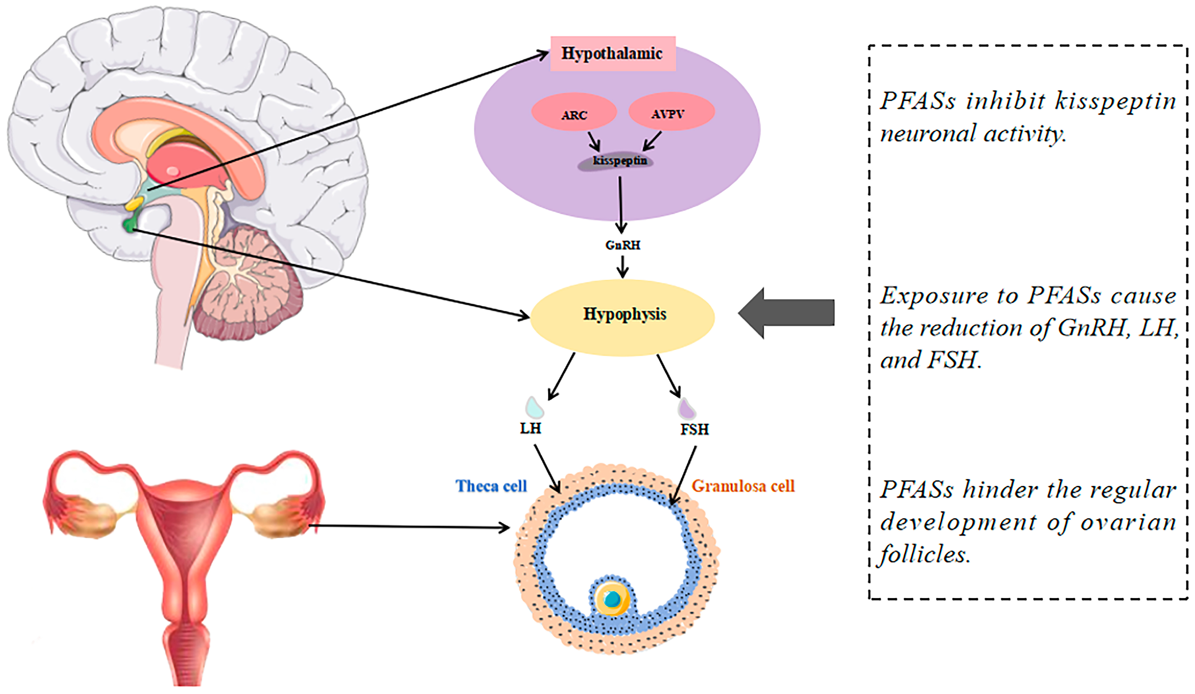
4.1. PFAS Exposure Suppresses Kisspeptin Signaling and Impairs Reproductive Hormone Regulation

Kisspeptin promotes GnRH secretion by activating signaling pathways through its interaction with the Kisspeptin receptor (Kiss1r), thereby playing a critical role in regulating the hypothalamic–pituitary–gonadal (HPG) axis [66]. PFASs interfere with steroid hormones and synthesis and normal oocyte development by modulating the HPG axis (Figure 2). In a previous study, Du et al. used female neonatal rats of the Sprague–Dawley (SD) line subcutaneously injected with PFOA or PFOS at three different doses (0.1, 1, and 10 mg/kg/day) for 5 days (PND1~5 or PND26~30). Compared with the controls, Kiss1 and Kiss1r mRNA expression in the AVPV and hypothalamic arcuate nucleus (ARC) of post-pubertal adult rats was suppressed [67]. In another study, twelve-week-old female mice were divided into four groups, one of which was given PFOS (10 mg/kg) orally for 30 days. In comparison with the controls, reductions in progesterone (P4), GnRH, and LH were observed on the seventh day of PFOS exposure, with decreased levels of E2 and thyroxine (T4) detected on the fourteenth day. These changes may be attributed to significant decreases in the number of Kiss1 cells in the AVPV nucleus, as well as reductions in AVPV-Kiss1 mRNA (p < 0.01) and AVPV-Kiss1 protein levels (p < 0.05). Furthermore, the authors discovered that PFOS could inhibit the E2-induced surge in serum LH levels, further demonstrating that PFOS exposure directly inhibits E2-activated kisspeptin neurons in the AVPV, thereby suppressing the production of the LH surge [68]. The experiment demonstrated that chronic exposure to PFOS (0.1 mg/kg/day) in adult female rats inhibits the biosynthesis of E2 by reducing the expression of StAR mRNA, which is mediated by the decreased acetylation of histone H3K14, resulting in the decreased transport of cholesterol as an essential precursor for ovarian steroidogenesis [69,70].

4.2. Disruption of Steroid Hormone Synthesis Gene Expression and Hormonal Interference by PFASs

The synthesis of steroid hormones involves a complex pathway. Cholesterol, a precursor for steroid hormone synthesis, is transported via the StAR protein to the cytoplasm of follicular membrane cells where it is converted by the enzyme cholesterol side-chain cleavage enzyme (P450scc) to pregnenolone, which is catalyzed by the enzyme 3β-hydroxysteroid dehydrogenase (3β-HSD) to produce progesterone. Progesterone is synthesized into androstenedione by the enzymes 17α-hydroxylase (CYP17A1) and the 17,20-cleaving enzyme-catalyzed synthesis of androstenedione. Cytochrome P450 aromatase (CYP19A1) converts androgens (especially testosterone and androstenedione) to estrogens (estradiol and estrone).
PFASs may interfere with steroid hormone biosynthesis in the female ovary at the molecular level. Kang et al. measured the amounts of E2 and testosterone produced after exposure to PFOA and PFOS (10 pM-10 μM) using human adrenal carcinoma cell H295R. They observed an approximately 2-fold increase in E2 levels when PFOA concentrations were 10 and 100 μM, and an approximately 2-fold increase in E2 levels when the PFOS concentration was only 100 μM; on the other hand, 100 μM of PFOA and PFOS decreased testosterone levels. After assessing the transcript levels of steroidogenic genes using qPCR, they found that PFOA and PFOS exposure increased the gene expression of 3β-HSD and CYP19 by more than 2-fold, and that CYP17 was transcriptionally activated 1.5–2-fold [71]. In contrast, in an experiment involving the in vitro culture of mice follicles, Yang et al. delivered PFOA (100 μg/mL) by oral gavage to 10- to 30-day-old mice. They found that PFOA can lead to decreased levels of estrogen and progesterone and increased testosterone levels compared with vehicle controls by downregulating the expression levels of genes for StAR, 3β-HSD, CYP17A1, and CYP19A1, which may pose a risk of premature ovarian insufficiency [72]. Similar findings were reported for porcine follicular membrane cells and granulosa cells exposed to 1.2 μM PFOS or PFOA. Compared to the control group, they observed the reduced secretion of estradiol, progesterone, and androstenedione, which was attributed to the decreased expression and enzyme activity of CYP17A1 and 17β-hydroxysteroid dehydrogenase (17β-HSD) [73].

4.3. PFASs as Estrogen Receptor Agonists and Antagonists Disrupt Estrogen Signaling and Reproductive Effects

Several PFASs can act as agonists or antagonists of the estrogen receptor and exhibit ERα-mediated estrogenic activity [74]. Benninghoff and colleagues utilized Mt Shasta strain juvenile rainbow trout as an animal model. They observed that at the highest tested concentration (1000 ppm), the mixture of PFOA/PFNA/perfluoroundecanoic acid (PFDA)/PFUnDA induced levels of the estrogen-responsive biomarker protein vitellogenin (Vtg) higher than the sum of the individual compounds. This indicates that PFASs induce Vtg expression in vivo, particularly PFOA and PFNA, which exhibit significant estrogen-like activity. Additionally, compounds such as PFOA, PFNA, PFDA, and PFOS can effectively dock with the ERα of various species (humans, mice, and trout) and form hydrogen bonds at the Arg394/398/407 site. This binding behavior suggests that they may disrupt normal estrogen signaling, potentially affecting reproduction and other physiological processes regulated by estrogen [75]. Lending further support, Li et al. conducted a combination of in vitro experiments and computer simulations with MCF-7 BUS human breast adenocarcinoma cells and MVLN cells. Specific PFAS compounds, such as PFHxS and PFOS, were found to act as estrogen receptor agonists, meaning they can mimic estrogen and activate the receptor. In contrast, a positive correlation between short-chain (C-4~C-5) PFASs, such as perfluorobutanoic acid (PFBA), PFBS and perfluoropentanoic acid (PFPeA), and anti-estrogenic activity was observed. PFASs interact with estrogen receptors by binding to their ligand-binding domains, potentially acting as agonists or antagonists, thereby affecting the expressions of estrogen-responsive genes like TFF1 and EGR3 and disrupting normal hormonal signaling [76].

4.4. Activation of the PPAR Signaling Pathway and Induction of Oxidative Stress by PFASs

PPARs are a family of nuclear hormone receptors participating in various processes that may affect ovarian function [77]. A study by Zhang et al. revealed that exposure to PFOA in mice significantly reduces the number of primordial and antral follicles. PFOA disrupted mitochondrial physiological functions and the antioxidant system by downregulating key genes associated with mitochondrial complexes and glutathione transferase in mouse ovaries. Meanwhile, PFOA impairs the mitochondrial functionality within granulosa cells, elevates mitochondrial ROS (mtROS) levels in the ovaries, and impedes the proper development of ovarian follicles [78]. Zhang et al. investigated the oral administration of PFOA to adult female mice, resulting in decreased GnRH and LH levels. This phenomenon can be attributed to PFOA’s effect of diminishing the kisspeptin–reproductive endocrine system’s activity, achieved by amplifying the expression of hepatic fibroblast growth factor 21 (FGF21, a peptide hormone that is synthesized by several organs and regulates energy homeostasis) through PPAR-α activation. Consequently, this disruption in the liver–brain reproductive endocrine axis could prolong the inter-estrus phase and, adversely, affect ovulation [79,80]. Table 2 summarizes studies on the mechanism of PFASs exposure for female reproductive system diseases.

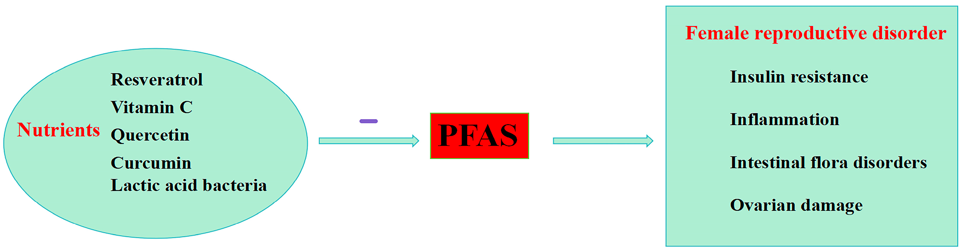
5. Nutritional Strategies to Reduce PFASs’ Effects in Female

PFASs induce oxidative stress by inhibiting mitochondrial energy production, causing mitochondrial dysfunction [81]. Antioxidants can scavenge excess ROS, helping to maintain the body’s oxidative/antioxidant balance [31]. Emerging research suggests that adopting positive lifestyle modifications, such as antioxidants and anti-inflammatory nutrients, may help reduce the vulnerability to diseases caused by environmental pollutants [82]. The intake of some nutrients can reduce the damage caused by PFASs on the female reproductive system (Figure 3).
Zhang et al. found that resveratrol supplementation reduces apoptosis by regulating FoxO1 via the SIRT1 and PI3K-AKT pathways. The antioxidant effect of resveratrol was confirmed by examining the RNA levels of mitochondrial electron transport chain-related genes in KGN cells. Resveratrol supplementation significantly increased the RNA levels of these genes, thereby reducing PFOA-induced oxidative stress. Resveratrol also contributed to the amelioration of PFOA-induced ovarian damage, including the protection of ovarian tissue structure and size, the reduction in follicular cell apoptosis, the enhancement of mitochondrial function, and the promotion of follicular development [78]. Nearly half of women diagnosed with PCOS have insulin resistance, which causes hyperinsulinemia [83]. A study from South Korea demonstrated that PFOS and perfluorododecanoic acid (PFDoDA) increase the risk of developing insulin resistance, and that vitamin C supplements protect against this adverse effect. However, this study was based on elderly subjects, and more research is still needed to prove this in those with PCOS [84]. Quercetin and curcumin help reduce the buildup and boost the elimination of PFOA in the body, lowering its bioavailability. They strengthen the intestinal barrier and reduce liver uptake by down-regulating key transport proteins. Quercetin also prevents PFOA from accumulating in the liver by decreasing certain binding proteins [85]. Curcumin, a primary polyphenolic compound found in turmeric, exerts beneficial effects on female reproductive disorders through multiple mechanisms. For PCOS, it reduces inflammation by inhibiting the nuclear factor kappa-light-chain-enhancer of activated B cells (NF-κB)-signaling pathway and lowers androgen levels, aiding follicle maturation and ovulation. In ovarian diseases, curcumin enhances ovarian health by reducing oxidative stress and preventing abnormal cell proliferation. For endometriosis, curcumin alleviates symptoms through its anti-inflammatory properties by inhibiting cyclooxygenase-2 (COX-2) and prostaglandin E2 (PGE2), reduces angiogenesis by downregulating vascular endothelial growth factor (VEGF), and induces apoptosis in endometrial cells by modulating the B-cell lymphoma 2/Bcl-2-associated X protein (Bcl-2/Bax) ratio [83]. Disturbances in intestinal flora play a role in the pathogenesis of PCOS, and PFOS accelerates the development of PCOS by disrupting intestinal environment [86]. Lactic acid bacteria can bind to PFOS and reduce the bioavailability and absorption of PFOS in the body. They also have strong antioxidant properties that help neutralize the oxidative stress caused by PFOS. In addition, LAB improve the intestinal barrier function by up-regulating the production of short-chain fatty acids (SCFAs) and enhancing the expression of tight junction proteins in the intestine [87].

6. Conclusions and Perspectives

In conclusion, the cumulative research elucidates a compelling link between PFAS exposure and various detrimental effects on female reproductive health. These include disruptions to the hormonal balance, such as those observed in cases of PCOS and endometriosis, as well as contributions to conditions like POI and DOR. These studies highlight the urgent need for further investigation into the mechanisms by which PFASs exert their effects on the female reproductive system. However, the underlying causes of these diseases are not clear. Most of the current studies are retrospective, and methodological issues limit the inference of causality in the association between exposure to PFASs and the aforementioned diseases in epidemiologic studies. Overall, there is insufficient evidence to establish a causal relationship between exposure to PFASs and diseases of the female reproductive system. Experimental studies and more prospective studies related to human exposure doses should be pursued. In this way, the detrimental effects of PFASs on women’s reproductive health are expected to be improved.

Author Contributions

Conceptualization, K.W. and Y.Y.; methodology, Y.Y.; software, Y.F.; validation, Y.S. and J.X.; formal analysis, M.L.; investigation, Y.F.; data curation, Y.Y.; writing—original draft preparation, Y.Y.; writing—review and editing, K.W.; visualization, Y.F.; supervision, J.X.; project administration, K.W.; funding acquisition, K.W. All authors have read and agreed to the published version of the manuscript.

Funding

This research was funded by Department of Science and Technology of Liaoning Province: No.2022-BS-248; the “1+X” program for Clinical Competency enhancement–Advancement in Clinical Technology Project, Second Affiliated Hospital of Dalian Medical University: No.2022LCJSGC15.

Institutional Review Board Statement

Not applicable.

Informed Consent Statement

Not applicable.

Data Availability Statement

Data availability is not applicable to this article as no new data were created in this study.

Acknowledgments

The materials used to create Figure 2 were taken from the webpage https://smart.servier.com/category/anatomy-and-the-human-body/nervous-system/brain/ (accessed on 22 July 2024).

Conflicts of Interest

The authors declare no conflicts of interest.

Abbreviations

The following abbreviations are used in this manuscript:
AMHAnti-mullerian hormone
ARCHypothalamic arcuate nucleus
AVPVAnteroventral periventricular nucleus
AFCAntral follicle count
Bcl-2/BaxB-cell lymphoma 2/Bcl-2-associated X protein
BPABisphenol A
C=F bondsCarbon-fluorine bonds
COX-2Cyclooxygenase-2
CYP17A117α-hydroxylase
CYP19A1Cytochrome P450 aromatase
DORDiminished ovarian reserve
E2Estradiol
EDCsEndocrine-disrupting chemicals
EREstrogen receptor
FGF21Fibroblast growth factor 21
FSHFollicle-stimulating hormone
FT3Free triiodothyronine
FT4Free thyroxine
GenXHexafluoropropylene oxide dimer acid
GnRHGonadotropin-releasing hormone
HFPO-DAHexafluoropropylene oxide dimer acids
HPG axisHypothalamic-Pituitary-Gonadal Axis
Kiss1rKisspeptin receptor
LHLuteinizing hormone
MtROSMitochondrial ROS
NF-κBNuclear factor kappa-light-chain-enhancer of activated B cells
NORNormal ovarian reserve
P450sccCholesterol side-chain cleavage enzyme
PCBsPolychlorinated biphenyls
PCOSPolycystic ovary syndrome
PFCAsPerfluoroalkyl carboxylic acids
PFASsPer- and polyfluoroalkyl substances
PFBAPerfluorobutanoic acid
PFBSPerfluorobutanesulfonic acid
PFDAPerfluoroundecanoic acid
PFDoAPerfluorododecanoic acid
PFDoDAPerfluorododecanoic acid
PFHxAPerfluorohexanoic acid
PFHxSPerfluorohexane sulfonate
PFNAPerfluorononanoic acid
PFOAPerfluorooctanoic acid
PFOSPerfluorooctane sulfonate
PFPeAPerfluoropentanoic acid
PFUnDAPerfluoroundecanoic acid
PFHxSPerfluorohexane sulfonate
PFOSPerfluorooctane sulfonate
PFSAsPerfluoroalkane sulfonates
PFPeAPerfluoropentanoic acid
POIPremature ovarian insufficiency
P4Progesterone
PGE2Prostaglandin E2
PPARsPeroxisome proliferators-activated receptors
PRLProlactin
SCFAsShort-chain fatty acids
TTestosterone
T4Thyroxine
TSHThyroid-stimulating hormone
UCAUrocanic acid
VEGFVascular endothelial growth factor
17β-HSD17β-hydroxysteroid dehydrogenase
3β-HSD3β-hydroxysteroid dehydrogenase
6:2 Cl-PFESA6:2 chlorinated polyfluorinated ether sulfonate

References

  1. Wallington, T.J.; Andersen, M.P.S.; Nielsen, O.J. The Case for a More Precise Definition of Regulated PFAS. Environ. Sci. Process. Impacts 2021, 23, 1834–1838. [Google Scholar] [CrossRef] [PubMed]
  2. Buck, R.C.; Franklin, J.; Berger, U.; Conder, J.M.; Cousins, I.T.; De Voogt, P.; Jensen, A.A.; Kannan, K.; Mabury, S.A.; Van Leeuwen, S.P. Perfluoroalkyl and Polyfluoroalkyl Substances in the Environment: Terminology, Classification, and Origins. Integr. Environ. Assess. Manag. 2011, 7, 513–541. [Google Scholar] [CrossRef] [PubMed]
  3. Ateia, M.; Maroli, A.; Tharayil, N.; Karanfil, T. The Overlooked Short- and Ultrashort-Chain Poly- and Perfluorinated Substances: A Review. Chemosphere 2019, 220, 866–882. [Google Scholar] [CrossRef] [PubMed]
  4. Panieri, E.; Baralic, K.; Djukic-Cosic, D.; Buha Djordjevic, A.; Saso, L. PFAS Molecules: A Major Concern for the Human Health and the Environment. Toxics 2022, 10, 44. [Google Scholar] [CrossRef] [PubMed]
  5. Sunderland, E.M.; Hu, X.C.; Dassuncao, C.; Tokranov, A.K.; Wagner, C.C.; Allen, J.G. A Review of the Pathways of Human Exposure to Poly- and Perfluoroalkyl Substances (PFASs) and Present Understanding of Health Effects. J. Expo. Sci. Environ. Epidemiol. 2019, 29, 131–147. [Google Scholar] [CrossRef]
  6. Evich, M.G.; Davis, M.J.B.; McCord, J.P.; Acrey, B.; Awkerman, J.A.; Knappe, D.R.U.; Lindstrom, A.B.; Speth, T.F.; Tebes-Stevens, C.; Strynar, M.J.; et al. Per- and Polyfluoroalkyl Substances in the Environment. Science 2022, 375, eabg9065. [Google Scholar] [CrossRef] [PubMed]
  7. Podder, A.; Sadmani, A.H.M.A.; Reinhart, D.; Chang, N.-B.; Goel, R. Per and Poly-Fluoroalkyl Substances (PFAS) as a Contaminant of Emerging Concern in Surface Water: A Transboundary Review of Their Occurrences and Toxicity Effects. J. Hazard. Mater. 2021, 419, 126361. [Google Scholar] [CrossRef] [PubMed]
  8. Lin, H.; Taniyasu, S.; Yamazaki, E.; Wu, R.; Lam, P.K.S.; Eun, H.; Yamashita, N. Fluorine Mass Balance Analysis and Per- and Polyfluoroalkyl Substances in the Atmosphere. J. Hazard. Mater. 2022, 435, 129025. [Google Scholar] [CrossRef]
  9. Bangma, J.; Guillette, T.C.; Bommarito, P.A.; Ng, C.; Reiner, J.L.; Lindstrom, A.B.; Strynar, M.J. Understanding the Dynamics of Physiological Changes, Protein Expression, and PFAS in Wildlife. Environ. Int. 2022, 159, 107037. [Google Scholar] [CrossRef] [PubMed]
  10. De Silva, A.O.; Armitage, J.M.; Bruton, T.A.; Dassuncao, C.; Heiger-Bernays, W.; Hu, X.C.; Kärrman, A.; Kelly, B.; Ng, C.; Robuck, A.; et al. PFAS Exposure Pathways for Humans and Wildlife: A Synthesis of Current Knowledge and Key Gaps in Understanding. Environ. Toxicol. Chem. 2021, 40, 631–657. [Google Scholar] [CrossRef]
  11. EFSA Panel on Contaminants in the Food Chain (CONTAM); Knutsen, H.K.; Alexander, J.; Barregård, L.; Bignami, M.; Brüschweiler, B.; Ceccatelli, S.; Cottrill, B.; Dinovi, M.; Edler, L.; et al. Risk to Human Health Related to the Presence of Perfluorooctane Sulfonic Acid and Perfluorooctanoic Acid in Food. EFSA J. 2018, 16, e05194. [Google Scholar] [CrossRef]
  12. Giffard, N.G.; Gitlin, S.A.; Rardin, M.; Petali, J.M.; Chen, C.Y.; Romano, M.E. Occurrence and Risks of Per- and Polyfluoroalkyl Substances in Shellfish. Curr. Environ. Health Rep. 2022, 9, 591–603. [Google Scholar] [CrossRef] [PubMed]
  13. Wee, S.Y.; Aris, A.Z. Environmental Impacts, Exposure Pathways, and Health Effects of PFOA and PFOS. Ecotoxicol. Environ. Saf. 2023, 267, 115663. [Google Scholar] [CrossRef] [PubMed]
  14. Zhang, Y.-T.; Zeeshan, M.; Su, F.; Qian, Z.-M.; Dee Geiger, S.; Edward McMillin, S.; Wang, Z.-B.; Dong, P.-X.; Ou, Y.-Q.; Xiong, S.-M.; et al. Associations between Both Legacy and Alternative Per- and Polyfluoroalkyl Substances and Glucose-Homeostasis: The Isomers of C8 Health Project in China. Environ. Int. 2022, 158, 106913. [Google Scholar] [CrossRef] [PubMed]
  15. UNEP. Stockholm Convention on Persistent Organic Pollutants (POPs). 2021. Available online: https://chm.pops.int/TheConvention/POPsReviewCommittee/Meetings/POPRC16/Overview/tabid/8472/Default.aspx (accessed on 5 May 2024).
  16. UNEP. Stockholm Convention on Persistent Organic Pollutants (POPs). 2022. Available online: http://chm.pops.int/TheConvention/POPsReviewCommittee/Meetings/POPRC17/Overview/tabid/8900/Default.aspx (accessed on 5 May 2024).
  17. Brendel, S.; Fetter, É.; Staude, C.; Vierke, L.; Biegel-Engler, A. Short-Chain Perfluoroalkyl Acids: Environmental Concerns and a Regulatory Strategy under REACH. Environ. Sci. Eur. 2018, 30, 9. [Google Scholar] [CrossRef] [PubMed]
  18. Fenton, S.E.; Ducatman, A.; Boobis, A.; DeWitt, J.C.; Lau, C.; Ng, C.; Smith, J.S.; Roberts, S.M. Per- and Polyfluoroalkyl Substance Toxicity and Human Health Review: Current State of Knowledge and Strategies for Informing Future Research. Environ. Toxicol. Chem. 2021, 40, 606–630. [Google Scholar] [CrossRef]
  19. Silva, A.B.P.; Carreiró, F.; Ramos, F.; Sanches-Silva, A. The Role of Endocrine Disruptors in Female Infertility. Mol. Biol. Rep. 2023, 50, 7069–7088. [Google Scholar] [CrossRef] [PubMed]
  20. Rickard, B.P.; Rizvi, I.; Fenton, S.E. Per- and Poly-Fluoroalkyl Substances (PFAS) and Female Reproductive Outcomes: PFAS Elimination, Endocrine-Mediated Effects, and Disease. Toxicology 2022, 465, 153031. [Google Scholar] [CrossRef] [PubMed]
  21. Vander Borght, M.; Wyns, C. Fertility and Infertility: Definition and Epidemiology. Clin. Biochem. 2018, 62, 2–10. [Google Scholar] [CrossRef] [PubMed]
  22. Wang, Y.; Chang, W.; Wang, L.; Zhang, Y.; Zhang, Y.; Wang, M.; Wang, Y.; Li, P. A Review of Sources, Multimedia Distribution and Health Risks of Novel Fluorinated Alternatives. Ecotoxicol. Environ. Saf. 2019, 182, 109402. [Google Scholar] [CrossRef]
  23. Chambers, W.S.; Hopkins, J.G.; Richards, S.M. A Review of Per- and Polyfluorinated Alkyl Substance Impairment of Reproduction. Front. Toxicol. 2021, 3, 732436. [Google Scholar] [CrossRef] [PubMed]
  24. Shi, W.; Zhang, Z.; Li, M.; Dong, H.; Li, J. Reproductive Toxicity of PFOA, PFOS and Their Substitutes: A Review Based on Epidemiological and Toxicological Evidence. Environ. Res. 2024, 250, 118485. [Google Scholar] [CrossRef] [PubMed]
  25. Roepke, T.A.; Sadlier, N.C. REPRODUCTIVE TOXICOLOGY: Impact of Endocrine Disruptors on Neurons Expressing GnRH or Kisspeptin and Pituitary Gonadotropins. Reproduction 2021, 162, F131–F145. [Google Scholar] [CrossRef] [PubMed]
  26. Ding, N.; Harlow, S.D.; Randolph, J.F., Jr.; Loch-Caruso, R.; Park, S.K. Perfluoroalkyl and Polyfluoroalkyl Substances (PFAS) and Their Effects on the Ovary. Hum. Reprod. Update 2020, 26, 724–752. [Google Scholar] [CrossRef] [PubMed]
  27. Grønnestad, R.; Johanson, S.M.; Müller, M.H.B.; Schlenk, D.; Tanabe, P.; Krøkje, Å.; Jaspers, V.L.B.; Jenssen, B.M.; Ræder, E.M.; Lyche, J.L.; et al. Effects of an Environmentally Relevant PFAS Mixture on Dopamine and Steroid Hormone Levels in Exposed Mice. Toxicol. Appl. Pharmacol. 2021, 428, 115670. [Google Scholar] [CrossRef] [PubMed]
  28. Dangudubiyyam, S.V.; Mishra, J.S.; Kumar, S. Perfluorooctane Sulfonic Acid Modulates Expression of Placental Steroidogenesis-Associated Genes and Hormone Levels in Pregnant Rats. Reprod. Toxicol. 2023, 118, 108390. [Google Scholar] [CrossRef] [PubMed]
  29. Fort, D.J.; Mathis, M.B.; Fort, C.E.; Fort, H.M.; Fort, T.D.; Guiney, P.D.; Weeks, J.A. Effect of Perfluorooctanesulfonate Exposure on Steroid Hormone Levels and Steroidogenic Enzyme Activities in Juvenile Silurana tropicalis. J. Appl. Toxicol. 2019, 39, 1066–1078. [Google Scholar] [CrossRef] [PubMed]
  30. Villeneuve, D.L.; Blackwell, B.R.; Cavallin, J.E.; Collins, J.; Hoang, J.X.; Hofer, R.N.; Houck, K.A.; Jensen, K.M.; Kahl, M.D.; Kutsi, R.N.; et al. Verification of In Vivo Estrogenic Activity for Four Per- and Polyfluoroalkyl Substances (PFAS) Identified as Estrogen Receptor Agonists via New Approach Methodologies. Environ. Sci. Technol. 2023, 57, 3794–3803. [Google Scholar] [CrossRef] [PubMed]
  31. Agarwal, A.; Aponte-Mellado, A.; Premkumar, B.J.; Shaman, A.; Gupta, S. The Effects of Oxidative Stress on Female Reproduction: A Review. Reprod. Biol. Endocrinol. 2012, 10, 49. [Google Scholar] [CrossRef] [PubMed]
  32. Rudnicka, E.; Duszewska, A.M.; Kucharski, M.; Tyczyński, P.; Smolarczyk, R. Oxidative Stress in Polycystic Ovary Syndrome. Reproduction 2022, 164, F145–F154. [Google Scholar] [CrossRef]
  33. Xi, Y.; Zhang, Y.; Zhu, S.; Luo, Y.; Xu, P.; Huang, Z. PPAR-Mediated Toxicology and Applied Pharmacology. Cells 2020, 9, 352. [Google Scholar] [CrossRef]
  34. Komar, C.M. Peroxisome Proliferator-Activated Receptors (PPARs) and Ovarian Function—Implications for Regulating Steroidogenesis, Differentiation, and Tissue Remodeling. Reprod. Biol. Endocrinol. 2005, 3, 41. [Google Scholar] [CrossRef]
  35. Escobar-Morreale, H.F. Polycystic Ovary Syndrome: Definition, Aetiology, Diagnosis and Treatment. Nat. Rev. Endocrinol. 2018, 14, 270–284. [Google Scholar] [CrossRef] [PubMed]
  36. Balen, A.H.; Morley, L.C.; Misso, M.; Franks, S.; Legro, R.S.; Wijeyaratne, C.N.; Stener-Victorin, E.; Fauser, B.C.J.M.; Norman, R.J.; Teede, H. The Management of Anovulatory Infertility in Women with Polycystic Ovary Syndrome: An Analysis of the Evidence to Support the Development of Global WHO Guidance. Hum. Reprod. Update 2016, 22, 687–708. [Google Scholar] [CrossRef]
  37. Merkin, S.S.; Phy, J.L.; Sites, C.K.; Yang, D. Environmental Determinants of Polycystic Ovary Syndrome. Fertil. Steril. 2016, 106, 16–24. [Google Scholar] [CrossRef]
  38. Gautam, R.; Prambil, A.M.; Patel, A.K.; Arora, T. Emerging Pollutants in Etiology and Pathophysiology of Polycystic Ovary Syndrome. Reprod. Toxicol. 2023, 123, 108515. [Google Scholar] [CrossRef] [PubMed]
  39. Rumph, J.T.; Stephens, V.R.; Martin, J.L.; Brown, L.K.; Thomas, P.L.; Cooley, A.; Osteen, K.G.; Bruner-Tran, K.L. Uncovering Evidence: Associations between Environmental Contaminants and Disparities in Women’s Health. Int. J. Environ. Res. Public Health 2022, 19, 1257. [Google Scholar] [CrossRef] [PubMed]
  40. Vagi, S.J.; Azziz-Baumgartner, E.; Sjödin, A.; Calafat, A.M.; Dumesic, D.; Gonzalez, L.; Kato, K.; Silva, M.J.; Ye, X.; Azziz, R. Exploring the Potential Association between Brominated Diphenyl Ethers, Polychlorinated Biphenyls, Organochlorine Pesticides, Perfluorinated Compounds, Phthalates, and Bisphenol a in Polycystic Ovary Syndrome: A Case–Control Study. BMC Endocr. Disord. 2014, 14, 86. [Google Scholar] [CrossRef]
  41. Heffernan, A.L.; Cunningham, T.K.; Drage, D.S.; Aylward, L.L.; Thompson, K.; Vijayasarathy, S.; Mueller, J.F.; Atkin, S.L.; Sathyapalan, T. Perfluorinated Alkyl Acids in the Serum and Follicular Fluid of UK Women with and without Polycystic Ovarian Syndrome Undergoing Fertility Treatment and Associations with Hormonal and Metabolic Parameters. Int. J. Hyg. Environ. Health 2018, 221, 1068–1075. [Google Scholar] [CrossRef]
  42. Zhan, W.; Qiu, W.; Ao, Y.; Zhou, W.; Sun, Y.; Zhao, H.; Zhang, J. Environmental Exposure to Emerging Alternatives of Per- and Polyfluoroalkyl Substances and Polycystic Ovarian Syndrome in Women Diagnosed with Infertility: A Mixture Analysis. Environ. Health Perspect. 2023, 131, 057001. [Google Scholar] [CrossRef]
  43. Radke, E.G.; Christensen, K. Invited Perspective: Challenges in Evaluating the Effect of Per- and Polyfluoroalkyl Substance Mixtures on Polycystic Ovarian Syndrome. Environ. Health Perspect. 2023, 131, 051301. [Google Scholar] [CrossRef] [PubMed]
  44. Li, S.; Li, G.; Lin, Y.; Sun, F.; Zheng, L.; Yu, Y.; Xu, H. Association between Perfluoroalkyl Substances in Follicular Fluid and Polycystic Ovary Syndrome in Infertile Women. Toxics 2024, 12, 104. [Google Scholar] [CrossRef]
  45. Bulun, S.E.; Yilmaz, B.D.; Sison, C.; Miyazaki, K.; Bernardi, L.; Liu, S.; Kohlmeier, A.; Yin, P.; Milad, M.; Wei, J. Endometriosis. Endocr. Rev. 2019, 40, 1048–1079. [Google Scholar] [CrossRef] [PubMed]
  46. Saunders, P.T.K.; Horne, A.W. Endometriosis: Etiology, Pathobiology, and Therapeutic Prospects. Cell 2021, 184, 2807–2824. [Google Scholar] [CrossRef] [PubMed]
  47. Louis, G.M.B.; Peterson, C.M.; Chen, Z.; Hediger, M.L.; Croughan, M.S.; Sundaram, R.; Stanford, J.B.; Fujimoto, V.Y.; Varner, M.W.; Giudice, L.C.; et al. Perfluorochemicals and Endometriosis: The ENDO Study. Epidemiology 2012, 23, 799–805. [Google Scholar] [CrossRef] [PubMed]
  48. Campbell, S.; Raza, M.; Pollack, A.Z. Perfluoroalkyl Substances and Endometriosis in US Women in NHANES 2003–2006. Reprod. Toxicol. 2016, 65, 230–235. [Google Scholar] [CrossRef] [PubMed]
  49. Wang, B.; Zhang, R.; Jin, F.; Lou, H.; Mao, Y.; Zhu, W.; Zhou, W.; Zhang, P.; Zhang, J. Perfluoroalkyl Substances and Endometriosis-Related Infertility in Chinese Women. Environ. Int. 2017, 102, 207–212. [Google Scholar] [CrossRef] [PubMed]
  50. Ao, J.; Zhang, R.; Huo, X.; Zhu, W.; Zhang, J. Environmental Exposure to Legacy and Emerging Per- and Polyfluoroalkyl Substances and Endometriosis in Women of Childbearing Age. Sci. Total Environ. 2024, 907, 167838. [Google Scholar] [CrossRef] [PubMed]
  51. McGlacken-Byrne, S.M.; Conway, G.S. Premature Ovarian Insufficiency. Best Pract. Res. Clin. Obstet. Gynaecol. 2022, 81, 98–110. [Google Scholar] [CrossRef]
  52. Chon, S.J.; Umair, Z.; Yoon, M.-S. Premature Ovarian Insufficiency: Past, Present, and Future. Front. Cell Dev. Biol. 2021, 9, 672890. [Google Scholar] [CrossRef]
  53. Tsiligiannis, S.; Panay, N.; Stevenson, J.C. Premature Ovarian Insufficiency and Long-Term Health Consequences. Curr. Vasc. Pharmacol. 2019, 17, 604–609. [Google Scholar] [CrossRef] [PubMed]
  54. Szeliga, A.; Calik-Ksepka, A.; Maciejewska-Jeske, M.; Grymowicz, M.; Smolarczyk, K.; Kostrzak, A.; Smolarczyk, R.; Rudnicka, E.; Meczekalski, B. Autoimmune Diseases in Patients with Premature Ovarian Insufficiency—Our Current State of Knowledge. Int. J. Mol. Sci. 2021, 22, 2594. [Google Scholar] [CrossRef] [PubMed]
  55. Zhu, Q.; Li, Y.; Ma, J.; Ma, H.; Liang, X. Potential Factors Result in Diminished Ovarian Reserve: A Comprehensive Review. J. Ovarian Res. 2023, 16, 208. [Google Scholar] [CrossRef] [PubMed]
  56. Ding, T.; Yan, W.; Zhou, T.; Shen, W.; Wang, T.; Li, M.; Zhou, S.; Wu, M.; Dai, J.; Huang, K.; et al. Endocrine Disrupting Chemicals Impact on Ovarian Aging: Evidence from Epidemiological and Experimental Evidence. Environ. Pollut. 2022, 305, 119269. [Google Scholar] [CrossRef] [PubMed]
  57. Wang, X.; Wang, L.; Xiang, W. Mechanisms of Ovarian Aging in Women: A Review. J. Ovarian Res. 2023, 16, 67. [Google Scholar] [CrossRef] [PubMed]
  58. Zhang, S.; Tan, R.; Pan, R.; Xiong, J.; Tian, Y.; Wu, J.; Chen, L. Association of Perfluoroalkyl and Polyfluoroalkyl Substances With Premature Ovarian Insufficiency in Chinese Women. J. Clin. Endocrinol. Metab. 2018, 103, 2543–2551. [Google Scholar] [CrossRef] [PubMed]
  59. Penzias, A.; Azziz, R.; Bendikson, K.; Falcone, T.; Hansen, K.; Hill, M.; Hurd, W.; Jindal, S.; Kalra, S.; Mersereau, J.; et al. Testing and Interpreting Measures of Ovarian Reserve: A Committee Opinion. Fertil. Steril. 2020, 114, 1151–1157. [Google Scholar] [CrossRef] [PubMed]
  60. Liao, K.; Wang, Y.; Zheng, L.; Lu, D.; Wu, J.; Wu, B.; Wu, Z.; Jiang, Z. Effect of Folic Acid Supplementation on Diminished Ovarian Reserve: Study Protocol of a Single-Centre, Open-Label, Randomised, Placebo-Controlled Clinical Trial. BMJ Open 2022, 12, e057689. [Google Scholar] [CrossRef] [PubMed]
  61. Björvang, R.D.; Hallberg, I.; Pikki, A.; Berglund, L.; Pedrelli, M.; Kiviranta, H.; Rantakokko, P.; Ruokojärvi, P.; Lindh, C.H.; Olovsson, M.; et al. Follicular Fluid and Blood Levels of Persistent Organic Pollutants and Reproductive Outcomes among Women Undergoing Assisted Reproductive Technologies. Environ. Res. 2022, 208, 112626. [Google Scholar] [CrossRef]
  62. Tian, T.; Hao, Y.; Wang, Y.; Xu, X.; Long, X.; Yan, L.; Zhao, Y.; Qiao, J. Mixed and Single Effects of Endocrine Disrupting Chemicals in Follicular Fluid on Likelihood of Diminished Ovarian Reserve: A Case-Control Study. Chemosphere 2023, 330, 138727. [Google Scholar] [CrossRef]
  63. Bellavia, A.; Zou, R.; Björvang, R.D.; Roos, K.; Sjunnesson, Y.; Hallberg, I.; Holte, J.; Pikki, A.; Lenters, V.; Portengen, L.; et al. Association between Chemical Mixtures and Female Fertility in Women Undergoing Assisted Reproduction in Sweden and Estonia. Environ. Res. 2023, 216 (Pt 1), 114447. [Google Scholar] [CrossRef]
  64. Shen, H.; Gao, M.; Li, Q.; Sun, H.; Jiang, Y.; Liu, L.; Wu, J.; Yu, X.; Jia, T.; Xin, Y.; et al. Effect of PFOA Exposure on Diminished Ovarian Reserve and Its Metabolism. Reprod. Biol. Endocrinol. 2023, 21, 16. [Google Scholar] [CrossRef] [PubMed]
  65. Hong, A.; Zhuang, L.; Cui, W.; Lu, Q.; Yang, P.; Su, S.; Wang, B.; Zhang, G.; Chen, D. Per- and Polyfluoroalkyl Substances (PFAS) Exposure in Women Seeking in Vitro Fertilization-Embryo Transfer Treatment (IVF-ET) in China: Blood-Follicular Transfer and Associations with IVF-ET Outcomes. Sci. Total Environ. 2022, 838 Pt 3, 156323. [Google Scholar] [CrossRef]
  66. Xie, Q.; Kang, Y.; Zhang, C.; Xie, Y.; Wang, C.; Liu, J.; Yu, C.; Zhao, H.; Huang, D. The Role of Kisspeptin in the Control of the Hypothalamic-Pituitary-Gonadal Axis and Reproduction. Front. Endocrinol. 2022, 13, 925206. [Google Scholar] [CrossRef] [PubMed]
  67. Du, G.; Hu, J.; Huang, Z.; Yu, M.; Lu, C.; Wang, X.; Wu, D. Neonatal and Juvenile Exposure to Perfluorooctanoate (PFOA) and Perfluorooctane Sulfonate (PFOS): Advance Puberty Onset and Kisspeptin System Disturbance in Female Rats. Ecotoxicol. Environ. Saf. 2019, 167, 412–421. [Google Scholar] [CrossRef] [PubMed]
  68. Wang, X.; Bai, Y.; Tang, C.; Cao, X.; Chang, F.; Chen, L. Impact of Perfluorooctane Sulfonate on Reproductive Ability of Female Mice through Suppression of Estrogen Receptor α-Activated Kisspeptin Neurons. Toxicol. Sci. 2018, 165, 475–486. [Google Scholar] [CrossRef] [PubMed]
  69. Feng, X.; Wang, X.; Cao, X.; Xia, Y.; Zhou, R.; Chen, L. Chronic Exposure of Female Mice to an Environmental Level of Perfluorooctane Sulfonate Suppresses Estrogen Synthesis Through Reduced Histone H3K14 Acetylation of the StAR Promoter Leading to Deficits in Follicular Development and Ovulation. Toxicol. Sci. 2015, 148, 368–379. [Google Scholar] [CrossRef] [PubMed]
  70. Fletcher, T.; Galloway, T.S.; Melzer, D.; Holcroft, P.; Cipelli, R.; Pilling, L.C.; Mondal, D.; Luster, M.; Harries, L.W. Associations between PFOA, PFOS and Changes in the Expression of Genes Involved in Cholesterol Metabolism in Humans. Environ. Int. 2013, 57–58, 2–10. [Google Scholar] [CrossRef] [PubMed]
  71. Kang, J.S.; Choi, J.-S.; Park, J.-W. Transcriptional Changes in Steroidogenesis by Perfluoroalkyl Acids (PFOA and PFOS) Regulate the Synthesis of Sex Hormones in H295R Cells. Chemosphere 2016, 155, 436–443. [Google Scholar] [CrossRef]
  72. Yang, M.; Lee, Y.; Gao, L.; Chiu, K.; Meling, D.D.; Flaws, J.A.; Warner, G.R. Perfluorooctanoic Acid Disrupts Ovarian Steroidogenesis and Folliculogenesis in Adult Mice. Toxicol. Sci. 2022, 186, 260–268. [Google Scholar] [CrossRef]
  73. Chaparro-Ortega, A.; Betancourt, M.; Rosas, P.; Vázquez-Cuevas, F.G.; Chavira, R.; Bonilla, E.; Casas, E.; Ducolomb, Y. Endocrine Disruptor Effect of Perfluorooctane Sulfonic Acid (PFOS) and Perfluorooctanoic Acid (PFOA) on Porcine Ovarian Cell Steroidogenesis. Toxicol. Vitr. 2018, 46, 86–93. [Google Scholar] [CrossRef] [PubMed]
  74. Tan, Y.; Zeng, Z.; Liang, H.; Weng, X.; Yao, H.; Fu, Y.; Li, Y.; Chen, J.; Wei, X.; Jing, C. Association between Perfluoroalkyl and Polyfluoroalkyl Substances and Women’s Infertility, NHANES 2013–2016. Int. J. Environ. Res. Public Health 2022, 19, 15348. [Google Scholar] [CrossRef] [PubMed]
  75. Benninghoff, A.D.; Bisson, W.H.; Koch, D.C.; Ehresman, D.J.; Kolluri, S.K.; Williams, D.E. Estrogen-Like Activity of Perfluoroalkyl Acids In Vivo and Interaction with Human and Rainbow Trout Estrogen Receptors In Vitro. Toxicol. Sci. 2011, 120, 42–58. [Google Scholar] [CrossRef] [PubMed]
  76. Li, J.; Cao, H.; Feng, H.; Xue, Q.; Zhang, A.; Fu, J. Evaluation of the Estrogenic/Antiestrogenic Activities of Perfluoroalkyl Substances and Their Interactions with the Human Estrogen Receptor by Combining In Vitro Assays and In Silico Modeling. Environ. Sci. Technol. 2020, 54, 14514–14524. [Google Scholar] [CrossRef] [PubMed]
  77. Komar, C.M.; Braissant, O.; Wahli, W.; Curry, T.E. Expression and Localization of PPARs in the Rat Ovary During Follicular Development and the Periovulatory Period. Endocrinology 2001, 142, 4831–4838. [Google Scholar] [CrossRef] [PubMed]
  78. Zhang, Z.; Tian, J.; Liu, W.; Zhou, J.; Zhang, Y.; Ding, L.; Sun, H.; Yan, G.; Sheng, X. Perfluorooctanoic Acid Exposure Leads to Defect in Follicular Development through Disrupting the Mitochondrial Electron Transport Chain in Granulosa Cells. Sci. Total Environ. 2023, 905, 166954. [Google Scholar] [CrossRef]
  79. Zhang, Y.; Cao, X.; Chen, L.; Qin, Y.; Xu, Y.; Tian, Y.; Chen, L. Exposure of Female Mice to Perfluorooctanoic Acid Suppresses Hypothalamic Kisspeptin-Reproductive Endocrine System through Enhanced Hepatic Fibroblast Growth Factor 21 Synthesis, Leading to Ovulation Failure and Prolonged Dioestrus. J. Neuroendocrinol. 2020, 32, e12848. [Google Scholar] [CrossRef] [PubMed]
  80. Fisher, F.M.; Maratos-Flier, E. Understanding the Physiology of FGF21. Annu. Rev. Physiol. 2016, 78, 223–241. [Google Scholar] [CrossRef] [PubMed]
  81. Bonato, M.; Corrà, F.; Bellio, M.; Guidolin, L.; Tallandini, L.; Irato, P.; Santovito, G. PFAS Environmental Pollution and Antioxidant Responses: An Overview of the Impact on Human Field. Int. J. Environ. Res. Public Health 2020, 17, 8020. [Google Scholar] [CrossRef]
  82. Hennig, B.; Petriello, M.C.; Gamble, M.V.; Surh, Y.-J.; Kresty, L.A.; Frank, N.; Rangkadilok, N.; Ruchirawat, M.; Suk, W.A. The Role of Nutrition in Influencing Mechanisms Involved in Environmentally Mediated Diseases. Rev. Environ. Health 2018, 33, 87–97. [Google Scholar] [CrossRef]
  83. Kamal, D.A.M.; Salamt, N.; Yusuf, A.N.M.; Kashim, M.I.A.M.; Mokhtar, M.H. Potential Health Benefits of Curcumin on Female Reproductive Disorders: A Review. Nutrients 2021, 13, 3126. [Google Scholar] [CrossRef]
  84. Kim, J.H.; Park, H.Y.; Jeon, J.D.; Kho, Y.; Kim, S.-K.; Park, M.-S.; Hong, Y.-C. The Modifying Effect of Vitamin C on the Association between Perfluorinated Compounds and Insulin Resistance in the Korean Elderly: A Double-Blind, Randomized, Placebo-Controlled Crossover Trial. Eur. J. Nutr. 2016, 55, 1011–1020. [Google Scholar] [CrossRef]
  85. Chen, Y.; Wu, H.; Cui, X. Influence of Dietary Bioactive Compounds on the Bioavailability and Excretion of PFOA and Its Alternative HFPO-TA: Mechanism Exploration. Sci. Total Environ. 2023, 899, 165560. [Google Scholar] [CrossRef]
  86. Wu, X.; Song, M.; Qiu, P.; Li, F.; Wang, M.; Zheng, J.; Wang, Q.; Xu, F.; Xiao, H. Lactic Acid Bacteria Alleviates Polycystic Ovarian Syndrome by Regulating Sex Hormone Related Gut Microbiota. Food Funct. 2018, 9, 87–95. [Google Scholar] [CrossRef]
  87. Chen, Q.; Sun, S.; Mei, C.; Zhao, J.; Zhang, H.; Wang, G.; Chen, W. Capabilities of Bio-Binding, Antioxidant and Intestinal Environmental Repair Jointly Determine the Ability of Lactic Acid Bacteria to Mitigate Perfluorooctane Sulfonate Toxicity. Environ. Int. 2022, 166, 107388. [Google Scholar] [CrossRef]
Figure 1. PFASs harm the female reproductive system through various routes of exposure. A: Seafood. B: Surface water. C: Factory. D: Water-repellent clothing, cosmetic, non-stick cookware. E: Fire-fighting foam.
Toxics 12 00539 g001
Figure 2. PFASs affect the HPG axis thereby inhibiting sex hormone and follicle production.
Toxics 12 00539 g002
Figure 3. Nutritional supplements mitigate the harmful effects of PFASs on the female reproductive system.
Toxics 12 00539 g003
Table 1. Epidemiologic evidence of the association between exposure to PFASs and four female reproductive disorders.
DiseaseSample Size
(Experimental Group/Controls)
ResultsRef.
PCOS52/50Those with PFOA and PFOS serum concentrations in the highest tertile were 6.9- and 5.8-fold more likely to develop PCOS compared to controls, respectively.[40]
PCOS30/29The geometric mean concentration of PFOS was higher in the PCOS group than in the control group.[41]
PCOS366/577Higher concentrations of 6:2 Cl-PFESA, HFPO-DA, PFDoA, and ∑3,4,5 m-PFOS enhanced the risk of PCOS, especially in obese/overweight women.[42]
PCOS73/218Compared to non-PCOS women, PCOS women had higher concentrations of LH and T in their follicular fluid, and PFOA is directly involved in the pathogenesis of PCOS.[44]
Endometriosis495/131PFOA and PFNA raise the risk of endometriosis in women.[47]
Endometriosis54/699Women with endometriosis have more PFOA, PFOS, and PFNA in their blood than women who do not suffer from this condition.[48]
Endometriosis157/178PFOA and PFNA raise the risk of endometriosis in women.[49]
Endometriosis240/344Women with endometriosis have more PFOA, PFOS, and PFNA in their blood than women who are not suffering from this condition.[50]
POI120/120High exposure to PFOA, PFOS, and PFHxS is associated with increased risk of POI. These also impact sex hormone and thyroid hormone levels.[58]
DOR64/86PFUnDA and PFOA were significantly negatively correlated with OSI.[62]
DOR185/148PFHxA was strongly associated with an increased risk of DOR.[63]
DOR25/25The DOR group had higher PFOA exposure than the group with normal ovarian reserve function.[64]
Table 2. In vivo experimental evidence of the effects of PFAS exposure on female reproductive health.
Experimental SubjectsResultsRef.
Sprague–Dawley female rats Neonatal and juvenile exposure to PFOA/PFOS in female rats accelerates puberty onset, increases estradiol and LH levels, disrupts estrous cycles, and downregulates Kisspeptin system gene expression.[67]
Twelve-week-old female ICR mice PFOS exposure suppresses ERα-induced activation of AVPV–Kisspeptin neurons, leading to prolonged diestrus, reduced corpora lutea, and diminished LH surge, ultimately impairing ovulation in female mice. [68]
Twelve-week-old female ICR mice Chronic low-dose PFOS exposure in adult female mice disrupts reproductive endocrine function by reducing the histone acetylation of StAR, leading to decreased estrogen biosynthesis, impaired follicular development, and ovulation failure. [69]
H295R cellPFOA and PFOS weakly antagonize ER transactivation, alter steroid hormone levels by inducing aromatase activity, and influence the transcription of genes involved in sex hormone and aldosterone synthesis.[71]
Adult female mice PFOA disrupts ovarian function in mice both in vitro and in vivo, causing alterations in hormone levels, steroidogenic gene expression, and folliculogenesis, suggesting a potential risk for premature ovarian failure. [72]
Porcine theca and granulosa cellsIn vitro analysis reveals that PFOS and PFOA disrupt steroidogenic secretion in porcine ovarian cells, inhibiting hormone secretion even under gonadotropic stimulation.[73]
Mt Shasta strain juvenile rainbow troutPFAAs exhibit estrogen-like activity in juvenile rainbow trout and bind to estrogen receptors in various species.[75]
MCF-7 BUS and MVLN cellsVarious PFASs exhibit both estrogenic and antiestrogenic effects.[76]
Six-week-old ICR micePFOA exposure impairs follicular development in mice, increases granulosa cell mtROS and apoptosis, and results in reduced follicular reserve.[78]
Twelve-week-old female ICR miceExposure to PFOA in mice leads to the down-regulation of the Kissin–reproductive endocrine system via enhanced PPARα-mediated hepatic FGF21 expression, potentially resulting in prolonged luteal phase and ovulation failure.[79]
Disclaimer/Publisher’s Note: The statements, opinions and data contained in all publications are solely those of the individual author(s) and contributor(s) and not of MDPI and/or the editor(s). MDPI and/or the editor(s) disclaim responsibility for any injury to people or property resulting from any ideas, methods, instructions or products referred to in the content.

