

Predictors of Serotonin Syndrome in Acute Poisoning with 5-Hydroxytryptamine Modulators
 , , , , ,
Abstract
:1. Introduction
2. Materials and Methods
2.1. Study Design and Setting
2.2. Sampling and Sample Size Calculations
2.3. Inclusion Criteria
2.4. Exclusion Criteria
2.5. Compliance with Ethical Standards
2.6. Data Collection Tool
2.7. Data Analysis
3. Results
4. Discussion
5. Conclusions
6. Limitations and Recommendations
Author Contributions
Funding
Institutional Review Board Statement
Informed Consent Statement
Data Availability Statement
Acknowledgments
Conflicts of Interest
References
- Talton, C. Serotonin Syndrome/Serotonin Toxicity. Fed. Pract. 2020, 37, 452–459. [Google Scholar] [CrossRef]
- Richerson, G.B.; Buchanan, G.F. The Serotonin Axis: Shared Mechanisms in Seizures, Depression, and SUDEP. Epilepsia 2011, 52, 28–38. [Google Scholar] [CrossRef]
- Lanthier, C.; Dallemagne, P.; Lecoutey, C.; Claeysen, S.; Rochais, C. Therapeutic Modulators of the Serotonin 5-HT4 Receptor: A Patent Review (2014-Present). Expert. Opin. Ther. Pat. 2020, 30, 495–508. [Google Scholar] [CrossRef]
- Kavanagh, J.J.; Taylor, J.L. Voluntary Activation of Muscle in Humans: Does Serotonergic Neuromodulation Matter? J. Physiol. 2022, 600, 3657–3670. [Google Scholar] [CrossRef]
- Juza, R.; Vojtechova, I.; Stefkova-Mazochova, K.; Dehaen, W.; Petrasek, T.; Prchal, L.; Kobrlova, T.; Janousek, J.; Vlcek, P.; Mezeiova, E. Novel D2/5-HT Receptor Modulators Related to Cariprazine with Potential Implication to Schizophrenia Treatment. Eur. J. Med. Chem. 2022, 232, 114193. [Google Scholar] [CrossRef]
- Prakash, S.; Rathore, C.; Rana, K.; Prakash, A. Fatal Serotonin Syndrome: A Systematic Review of 56 Cases in the Literature. Clin. Toxicol. 2021, 59, 89–100. [Google Scholar] [CrossRef]
- Moss, M.J.; Hendrickson, R.G. Serotonin Toxicity: Associated Agents and Clinical Characteristics. J. Clin. Psychopharmacol. 2019, 39, 628–633. [Google Scholar] [CrossRef]
- Little, K.; Lin, C.M.; Reynolds, P.M. Delayed Serotonin Syndrome in the Setting of a Mixed Fluoxetine and Serotonin Antagonist Overdose. Am. J. Case Rep. 2018, 19, 604–607. [Google Scholar] [CrossRef]
- Scotton, W.J.; Hill, L.J.; Williams, A.C.; Barnes, N.M. Serotonin Syndrome: Pathophysiology, Clinical Features, Management, and Potential Future Directions. Int. J. Tryptophan Res. 2019, 12, 1178646919873925. [Google Scholar] [CrossRef] [PubMed]
- Mikkelsen, N.; Damkier, P.; Pedersen, S.A. Serotonin Syndrome—A Focused Review. Basic. Clin. Pharmacol. Toxicol. 2023, 133, 124–129. [Google Scholar] [CrossRef] [PubMed]
- Dunkley, E.J.C.; Isbister, G.K.; Sibbritt, D.; Dawson, A.H.; Whyte, I.M. The Hunter Serotonin Toxicity Criteria: Simple and Accurate Diagnostic Decision Rules for Serotonin Toxicity. QJM Int. J. Med. 2003, 96, 635–642. [Google Scholar] [CrossRef] [PubMed]
- Spadaro, A.; Scott, K.R.; Koyfman, A.; Long, B. High Risk and Low Prevalence Diseases: Serotonin Syndrome. Am. J. Emerg. Med. 2022, 61, 90–97. [Google Scholar] [CrossRef]
- Zein, M.; Khan, S.; Legha, J.; Wasserman, L. Toxicological Exposures and Nutritional Deficiencies in the Psychiatric Patient. In Textbook of Medical Psychiatry; American Psychiatric Pub.: Washington, DC, USA, 2020; pp. 94–95. [Google Scholar]
- Sun-edelstein, C.; Tepper, S.J.; Shapiro, R.E. Drug-Induced Serotonin: A Review. Expert Opin. Drug Saf. 2008, 7, 587–596. [Google Scholar] [CrossRef] [PubMed]
- Mackay, F.J.; Dunn, N.R.; Mann, R.D. Antidepressants and the Serotonin Syndrome in General Practice. Br. J. Gen. Pract. 1999, 49, 871–874. [Google Scholar]
- Koury, K.M.; Tsui, B.; Gulur, P. Incidence of Serotonin Syndrome in Patients Treated with Fentanyl on Serotonergic Agents. Pain. Physician 2015, 18, E27–E30. [Google Scholar]
- Isbister, G.K.; Bowe, S.J.; Dawson, A.; Whyte, I.M. Relative Toxicity of Selective Serotonin Reuptake Inhibitors (SSRIs) in Overdose. J. Toxicol. Clin. Toxicol. 2004, 42, 277–285. [Google Scholar] [CrossRef]
- Prakash, S.; Rathore, C.; Rana, K. The Prevalence of Serotonin Syndrome in an Intensive Care Unit: A Prospective Observational Study. J. Crit. Care 2021, 63, 92–97. [Google Scholar] [CrossRef]
- Sternbach, H. The Serotonin Syndrome. Am. J. Psychiatry 1991, 148, 705–713. [Google Scholar] [PubMed]
- Carlsson, A.; Svennerholm, L.; Winblad, B. Seasonal and Circadian Monoamine Variations in Human Brains Examined Post Mortem. Acta Psychiatr. Scand. 1980, 61, 75–85. [Google Scholar] [CrossRef]
- Gupta, A.; Sharma, P.K.; Garg, V.K.; Singh, A.K.; Mondal, S.C. Role of Serotonin in Seasonal Affective Disorder. Eur. Rev. Med. Pharmacol. Sci. 2013, 17, 49–55. [Google Scholar]
- Francescangeli, J.; Karamchandani, K.; Powell, M.; Bonavia, A. The Serotonin Syndrome: From Molecular Mechanisms to Clinical Practice. Int. J. Mol. Sci. 2019, 20, 2288. [Google Scholar] [CrossRef]
- Gillman, P.K. The Serotonin Syndrome and Its Treatment. J. Psychopharmacol. 1999, 13, 100–109. [Google Scholar] [CrossRef]
- Koekkoek, W.A.C.; Tjan, D.H.T. The Serotonin Syndrome—A Narrative Review. Hemoglobin 2017, 9, 7. [Google Scholar]
- Oates, J.A.; Sjoerdsma, A. Neurologic Effects of Tryptophan in Patients Receiving a Monoamine Oxidase Inhibitor. Neurology 1960, 10, 1076. [Google Scholar] [CrossRef] [PubMed]
- GK, I. Comment: Serotonin Syndrome and 5-HT2A Antagonism. Ann. Pharmacother. 2001, 35, 1143–1144. [Google Scholar]
- Bodner, R.A.; Lynch, T.; Lewis, L.; Kahn, D. Serotonin Syndrome. Neurology 1995, 45, 219–223. [Google Scholar] [CrossRef]
- Isbister, G.K.; Buckley, N.A. The Pathophysiology of Serotonin Toxicity in Animals and Humans: Implications for Diagnosis and Treatment. Clin. Neuropharmacol. 2005, 28, 205–214. [Google Scholar] [CrossRef]
- Fraser, J.; South, M. Life-Threatening Fluvoxamine Overdose in a 4-Year-Old Child. Intensive Care Med. 1999, 25, 548. [Google Scholar] [CrossRef] [PubMed]
- Martin, T.G. Serotonin Syndrome. Ann. Emerg. Med. 1996, 28, 520–526. [Google Scholar] [CrossRef]
- Sporer, K.A. The Serotonin Syndrome: Implicated Drugs, Pathophysiology and Management. Drug Saf. 1995, 13, 94–104. [Google Scholar] [CrossRef]
- Gillman, P.K. A Review of Serotonin Toxicity Data: Implications for the Mechanisms of Antidepressant Drug Action. Biol. Psychiatry 2006, 59, 1046–1051. [Google Scholar]
- Schifano, F.; Chiappini, S.; Miuli, A.; Corkery, J.M.; Scherbaum, N.; Napoletano, F.; Arillotta, D.; Zangani, C.; Catalani, V.; Vento, A. New Psychoactive Substances (NPS) and Serotonin Syndrome Onset: A Systematic Review. Exp. Neurol. 2021, 339, 113638. [Google Scholar] [CrossRef]
- Lizer, M.H.; Masters, K.P. Combination of Methylphenidate, Venlafaxine, and Escitalopram Resulting in Serotonin Syndrome. J. Pharm. Technol. 2006, 22, 110–113. [Google Scholar] [CrossRef]
- Silins, E.; Copeland, J.; Dillon, P. Recreational Ecstasy/MDMA, the Serotonin Syndrome, and Serotonergic Neurotoxicity. Pharmacol. Biochem. Behav. 2002, 71, 837–844. [Google Scholar]
- Mir, S.; Taylor, D. Serotonin Syndrome. Psychiatr. Bull. 1999, 23, 742–747. [Google Scholar] [CrossRef]
- Silins, E.; Copeland, J.; Dillon, P. Qualitative Review of Serotonin Syndrome, Ecstasy (MDMA) and the Use of Other Serotonergic Substances: Hierarchy of Risk. Aust. N. Z. J. Psychiatry 2007, 41, 649–655. [Google Scholar]
- Foong, A.-L.; Grindrod, K.A.; Patel, T.; Kellar, J. Demystifying Serotonin Syndrome (or Serotonin Toxicity). Can. Fam. Physician 2018, 64, 720–727. [Google Scholar]
- Volpi-Abadie, J.; Kaye, A.M.; Kaye, A.D. Serotonin Syndrome. Ochsner J. 2013, 13, 533–540. [Google Scholar]
- Paz-Ramos, M.I.; Cruz, S.L.; Violante-Soria, V. Amphetamine-Type Stimulants: Novel Insights into Their Actions and Use Patterns. Rev. Investig. Clínica 2023, 75, 143–157. [Google Scholar] [CrossRef]
- Mason, P.J.; Morris, V.A.; Balcezak, T.J. Serotonin Syndrome Presentation of 2 Cases and Review of the Literature. Medicine 2000, 79, 201–209. [Google Scholar] [CrossRef]
- Park, H.W.; Park, H.Y.; Kim, H.B.; Park, K.W.; Lee, S.H.; Lee, H.W.; Lee, J.W.; Hwang, T.S. Classify the Acute Drug Intoxication Patients with Poisoning Severity Score (PSS) and Calculate the Optimal Cutoff Value of PSS, PSSsum to Predict Poor Prognosis. J. Korean Soc. Clin. Toxicol. 2018, 16, 75–85. [Google Scholar] [CrossRef]
- Schwarz, E.S.; Kopec, K.T.; Wiegand, T.J.; Wax, P.M.; Brent, J. Should We Be Using the Poisoning Severity Score? J. Med. Toxicol. 2017, 13, 135–145. [Google Scholar] [CrossRef] [PubMed]
- Ponnusankar, S.; Rangadham, P.; Roy, D.; Kutty, J.J.; Manu, P.; Stanly, S. Assessment of Poisoning Pattern, Severity and Clinical Outcome Using Clinical Scoring Systems in Secondary Care Public Hospital in South India. Res. J. Pharm. Technol. 2019, 12, 4767–4770. [Google Scholar] [CrossRef]
- Sharif, A.F.; Shaheen, R.S.; Alsubaie, D.S.; Alshabibi, R.A.; Abusamak, F.W.; AlNasser, S.; Al-Mulhim, K.A.; Abdelgawad, I.I. Performance of Several Clinical Scoring Systems as Predictors of Adverse Outcomes in Acute Exposure to Toxic Alcohols. Toxicol. Res. 2024, 13, tfae069. [Google Scholar] [CrossRef]
- Seok, S.-J.; Choi, S.-C.; Gil, H.-W.; Yang, J.-O.; Lee, E.-Y.; Song, H.-Y.; Hong, S.-Y. Acute Oral Poisoning Due to Chloracetanilide Herbicides. J. Korean Med. Sci. 2012, 27, 111–114. [Google Scholar] [CrossRef]
- Sharif, A.F.; Kasemy, Z.A.; Alshabibi, R.A.; Almufleh, S.J.; Abousamak, F.W.; Alfrayan, A.A.; Alshehri, M.; Alemies, R.A.; Almuhsen, A.S.; AlNasser, S.N.; et al. Prognostic Factors in Acute Poisoning with Central Nervous System Xenobiotics: Development of a Nomogram Predicting Risk of Intensive Care Unit Admission. Toxicol. Res. 2023, 12, 62–75. [Google Scholar] [CrossRef] [PubMed]
- Sundari, S.P.; Adithyan, C. Comparison of apache II, sofa, poison severity scoring system in predicting outcome of the patients admitted in toxicology icu in tertiary care hospital. Int. J. Acad. Med. Pharm. 2023, 5, 1474–1478. [Google Scholar]
- Lashin, H.I.; Sharif, A.F. Evaluation of Various Scoring Systems as Predictors of the Need for Intensive Care Unit Admission and Other Adverse Outcomes among Patients with Acute Clozapine Poisoning. Toxicol. Res. 2023, 12, 468–479. [Google Scholar] [CrossRef] [PubMed]
- Berger, M.; Gray, J.A.; Roth, B.L. The Expanded Biology of Serotonin. Annu. Rev. Med. 2009, 60, 355–366. [Google Scholar] [CrossRef]
- van Ewijk, C.E.; Jacobs, G.E.; Girbes, A.R.J. Unsuspected Serotonin Toxicity in the ICU. Ann. Intensive Care 2016, 6, 85. [Google Scholar] [CrossRef]
- Tomaselli, G.; Modestin, J. Repetition of Serotonin Syndrome after Reexposure to SSRI. Pharmacopsychiatry 2004, 37, 236–238. [Google Scholar] [CrossRef] [PubMed]
- Côté, F.; Thévenot, E.; Fligny, C.; Fromes, Y.; Darmon, M.; Ripoche, M.-A.; Bayard, E.; Hanoun, N.; Saurini, F.; Lechat, P. Disruption of the Nonneuronal Tph1 Gene Demonstrates the Importance of Peripheral Serotonin in Cardiac Function. Proc. Natl. Acad. Sci. USA 2003, 100, 13525–13530. [Google Scholar] [CrossRef] [PubMed]
- Neumann, J.; Hofmann, B.; Dhein, S.; Gergs, U. Cardiac Roles of Serotonin (5-HT) and 5-HT-Receptors in Health and Disease. Int. J. Mol. Sci. 2023, 24, 4765. [Google Scholar] [CrossRef] [PubMed]
- De Maeyer, J.H.; Straetemans, R.; Schuurkes, J.A.J.; Lefebvre, R.A. Porcine Left Atrial and Sinoatrial 5-HT4 Receptor-induced Responses: Fading of the Response and Influence of Development. Br. J. Pharmacol. 2006, 147, 140–157. [Google Scholar] [CrossRef] [PubMed]
- Lonardo, G.; Cerbai, E.; Casini, S.; Giunti, G.; Bonacchi, M.; Battaglia, F.; Fiorani, B.; Stefano, P.L.; Sani, G.; Mugelli, A. Pharmacological Modulation of the Hyperpolarization-Activated Current (If) in Human Atrial Myocytes: Focus on G Protein-Coupled Receptors. J. Mol. Cell Cardiol. 2005, 38, 453–460. [Google Scholar] [CrossRef] [PubMed]
- Pino, R.; Cerbai, E.; Calamai, G.; Alajmo, F.; Borgioli, A.; Braconi, L.; Cassai, M.; Montesi, G.F.; Mugelli, A. Effect of 5-HT4 Receptor Stimulation on the Pacemaker Current I f in Human Isolated Atrial Myocytes. Cardiovasc. Res. 1998, 40, 516–522. [Google Scholar] [CrossRef]
- Abraham, M.; Bhattacharjee, S.; Ram, A.; Maramattom, B.V.; Padmanabhan, S.; Soman, A. Understanding and Managing Autonomic Disorders in the Neurocritical Care Unit: A Comprehensive Review. Neurol. India 2022, 70, 485–490. [Google Scholar] [PubMed]
- Chrétien, B.; Bourgine, J.; Sénécal, L.H.; Bretaudeau-Deguigne, M.; Boels, D.; Lelong-Boulouard, V.; Le Boisselier, R. Severe Serotonin Syndrome in an Autistic New Psychoactive Substance User after Consumption of Pills Containing Methoxphenidine and α-Methyltryptamine. J. Clin. Psychopharmacol. 2018, 38, 94–96. [Google Scholar] [CrossRef] [PubMed]
- Spoelder, A.S.; Louwerens, J.K.G.; Krens, S.D.; Jager, N.; LeCouffe, N.E.; de Ruijter, W.; Brunt, T.M. Unexpected Serotonin Syndrome, Epileptic Seizures, and Cerebral Edema Following 2,5-dimethoxy-4-bromophenethylamine Ingestion. J. Forensic Sci. 2019, 64, 1950–1952. [Google Scholar]
- Umemura, Y.; Andrew, T.A.; Jacobs, V.L.; Giustini, A.J.; Lewis, L.D.; Filiano, J.J. Fatal 251-NBOMe Intoxication: A New Recreational Risk. Acad. Forensic Pathol. 2015, 5, 91–97. [Google Scholar]
- Morris, R.; Matthes, J. Serotonin Syndrome in a Breast-Fed Neonate. Case Rep. 2015, 2015, bcr2015209418. [Google Scholar] [CrossRef] [PubMed]
- Zaitsu, K.; Noda, S.; Iguchi, A.; Hayashi, Y.; Ohara, T.; Kimura, Y.; Koketsu, Y.; Kosaki, T.; Kusano, M.; Sato, T. Metabolome Analysis of the Serotonin Syndrome Rat Model: Abnormal Muscular Contraction Is Related to Metabolic Alterations and Hyper-Thermogenesis. Life Sci. 2018, 207, 550–561. [Google Scholar] [CrossRef]
- Richerson, G.B. Serotonergic Neurons as Carbon Dioxide Sensors That Maintain Ph Homeostasis. Nat. Rev. Neurosci. 2004, 5, 449–461. [Google Scholar] [CrossRef]
- Buchanan, G.F.; Richerson, G.B. Central Serotonin Neurons Are Required for Arousal to CO2. Proc. Natl. Acad. Sci. USA 2010, 107, 16354–16359. [Google Scholar] [CrossRef]
- Corcoran, A.E.; Hodges, M.R.; Wu, Y.; Wang, W.; Wylie, C.J.; Deneris, E.S.; Richerson, G.B. Medullary Serotonin Neurons and Central CO2 Chemoreception. Respir. Physiol. Neurobiol. 2009, 168, 49–58. [Google Scholar] [CrossRef]
- Leibold, N.K.; van den Hove, D.L.A.; Esquivel, G.; De Cort, K.; Goossens, L.; Strackx, E.; Buchanan, G.F.; Steinbusch, H.W.M.; Lesch, K.P.; Schruers, K.R.J. The Brain Acid-Base Homeostasis and Serotonin: A Perspective on the Use of Carbon Dioxide as Human and Rodent Experimental Model of Panic. Prog. Neurobiol. 2015, 129, 58–78. [Google Scholar] [CrossRef] [PubMed]
- Mondal, H.; Lotfollahzadeh, S. Hematocrit; StatPearls Publishing: Treasure Island, FL, USA, 2023. [Google Scholar]
- Amireault, P.; Hatia, S.; Bayard, E.; Bernex, F.; Collet, C.; Callebert, J.; Launay, J.-M.; Hermine, O.; Schneider, E.; Mallet, J. Ineffective Erythropoiesis with Reduced Red Blood Cell Survival in Serotonin-Deficient Mice. Proc. Natl. Acad. Sci. USA 2011, 108, 13141–13146. [Google Scholar] [CrossRef]
- Amireault, P.; Bayard, E.; Launay, J.M.; Sibon, D.; Le Van Kim, C.; Colin, Y.; Dy, M.; Hermine, O.; Côté, F. Serotonin Is a Key Factor for Mouse Red Blood Cell Survival. PLoS ONE 2013, 8, e83010. [Google Scholar] [CrossRef] [PubMed]


| Diagnosis of Serotonin Syndrome Based on the Hunter Serotonin Toxicity Criteria | |||||
|---|---|---|---|---|---|
| No | Yes | Total | Accuracy | ||
| Actual | 91 | 21 | 112 | ||
| Diagnosis of serotonin syndrome by a clinical toxicologist, as per medical records | No | 91 | 5 | 96 | Sensitivity = 76.2% |
| Yes | 0 | 16 | 16 | Specificity = 100% | |
| Total | 91 | 21 | 112 | ||
| Diagnosis of serotonin toxicity according to the treatment with a 5-HT2A antagonist | No | 83 | 9 | 92 | Sensitivity = 57.1% |
| Yes | 8 | 12 | 20 | Specificity = 91.2% | |
| Total | 91 | 21 | 112 | ||
| Total (n = 112) | No serotonin Syndrome (n = 91) | Serotonin Syndrome (n = 21) | Test of sig. | p | ||||
|---|---|---|---|---|---|---|---|---|
| Age (years) | U 472.5 | <0.001 * | ||||||
| Mean ± SD. | 29.2 ± 12.47 | 27.3 ± 12.13 | 37.1 ± 10.91 | |||||
| Min.–Max. | 18.0–76.0 | 18.0–76.0 | 18.0–54.0 | |||||
| Median (IQR) | 25.0 (19.0–35.75) | 24.0 (18.0–32.0) | 39.0 (29.5–46.0) | |||||
| Sex | No. | % | No. | % | No. | % | χ2 2.999 | 0.083 |
| Female | 51 | 45.5 | 45 | 49.5 | 6 | 28.6 | ||
| Male | 61 | 54.5 | 46 | 50.5 | 15 | 71.4 | ||
| Manner of exposure | MC | 0.004 * | ||||||
| Unintentional | 23 | 20.5 | 20 | 22.0 | 3 | 14.3 | ||
| Suicidal | 46 | 41.1 | 35 | 38.5 | 11 | 52.4 | ||
| To obtain inebriation | 12 | 10.7 | 6 | 6.6 | 6 | 28.6 | ||
| Undetermined | 31 | 27.7 | 30 | 33.0 | 1 | 4.8 | ||
| Long-term co-ingestion | 49 | 43.8 | 35 | 38.5 | 14 | 66.7 | χ2 6.476 | 0.011 * |
| Delay time between exposure and receiving treatment (hr.) | U 858.5 | 0.461 | ||||||
| Mean ± SD. | 9.4 ± 15.53 | 9.7 ± 15.77 | 8.3 ± 14.74 | |||||
| Min.–Max. | 0.5–120.0 | 0.5–120.0 | 3.0–72.0 | |||||
| Median (IQR) | 6.0 (4.0–6.75) | 6.0 (4.0–7.0) | 5.0 (4.0–6.0) | |||||
| Systolic blood pressure | t 2.028 | 0.045 * | ||||||
| Mean ± SD. | 117.6 ± 18.38 | 115.9 ± 18.85 | 124.8 ± 14.44 | |||||
| Min.–Max. | 80.0–176.0 | 80.0–176.0 | 100.0–150.0 | |||||
| Median (IQR) | 116.0 (105.0–129.75) | 115.0 (102.0–125.0) | 126.0 (110.5–138.0) | |||||
| Diastolic blood pressure | t 2.301 | 0.023 * | ||||||
| Mean ± SD. | 74.3 ± 13.71 | 72.8 ± 13.88 | 80.4 ± 11.31 | |||||
| Min.–Max. | 42.0–102.0 | 42.0–102.0 | 65.0–102.0 | |||||
| Median (IQR) | 75.0 (66.0–82.75) | 75.0 (60.0–80.0) | 83.0 (70.0–90.0) | |||||
| Axillary body Temperature °C | t 12.221 | <0.001 * | ||||||
| Mean ± SD. | 37.3 ± 1.07 | 36.9 ± 0.68 | 39.0 ± 0.80 | |||||
| Min.–Max. | 34.5–40.1 | 34.5–40.0 | 36.6–40.1 | |||||
| Median (IQR) | 37.0 (36.7–37.7) | 36.9 (36.7–37.1) | 39.0 (38.8–39.5) | |||||
| O2 saturation | t 1.784 | 0.077 | ||||||
| Mean ± SD. | 97.8 ± 2.43 | 97.9 ± 2.45 | 96.9 ± 2.21 | |||||
| Min.–Max. | 88.0–100.0 | 88.0–100.0 | 90.0–100.0 | |||||
| Median (IQR) | 98.0 (97.0–99.0) | 98.0 (97.0–100.0) | 97.0 (95.5–98.0) | |||||
| Respiratory rate (cycle/min) | t 4.154 | <0.001 * | ||||||
| Mean ± SD. | 21.0 ± 3.90 | 20.3 ± 3.50 | 24.0 ± 4.21 | |||||
| Min.–Max. | 12.0–37.0 | 12.0–30.0 | 18.0–37.0 | |||||
| Median (IQR) | 20.0 (18.0–24.0) | 20.0 (18.0–23.0) | 24.0 (21.0–26.5) | |||||
| Pulse (beat/minute) | t 4.610 | <0.001 * | ||||||
| Mean ± SD. | 101.2 ± 19.04 | 97.5 ± 17.89 | 117.1 ± 15.71 | |||||
| Min.–Max. | 57.0–151.0 | 57.0–140.0 | 99.0–151.0 | |||||
| Median (IQR) | 100.0 (90.0–115.0) | 95.0 (86.0–109.0) | 118.0 (100.0–129.0) | |||||
| GCS | U 408.0 | <0.001 * | ||||||
| Min.–Max. | 3.0–15.0 | 3.0–15.0 | 8.0–15.0 | |||||
| Median (IQR) | 15.0 (13.0–15.0) | 15.0 (13.0–15.0) | 13.0 (9.0–13.0) | |||||
| PSS | No. | % | No. | % | No. | % | χ2 51.091 | <0.001 * |
| Minor | 53 | 47.3 | 52 | 57.1 | 1 | 4.8 | ||
| Moderate | 43 | 38.4 | 36 | 39.6 | 7 | 33.3 | ||
| Severe | 16 | 14.3 | 3 | 3.3 | 13 | 61.9 | ||
| Total (n = 112) | No Serotonin Syndrome (n = 91) | Serotonin Syndrome (n = 21) | Test of sig. | p | ||||
|---|---|---|---|---|---|---|---|---|
| Gastrointestinal symptoms | No. | % | No. | % | No. | % | χ2 17.699 | <0.001 * |
| Vomiting, colic, diarrhea, constipation | 55 | 49.1 | 36 | 39.6 | 19 | 90.5 | ||
| Respiratory affection | MC | 0.392 | ||||||
| Chest tightness and breathlessness | 9 | 8.0 | 6 | 6.6 | 3 | 14.3 | ||
| Cough | 2 | 1.8 | 2 | 2.2 | 0 | 0.0 | ||
| Cardiovascular manifestations | MC | 0.010 * | ||||||
| Chest pain | 5 | 4.5 | 4 | 4.4 | 1 | 4.8 | ||
| Palpitation | 21 | 18.8 | 12 | 13.2 | 9 | 42.9 | ||
| Central nervous system manifestations | MC | <0.001 * | ||||||
| Decreased consciousness level | 43 | 38.4 | 27 | 29.7 | 16 | 76.2 | ||
| Lost consciousness | 17 | 15.2 | 13 | 14.3 | 4 | 19.0 | ||
| Seizures | 27 | 24.1 | 16 | 17.6 | 11 | 52.4 | χ2 11.293 | <0.001 * |
| Extrapyramidal manifestations | χ2 26.266 | <0.001 * | ||||||
| Rolling of eye, clenched jaw, dystonia, neck spasm, etc. | 30 | 26.8 | 15 | 16.5 | 15 | 71.4 | ||
| Clonus | MC | <0.001 * | ||||||
| Inducible | 12 | 10.7 | 4 | 4.4 | 8 | 38.1 | ||
| Spontaneous | 3 | 2.7 | 0 | 0.0 | 3 | 14.3 | ||
| Diaphoresis | 24 | 21.4 | 12 | 13.2 | 12 | 57.1 | FE | <0.001 * |
| Agitation | 23 | 20.5 | 12 | 13.2 | 11 | 52.4 | FE | <0.001 * |
| Shivering | 34 | 30.4 | 18 | 19.8 | 16 | 76.2 | χ2 25.682 | <0.001 * |
| Ocular clonus | 17 | 15.2 | 5 | 5.5 | 12 | 57.1 | FE | <0.001 * |
| Tremors | 16 | 14.3 | 8 | 8.8 | 8 | 38.1 | FE | 0.002 * |
| Hyperreflexia | 5 | 4.5 | 0 | 0.0 | 5 | 23.8 | FE | <0.001 * |
| Hypertonicity | 8 | 7.1 | 0 | 0.0 | 8 | 38.1 | FE | <0.001 * |
| Pupil | χ2 59.134 | <0.001 * | ||||||
| Normal | 75 | 67.0 | 73 | 80.2 | 2 | 9.5 | ||
| Miosis | 9 | 8.0 | 9 | 9.9 | 0 | 0.0 | ||
| Mydriasis | 28 | 25.0 | 9 | 9.9 | 19 | 90.5 | ||
| ECG findings | χ2 33.924 | <0.001 * | ||||||
| Normal sinus rhythm | 68 | 60.7 | 67 | 73.6 | 1 | 4.8 | ||
| One or more forms of arrhythmias | 44 | 39.3 | 24 | 26.4 | 20 | 95.2 | ||
| Total (n = 112) | No Serotonin Syndrome (n = 91) | Serotonin Syndrome (n = 21) | Test of sig. | p | |
|---|---|---|---|---|---|
| Random blood glucose level (mmol/L) | t 1.459 | 0.147 | |||
| Mean ± SD. | 4.9 ± 1.09 | 4.9 ± 1.06 | 4.6 ± 1.20 | ||
| Min.–Max. | 2.3–8.9 | 2.3–8.9 | 2.9–6.7 | ||
| Median (IQR) | 4.8 (4.2–5.6) | 4.9 (4.3–5.8) | 4.4 (3.64–5.6) | ||
| Na (mmol\L) | t 0.081 | 0.936 | |||
| Mean ± SD. | 137.1 ± 3.53 | 137.1 ± 3.45 | 137.2 ± 3.95 | ||
| Min.–Max. | 121.0–145.0 | 121.0–145.0 | 128.0–145.0 | ||
| Median (IQR) | 138.0 (136.0–139.0) | 138.0 (135.0–139.0) | 138.0 (136.0–139.0) | ||
| K (mmol\L) | t 0.119 | 0.905 | |||
| Mean ± SD. | 3.9 ± 0.43 | 3.9 ± 0.42 | 3.9 ± 0.47 | ||
| Min.–Max. | 2.8–5.5 | 3.0–5.5 | 2.8–4.68 | ||
| Median (IQR) | 3.9 (3.7–4.2) | 3.9 (3.7–4.2) | 4.09 (3.76–4.3) | ||
| Total bilirubin level (umol/L) | U 789.5 | 0.216 | |||
| Mean ± SD. | 10.1 ± 5.61 | 9.9 ± 5.73 | 10.9 ± 5.07 | ||
| Min.–Max. | 2.6–25.9 | 2.6–25.9 | 3.8–22.0 | ||
| Median (IQR) | 8.9 (5.8–13.0) | 8.4 (5.7–12.0) | 10.5 (6.55–14.55) | ||
| SGOT (U/L) | U 952.5 | 0.982 | |||
| Mean ± SD. | 66.1 ± 394.42 | 74.4 ± 437.56 | 29.9 ± 11.84 | ||
| Min.–Max. | 10.0–4202.0 | 10.0–4202.0 | 15.0–58.0 | ||
| Median (IQR) | 29.0 (22.0–33.0) | 29.0 (22.0–33.0) | 30.0 (19.5–33.5) | ||
| SGPT (U/L) | U 675.5 | 0.036 * | |||
| Mean ± SD. | 55.4 ± 366.60 | 62.6 ± 406.76 | 24.4 ± 10.26 | ||
| Min.–Max. | 7.0–3899.0 | 7.0–3899.0 | 7.0–43.0 | ||
| Median (IQR) | 19.0 (16.0–22.0) | 19.0 (15.0–22.0) | 22.0 (18.0–34.0) | ||
| Serum urea (mmol/L) | U 921.5 | 0.800 | |||
| Mean ± SD. | 4.1 ± 1.83 | 4.0 ± 1.67 | 4.2 ± 2.45 | ||
| Min.–Max. | 1.1–13.2 | 1.1–10.0 | 1.8–13.2 | ||
| Median (IQR) | 3.6 (2.9–5.0) | 3.6 (2.9–5.0) | 3.4 (2.65–5.05) | ||
| Serum creatinine (umol/L) | U 917.0 | 0.773 | |||
| Mean ± SD. | 66.9 ± 24.24 | 66.3 ± 24.08 | 69.3 ± 25.40 | ||
| Min.–Max. | 40.0–181.0 | 40.0–181.0 | 42.0–135.0 | ||
| Median (IQR) | 59.0 (49.0–78.0) | 58.0 (49.0–78.0) | 62.0 (49.0–88.5) | ||
| pH | 0.407 | 0.684 | |||
| Mean ± SD. | 7.4 ± 0.08 | 7.4 ± 0.08 | 7.4 ± 0.08 | ||
| Min.–Max. | 6.9–7.503 | 6.9–7.49 | 7.2–7.503 | ||
| Median (IQR) | 7.37 (7.35–7.41) | 7.37 (7.35–7.41) | 7.36 (7.31–7.4) | ||
| HCO3 (mEq/L) | 3.816 | <0.001 * | |||
| Mean ± SD. | 22.2 ± 2.92 | 22.6 ± 2.81 | 20.1 ± 2.53 | ||
| Min.–Max. | 14.0–32.0 | 14.0–32.0 | 16.0–24.2 | ||
| Median (IQR) | 22.85 (20.0–24.0) | 23.1 (20.5–24.2) | 19.0 (18.5–22.5) | ||
| PCO2 (mmHg) | 4.567 | <0.001 * | |||
| Mean ± SD. | 40.0 ± 8.51 | 41.7 ± 7.93 | 33.0 ± 7.40 | ||
| Min.–Max. | 20.0–62.0 | 23.0–62.0 | 20.0–51.0 | ||
| Median (IQR) | 39.0 (35.0–45.0) | 40.0 (36.0–45.0) | 31.0 (29.0–34.85) | ||
| Hemoglobin (gram/dL) | t 1.433 | 0.155 | |||
| Mean ± SD. | 13.5 ± 1.61 | 13.4 ± 1.61 | 13.9 ± 1.56 | ||
| Min.–Max. | 9.4–17.7 | 9.4–17.7 | 10.72–16.8 | ||
| Median (IQR) | 13.2 (12.6–14.6) | 13.2 (12.5–14.5) | 13.8 (12.7–15.2) | ||
| RBCs (million/microliter) | t 3.234 | 0.002 * | |||
| Mean ± SD. | 4.8 ± 0.61 | 4.7 ± 0.50 | 5.1 ± 0.89 | ||
| Min.–Max. | 3.82–8.32 | 3.82–6.48 | 4.29–8.32 | ||
| Median (IQR) | 4.63 (4.35–4.95) | 4.54 (4.34–4.90) | 4.9 (4.60–5.33) | ||
| WBCs * 1000/microliter | U 747.0 | 0.120 | |||
| Mean ± SD. | 9.6 ± 3.91 | 9.3 ± 3.57 | 11.2 ± 4.94 | ||
| Min.–Max. | 3.73–21.17 | 3.73–19.4 | 5.26–21.17 | ||
| Median (IQR) | 8.26 (6.80–11.83) | 8.05 (6.52–11.8) | 9.81 (7.08–15.85) | ||
| Platelet count * 1000/microliter | U 944.5 | 0.935 | |||
| Mean ± SD. | 305.0 ± 86.94 | 306.7 ± 89.67 | 297.9 ± 75.51 | ||
| Min.–Max. | 152.0–622.0 | 152.0–622.0 | 152.0–415.0 | ||
| Median (IQR) | 299.5 (239.25–364.0) | 297.0 (240.0–364.0) | 305.0 (227.0–371.0) |
| Total (n = 112) | No Serotonin Syndrome (n = 91) | Serotonin Syndrome (n = 21) | Test of sig. | p | ||||
|---|---|---|---|---|---|---|---|---|
| No. | % | No. | % | No. | % | |||
| Treatment with 5-HT2A antagonist | 20 | 17.9 | 8 | 8.8 | 12 | 57.1 | FE | <0.001 * |
| Need for mechanical ventilation | 7 | 6.3 | 0 | 0.0 | 7 | 33.3 | FE | <0.001 * |
| ICU admission | 31 | 27.7 | 10 | 11.0 | 21 | 100.0 | χ2 67.533 | <0.001 * |
| Length of hospital stay from admission until discharge | U 281.0 | <0.001 * | ||||||
| Mean ± SD. | 26.0 ± 34.73 | 18.0 ± 14.56 | 60.6 ± 64.77 | |||||
| Min.–Max. | 4.0–308.0 | 4.0–72.0 | 6.0–308.0 | |||||
| Median (IQR) | 17.0 (8.0–29.75) | 13.0 (7.0–24.0) | 48.0 (24.0–72.0) | |||||
| Serotonin Syndrome (Single Administration) (n = 7) | Serotonin Syndrome (Drug Combination) (n = 14) | Test of sig. | p | |||
|---|---|---|---|---|---|---|
| Systolic blood pressure | t 1.437 | 0.167 | ||||
| Mean ± SD. | 118.6 ± 13.25 | 127.9 ± 14.43 | ||||
| Min.–Max. | 100.0–141.0 | 103.0–150.0 | ||||
| Median (IQR) | 119.0 (110.0–126.0) | 130.0 (117.5–139.25) | ||||
| Diastolic blood pressure | t 0.967 | 0.346 | ||||
| Mean ± SD. | 77.0 ± 13.88 | 82.1 ± 9.93 | ||||
| Min.–Max. | 65.0–102.0 | 67.0–95.0 | ||||
| Median (IQR) | 71.0 (67.0–90.0) | 86.0 (70.75–90.0) | ||||
| Axillary body Temperature C0 | t 1.002 | 0.329 | ||||
| Mean ± SD. | 39.3 ± 0.52 | 38.9 ± 0.90 | ||||
| Min.–Max. | 38.8–40.0 | 36.6–40.1 | ||||
| Median (IQR) | 39.1 (38.8–40.0) | 39.0 (38.57–39.32) | ||||
| O2 saturation | t 1.199 | 0.245 | ||||
| Mean ± SD. | 97.7 ± 1.60 | 96.5 ± 2.41 | ||||
| Min.–Max. | 95.0–100.0 | 90.0–99.0 | ||||
| Median (IQR) | 98.0 (97.0–99.0) | 97.0 (95.0–98.0) | ||||
| Respiratory rate (cycle/min) | t 0.215 | 0.832 | ||||
| Mean ± SD. | 24.3 ± 2.69 | 23.9 ± 4.88 | ||||
| Min.–Max. | 20.0–28.0 | 18.0–37.0 | ||||
| Median (IQR) | 24.0 (22.0–26.0) | 22.5 (20.0–27.0) | ||||
| Pulse (beat/minute) | t 2.093 | 0.052 | ||||
| Mean ± SD. | 126.4 ± 17.35 | 112.4 ± 13.02 | ||||
| Min.–Max. | 99.0–151.0 | 100.0–135.0 | ||||
| Median (IQR) | 122.0 (120.0–145.0) | 110.0 (100.0–127.0) | ||||
| GCS | U 38.0 | 0.392 | ||||
| Min.–Max. | 9.0–13.0 | 8.0–15.0 | ||||
| Median (IQR) | 12.0 (9.0–13.0) | 13.0 (9.0–13.25) | ||||
| PSS | No. | % | No. | % | MC | 1.000 |
| Minor | 0 | 0.0 | 1 | 7.1 | ||
| Moderate | 2 | 28.6 | 5 | 35.7 | ||
| Severe | 5 | 71.4 | 8 | 57.1 | ||
| Gastrointestinal symptoms | No. | % | No. | % | FE | 0.533 |
| Vomiting, colic, diarrhea, constipation | 7 | 100.0 | 12 | 85.7 | ||
| Respiratory affection | FE | 1.000 | ||||
| Chest tightness and breathlessness | 1 | 14.3 | 2 | 14.3 | ||
| Cough | 0 | 0.0 | 0 | 0.0 | ||
| Cardiovascular manifestations | MC | 0.028 * | ||||
| Chest pain | 1 | 14.3 | 0 | 0.0 | ||
| Palpitation | 5 | 71.4 | 4 | 28.6 | ||
| Central nervous system manifestations | MC | 0.152 | ||||
| Decreased consciousness level | 4 | 57.1 | 12 | 85.7 | ||
| Lost consciousness | 3 | 42.9 | 1 | 7.1 | ||
| Seizures | 3 | 42.9 | 8 | 57.1 | FE | 0.659 |
| Extrapyramidal manifestations | FE | 1.000 | ||||
| Rolling of eye, clenched jaw, dystonia, neck spasm, etc. | 5 | 71.4 | 10 | 71.4 | ||
| Clonus | MC | 0.574 | ||||
| Inducible | 2 | 28.6 | 6 | 42.9 | ||
| Spontaneous | 2 | 28.6 | 1 | 7.1 | ||
| Diaphoresis | 5 | 71.4 | 7 | 50.0 | FE | 0.642 |
| Agitation | 4 | 57.1 | 7 | 50.0 | FE | 1.000 |
| Shivering | 5 | 71.4 | 11 | 78.6 | FE | 1.000 |
| Ocular clonus | 5 | 71.4 | 7 | 50.0 | FE | 0.642 |
| Tremors | 1 | 14.3 | 7 | 50.0 | FE | 0.174 |
| Hyperreflexia | 2 | 28.6 | 3 | 21.4 | FE | 1.000 |
| Hypertonicity | 5 | 71.4 | 3 | 21.4 | FE | 0.056 |
| Pupil | FE | 0.533 | ||||
| Normal | 0 | 0.0 | 2 | 14.3 | ||
| Mydriasis | 7 | 100.0 | 12 | 85.7 | ||
| ECG findings | FE | 1.000 | ||||
| Normal sinus rhythm | 0 | 0.0 | 1 | 7.1 | ||
| One or more forms of arrhythmias | 7 | 100.0 | 13 | 92.9 | ||
| Parameters | Univariate | Multivariate | ||
|---|---|---|---|---|
| B (95% CI) | p | B (95% CI) | p | |
| Age (years) | 0.010 (0.004–0.015) | <0.001 * | 0.004 (0.0–0.009) | 0.069 |
| Manner of exposure | −0.034 (−0.1–0.033) | 0.320 | ||
| Systolic blood pressure | 0.004 (0.00009–0.008) | 0.045 * | −0.002 (−0.006–0.002) | 0.372 |
| Diastolic blood pressure | 0.006 (0.001–0.011) | 0.023 * | 0.002 (−0.003–0.008) | 0.392 |
| Respiratory rate (cycle/min) | 0.037 (0.019–0.055) | <0.001 * | 0.008 (−0.009–0.026) | 0.329 |
| Pulse (beat/minute) | 0.008 (0.005–0.012) | <0.001 * | 0.005 (0.001–0.008) | 0.009 * |
| GCS | −0.037 (−0.062–−0.011) | 0.005 * | −0.037 (−0.058–−0.016) | <0.001 * |
| SGPT (ALT) (U/L) | 0.0 (0.0–0.0) | 0.669 | ||
| HCO3 (mEq/L) | −0.046 (−0.07–−0.022) | <0.001 * | −0.033 (−0.053–−0.013) | 0.001 * |
| PCO2 (mmHg) | −0.018 (−0.026–−0.01) | <0.001 * | −0.012 (−0.019–−0.004) | 0.003 * |
| RBC count (million/microliter) | 0.188 (0.073–0.304) | 0.002 * | 0.145 (0.051–0.239) | 0.003 * |
| PSS | 0.334 (0.251–0.416) | <0.001 * | ||
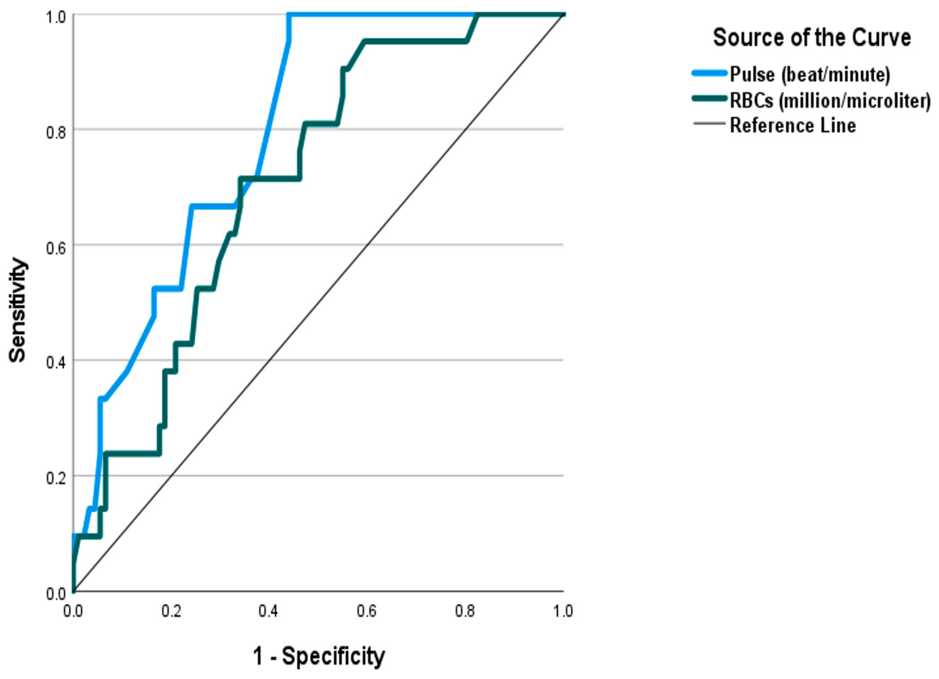
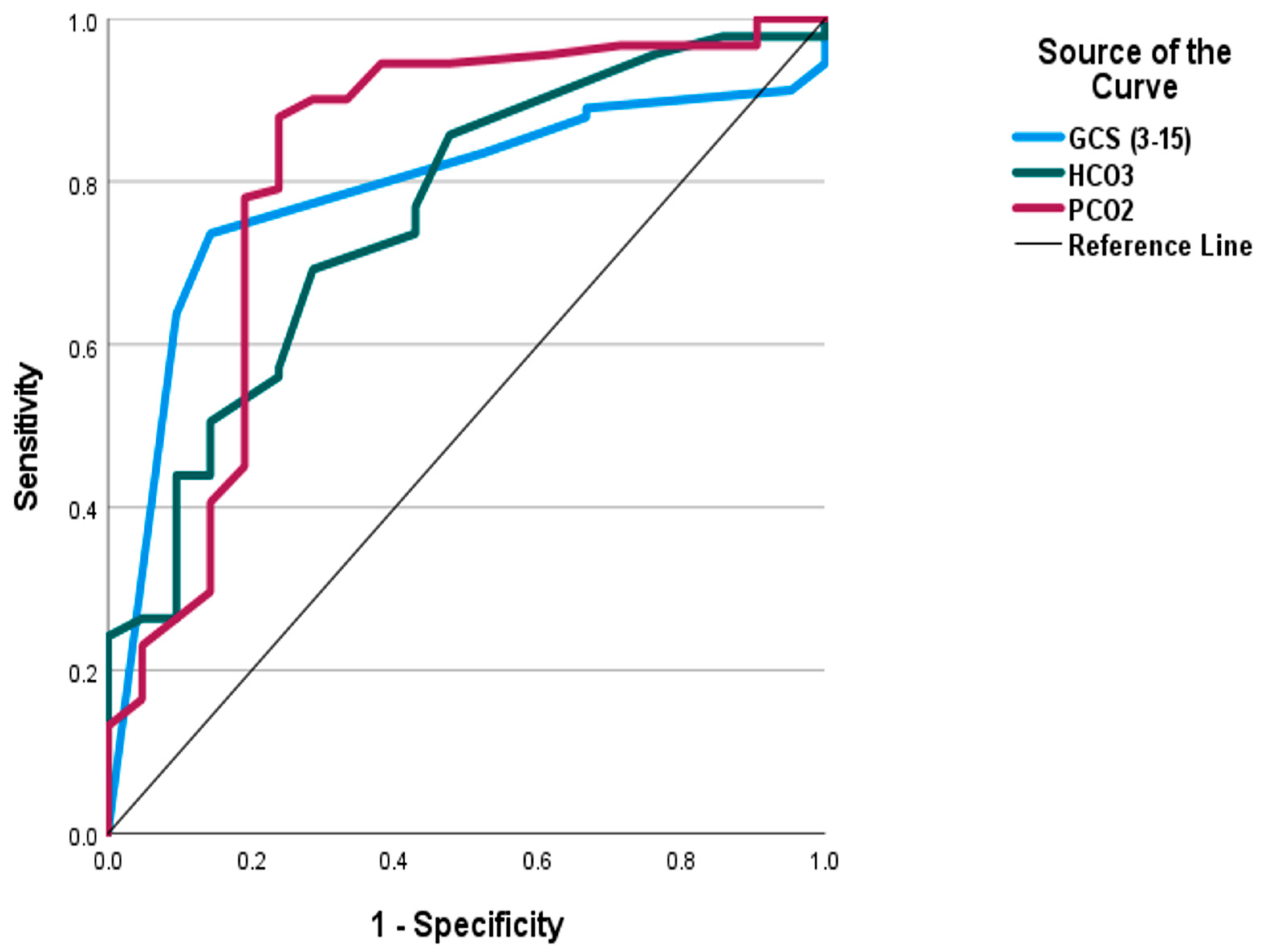
| AUC | p | Cut Off | Sensitivity | Specificity | PPV | NPV | Accuracy | |
|---|---|---|---|---|---|---|---|---|
| PSS | 0.879 | <0.001 * | >2.5 | 61.9% | 96.7% | 81.3% | 91.7% | 90.2% |
| PCO2 (mmHg) | 0.816 | <0.001 * | <35.85 | 78.0% | 81.0% | 94.7% | 45.9% | 78.6% |
| Pulse (beat/minute) | 0.796 | <0.001 * | >109.5 | 66.7% | 75.8% | 38.9% | 90.8% | 74.1% |
| GCS | 0.786 | <0.001 * | <14.0 | 73.6% | 85.7% | 95.7% | 42.9% | 75.8% |
| HCO3 (mEq/L) | 0.758 | <0.001 * | <21.5 | 69.2% | 71.4% | 91.3% | 34.9% | 69.6% |
| RBCs (million/microliter) | 0.708 | 0.003 * | >4.705 | 71.4% | 65.9% | 32.6% | 90.9% | 66.9% |
| Pair wise comparison of AUC | ||||||||
| PSS | PCO2 | Pulse (beat/minute) | GCS | HCO3 | RBCs (million/microliter) | |||
| PSS | - | 0.404 | 0.182 | 0.159 | 0.087 | 0.016 * | ||
| PCO2 (mmHg) | 0.404 | - | 0.794 | 0.706 | 0.488 | 0.196 | ||
| Pulse (beat/minute) | 0.182 | 0.794 | - | 0.894 | 0.597 | 0.221 | ||
| GCS | 0.159 | 0.706 | 0.894 | - | 0.709 | 0.299 | ||
| HCO3 (mEq/L) | 0.087 | 0.488 | 0.597 | 0.709 | - | 0.528 | ||
| RBCs (million/microliter) | 0.016 * | 0.196 | 0.221 | 0.299 | 0.528 | - | ||
| Potential Predictors | Wald | Sig. | Odds Ratio | 95% C.I for Odds Ratio | |
|---|---|---|---|---|---|
| Lower | Upper | ||||
| PSS | 23.460 | <0.001 * | 17.380 | 5.474 | 55.189 |
| PCO2 (mmHg) | 7.650 | 0.006 * | 0.847 | 0.754 | 0.953 |
| Pulse (beat/minute) | 7.245 | 0.007 * | 1.063 | 1.017 | 1.111 |
| GCS | 9.848 | 0.002 * | 0.680 | 0.534 | 0.865 |
| HCO3 (mEq/L) | 5.351 | 0.021 * | 0.739 | 0.572 | 0.955 |
| RBCs (million/microliter) | 7.022 | 0.008 * | 3.806 | 1.416 | 10.230 |
Disclaimer/Publisher’s Note: The statements, opinions and data contained in all publications are solely those of the individual author(s) and contributor(s) and not of MDPI and/or the editor(s). MDPI and/or the editor(s) disclaim responsibility for any injury to people or property resulting from any ideas, methods, instructions or products referred to in the content. |
© 2024 by the authors. Licensee MDPI, Basel, Switzerland. This article is an open access article distributed under the terms and conditions of the Creative Commons Attribution (CC BY) license (https://creativecommons.org/licenses/by/4.0/).
Share and Cite
Sharif, A.F.; Almulhim, M.N.M.; Almosabeh, H.M.A.; Alshammasy, M.E.A.; Aljeshi, A.M.A.; Mufti, T.M.A.; AlNasser, S.; Al-Mulhim, K.A.; AlMubarak, Y.A. Predictors of Serotonin Syndrome in Acute Poisoning with 5-Hydroxytryptamine Modulators. Toxics 2024, 12, 550. https://doi.org/10.3390/toxics12080550
Sharif AF, Almulhim MNM, Almosabeh HMA, Alshammasy MEA, Aljeshi AMA, Mufti TMA, AlNasser S, Al-Mulhim KA, AlMubarak YA. Predictors of Serotonin Syndrome in Acute Poisoning with 5-Hydroxytryptamine Modulators. Toxics. 2024; 12(8):550. https://doi.org/10.3390/toxics12080550
Chicago/Turabian StyleSharif, Asmaa F., Mubarak Nasir M. Almulhim, Hadi Mohamed A. Almosabeh, Mohammed Essam A. Alshammasy, Ali Mohammed A. Aljeshi, Taher Mohammed A. Mufti, Shahd AlNasser, Khalid A. Al-Mulhim, and Yousef A. AlMubarak. 2024. "Predictors of Serotonin Syndrome in Acute Poisoning with 5-Hydroxytryptamine Modulators" Toxics 12, no. 8: 550. https://doi.org/10.3390/toxics12080550