Review

Radiotherapy Advances in Renal Disease—Focus on Renal Ischemic Preconditioning

1
Groupe Interdisciplinaire de Génoprotéomique Appliquée (GIGA), Cardiovascular Sciences, University of Liège (ULiège), 4000 Liège, Belgium
2
III. Department of Medicine, University Medical Center Hamburg-Eppendorf, 20251 Hamburg, Germany
3
Zhongshan Hospital, Fudan University, Shanghai 200032, China
4
Cyclotron Research Center, University of Liège, 4000 Liège, Belgium
5
Division of Radiotherapy, CHU of Liège, University of Liège (CHU ULiège), 4000 Liège, Belgium
6
Division of Nephrology, CHU of Liège, University of Liège (CHU ULiège), Avenue Hippocrate, 13, 4000 Liège, Belgium
*
Author to whom correspondence should be addressed.
Bioengineering 2023, 10(1), 68; https://doi.org/10.3390/bioengineering10010068
Submission received: 25 September 2022 / Revised: 23 December 2022 / Accepted: 30 December 2022 / Published: 5 January 2023

Abstract

:
Ionizing irradiation is widely applied as a fundamental therapeutic treatment in several diseases. Acute kidney injury (AKI) represents a global public health problem with major morbidity and mortality. Renal ischemia/reperfusion (I/R) is the main cause of AKI. I/R injury occurs when blood flow to the kidney is transiently interrupted and then restored. Such an ischemic insult significantly impairs renal function in the short and long terms. Renal ischemic preconditioning (IPC) corresponds to the maneuvers intended to prevent or attenuate the ischemic damage. In murine models, irradiation-induced preconditioning (IP) renders the renal parenchyma resistant to subsequent damage by activating defense pathways involved in oxidative stress, angiogenesis, and inflammation. Before envisioning translational applications in patients, safe irradiation modalities, including timing, dosage, and fractionation, need to be defined.

Graphical Abstract

1. Introduction

Ionizing radiation is the energy released by atoms, which travels in the form of electromagnetic waves (gamma or X-rays) or particles (neutrons, beta, or alpha). It has been recognized as a risk factor for cancer in cellular and animal models, as well as in large epidemiological studies of populations exposed to extremely high doses of radiation [1,2,3]. Nevertheless, in recent decades, the use of ionizing irradiation as a therapeutic treatment has been widely applied. In particular, radiotherapy (RT) has become a fundamental tool against several diseases, especially some forms of cancer [4].
In the context of kidney diseases, several studies based only on animal models showed a protective effect of radiation therapy. In this review, we particularly focus on acute kidney injury (AKI), a known global public health problem that affects ~1000 people per million/year and is associated with a significant mortality rate [5,6]. Renal ischemia/reperfusion (I/R) injury is the leading cause of AKI [7] and corresponds to the transient interruption of the renal blood flow, with an abrupt drop in oxygen pressure and nutrient delivery leading to vascular and tubular dysfunction [8,9,10]. Here, we will provide an overarching view of recent advances in radiation therapy relevant to renal I/R and kidney diseases, as suggested in various murine models. We eventually envision the challenges and limitations of RT in the management of AKI in patients.

2. Renal Ischemia-Reperfusion

Renal I/R is an unavoidable event in kidney transplantation and cardio-thoracic surgery, with a negative impact on short- and long-term kidney outcomes [11,12]. Renal blood flow at rest is 1200 mL/min, which corresponds to 20% of cardiac output, while both kidneys represent less than 1% of total body weight [13]. Under physiological conditions, the renal blood flow remains constant up to a systolic arterial pressure of 80 mmHg due to the fine regulation of the sympathetic nervous system, the hormonal renin/angiotensin/aldosterone axis, and the synthesis of prostaglandins [13]. The prolonged interruption of renal blood perfusion leads to the cessation of nutrient supply and a drop in the partial pressure of oxygen. This transient interruption/reduction of renal blood flow, followed by its restoration and re-oxygenation, causes a cascade of cellular and tissue events grouped under the term “ischemic damage” [14,15].
Different I/R mouse models are used to mimic different aspects of the pathophysiology. Currently, two kinds of warm renal I/R models are mainly used: bilateral renal and unilateral renal I/R [16]. The unilateral renal I/R model has the advantage of a left nephrectomy being undertaken at the time of surgery. The left nephrectomy tissue serves as valuable control tissue in studies involving a pretreatment step, like irradiation, that modulates certain pathways [17,18,19]. The uniqueness of this model also makes it important for adhering to the three Rs rule (Replacement, Reduction, and Refinement) for a more ethical use of animals in research (Figure 1).

3. Renal Ischemic Preconditioning

Renal ischemic preconditioning (IPC) is an evolving concept that regroups all maneuvers aimed at preventing or attenuating the severity of the renal I/R injury. The principle of the “original” IPC was to mechanically expose the organ to brief episodes (3–5 min) of ischemia before conservation in prolonged ischemia in order to reduce ischemic damage and accelerate functional recovery [20,21]. In vivo, the renal IPC makes it possible to maintain the organization of the actin network of the cytoskeleton and the cell polarity, thereby preserving the polarized distribution of ion transporters such as the basolateral Na+/K+-ATPase. Tissue infiltrations by leukocytes, as well as the degree of apoptosis and necrosis, are significantly lower when IPC has been performed [22].
On the basis of the identified cellular targets of key pathways in renal IPC, the concept of pharmacological conditioning has emerged in the field of renal I/R. The use of pharmacological agents aims at stimulating the biochemical pathways of IPC [23]. Several drugs have been hypothesized to reproduce these protective mechanisms and have subsequently been tested in different settings (Table 1).
The current therapeutic strategies are directed to interact with the major I/R signaling pathways, such as inflammation, vascularization, energy metabolism, or oxygen transport [23,24,25].

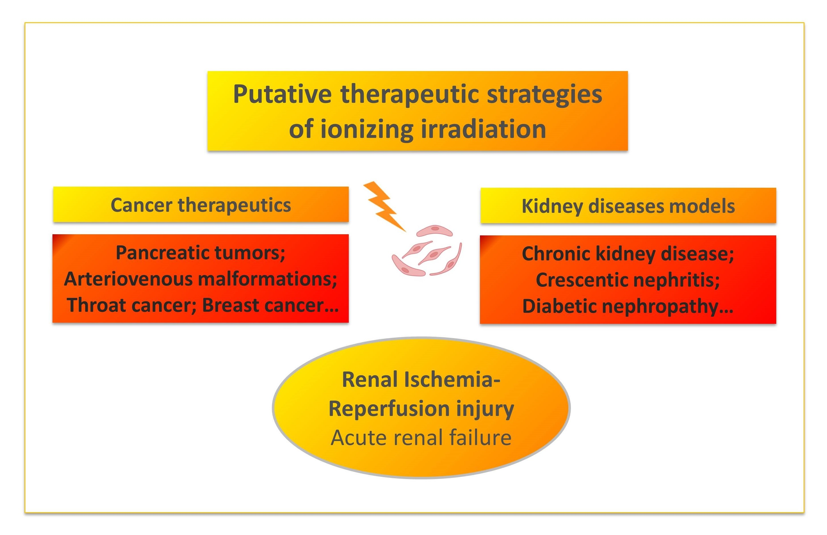
4. Radiation-Induced Effects on the Kidney

The impacts of radiation on the tissue and/or organs depend on the dose of radiation received and absorbed. It is expressed in gray units (Gy). The potential impact of an absorbed dose depends on the type of radiation and the variable sensitivity of organs [26]. At the cellular level, the effects of ionizing radiation can manifest as cell death or changes in the cytogenetic information [26]. These events can lead to adverse tissue reactions, in which manifestations depend on exceeding the dose threshold, or to stochastic effects, when the effect increases with the dose (Figure 2).
Basically, ionizing radiation can cause double-stranded breaks (DSB) in the DNA, followed by cell death including apoptosis and necrosis of renal endothelial, tubular, and glomerular cells in the case of radiation nephropathy (RN) [27]. Kidney toxicity depends on the use and intensity of RT. Protective genetic and biochemical effects can also be induced by DNA damage response (DDR) activation after sensing the DSB. This system reacts with cell death induction or cell cycle arrest and DNA repair based on the affected tissue and the severity of the damage. The oxidative stress [28], the renin–angiotensin (RAS) system [29], cellular senescence [30], proliferation, angiogenesis [18,31,32], and endothelial and hemodynamic modulations [33] might be involved in the putative mechanisms in radiation-induced effects on the renal parenchyma.

5. Radiation-Based Therapy in Kidney Diseases

Radiation therapy relies on ionizing radiation and is usually used as part of cancer treatment to control or eradicate malignant cells [34]. Irradiation-mediated renal IPC, although not yet well reported, would aim to render cells or tissues resistant to subsequent damage by activating their intracellular defense system [35]. Indeed, despite the potential for radiation exposure to cause kidney damage, a certain dose of irradiation has also been shown to have a protective effect in animal studies. A summary of the literature suggesting the renal protective role of kidney-centered ionizing irradiation in rodent models of kidney diseases is listed in Table 2. Note that there is no translational data in humans.
In the global context of kidney diseases, it has been shown that continuous whole-body low-dose-rate gamma irradiation ameliorates diabetic nephropathy and increases the lifespan in mice through the activation of renal antioxidants [41]. Taylor et al. also showed that the incidence of kidney disease was significantly reduced following a 10 mGy irradiation compared to non-irradiated mice [42]. Histopathological changes in the renal parenchyma of several animal models of kidney diseases showed a protective effect after local or whole-body irradiation to the kidneys, such as in CKD [37], crescentic nephritis [38], and diabetic nephropathy rodent models [40]. Aunapuu et al. found that the CKD rat model through 5/6 nephrectomy (NPX) manifested a high level of proliferating and apoptotic markers, which could be reduced significantly in glomeruli and distal tubular cells following a 3 Gy dose of γ radiation on the left kidney [36]. The indicators of renal function, such as proteinuria, systolic blood pressure, and serum creatinine (SCr), were also improved after the irradiation in these kinds of CKD rodent models [36,37]. Anti-oxidative stress, anti-inflammatory, and anti-renal fibrosis may be considered as the potential benefits of irradiation therapy.
Cheng et al. [40] found that the whole-body exposure of type 1 diabetic mice (DM) to 25 mGy X-rays weekly decreased the microalbuminuria, the renal accumulation of 3-nitrotyrosine and 4-hydroxynonenal, and the renal expression of collagen IV and fibronectin, suggesting that low-dose radiation (LDR) improved DM-induced oxidative/nitrosative damage and renal fibrosis effectively. Shao et al. [43] also showed that frequent LDR X-ray treatment attenuated dyslipidemia and insulin resistance followed by renal inflammation and oxidative stress in mice with type 2 diabetes. A low dose of gamma radiation has also been found to protect against D-GalN-induced renal damage in Swiss albino rats. The results revealed increases in antioxidant activities and decreases in inflammatory markers (tumor necrosis factor-alpha (TNF-α) and nuclear factor kappa-light-chain-enhancer of activated B cells (NF-κB). Additionally, this treatment was associated with an upregulation of Nrf-2 gene expression, which relates to the prevention of oxidative stress, and decreased lipid peroxidation levels, which were concordant with histopathological findings following the irradiation therapy [39]. In an experimental model of crescentic nephritis in rats [38], the overexpression of active caspases 3 and 7, elevation in the TUNEL assay, and decrease in PCNA were noted after local, bilateral irradiation treatment. The improvement in morphology, the decrease in the proliferation marker, and the elevation in the apoptosis assay have also been shown in a CKD rat model [37].
In the context of I/R diseases, several studies have reported a protective effect of radiation therapy against I/R injury. Yuan-Po et al. showed that far-infrared radiation attenuated I/R injury in rat testis by inducing heme oxygenase (HO1) expression [44]. Lakyova et al. similarly demonstrated that low-level laser irradiation causes a profound reduction in the amount of necrotic tissue and enhances recovery after I/R muscle injury in rat hindlimbs by attenuating the inflammatory reaction and facilitating angiogenesis [45].

6. Irradiation Preconditioning against AKI

Interestingly enough, in the context of in vivo studies of ischemic AKI using renal I/R mouse models, whole-body irradiation (WBI) has been suggested to induce renal IPC. Indeed, the 8 Gy WBI significantly attenuated the elevations of SCr and BUN concentrations, structural damage, lipid peroxidation, expression, and activity of NADPH oxidase (NOX)-2, nitrotyrosine levels, and hydrogen peroxide production following renal I/R [35]. This protection was inhibited by the treatment of animals with manganese (III) tetrakis (1-methyl-4-pyridyl) porphyrin (MnTMPyP), a superoxide scavenger, indicating that irradiation-induced preconditioning (IP)-rendered protection was triggered by superoxide formation, enhancing manganese superoxide dismutase (MnSOD) activity and expression, and HSP-27 expression.
Advances in conformational radiology and preclinical radiotherapy research have recently spurred the development of precise micro-irradiators for small animals, including rodents. These devices are often kilovolt X-ray radiation sources combined with high-resolution computed tomography (CT) imaging equipment for image guidance, as the latter allows precise and accurate beam positioning. These devices are similar to modern human radiotherapy machines and are considered a major step forward in radiobiology research [46,47].
In order to investigate the impact of kidney-focused irradiation in mice before renal I/R, we have recently used the small animal irradiator and scanner (SmART) instrument from precision X-ray (North Branford, CT) designed to image, target, and irradiate cells and small animals up to the size of rodents [48,49,50]. This scanner provides images that have a resolution of 0.1 mm. A prescan of the whole body helped to precisely locate the volume to be investigated, and this volume was then defined in all three dimensions by moving cursors on the computer screen with the mouse. The radiation exposure focused on the kidneys, with beams of 225 KV and 13 mA (Figure 3A,B).

7. Kidney-Centered Irradiation Mediates IPC

The bilateral X-ray renal irradiation with a single dose of 8 Gy induced IPC in mice, with reduced macrophage infiltration and attenuated tubular necrosis following renal I/R. The metabolomics signature of renal I/R was attenuated in pre-irradiated mice. BUN and SCr plasma assays showed stable circulating levels of these two AKI biomarkers, which reflect preserved renal function in pre-irradiated mice exposed to I/R. Such a renal IPC was significantly observed as early as 14 days post-irradiation, preceding the I/R episode. Moreover, the physiological follow-up study of irradiated mice showed no structural or functional complications 3 months after kidney-centered irradiation. The comparative high-throughput RNA-seq between the irradiated and control kidneys reveals that renal irradiation was associated with an upregulation of signaling pathways involved in angiogenesis, cell proliferation, and stress response and a downregulation of the JNK cascade [18].
Several works reported a protective effect of radiation therapy against I/R injuries [36,37]. By extrapolating from these studies, one may speculate that even the exposure of animals to low doses (0.1–2 Gy) could have a similar degree of protective effect in the case of renal I/R. Recently, multiple exposures to low-dose whole body irradiation (LD-WBI) in C57BL/6J mice have been reported to significantly suppress the diabetes-induced systemic, renal inflammatory response, and renal oxidative damage, thereby preventing renal dysfunction and fibrosis [40]. Aunapuu et al. also showed that low-dose-rate gamma irradiation slows the process of CKD through the reduction of apoptotic cells [41]. With the application of higher irradiation doses like the one used in our study, acute toxic effects and potentially late long-term effects could be induced. In the perspective of therapeutic irradiation in humans, future studies will be required to determine whether kidney-centered low-dose irradiation at decreasing doses (upon a 10-based order of magnitude) could have a similar protective impact against renal I/R injury. In our murine model, the post-irradiation follow-up at 3 months did not reveal complications regarding the physiological functions of the irradiated mice or the establishment of renal fibrosis. However, this follow-up study did not go beyond 3 months after irradiation. A larger study, including the evaluation of overall physiological parameters with a longer follow-up period, is therefore required.
Another important issue in radiotherapy concerns the therapeutic window. In the work of Kim et al., WBI with 8 Gy induced IPC against renal I/R 6 days after irradiation [35]. In our kinetics study of pre-irradiation conditioning, we compared the impact of kidney-focused irradiation 7, 14, and 28 days before renal I/R. The IPC was significantly reached at day 14, and a trend was observed at day 7 [18]. As a result, future research will concentrate on characterizing the kinetics of irradiation-mediated IPC from 7 days to 1 day after renal irradiation in order to mimic clinical settings as closely as possible, such as kidney transplantation or cardiothoracic surgery.

8. Conclusions

AKI is a growing medical challenge with a high incidence that affects numerous patients worldwide. The occurrence of AKI is associated with an increased complication rate and the development of CKD, as well as short- and long-term mortality. The reported demonstrations highlight new evidence for the development of innovative therapeutic approaches. Still, we need to further improve the safety of the efficacy/toxicity ratio. Taken together, the modulation of the molecular targets of irradiation-induced renal IPC may open new avenues in the development of innovative pharmacological strategies against a major medical problem, which is acute ischemic kidney injury.

Author Contributions

Conceptualization, B.K. and F.J.; writing—original draft preparation, B.K. and S.G.; writing—review and editing, B.K., S.G., T.P.C., F.L. and F.J. All authors have read and agreed to the published version of the manuscript.

Funding

The Belgian National Fund for Scientific Research and the University of Liège (Fonds Léon Fredericq and Fonds Spéciaux à la Recherche) 2020 to B.K. and F.J.

Institutional Review Board Statement

Not applicable.

Informed Consent Statement

Not applicable.

Data Availability Statement

Data sharing is not applicable.

Conflicts of Interest

The authors declare no conflict of interest.

References

  1. Havránková, R. Biological effects of ionizing radiation. Cas. Lek. Cesk. 2020, 159, 258–260. [Google Scholar]
  2. Tomonaga, M. Radiation injury to human body: Atomic bombs, Chernobyl and Fukushima. Rinsho Ketsueki 2011, 52, 1740–1747. [Google Scholar] [PubMed]
  3. Muirhead, C.R. Studies on the Hiroshima and Nagasaki survivors, and their use in estimating radiation risks. Radiat. Prot. Dosim. 2003, 104, 331–335. [Google Scholar] [CrossRef] [PubMed] [Green Version]
  4. Furdui, C.M. Ionizing Radiation: Mechanisms and Therapeutics. Antioxid. Redox Signal. 2014, 21, 218–220. [Google Scholar] [CrossRef] [PubMed] [Green Version]
  5. Ali, T.; Khan, I.; Simpson, W.; Prescott, G.; Townend, J.; Smith, W.; MacLeod, A. Incidence and Outcomes in Acute Kidney Injury: A Comprehensive Population-Based Study. J. Am. Soc. Nephrol. 2007, 18, 1292–1298. [Google Scholar] [CrossRef] [Green Version]
  6. Hoste, E.A.J.; Kellum, J.A.; Selby, N.M.; Zarbock, A.; Palevsky, P.M.; Bagshaw, S.M.; Goldstein, S.L.; Cerdá, J.; Chawla, L.S. Global epidemiology and outcomes of acute kidney injury. Nat. Rev. Nephrol. 2018, 14, 607–625. [Google Scholar] [CrossRef] [PubMed]
  7. Singh, A.P.; Muthuraman, A.; Jaggi, A.S.; Singh, N.; Grover, K.; Dhawan, R. Animal models of acute renal failure. Pharmacol. Rep. 2012, 64, 31–44. [Google Scholar] [CrossRef]
  8. Bonventre, J.J.V.; Yang, L. Cellular pathophysiology of ischemic acute kidney injury. J. Clin. Investig. 2011, 121, 4210–4221. [Google Scholar] [CrossRef]
  9. Rowart, P.; Erpicum, P.; Detry, O.; Weekers, L.; Grégoire, C.; Lechanteur, C.; Briquet, A.; Beguin, Y.; Krzesinski, J.-M.; Jouret, F. Mesenchymal Stromal Cell Therapy in Ischemia/Reperfusion Injury. J. Immunol. Res. 2015, 2015, 602597. [Google Scholar] [CrossRef] [Green Version]
  10. Erpicum, P.; Rowart, P.; Poma, L.; Krzesinski, J.-M.; Detry, O.; Jouret, F. Administration of mesenchymal stromal cells before renal ischemia/reperfusion attenuates kidney injury and may modulate renal lipid metabolism in rats. Sci. Rep. 2017, 7, 8687. [Google Scholar] [CrossRef] [Green Version]
  11. Erpicum, P.; Rowart, P.; Defraigne, J.-O.; Krzesinski, J.-M.; Jouret, F. What we need to know about lipid-associated injury in case of renal ischemia-reperfusion. Am. J. Physiol. Physiol. 2018, 315, F1714–F1719. [Google Scholar] [CrossRef] [PubMed]
  12. Legrand, M.; Rossignol, P. Cardiovascular Consequences of Acute Kidney Injury. N. Engl. J. Med. 2020, 382, 2238–2247. [Google Scholar] [CrossRef] [PubMed]
  13. Dalal, R.; Bruss, Z.S.; Sehdev, J.S. Physiology, Renal Blood Flow and Filtration. In StatPearls [Internet]; StatPearls Publishing: Treasure Island, FL, USA, 2022. [Google Scholar]
  14. Schrier, R.W.; Wang, W.; Poole, B.; Mitra, A. Acute renal failure: Definitions, diagnosis, pathogenesis, and therapy. J. Clin. Investig. 2004, 114, 5–14. [Google Scholar] [CrossRef] [PubMed] [Green Version]
  15. Nieuwenhuijs-Moeke, G.J.; Pischke, S.E.; Berger, S.P.; Sanders, J.S.F.; Pol, R.A.; Struys, M.M.R.F.; Ploeg, R.J.; Leuvenink, H.G.D. Ischemia and Reperfusion Injury in Kidney Transplantation: Relevant Mechanisms in Injury and Repair. J. Clin. Med. 2020, 9, 253. [Google Scholar] [CrossRef] [PubMed] [Green Version]
  16. Wei, Q.; Dong, Z. Mouse model of ischemic acute kidney injury: Technical notes and tricks. Am. J. Physiol. Physiol. 2012, 303, F1487–F1494. [Google Scholar] [CrossRef] [PubMed] [Green Version]
  17. Le Clef, N.; Verhulst, A.; D’Haese, P.C.; Vervaet, B.A. Unilateral Renal Ischemia-Reperfusion as a Robust Model for Acute to Chronic Kidney Injury in Mice. PLoS ONE 2016, 11, e0152153. [Google Scholar] [CrossRef] [Green Version]
  18. Khbouz, B.; Lallemand, F.; Cirillo, A.; Rowart, P.; Legouis, D.; Sounni, N.E.; Noël, A.; De Tullio, P.; de Seigneux, S.; Jouret, F. Kidney-targeted irradiation triggers renal ischemic preconditioning in mice. Am. J. Physiol. Physiol. 2022, 323, F198–F211. [Google Scholar] [CrossRef]
  19. Jouret, F.; Leenders, J.; Poma, L.; Defraigne, J.-O.; Krzesinski, J.-M.; de Tullio, P. Nuclear Magnetic Resonance Metabolomic Profiling of Mouse Kidney, Urine and Serum Following Renal Ischemia/Reperfusion Injury. PLoS ONE 2016, 11, e0163021. [Google Scholar] [CrossRef] [Green Version]
  20. Murry, C.E.; Jennings, R.B.; Reimer, K.A. Preconditioning with ischemia: A delay of lethal cell injury in ischemic myocardium. Circulation 1986, 74, 1124–1136. [Google Scholar] [CrossRef] [Green Version]
  21. Bonventre, J.V. Kidney ischemic preconditioning. Curr. Opin. Nephrol. Hypertens. 2002, 11, 43–48. [Google Scholar] [CrossRef]
  22. Park, K.M.; Chen, A.; Bonventre, J.V. Prevention of Kidney Ischemia/Reperfusion-induced Functional Injury and JNK, p38, and MAPK Kinase Activation by Remote Ischemic Pretreatment. J. Biol. Chem. 2001, 276, 11870–11876. [Google Scholar] [CrossRef]
  23. Erpicum, P.; Krzesinski, J.-M.; Jouret, F. Place de l’AMP-activated protein kinase dans le préconditionnement ischémique rénal. Néphrologie Thérapeutique 2014, 10, 17–24. [Google Scholar] [CrossRef] [Green Version]
  24. Khbouz, B.; Rowart, P.; Poma, L.; Dahlke, E.; Bottner, M.; Stokes, M.; Bolen, G.; Rahmouni, S.; Theilig, F.; Jouret, F. The genetic deletion of the Dual Specificity Phosphatase 3 (DUSP3) attenuates kidney damage and inflammation following ischaemia/reperfusion injury in mouse. Acta Physiol. 2022, 234, e13735. [Google Scholar] [CrossRef] [PubMed]
  25. Yakulov, T.A.; Todkar, A.P.; Slanchev, K.; Wiegel, J.; Bona, A.; Groß, M.; Scholz, A.; Hess, I.; Wurditsch, A.; Grahammer, F.; et al. CXCL12 and MYC control energy metabolism to support adaptive responses after kidney injury. Nat. Commun. 2018, 9, 3660. [Google Scholar] [CrossRef] [Green Version]
  26. Reisz, J.A.; Bansal, N.; Qian, J.; Zhao, W.; Furdui, C.M. Effects of Ionizing Radiation on Biological Molecules—Mechanisms of Damage and Emerging Methods of Detection. Antioxid. Redox Signal. 2014, 21, 260–292. [Google Scholar] [CrossRef] [PubMed] [Green Version]
  27. Klaus, R.; Niyazi, M.; Lange-Sperandio, B. Radiation-induced kidney toxicity: Molecular and cellular pathogenesis. Radiat. Oncol. 2021, 16, 43. [Google Scholar] [CrossRef]
  28. Kim, J.H.; Jenrow, K.A.; Brown, S.L. Mechanisms of radiation-induced normal tissue toxicity and implications for future clinical trials. Radiat. Oncol. J. 2014, 32, 103–115. [Google Scholar] [CrossRef] [Green Version]
  29. Datta, P.K. TGF-beta1 production in radiation nephropathy: Role of angiotensin II. Int. J. Radiat. Biol. 1999, 75, 473–479. [Google Scholar] [CrossRef]
  30. Kim, D.Y.; Lee, M.; Kim, E.J. Involvement of Klotho, TNF-α and ADAMs in radiation-induced senescence of renal epithelial cells. Mol. Med. Rep. 2021, 23, 22. [Google Scholar] [CrossRef] [PubMed]
  31. Scharpfenecker, M.; Floot, B.; Russell, N.S.; Coppes, R.P.; Stewart, F.A. Endoglin haploinsufficiency attenuates radiation-induced deterioration of kidney function in mice. Radiother. Oncol. 2013, 108, 464–468. [Google Scholar] [CrossRef]
  32. Kruse, J.J.C.M.; Floot, B.G.J.; Poele, J.A.M.T.; Russell, N.S.; Stewart, F.A. Radiation-Induced Activation of TGF-β Signaling Pathways in Relation to Vascular Damage in Mouse Kidneys. Radiat. Res. 2009, 171, 188–197. [Google Scholar] [CrossRef] [PubMed]
  33. Abou Daher, A.; Francis, M.; Azzam, P.; Ahmad, A.; Eid, A.A.; Fornoni, A.; Marples, B.; Zeidan, Y.H. Modulation of radiation-induced damage of human glomerular endothelial cells by SMPDL3B. FASEB J. 2020, 34, 7915–7926. [Google Scholar] [CrossRef] [PubMed] [Green Version]
  34. Skliarenko, J.; Warde, P. Practical and clinical applications of radiation therapy. Medicine 2016, 44, 15–19. [Google Scholar] [CrossRef]
  35. Kim, J.; Park, J.-W.; Park, K.M. Increased superoxide formation induced by irradiation preconditioning triggers kidney resistance to ischemia-reperfusion injury in mice. Am. J. Physiol. Physiol. 2009, 296, F1202–F1211. [Google Scholar] [CrossRef] [Green Version]
  36. Aunapuu, M.; Pechter, Ü.; Gerskevits, E.; Marjamägi, M.-M.; Suuroja, S.; Arend, A.; Kolts, I.; Kühnel, W.; Ots, M. Low-dose radiation modifies the progression of chronic renal failure. Ann. Anat.-Anat. Anz. 2004, 186, 277–282. [Google Scholar] [CrossRef]
  37. Aunapuu, M.; Arend, A.; Ots, M.; Pilmane, M. Cell proliferation and apoptosis in Wistar rat kidney after renal mass ablation and low-dose irradiation. Medicina 2010, 46, 204–210. [Google Scholar] [CrossRef] [Green Version]
  38. Liu, D.; Nazneen, A.; Taguchi, T.; Razzaque, M.S. Low-Dose Local Kidney Irradiation Inhibits Progression of Experimental Crescentic Nephritis by Promoting Apoptosis. Am. J. Nephrol. 2008, 28, 555–568. [Google Scholar] [CrossRef]
  39. El-Sonbaty, S.M.; Moawed, F.; Elbakry, M.M.M. Amphora algae with low-level ionizing radiation exposure ameliorate D-galactosamine-induced inflammatory impairment in rat kidney. Environ. Toxicol. 2021, 36, 451–459. [Google Scholar] [CrossRef]
  40. Cheng, J.; Li, F.; Wang, G.; Guo, W.; Huang, S.; Wang, B.; Li, C.; Jiang, Q.; Cai, L.; Cui, J. Optimal LDR to Protect the Kidney From Diabetes: Whole-Body Exposure to 25 mGy X-rays Weekly for 8 Weeks Efficiently Attenuates Renal Damage in Diabetic Mice. Dose-Response 2018, 16. [Google Scholar] [CrossRef]
  41. Nomura, T.; Li, X.-H.; Ogata, H.; Sakai, K.; Kondo, T.; Takano, Y.; Magae, J. Suppressive Effects of Continuous Low-Dose-Rate γ Irradiation on Diabetic Nephropathy in Type II Diabetes Mellitus Model Mice. Radiat. Res. 2011, 176, 356–365. [Google Scholar] [CrossRef]
  42. Taylor, K.; Lemon, J.A.; Phan, N.; Boreham, D.R. Low-dose radiation from 18F-FDG PET does not increase cancer frequency or shorten latency but reduces kidney disease in cancer-prone Trp53+/− mice. Mutagenesis 2014, 29, 289–294. [Google Scholar] [CrossRef] [PubMed]
  43. Shao, M.; Lu, X.; Cong, W.; Xing, X.; Tan, Y.; Li, Y.; Li, X.; Jin, L.; Wang, X.; Dong, J.; et al. Multiple low-dose radiation prevents type 2 diabetes-induced renal damage through attenuation of dyslipidemia and insulin resistance and subsequent renal inflammation and oxidative stress. PLoS ONE 2014, 9, e92574. [Google Scholar] [CrossRef] [PubMed] [Green Version]
  44. Tu, Y.-P.; Chen, S.-C.; Liu, Y.-H.; Chen, C.-F.; Hour, T.-C. Postconditioning with far-infrared irradiation increases heme oxygenase-1 expression and protects against ischemia/reperfusion injury in rat testis. Life Sci. 2013, 92, 35–41. [Google Scholar] [CrossRef]
  45. Lakyová, L.; Toporcer, T.; ScD, V.T.; Sabo, J.; Radoňak, J. Low-level laser therapy for protection against skeletal muscle damage after ischemia-reperfusion injury in rat hindlimbs. Lasers Surg. Med. 2010, 42, 825–832. [Google Scholar] [CrossRef]
  46. Verhaegen, F.; Granton, P.; Tryggestad, E. Small animal radiotherapy research platforms. Phys. Med. Biol. 2011, 56, R55–R83. [Google Scholar] [CrossRef] [PubMed]
  47. Grandinetti, J.; Zhong, Y.; Shen, C.; Jia, X. Design and experimental validation of a unilateral magnet for MRI-guided small animal radiation experiments. J. Magn. Reson. 2021, 332, 107062. [Google Scholar] [CrossRef]
  48. Chiu, T.D.; Arai, T.J.; Iii, J.C.; Jiang, S.B.; Mason, R.P.; Stojadinovic, S. MR-CBCT image-guided system for radiotherapy of orthotopic rat prostate tumors. PLoS ONE 2018, 13, e0198065. [Google Scholar] [CrossRef] [PubMed] [Green Version]
  49. Lallemand, F.; Chiver, I.; dos Santos, E.B.; Ball, G.F.; Balthazart, J. Repeated assessment of changes in testes size in canaries by X-ray computer tomography. Gen. Comp. Endocrinol. 2021, 310, 113808. [Google Scholar] [CrossRef]
  50. Lallemand, F.; Leroi, N.; Blacher, S.; Bahri, M.A.; Balteau, E.; Coucke, P.; Noël, A.; Plenevaux, A.; Martinive, P. Tumor Microenvironment Modifications Recorded with IVIM Perfusion Analysis and DCE-MRI after Neoadjuvant Radiotherapy: A Preclinical Study. Front. Oncol. 2021, 11, 784437. [Google Scholar] [CrossRef]
Figure 1. Mouse model of I/R-induced AKI. (1) Laparotomy, followed by (2) 30 min of right renal ischemia, in parallel to (3) left nephrectomy (control sham). Forty-eight hours after reperfusion, blood and kidneys are harvested for the assessment of the ischemic damage. (Created with BioRender.com).
Bioengineering 10 00068 g001
Figure 2. Onset of radiation-induced effects at cellular and tissue levels. Immediate radiation effects lead to signaling events such as DNA damage and repair mechanisms, tissue inflammation, and apoptosis (left). Moreover, radiation is associated with bystander effects (biological effects expressed by non-irradiated cells through signals produced by the irradiated area) and (epi)genomic modulations (middle). The cellular epigenetic modifications can trigger particular circuits after radiation exposure (right). (Created with BioRender.com).
Bioengineering 10 00068 g002
Figure 3. PXi X-Rad SmART renal radiation therapy: (A) bilateral renal irradiation with two beams of X-rays targeting both kidneys and (B) unilateral renal irradiation with three beams of X-rays targeting the left kidney.
Bioengineering 10 00068 g003
Table 1. Examples of pharmacological renal ischemic preconditioning in rodents. MAPK: mitogen-activated protein kinases; GPCR: G protein-coupled receptor; HIF-1: hypoxia-inducible factor 1; NOS: nitric oxide synthase; and AMPK: AMP-activated protein kinase (adapted from [23]).
DrugsProtein TargetsPathways
Cyclosporine; FK-506
Mesenchymal stem cells (MSC); Adenosine; Apyrase; and Catecholamines
MAPK
GPCR
Inflammation
Erythropoıetin; IsofluraneHIF-1aHypoxia
L-carnitine; Lithium; Danshen; N-acetylcysteine; and Spermine NONOateFree radicals; NOSOxidative stress
Metformin; AICAR; and
Hemin
AMPK
Hmox1 inducer
Metabolism
Table 2. Summary of the literature exhibiting renal protection following ionizing irradiation on different assays at different doses in rodent experimental disease models: monocyte chemoattractant protein-1(MCP-1), dUTP nick-end labeling (TUNEL), nuclear factor erythroid 2-related factor 2 (Nrf-2), urinary microalbumin (Malb), blood urea nitrogen (BUN), serum creatinine (SCr), 3-nitrotyrosine (3-NT), 4-hydroxynonenal (4-HNE), superoxide dismutase (CuZnSOD), manganese superoxide dismutase (MnSOD), glutathione peroxidase GPx, heme oxygenase 1 (HO1), heat shock protein (HSP), platelet and endothelial cell adhesion molecule 1 (PECAM1), collagen IV (Col IV), ↑: significant functional and morphological improvement, and ↓: significant decrease in degradation markers.
Disease ModelRadiationSiteDoseTimeAspectTendencyReference
Adult Wistar rats (CKD)ΓLeft kidney3 Gy2 wFunction (urinary proteins excretion rate, awake systolic blood pressure, and SCr)
Morphology
MCP-1


[36]
Adult Wistar rats (CKD)ΓLeft kidney3 Gy1/2 wFunction (Urinary proteins, systolic blood pressure, and SCr)
Morphology
Apoptosis (TUNEL)


[37]
Inbred male Sprague- Dawley rats
(Crescentic nephritis)
XLocal bilateral kidneys0.5 Gy1/2/3/4 wFunction (SCr)
Morphology
PCNA ED-1
Apoptosis (Caspase 3/7 and TUNEL)


[38]
Adult male C57BL/6 mice (I/R induced AKI)XWhole body8 Gy1 wFunction (BUN and SCr)
Morphology
Oxidative stress (MnSOD and HSP27)


[35]
Adult male Swiss albino rats (D-GalN-induced renal damage)ΓWhole body0.25 GyN/AAntioxidant activities (CuZnSOD and GPx)
Lipid peroxidation level
Function
Inflammation (TNF-a and NF-KB)
Morphology
Nrf-2 gene


[39]
Adult male C57BL/6J mice (Type I diabetes)XWhole body12.5 or 25 mGy4/8 wFunction (SCr and urinary microalbumin)
Morphology (PAS staining)
Nitrosative damage (3-NT and 4-HNE)
Renal fibrosis (Col IV and fibronectin)


[40]
Adult male C57BL/6 mice (I/R-induced AKI)XLocal bilateral kidneys8 Gy2 wFunction (BUN and SCr)
Morphology (PAS staining)
Inflammation (Cd11b; F4/80)
Angiogenesis (PECAM1)
Oxidative stress (HSP70; HO1)


[18]
Disclaimer/Publisher’s Note: The statements, opinions and data contained in all publications are solely those of the individual author(s) and contributor(s) and not of MDPI and/or the editor(s). MDPI and/or the editor(s) disclaim responsibility for any injury to people or property resulting from any ideas, methods, instructions or products referred to in the content.

