Article

Bioactive Aspergteroids G–J from Soft-Coral-Associated Symbiotic and Epiphytic Fungus Aspergillus terreus EGF7-0-1

1
School of Pharmaceutical Sciences, Guangzhou University of Chinese Medicine, Guangzhou 510006, China
2
Research Center of Integrative Medicine, School of Basic Medical Sciences, Guangzhou University of Chinese Medicine, Guangzhou 510006, China
*
Authors to whom correspondence should be addressed.
These authors contributed equally to this work.
Bioengineering 2023, 10(7), 805; https://doi.org/10.3390/bioengineering10070805
Submission received: 2 June 2023 / Revised: 21 June 2023 / Accepted: 3 July 2023 / Published: 5 July 2023

Abstract

:
Two new disubstituted maleimides, aspergteroids G–H (12), and two trisubstituted butenolides aspergteroids I–J (34), along with four known analogs, were isolated and structurally identified from the fermentation extract of soft-coral-associated symbiotic and epiphytic fungus Aspergillus terreus EGF7-0-1. The structures of the new compounds were established mainly via spectroscopic data analyses, and their absolute configurations were determined via X-ray diffraction analysis and comparison of the calculated and experimental electronic circular dichroism. Myocardial protection assays showed that compounds 1, 2, 5, and 6 possess protective effects against tert-butyl hydroperoxide (TBHP)-induced H9c2 (rat myocardial cells) apoptosis at low concentrations. Based on the analyses of the protein–protein interaction (PPI) network and Western blotting, compound 1 may inhibit the apoptosis and inflammatory response of cardiomyocytes after TBHP induction and improve the antioxidant capacity of cardiomyocytes. We speculate that the anti-inflammatory response of compound 1 is suppressed by the glycogen synthase kinase-3 beta (GSK-3β), downregulated by the NOD-like receptor thermal protein domain associated protein 3 (NLRP3) inflammasome activation, and suppressed by the expression of cysteinyl aspartate specific proteinase-3 (caspase-3) and B-cell lymphoma-2 associated X protein (Bax).

1. Introduction

Cardiovascular diseases (CVDs) seriously threaten human health; more than 31% of all deaths worldwide are caused by CVDs [1]. Among them, GSK-3β is considered to be an important target [2]. New drugs and targets for cardiovascular diseases are still the focus of research. Therefore, it is critical to investigate the pathogenesis and therapeutic targets of cardiovascular diseases and identify new therapeutic agents. The trisubstituted butenolides refer to a group of natural products with -phenyl and -benzyl substituents on the butenolide ring, which have been isolated mostly from fungi [3,4,5,6,7,8,9,10]. At present, 176 tri-substituted butenolides analogues have been reported [11]. Phenyl- and benzyl-disubstituted γ-butenolides from microorganisms can be classified into 4,5-, 3,4-, and 3,5-disubstituted according to the substituted patterns of the lactone core. Among 32 3,4-disubstituted butenolides, there are no reports on the phenyl- and benzyl-disubstituted maleimides from marine organisms. According to our research, only four phenyl- and benzyl- disubstituted maleimides have been isolated, asperimides A–D, from a tropical endophytic fungus Aspergillus terreus [12]. Trisubstituted butenolides exhibit diverse biological activities such as antibacterial [13], cytotoxic [14,15], anti-inflammatory [16], and antioxidant [17] activities.
As a continuation of our investigation into novel and bioactive secondary metabolites derived from marine microorganisms [18,19,20], we first isolated two disubstituted maleimides, aspergteroids G–H (12), from microorganisms of marine origin. Two new 4,5-disubstituted aspergteroids (I–J (34)) and four known 4,5-disubstituted analogs (asperimide A (5) [12], asperteretal D (6) [21], butyrolactone VII (7) [22], and versicolactone B (8) [5]) were isolated from soft-coral-associated symbiotic and epiphytic fungus Aspergillus terreus EGF7-0-1 (Figure 1). We further studied their protective effects on cardiomyocytes. This study used TBHP to induce oxidative stress injury in cardiomyocytes and explored the protective effects of compounds on cardiomyocytes at the cellular and molecular levels.

2. Materials and Methods

2.1. General Experimental Procedures

Details of the instrumentations and materials used in this work are included in the Supplementary Materials.

2.2. Fungal Materials, Extraction, and Fermentation

Strain EGF7-0-1 was isolated from soft coral in the South China Sea and identified as Aspergillus terreus based on the sequencing of its ITS region (GenBank no. KY425727.1) with 100% similarity. The soft coral was taxonomically identified as Sinularia scabra Tixier-Durivault. by associate research fellow Xin-ming Liu (Institute of Marine Drugs, Guangxi Key Laboratory of Marine Drugs, Guangxi University of Chinese Medicine). The strain and soft coral voucher specimen were stored in the Laboratory of Marine Natural Medicine, School of Pharmaceutical Sciences, Guangzhou University of Chinese Medicine (Strain No. EGF7-0-1).
The rice culture medium included 100 g of rice, 3.3% sea salt, and 110 mL of pure water and was pH-neutral. A 10 mL seed solution was inoculated into the rice culture medium (100 g/1 L/flask) at 28 °C for 30 days, and a total of 200 L was cultured. The fermented media were exhaustively extracted with EtOAc (500 mL) and evaporated under reduced pressure to afford the EtOAc extracts (500 g).

2.3. Isolation

The EtOAc extract (500 g) of the rice medium was chromatographed on a silica gel column (CC) with a gradient mixture of petroleum ether/ethyl acetate (VPE:VEtOAc = 100:0 to 0:100, v/v) to afford eleven fractions (Fr.1 (8.1 g), Fr.2 (16.5 g), Fr.3 (6.7 g), Fr.4 (17.1 g), Fr.5 (200.0 g), Fr.6 (28.3 g), Fr.7 (68.1 g), Fr.8 (19.9 g), Fr.9 (11.9 g), Fr.10 (7.4 g), and Fr.11 (3.3 g)). Fr.5 (200.0 g) was separated via CC on silica gel and eluted with CH2Cl2–MeOH (80:1 to 0:100) to produce six subfractions (Fr.5-1–Fr.5-6). Fr.5-1-2 (3.0 g) was separated by Sephadex LH-20 gel CC and eluted with methyl alcohol to yield five subfractions (Fr.5-1-2-1–Fr.5-1-2-5). Fr.5-1-2-2 (1.2 g) was purified via semipreparative high-performance liquid chromatography (SP-HPLC) (flow rate: 3 mL/min) with VMeOH:VH2O = 65:35 as the mobile phase, yielding compounds 7 (tR = 16.5 min, 12.6 mg), 3 (tR = 23.0 min, 15.5 mg), 8 (tR = 36.5 min, 10.2 mg), and 4 (tR = 45.4 min, 12.2 mg). Fr. 5-1-2-4 (2.0 g) was purified via SP-HPLC (flow rate: 3 mL/min) with VMeOH:VH2O = 55:45 as the mobile phase, yielding compound 2 (tR = 16.5 min, 2.5 mg) and compound 1 (tR = 40.2 min, 4.0 mg). Fr.5-5 (1.5 g) was separated via Sephadex LH-20 gel CC and eluted with methyl alcohol to yield four subfractions (Fr.5-5-1–Fr.5-1-4). Fr.5-5-1 (0.4 g) was purified via SP-HPLC (flow rate: 3 mL/min) with VMeOH:VH2O = 60:40 as the mobile phase, yielding compounds 5 (tR = 23.2 min, 8.0 mg) and 6 (tR = 42.0 min, 25.8 mg).

2.4. Structural Characterizations of Compounds 18

Aspergteroid G (1): colorless orthorhombic crystals (MeOH), mp 198–199 °C, [α ] D 25 + 64.6 (c 0.10, MeOH); UV (MeOH) λmax (log ε): 229 (3.69), 278 (3.08); IR (neat) νmax: 3292, 1705, 1515, 1496 cm−1; HRESIMS m/z: 364.1542 [M − H] (calcd for C22H23NO4, 364.1549 [M − H]); 1H NMR (400 MHz) and 13C NMR (100 MHz) data in CD3OD, see Table 1.
Aspergteroid H (2): pale-yellow gum (MeOH), UV (MeOH) λmax (log ε): 224 (3.95), 276 (2.76); IR (neat) νmax: 3321, 1705, 1514, 1240 cm−1; HRESIMS m/z: 294.0768 [M − H] (calcd for C17H13NO4, 294.0766 [M − H]); 1H NMR (400 MHz) and 13C NMR (100 MHz) data in CD3OD, see Table 1.
Aspergteroid I (3): pale-yellow gum (MeOH), [α ] D 25 + 65.8 (c 0.10, MeOH); UV (MeOH) λmax (log ε): 230 (3.40), 306 (4.43) nm; IR (neat) νmax: 3336, 1734, 1519, 1255 cm−1; HRESIMS m/z: 437.1602 [M − H] (calcd for C25H26O7, 437.1600 [M − H]); 1H NMR (400 MHz) and 13C NMR (100 MHz) data in CD3OD, see Table 1.
Aspergteroid J (4): pale-yellow gum (MeOH), [α ] D 25 + 83.5 (c 0.10, MeOH); UV (MeOH) λmax (log ε): 226 (4.15), 306 (4.33), 320 (3.02) nm; IR (neat) νmax: 3307, 1739, 1498, 1257 cm−1; HRESIMS m/z: 407.1493 [M − H] (calcd for C24H24O6, 407.1495 [M − H]); 1H NMR (400 MHz) and 13C NMR (100 MHz) data in CD3OD, see Table 1.
Crystal data for 1. C22H23NO4 (M = 365.41 g/mol): orthorhombic, space group P212121 (no. 19), a = 6.04800(10) Å, b = 8.6808(2) Å, c = 35.8269(8) Å, V = 1880.97(7) Å3, Z = 4, T = 173.00(10) K, μ (Cu Kα) = 0.719 mm−1, Dcalc = 1.290 g/cm3, 17,318 reflections measured (4.934° ≤ 2Θ ≤ 147.694°), 3749 unique (Rint = 0.0508, Rsigma = 0.0449), which were used in all calculations. The final R1 was 0.0514 (I > 2σ(I)) and wR2 was 0.1072 (all data). Flack parameter of 0.02 (11). The standard CIF files of 1 was deposited with the Cambridge Crystallographic Data Centre with CCDC number 2262868.
For structural characterizations of known analogs 58, see Supplementary Materials Table S1.

2.5. Quantum Chemical Calculations

Conformational searches were carried out on compounds 3 and 4 using the MMFF94 molecular force field via the SYBYL X 2.1.1 program with an energy cutoff of 10 kcal/mol. The optimized structures were obtained at the B3LYP/6–31+G(d) gas-phase level by using Gaussian09 software (Semichem Inc, Kansas, MO, USA). Furthermore, the electron circular dichroism (ECD) of methanol was calculated at the B3LYP/6–31+G(d) level. The resulting ECD spectra of compounds 3 and 4 were then weighted by Boltzmann distribution and compared with experimental spectra, which were simulated using SpecDis 1.70.1 software (Torsten Bruhn, Berlin, Germany).

2.6. Cell Viability Assays

The cell viability of compounds 18, a Cell Counting Kit-8 (CCK-8) was used in accordance with the manufacturer’s instructions. H9c2 cells were plated in a 96-well plate at a density of 5000 cells per well and incubated with the test samples for 24 h. Following this 24 h incubation, 10 μL of the CCK-8 solution was added to each well, and the plate was further incubated for 2 h. The optical density values were then measured at 450 nm using a microplate reader, and cell viability was calculated using the following equation:
cell viability (%) = (experimental value − control value)/(control value − blank control value) × 100%.

2.7. Targets Prediction

The prediction of potential targets of compound 1 and the construction of PPI networks were modeled in accordance with the methods we previously reported [20].

2.8. Western Blot Assays

H9c2 cells were cultured in a 6-well plate at a density of 2 × 10⁵ cells/per well and incubated at 37 °C under a 5% CO2 atmosphere. After 24 h, the cells were stimulated at concentrations of 1 µM, 5 µM, and 10 µM for an additional 24 h. Following a 1 h incubation with DMEM medium (including 10% FBS and 1% penicillin)containing 200 μM TBHP, the cells were washed with PBS before protein was extracted using RIPA lysis buffer containing protease or phosphatase inhibitors. Protein concentration was quantified using a bicinchoninic acid (BCA) protein assay kit. Next, equal amounts of protein extract were separated on a 12% dodecyl sulfate-polyacrylamide gel electrophoresis (SDS-PAGE) gel and transferred to a 0.45 μm polyvinylidene fluoride (PVDF) membrane. The membranes were then blocked with 5% nonfat milk before being incubated overnight at 4 °C with specific antibodies targeting B-cell lymphoma-2 (Bcl-2), Bax, caspase-3, NLRP3, GSK-3β, and β-actin. After incubation with the appropriate HRP-conjugated secondary antibodies, protein bands were detected using an enhanced chemiluminescence kit and imaged, and band intensities were quantified using Image J 1.46 r (National Institutes of Health, Bethesda MD, USA).

3. Results and Discussion

3.1. Structural Identifications of New Compounds

Aspergteroid G (1) was isolated as colorless crystals with a molecular formula of C22H23NO4, determined by its HR–ESI–MS m/z 364.1542 [M − H] (calculated: 364.1549), indicating 12 degrees of unsaturation. The 1H NMR spectrum indicated the signals of a para-substituted phenol (A2B2 system) at δH 6.75 (d, 8.6 Hz, 2H) and 6.62 (d, 8.6 Hz, 2H), a 3,4-disubstituted benzyl ring. Additionally, three aromatic signals, δH 6.83 (s, 1H), 6.58 (d, 8.2 Hz, 1H), and 6.85 (d, 8.2 Hz, 1H), were indicative of the presence of an additional 1,3,5-trisubstituted benzene ring in the molecule. Inspection of the 13C NMR spectrum showed 22 well-resolved resonance peaks, of which two carbonyl carbons (δC 181.3 (C) and 181.7 (C)) and two methines (δC 53.2 (CH) and 53.6 (CH)) are characteristic of a maleimide nucleus [23,24]. The presence was noted of 12 aromatic signals for 2 aromatic rings, 3 methylene signals (δC 23.3 (CH2), 33.8 (CH2), and 35.6 (CH2)), and 1 oxygenated quaternary (δC 75.2 (C)).
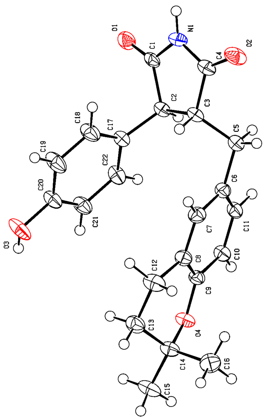
HSQC was used to assign attribution to their NMR data (Table 1). The 1H–1H COSY correlation considered information between H–3 (δH 3.64, d, 5.7)/H–4 (δH 3.16, m); H–2′ (6′) (δH 6.75, d, 8.6)/H–3′ (5′) (δH 6.62, d, 8.6), H–5″ (δH 6.58, d, 8.2)/H–6″ (δH 6.85, d, 8.2), and H–7″ (δH 2.66, m)/H–8″ (δH 1.76, t, 6.8) and HMBC correlations from H–3 (δH 3.64, d, 5.7) to C–2 (δC 181.2, C), C–5 (δC 181.7, C) and C–1′ (δC 129.7, C); H–4 (δH 3.16, m) to C–2 (δC 181.3, C), C–5 (δC 181.7, C), and C–1″ (δC 129.7, C); Ha–6 (δH 2.88, dd, 13.8, 8.5) and Hb–6 (δH 3.13, m) to C–3 (δC 53.2, CH), C–4 (δC 53.6, CH), C–5 (δC 181.7, C), C–1″ (δC 129.7, C), C–2″ (δC 131.4, C) and C–6″ (δC 129.3, C); H–7″ (δH 2.66, m) to C–2″ (δC 131.4, C), C–3″ (δC 123.3, C), C–4″ (δC 154.1, C) and C–9″ (δC 75.2, C); H–8″ (δH 1.76, t, 6.8) to C–3″ (δC 123.3, C), C–9″ (δC 75.2, C) and C–10″ (δC 26.9, C); H–11″ (δH 1.28, s) to C–9″ (δC 75.2, C) and C–10″ (δC 26.9, C). This completed the gross structure of compound 1 (Figure 2). The NOESY correlation of H–3 and H–6 indicated these protons were cofacial. The absolute configurations of 1 were established based on a single-crystal X-ray diffraction experiment. With a Flack parameter of 0.02(11), the absolute configuration of 1 was definitively assigned as 3S, 4R. (Figure 3 and Figure 4).
Aspergteroid H (2) was isolated as pale-yellow gum with a molecular formula of C17H13NO4, determined by its HR–ESI–MS m/z 294.0768 [M − H] (calculated: 294.0766), indicating 11 degrees of unsaturation. The NMR data of compounds 1 and 2 are extremely similar (see Table 1), indicating the same disubstituted maleimides fragment. Compared with 1, 2 showed additional signals of a para-substituted phenol (A2B2 system) at δH 6.99 (d, 8.6 Hz, 2H) and 6.83 (d, 8.6 Hz, 2H) and two olefinic carbons (δC 139.7 (C) and 138.2 (C)), while the three methylene signals (δC 23.3 (CH2), 33.8 (CH2), and 35.6 (CH2)) and one oxygenated quaternary δC 75.2 (C)) in 1 were absent in 2. The 1H–1H COSY correlation information between H–2′ (6′) (δH 6.76, d, 8.6)/H–3′ (4′) (δH 6.63, d, 8.6), H–2″ (6″) (δH 6.99, d, 8.6)/H–3″ (5″) (δH 6.83, d, 8.6) and the HMBC correlations from H–6 (δH 2.80, s) to C–3 (δC 139.7, C), C–4 (δC 138.2, C), C–5 (δC 174.0, C), C–1″ (δC 129.8, C), and C–2″ (6″) (δC 130.3, C) were further confirmed in the structure of 2 (Figure 2 and Table 1).
Aspergteroid I (3) was also isolated as a pale-yellow gum with a molecular formula of C25H26O7, determined by its HR–ESI–MS m/z 437.1602 [M − H] (calculated: 437.1600), indicating 13 degrees of unsaturation. The NMR data of compounds 3 and 7 [22] are extremely similar, indicating the same trisubstituted butenolides fragment. The significant difference observed in the NMR spectra of 3 compared with that of 7 was the absence of an olefinic proton signal (δH 5.11, m, 1H), two olefinic carbon signals (δc 122.6 d and 131.5 s), and the presence of three methylenes and two oxygenated sp3 quaternary carbons compared ith two methylenes and one oxygenated sp3 quaternary carbon in 7 in both 13C NMR and DEPT spectra. These data were indicative of the presence of a dihydropyran ring fused to a trisubstituted benzene ring in place of the open prenyl chain present in 7. The HMBC data were in good agreement with structure 3 (Figure 2). Key 1H–1H COSY correlation information between H–7″ (δH 2.57, m)/H–8″ (1.71, t (6.5)) and the HMBC correlations from H–7″ (δH 2.57, m) to C–2″ (δC 132.6, CH), C–3″ (δC 121.4, C), C–4″ (δC 154.3, C), C–8″ (δC 33.7, CH2) and C–9″ (δC 75.1, C); H–8″ (1.71, t (6.5)])to C–3″ (δC 121.4, C), C–7″ (δC 23.2, CH2), C–9″ (δC 75.1, C) and C–10″ (δC 26.9, CH3) established a dihydropyran ring fused through the C3″–C4″ bond of a benzene ring.
Similarly, the absolute configuration of 3 was established as 5R according to biological pathways [25] and the quantum chemical ECD calculation.
Aspergteroid I (4) was obtained as pale-yellow gum with a molecular formula of C24H24O6, determined by its HR–ESI–MS m/z 407.1493 [M − H] (calculated: 407.1495), indicating 13 degrees of unsaturation. Comprehensive analyses of the 1D NMR spectra of 4 with 8 [5] suggested the presence of a dihydropyran ring fused to a trisubstituted benzene ring in place of the open isopentenyl chain present in 8. Similarly, the HMBC data were in good agreement with structure 4 (Figure 2). Key 1H–1H COSY correlation information between H–7″ (δH 2.53, m)/H–8″ (1.68, t (6.5)), and the HMBC correlations from H–7″ (δH 2.53, m) to C–2″ (δC 132.6, CH), C–3″ (δC 121.4, C), C–4″ (δC 154.3, C), C–8″ (δC 33.6, CH2) and C–9″ (δC 75.1, C); H–8″ [1.68, t (6.5)] to C–3″ (δC 121.4, C), C–7″ (δC 23.1, CH2), C–9″ (δC 75.1, C) and C–10″ (δC 26.9, CH3) established a dihydropyran ring fused through the C3″–C4″ bond of a benzene ring.
Similarly, the absolute configuration of 4 was determined to be 5R through both the biological pathways [25] and the quantum chemical ECD calculations.

3.2. Evaluation of Cell Viability

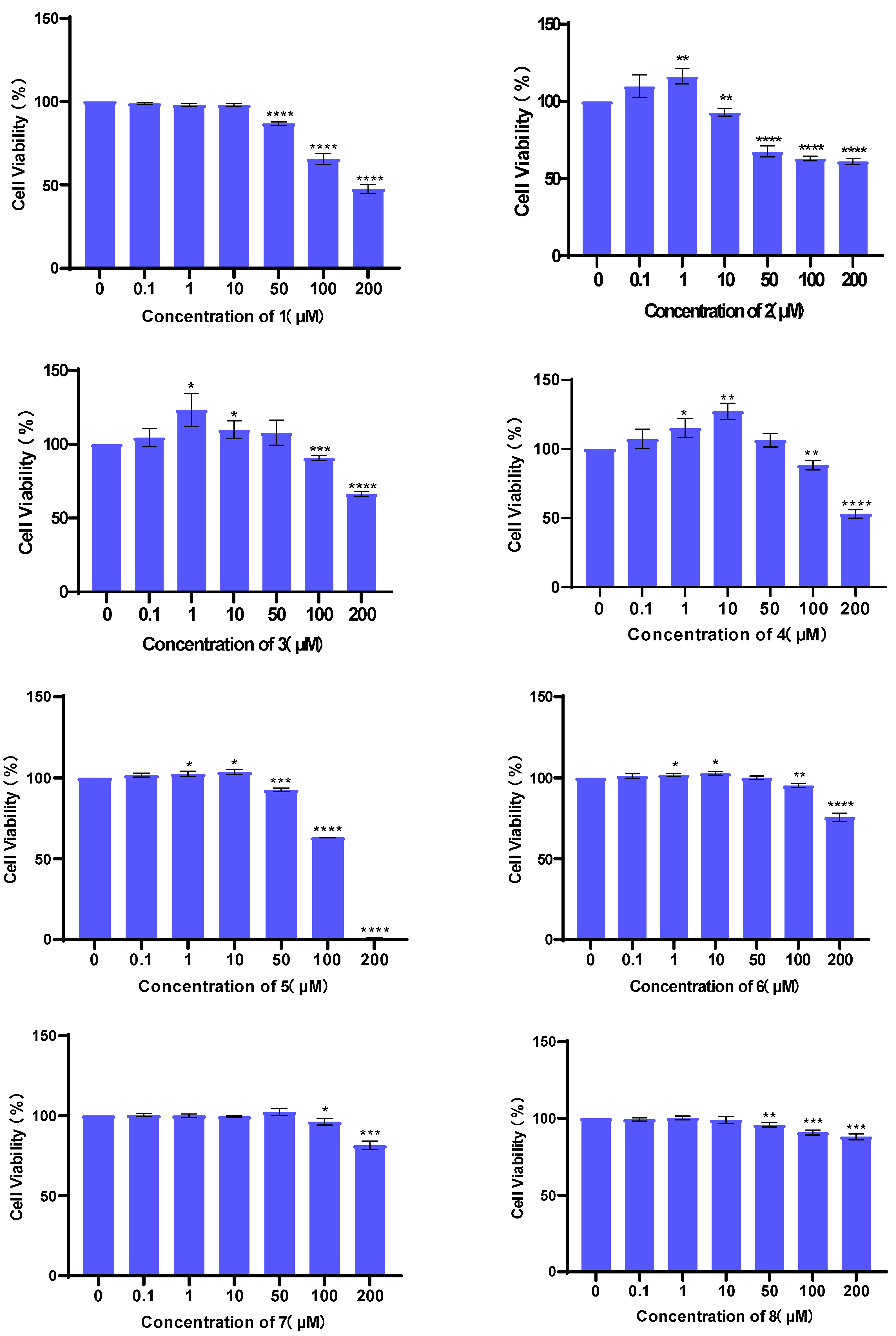
The cytotoxicities of compounds 18 against H9c2 cell lines were tested using a CCK-8 assay kit. The results showed that compounds 18 did not exhibit obvious cytotoxic effects in concentrations below 10 µM (Figure 5).

3.3. Evaluation of Cardioprotective Effects

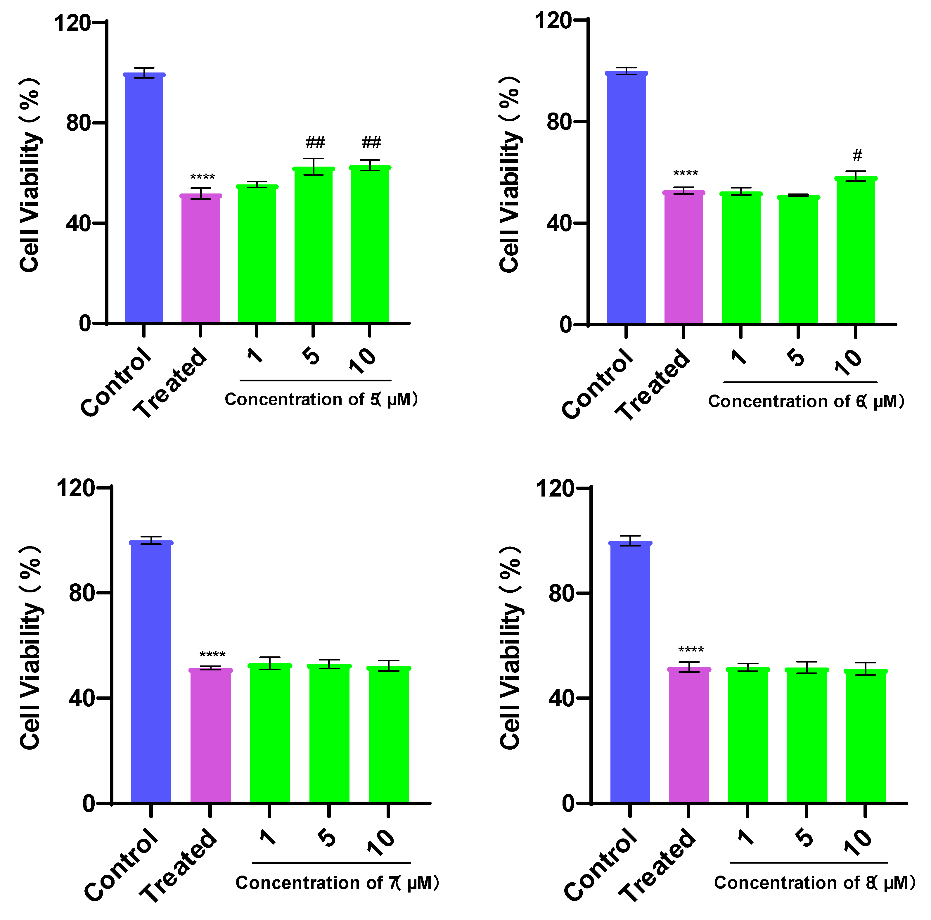
Since oxidative stress contributes to many cardiovascular diseases [26], the antioxidant effects of butenolides were the focus of our attention. All compounds in this study were evaluated for their protective effects against TBHP-induced H9c2 apoptosis. H9c2 cells were treated with TBHP-induced cell injury. The cell injury was estimated in terms of cell viability. Treatment with TBHP significantly decreased cell viabilities (p < 0.05, Figure 6). As shown in Figure 6, compounds 1, 2, 5, and 6 showed protective effects while compounds 3, 4, 7, and 8 could not improve the cell viability at these three concentrations, suggesting that the cardioprotective effects of disubstituted maleimides and trisubstituted butenolides could be related to their phenyl- and benzyl-disubstituted types and nitrogenous element.
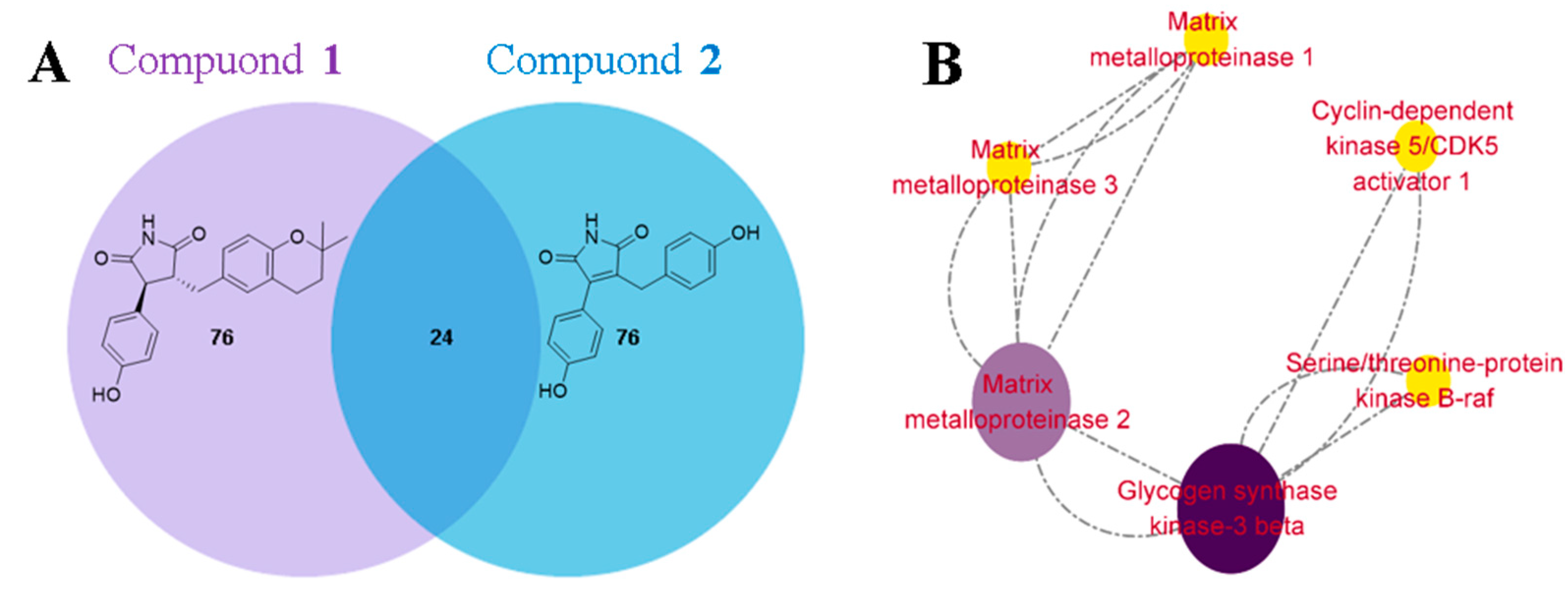
To investigate the mechanism underlying the cardioprotective effects of compound 1, the top 100 genes associated with myocardial protection for compounds 1 and 2 were predicted [27]. A Venn diagram approach identified 24 overlapping genes. Subsequently, a protein–protein interaction (PPI) network was constructed using the STRING 11.5 database [28] and Cytoscape 3.9.0 software [29]. The PPI analysis revealed that glycogen synthase kinase-3β (GSK-3β) had the highest degree score, suggesting it may play a crucial role in mediating the cardioprotective effects of compound 1 (Figure 7).
Many studies have indicated that GSK-3β is a promising target for the treatment of cardiovascular diseases [30]. Additionally, the Bcl-2/Bax and caspase-3 proteins are important regulators of cell survival and apoptosis. We found that after TBHP induced oxidative stress injury of cardiomyocytes, 1 could inhibit GSK-3β activity to downregulate the activation of NLRP3 inflammatorome and reduce the inflammatory response. The expression changes in Bcl-2/Bax and caspase-3 indicated that 1 could effectively inhibit cell apoptosis and protect myocardial cells, as shown in Figure 8.

4. Conclusions

Four new and four known compounds were isolated from the fermentation extract of the soft-coral-associated symbiotic and epiphytic fungus Aspergillus terreus EGF7-0-1. The new natural disubstituted maleimides identified in this research expand the knowledge of the chemical space and biological diversity of microorganisms of marine origin. Because of structural specificities, compound 1 showed significant protective effects against the TBHP-induced H9c2 apoptosis at low concentrations. We further attempted to explore the possible mechanism of oxidative stress injury in cardiomyocytes through network pharmacology and the TBHP-induced injury model. The results suggested that compound 1 may reduce the inflammatory response and protect cardiomyocytes through GSK-3β/NLRP3 pathway. At the same time, the expression of caspase-3 and Bax was regulated, the antiapoptotic gene Bcl-2 was upregulated, the antioxidant capacity of cells was improved, and apoptosis was inhibited.

Supplementary Materials

The following supporting information can be downloaded at: https://www.mdpi.com/article/10.3390/bioengineering10070805/s1, Figures S1 and S2: Key molecular orbitals involved in important transitions regarding the ECD spectrum of the dominant conformer of 3 and 4; Figures S3–S35: the HRESIMS, IR, 1D NMR, and 2D NMR spectra of compounds 1–4; Figure S36: The whole Western blot; Table S1: 1H NMR data (400 MHz) and 13C NMR data (100 MHz) of 5–8 (δ in ppm, J in Hz) in CD3OD; Tables S2–S5: Cartesian coordinates of the dominant conformer and key transitions and their related rotatory and oscillator strengths of the dominant conformer of 3 and 4 at the B3LYP/6-31+g(d) level; Tables S6–S10: Densitometry readings/intensity ratio of Bax, Bcl-2, caspase-3, GSK-3β, and NLRP3.

Author Contributions

Conceptualization, C.-X.Z.; methodology, H.F. and L.W.; software, Z.-K.Z.; formal analysis, H.F., L.W., Q.W. and Z.-K.Z.; investigation, P.-P.W., L.-Y.C. and Y.-P.H.; data curation, H.F., L.W. and Z.-K.Z.; writing—original draft preparation, H.F. and L.W.; writing—review and editing, C.-X.Z. and Q.W.; supervision, C.-X.Z.; funding acquisition, C.-X.Z. All authors have read and agreed to the published version of the manuscript.

Funding

This research was funded by Marine Economic Department of Natural Resources of Guangdong Province (GDNRC [2023]37, GDNRC [2021]48 and GDNRC [2020]039), the National Natural Science Foundation of China (No. 82273845 and No. 81741160), the Science and Technology Planning Project of Guangdong Province (No. 2017A020217008 and No. 2015A020216017), and Guangdong Provincial Science and Technology Innovation Strategy Special Fund-Guangdong ‘Climbing Plan’ (pdjh2023b0126).

Institutional Review Board Statement

Not applicable.

Informed Consent Statement

Not applicable.

Data Availability Statement

Not applicable.

Conflicts of Interest

The authors declare no conflict of interest.

References

  1. Read, S.H.; Wild, S.H. Prevention of premature cardiovascular death worldwide. Lancet 2020, 395, 758–760. [Google Scholar] [CrossRef] [PubMed]
  2. Umbarkar, P.; Tousif, S.; Singh, A.P.; Anderson, J.C.; Zhang, Q.; Tallquist, M.D.; Woodgett, J.; Lal, H. Fibroblast GSK-3α promotes fibrosis via RAF-MEK-ERK pathway in the injured heart. Circ. Res. 2022, 131, 620–636. [Google Scholar] [CrossRef] [PubMed]
  3. Hosoe, T.; Iizuka, T.; Komai, S.; Wakana, D.; Itabashi, T.; Nozawa, K.; Fukushima, K.; Kawai, K. 4-Benzyl-3-phenyl-5H-furan-2-one, a vasodilator isolated from Malbranchea filamentosa IFM 41300. Phytochemistry 2005, 66, 2776–2779. [Google Scholar] [CrossRef]
  4. San-Martín, A.; Rovirosa, J.; Vaca, I.; Vergara, K.; Acevedo, L.; Vina, D.; Orallo, F.; Chamy, M.C. New butyrolactine from a marine-derived fungus Aspergillus sp. J. Chil. Chem. Soc. 2011, 56, 625–627. [Google Scholar] [CrossRef] [Green Version]
  5. Peng, Q.Y.; Chen, W.H.; Lin, X.P.; Xiao, J.; Liu, Y.H.; Zhou, X.F. Butenolides from the Coral-Derived fungus Aspergillius terreus SCSIO41404. Mar. Drugs 2022, 20, 212. [Google Scholar] [CrossRef] [PubMed]
  6. Luo, X.W.; Lin, Y.; Lu, Y.J.; Zhou, X.F.; Liu, Y.H. Peptides and polyketides isolated from the marine sponge-derived fungus Aspergillus terreus SCSIO 41008. Chin. J. Nat. Med. 2019, 17, 149–154. [Google Scholar] [CrossRef]
  7. He, F.; Bao, J.; Zhang, X.Y.; Tu, Z.C.; Shi, Y.M.; Qi, S.H. Asperterrestide A, a cytotoxic cyclic tetrapeptide from the marine-derived fungus Aspergillus terreus SCSGAF0162. J. Nat. Prod. 2013, 76, 1182–1186. [Google Scholar] [CrossRef]
  8. Hamed, A.; Abdel-Razek, A.S.; Omran, D.A.; EiMetwally, M.M.; Ei-Hosari, D.G.; Frese, M.; Soliman, S.M.; Sewald, N.; Shaaban, M. Terretonin O: A new meroterpenoid from Aspergillus terreus. Nat. Prod. Res. 2019, 34, 965–974. [Google Scholar] [CrossRef]
  9. Liu, C.; Liu, H.; Wen, Y.Z.; Huang, H.Q.; Hao, J.; Lv, Y.B.; Qin, R.; Yang, X.Z. Aspernolide a inhibits the proliferation of human laryngeal carcinoma cells through the mitochondrial apoptotic and STAT3 signaling pathways. Molecules 2019, 24, 1074. [Google Scholar] [CrossRef] [Green Version]
  10. Dewi, R.T.; Tachibana, S.; Darmawan, A. Effect on α-glucosidase inhibition and antioxidant activities of butyrolactone derivatives from Aspergillus terreus MC751. Med. Chem. Res. 2014, 23, 454–460. [Google Scholar] [CrossRef]
  11. Fan, H.; Wei, X.; Si-Tu, M.X.; Lei, Y.H.; Zhou, F.G.; Zhang, C.X. γ-aromatic butenolides of microbial source—A review of their structures, biological activities and biosynthesis. Chem. Biodiv. 2022, 19, e202200208. [Google Scholar] [CrossRef]
  12. Liao, G.F.; Wu, P.; Xue, J.H.; Liu, L.; Li, H.X.; Wei, X.Y. Asperimides A–D, anti-inflammatory aromatic butenolides from a tropical endophytic fungus Aspergillus terreus. Fitoterapia 2018, 131, 50–54. [Google Scholar] [CrossRef] [PubMed]
  13. Ibrahim, S.R.; Elkhayat, E.S.; Mohamed, G.A.; Khedr, A.I.; Fouad, M.A.; Kotb, M.H.; Ross, S.A. Aspernolides F and G, new butyrolactones from the endophytic fungus Aspergillus terreus. Phytochem. Lett. 2015, 14, 84–90. [Google Scholar] [CrossRef]
  14. Gu, W.; Qiao, C. Furandiones from an endophytic Aspergillus terreus residing in Malus halliana. Chem. Pharm. Bull. 2012, 60, 1474–1477. [Google Scholar] [CrossRef] [PubMed] [Green Version]
  15. Rao, K.; Sadhukhan, A.; Veerender, M.; Ravikumar, V.; Mohan, E.; Dhanvantri, S.; Sitaramkumar, M.; Babu, J.M.; Vyas, K.; Reddy, G.O. Butyrolactones from Aspergillus terreus. Chem. Pharm. Bull. 2000, 48, 559–562. [Google Scholar] [CrossRef] [Green Version]
  16. Guo, F.; Li, Z.; Xu, X.; Wang, K.; Shao, M.; Zhao, F.; Wang, H.; Hua, H.; Pei, Y.; Bai, J. Butenolide derivatives from the plant endophytic fungus Aspergillus terreus. Fitoterapia 2016, 113, 44–50. [Google Scholar] [CrossRef]
  17. Li, L.J.; Li, T.X.; Kong, L.Y.; Yang, M.H. Antioxidant aromatic butenolides from an insect-associated Aspergillus iizukae. Phytochem. Lett. 2016, 16, 134–140. [Google Scholar] [CrossRef]
  18. Wei, X.; Su, J.C.; Hu, H.S.; He, X.X.; Lin, S.J.; Zhang, D.M.; Ye, W.C.; Chen, M.F.; Lin, H.W.; Zhang, C.X. Probing indole diketopiperazine-based hybrids as environmental-2 induced products from Aspergillus sp. EGF 15-0-3. Org. Lett. 2022, 24, 158–163. [Google Scholar] [CrossRef]
  19. Cheng, Y.J.; Chen, N.N.; Li, J.; Su, J.C.; Yang, J.Y.; Zhang, C.X.; Lin, H.W.; Zhou, Y.J. Antimicrobial chlorinated carbazole alkaloids from the sponge-associated actinomycete streptomyces diacarni LHW51701. Chin. J. Chem. 2021, 39, 1188–1192. [Google Scholar] [CrossRef]
  20. Fan, H.; Shi, Z.M.; Lei, Y.H.; Si-Tu, M.X.; Zhou, F.G.; Feng, C.; Wei, X.; Shao, X.H.; Zhang, C.X. Rare carbon-bridged citrinin dimers from the starfish-derived symbiotic fungus Penicillium sp. GGF16-1-2. Mar. Drugs 2022, 20, 443. [Google Scholar] [CrossRef]
  21. Sun, Y.T.; Liu, J.T.; Li, L.; Gong, C.; Wang, S.P.; Yang, F.; Hua, H.M.; Lin, H.W. New butenolide derivatives from the marine sponge-derived fungus Aspergillus terreus. Bioorg. Med. Chem. Lett. 2018, 28, 315–318. [Google Scholar] [CrossRef] [PubMed]
  22. Rachada, H.; Pranee, R.; Rungtiwa, C.; Nattawut, B.; Masahiko, I. Butyrolactones from the Fungus Aspergillus terreus BCC 4651. Chem. Pharm. Bull. 2010, 58, 1545–1548. [Google Scholar]
  23. Nakamura, N.; Hirakawa, A.; Gao, J.J.; Kakuda, H.; Shiro, M.; Komatsu, Y.; Sheu, C.C.; Hattori, M. Five new maleic and succinic acid derivatives from the mycelium of Antrodia camphorata and their cytotoxic effects on LLC tumor cell line. J. Nat. Prod. 2004, 67, 46–48. [Google Scholar] [CrossRef]
  24. Wu, M.D.; Cheng, M.J.; Wang, B.C.; Yech, Y.J.; Lai, J.T.; Kuo, Y.H.; Yuan, G.F.; Chen, I.S. Maleimide and maleic anhydride derivatives from the mycelia of Antrodia cinnamomea and their nitric oxide inhibitory activities in macrophages. J. Nat. Prod. 2008, 71, 1258–1261. [Google Scholar] [CrossRef] [PubMed]
  25. Bao, J.; Li, X.; He, F.; Zhang, X.Y.; Zhu, K.K.; Tao, H.R.; Yu, J.H.; Liu, H.S.; Zhang, H. Asperbutenolide A, an unusual aromatic butenolide dimer with diverse bioactivities from a marine-derived fungus Aspergillus terreus SCAU011. Tetrahedron Lett. 2020, 61, 152193. [Google Scholar] [CrossRef]
  26. Senoner, T.; Dichtl, W. Oxidative stress in cardiovascular diseases: Still a therapeutic target? Nutrients 2019, 11, 2090. [Google Scholar] [CrossRef] [PubMed] [Green Version]
  27. Daina, A.; Michielin, O.; Zoete, V. Swiss Target Prediction: A web server for target prediction of bioactive small molecules. Nucleic Acids. Res. 2019, 47, 32–38. [Google Scholar]
  28. Szklarczyk, D.; Gable, A.L.; Lyon, D.; Junge, A.; Wyder, S.; Huerta-Cepas, J.; Simonovic, M.; Doncheva, N.T.; Morris, J.H.; Bork, P.; et al. STRING v11: Protein–protein association networks with increased coverage, supporting functional discovery in genome-wide experimental datasets. Nucleic Acids. Res. 2019, 47, 607–613. [Google Scholar] [CrossRef] [Green Version]
  29. Shannon, P.; Markiel, A.; Ozier, O.; Baliga, N.S.; Wang, J.T.; Ramage, D.; Amin, N.; Schwikowski, B.; Ideker, T. Cytoscape: A software environment for integrated models of biomolecular interaction networks. Genome Res. 2003, 13, 2498–2504. [Google Scholar] [CrossRef]
  30. Wang, Z.R.; Yao, M.R.; Wang, L.Y.; Yang, Y.Q.; Wang, Q.; Qian, X.; Zhao, Y.; Qiao, J.Q. Dexmedetomidine attenuates myocardial ischemia/reperfusion-induced ferroptosis via AMPK/GSK-3β/Nrf2 axis. Biomed. Pharmacother. 2022, 154, 113572. [Google Scholar] [CrossRef]
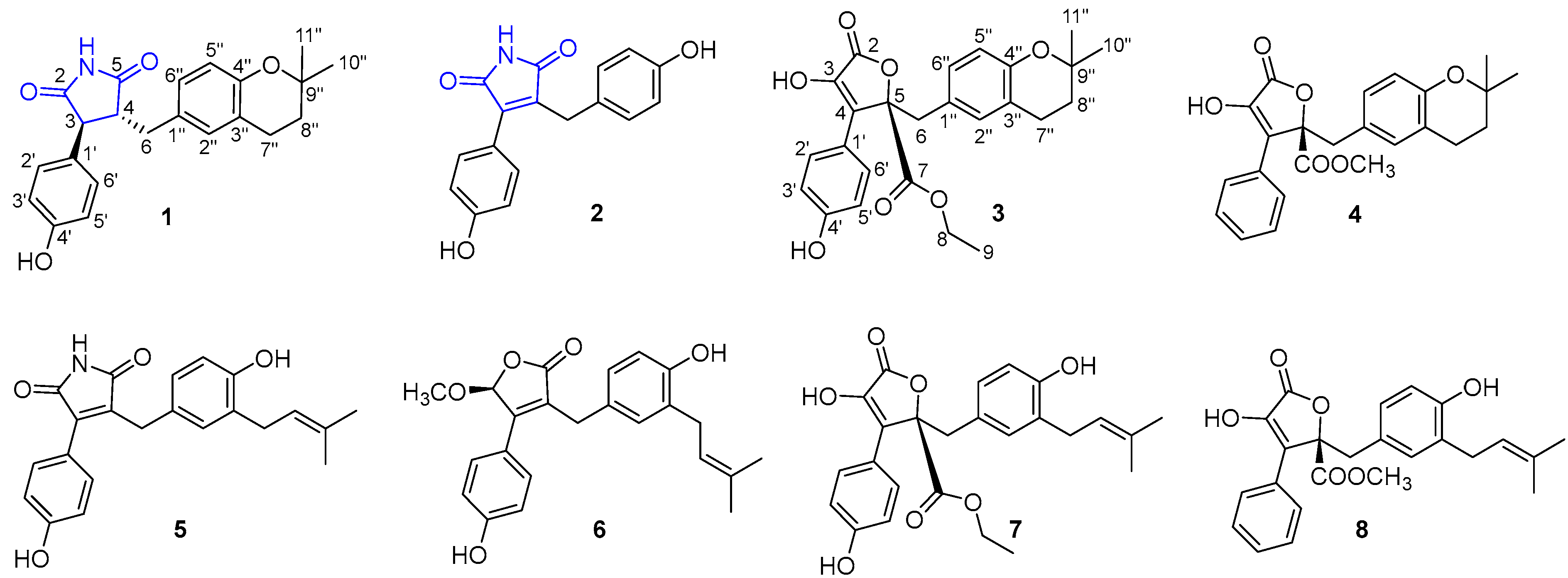
Figure 1. The structures of compounds 18 isolated from Aspergillus terreus EGF7-0-1.
Bioengineering 10 00805 g001
Figure 2. Correlation diagram of main 1H–1H COSY and HMBC of compounds 14.
Bioengineering 10 00805 g002
Figure 3. Crystal structure of compound 1.
Bioengineering 10 00805 g003
Figure 4. Experimental and calculated ECD spectra of 3 and 4.
Bioengineering 10 00805 g004
Figure 5. H9c2 cell viability of compounds 18 at concentrations of 0, 0.1, 1, 10, 50, 100, and 200 µM at 37 °C for 24 h. Cytotoxicity was assessed via a CCK-8 assay. Values are expressed as mean ± standard deviation (SD), n = 3. * p < 0.05, ** p < 0.01, *** p < 0.001, **** p < 0.0001 vs. control group.
Bioengineering 10 00805 g005
Figure 6. Protective effects of compounds 18 on TBHP-induced injury in H9c2 cells. Values are expressed as mean ± standard deviation (SD), n = 3. **** p < 0.0001 vs. control group. # p < 0.05, ## p < 0.01, ### p < 0.001 vs. treated group.
Bioengineering 10 00805 g006aBioengineering 10 00805 g006b
Figure 7. (A) The Venn diagram of the number of overlapping genes. (B) The PPI networks of 1 and 2.
Bioengineering 10 00805 g007
Figure 8. The effects of 1 on Bcl-2, Bax, caspase-3, NLRP3, and GSK-3β proteins in H9c2 cells were determined by Western blot analysis. All data are expressed as means ± SD. ** p < 0.01, *** p < 0.001, **** p < 0.0001 vs. control group. # p < 0.05, ## p < 0.01, ### p < 0.001 vs. treated group.
Bioengineering 10 00805 g008
Table 1. NMR data of 14 in CD3OD.
No.1 2 3 4
δH, mult, (J in Hz)δC, TypeNo.δH, mult, (J in Hz)δC, TypeNo.δH, mult, (J in Hz)δC, TypeNo.δH, mult, (J in Hz)δC, Type
2 181.3, C2 174,7 C2 170.8, C2 169.8, C
33.64, d (5.7)53.2, CH3 139.7, C3 140.3, C3 141.8, C
43.16, m53.6, CH4 138.2, C4 125.7, C4 125.4, C
5 181.7, C5 174.0, C5 86.9, C5 86.9, C
6a 2.88, dd (13.8, 8.5)
b 3.13, m
35.6, CH262.80, s29.4, CH263.43, s39.6, CH263.44, d (5.20)39.4, CH2
1′ 121.5, C7 171.1, C7 171.3, C
1′ 129.7, C2′ (6′)7.43, d (8.6)132.4, CH84.25, q (7.0)63.6, CH21′ 130.2, C
2′ (6′)6.75, d (8.6)130.2, CH3′ (5′)6.69, d (8.6)116.5, CH91.21, t (7.0)14.3, CH32′ (6′)7.66, d (8.5)128.6, CH
3′ (5′)6.62, d (8.6)116.4, CH4′ 160.4, C1′ 123.4, C3′ (5′)6.44, m129.8, CH
4′ 157.8, C1″ 129.8, C2′ (6′) 7.59, d (8.8)130.3, CH4′7.37, m130.2, CH
1″ 129.7, C2″ (6″)6.99, d (8.6)130.3, CH3′ (5′) 6.87, d (8.8)116.5, CH1″ 131.8, C
2″6.83, s131.4, CH3″ (5″)6.83, d (8.6)116.4, CH4′ 159.2, C2″6.45, s132.6, CH
3″ 122.3, C4″ 157.1, C1″ 129.0, C3″ 121.4, C
4″ 154.1, C 2″6.49, s132.6, CH4″ 154.3, C
5″6.58, d (8.2)118.2, CH 3″ 121.4, C5″6.42, d (8.0)117.4, CH
6″6.85, d (8.2)129.3, CH 4″ 154.3, C6″6.48, d (8.0)130.2, CH
7″2.66, m23.3, CH2 5″6.43, d (8.0)117.4, CH7″2.53, m23.1, CH2
8″1.76, t (6.8)33.8, CH2 6″6.52, d (8.0)130.2, CH8″1.68, t (6.5)33.6, CH2
9″ 75.2, C 7″2.57, m23.2, CH29″ 75.1, C
10″1.28, s26.9, CH3 8″1.71, t (6.5)33.7, CH210″1.22, s26.9, CH3
11″1.28, s27.1, CH3 9″ 75.1, C11″1.22, s27.0, CH3
10″1.24, s26.9, CH3-OMe3,49, s53.9
11″1.24, s27.0, CH3
Disclaimer/Publisher’s Note: The statements, opinions and data contained in all publications are solely those of the individual author(s) and contributor(s) and not of MDPI and/or the editor(s). MDPI and/or the editor(s) disclaim responsibility for any injury to people or property resulting from any ideas, methods, instructions or products referred to in the content.