

Comparison of Left Ventricular Function Derived from Subject-Specific Inverse Finite Element Modeling Based on 3D ECHO and Magnetic Resonance Images


Abstract
1. Introduction
2. Methods
2.1. Data Acquisition and Preprocessing
2.2. Left Ventricular Mechanics Finite Element Model
2.2.1. Constitutive Law of the LV
2.2.2. Finite Element Formulation of the Left Ventricular
2.2.3. Estimation of the Model Parameters
Estimation of Passive Parameters
Estimation of the Active Parameter
Implementation
2.3. Statistical Analysis
2.4. Scientific Contribution
3. Results
3.1. Left Ventricular Wall Thickness and Volumes
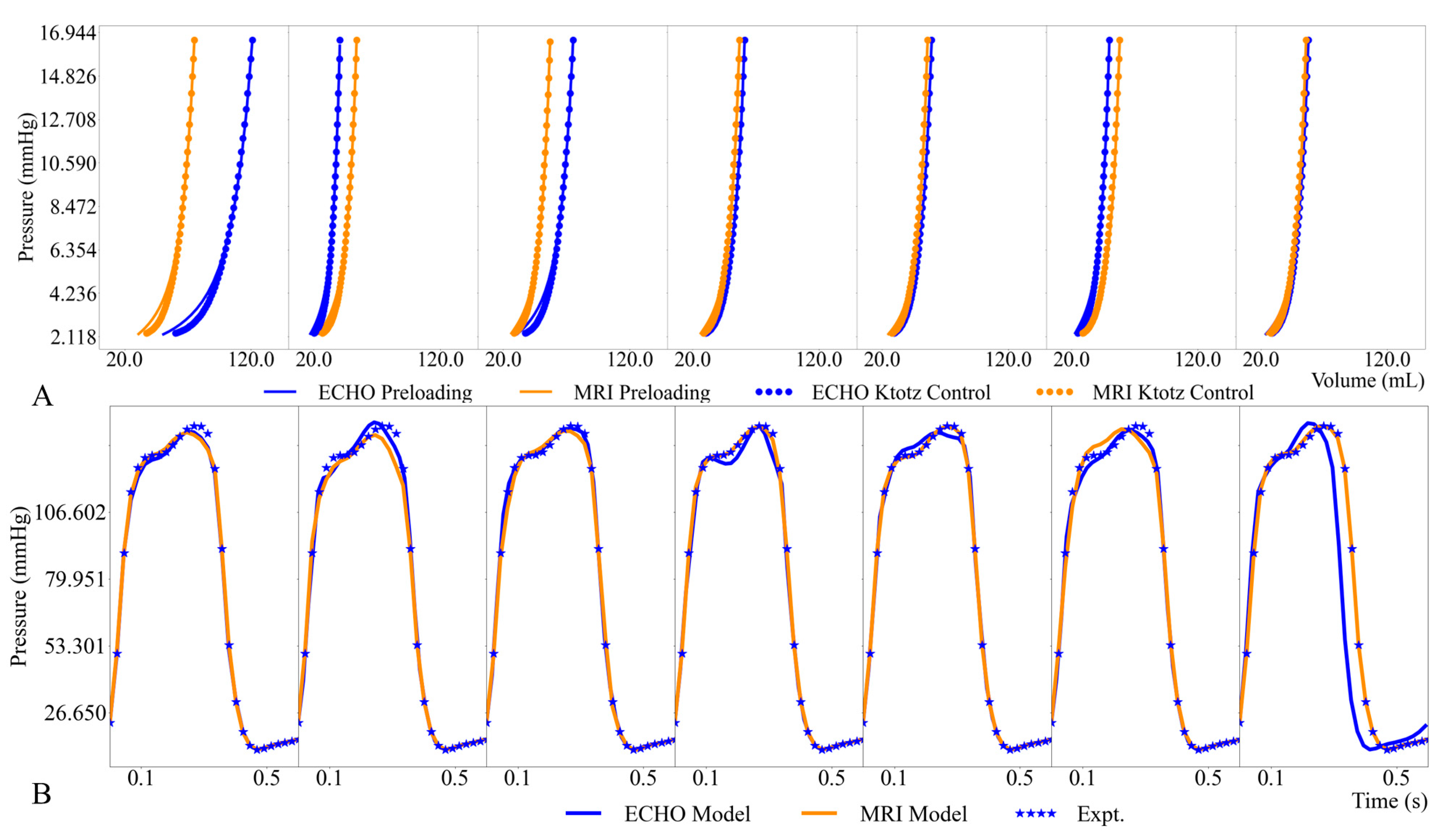
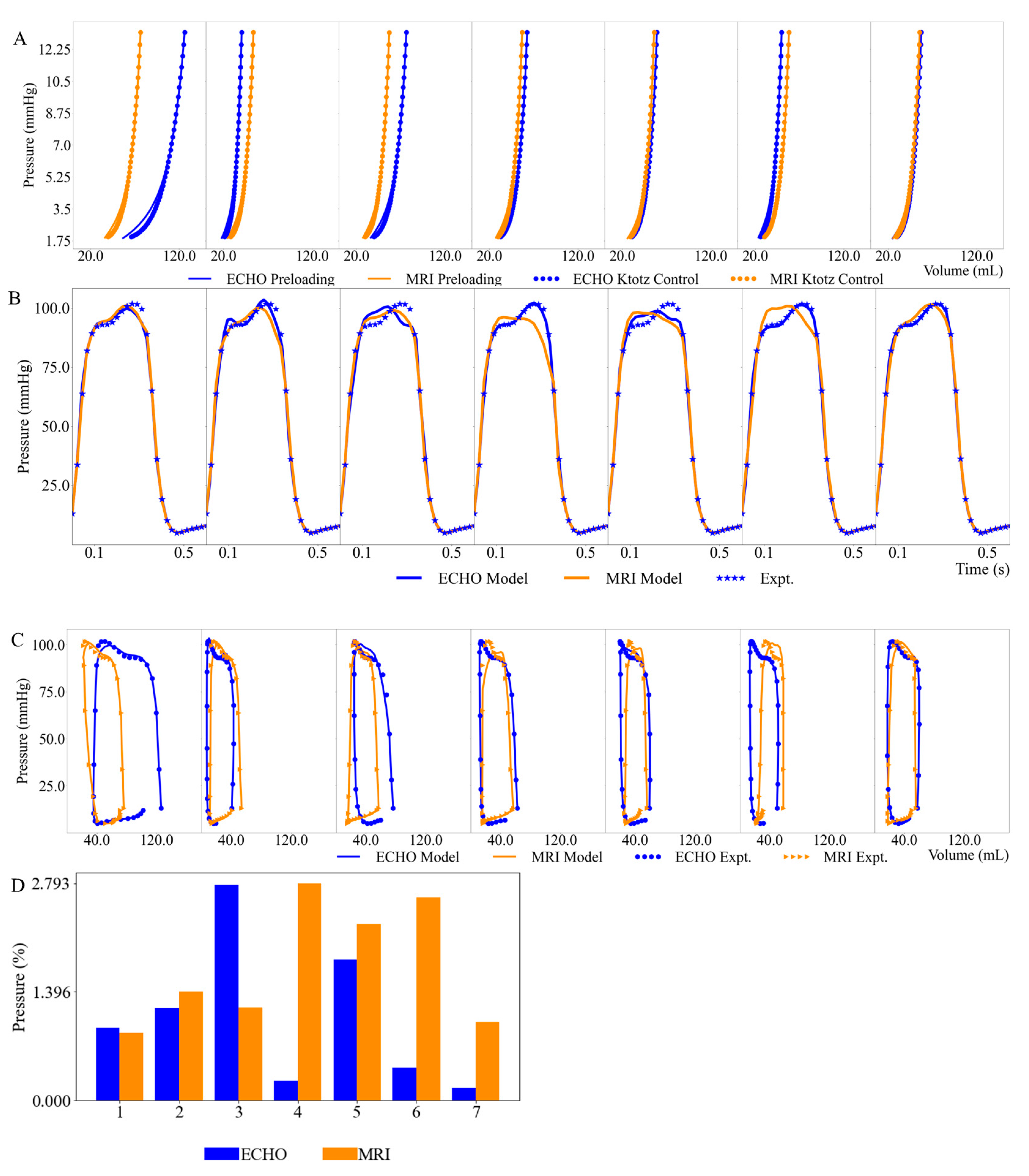
3.2. Left Ventricular Pressure–Volume Loops
3.3. Myocardial Contractility
4. Discussion
4.1. Left Ventricular Geometry and Volumes
4.2. Left Ventricular Function
5. Limitations
6. Conclusions
Author Contributions
Funding
Institutional Review Board Statement
Informed Consent Statement
Data Availability Statement
Conflicts of Interest
Appendix A


References
- Obokata, M.; Nagata, Y.; Wu, V.C.C.; Kado, Y.; Kurabayashi, M.; Otsuji, Y.; Takeuchi, M. Direct Comparison of Cardiacmagnetic Resonance Feature Tracking and 2D/3D Echocardiography Speckle Tracking for Evaluation of Global Left Ventricular Strain. Eur. Heart J. Cardiovasc. Imaging 2016, 17, 525–532. [Google Scholar] [CrossRef] [PubMed]
- Sugeng, L.; Mor-Avi, V.; Weinert, L.; Niel, J.; Ebner, C.; Steringer-Mascherbauer, R.; Schmidt, F.; Galuschky, C.; Schummers, G.; Lang, R.M.; et al. Quantitative Assessment of Left Ventricular Size and Function: Side-by-Side Comparison of Real-Time Three-Dimensional Echocardiography and Computed Tomography with Magnetic Resonance Reference. Circulation 2006, 114, 654–661. [Google Scholar] [CrossRef]
- Onishi, T.; Saha, S.K.; Delgado-Montero, A.; Ludwig, D.R.; Onishi, T.; Schelbert, E.B.; Schwartzman, D.; Gorcsan, J. Global Longitudinal Strain and Global Circumferential Strain by Speckle-Tracking Echocardiography and Feature-Tracking Cardiac Magnetic Resonance Imaging: Comparison with Left Ventricular Ejection Fraction. J. Am. Soc. Echocardiogr. 2015, 28, 587–596. [Google Scholar] [CrossRef] [PubMed]
- Brown, J.; Jenkins, C.; Marwick, T.H. Use of Myocardial Strain to Assess Global Left Ventricular Function: A Comparison with Cardiac Magnetic Resonance and 3-Dimensional Echocardiography. Am. Heart J. 2009, 157, 102.e1–102.e5. [Google Scholar] [CrossRef] [PubMed]
- Wang, V.Y.; Nielsen, P.M.F.; Nash, M.P. Image-Based Predictive Modeling of Heart Mechanics. Annu. Rev. Biomed. Eng. 2015, 17, 351–383. [Google Scholar] [CrossRef]
- Lee, L.C.; Genet, M.; Dang, A.B.; Ge, L.; Guccione, J.M.; Ratcliffe, M.B. Applications of Computational Modeling in Cardiac Surgery. J. Card. Surg. 2014, 29, 293–302. [Google Scholar] [CrossRef]
- Lee, L.C.; Wenk, J.F.; Zhong, L.; Klepach, D.; Zhang, Z.; Ge, L.; Ratcliffe, M.B.; Zohdi, T.I.; Hsu, E.; Navia, J.L.; et al. Analysis of Patient-Specific Surgical Ventricular Restoration: Importance of an Ellipsoidal Left Ventricular Geometry for Diastolic and Systolic Function. J. Appl. Physiol. 2013, 115, 136–144. [Google Scholar] [CrossRef] [PubMed]
- Lasso, A.; Herz, C.; Nam, H.; Cianciulli, A.; Pieper, S.; Drouin, S.; Pinter, C.; St-Onge, S.; Vigil, C.; Ching, S.; et al. SlicerHeart: An Open-Source Computing Platform for Cardiac Image Analysis and Modeling. Front. Cardiovasc. Med. 2022, 9, 886549. [Google Scholar] [CrossRef]
- Finsberg, H.; Xi, C.; Zhao, X.; Tan, J.L.; Genet, M.; Sundnes, J.; Lee, L.C.; Zhong, L.; Wall, S.T. Computational Quantification of Patient-Specific Changes in Ventricular Dynamics Associated with Pulmonary Hypertension. Am. J. Physiol.-Hear. Circ. Physiol. 2019, 317, H1363–H1375. [Google Scholar] [CrossRef]
- Fan, L.; Namani, R.; Choy, J.S.; Awakeem, Y.; Kassab, G.S.; Lee, L.C. Role of Coronary Flow Regulation and Cardiac-Coronary Coupling in Mechanical Dyssynchrony Associated with Right Ventricular Pacing. Am. J. Physiol.-Hear. Circ. Physiol. 2021, 320, H1037–H1054. [Google Scholar] [CrossRef]
- Fan, L.; Namani, R.; Choy, S.; Kassab, G.S.; Lee, L.C. Effects of Mechanical Dyssynchrony on Coronary Flow: Insights from a Computational Model of Coupled Coronary Perfusion with Systemic Circulation. Front. Physiol. 2020, 11, 915. [Google Scholar] [CrossRef]
- Fan, L.; Choy, J.S.; Raissi, F.; Kassab, G.S.; Lee, L.C. Optimization of Cardiac Resynchronization Therapy Based on a Cardiac Electromechanics-Perfusion Computational Model. Comput. Biol. Med. 2021, 141, 105050. [Google Scholar] [CrossRef]
- Fan, L.; Namani, R.; Choy, J.S.; Kassab, G.S.; Lee, L.C. Transmural Distribution of Coronary Perfusion and Myocardial Work Density Due to Alterations in Ventricular Loading, Geometry and Contractility. Front. Physiol. 2021, 12, 744855. [Google Scholar] [CrossRef]
- Fan, L.; Choy, J.S.; Lee, S.; Campbell, K.S.; Wenk, J.F.; Kassab, G.S.; Burkhoff, D.; Lee, L.C. An in Silico Study of the Effects of Left Ventricular Assist Device on Right Ventricular Function and Inter-Ventricular Interaction. Artif. Organs 2023, 47, 1831–1847. [Google Scholar] [CrossRef]
- Guccione, J.M.; McCulloch, A.D.; Waldman, L.K. Passive Material Properties of Intact Ventricular Myocardium Determined from a Cylindrical Model. J. Biomech. Eng. 1991, 113, 42–55. [Google Scholar] [CrossRef] [PubMed]
- Guccione, J.M.; Waldman, L.K.; McCulloch, A.D. Mechanics of Active Contraction in Cardiac Muscle: Part II-Cylindrical Models of the Systolic Left Ventricle. J. Biomech. Eng. 1993, 115, 82–90. [Google Scholar] [CrossRef]
- Pezzuto, S.; Ambrosi, D. Active Contraction of the Cardiac Ventricle and Distortion of the Microstructural Architecture. Int. J. Numer. Method. Biomed. Eng. 2014, 30, 1578–1596. [Google Scholar] [CrossRef]
- Pezzuto, S.; Ambrosi, D.; Quarteroni, A. An Orthotropic Active-Strain Model for the Myocardium Mechanics and Its Numerical Approximation. Eur. J. Mech. A/Solids 2014, 48, 83–96. [Google Scholar] [CrossRef]
- Bols, J.; Degroote, J.; Trachet, B.; Verhegghe, B.; Segers, P.; Vierendeels, J. A Computational Method to Assess the in Vivo Stresses and Unloaded Configuration of Patient-Specific Blood Vessels. J. Comput. Appl. Math. 2013, 246, 10–17. [Google Scholar] [CrossRef]
- Sellier, M. An Iterative Method for the Inverse Elasto-Static Problem. J. Fluids Struct. 2011, 27, 1461–1470. [Google Scholar] [CrossRef]
- Klotz, S.; Hay, I.; Dickstein, M.L.; Yi, G.H.; Wang, J.; Maurer, M.S.; Kass, D.A.; Burkhoff, D. Single-Beat Estimation of End-Diastolic Pressure-Volume Relationship: A Novel Method with Potential for Noninvasive Application. Am. J. Physiol.-Hear. Circ. Physiol. 2006, 291, 403–412. [Google Scholar] [CrossRef]
- Klotz, S.; Dickstein, M.L.; Burkhoff, D. A Computational Method of Prediction of the End-Diastolic Pressure-Volume Relationship by Single Beat. Nat. Protoc. 2007, 2, 2152–2158. [Google Scholar] [CrossRef]
- Finsberg, H.; Xi, C.; Tan, J.L.; Zhong, L.; Genet, M.; Sundnes, J.; Lee, L.C.; Wall, S.T. Efficient Estimation of Personalized Biventricular Mechanical Function Employing Gradient-Based Optimization. Int. J. Numer. Method. Biomed. Eng. 2018, 34, e2982. [Google Scholar] [CrossRef]
- Logg, A.; Mardal, K.A.; Wells, G. Automated Solution of Differential Equations by the Finite Element Method: The FEniCS Book; Lecture Notes in Computational Science and Engineering; Springer Science & Business Media: Berlin, Germany, 2012; Volume 84, ISBN 3642230989. [Google Scholar]
- Li, X.S.; Demmel, J.W. SuperLU_DIST: A Scalable Distributed-Memory Sparse Direct Solver for Unsymmetric Linear Systems. ACM Trans. Math. Softw. 2003, 29, 110–140. [Google Scholar] [CrossRef]
- Fletcher, R. Practical Methods of Optimization; John Wiley & Sons: Hoboken, NJ, USA, 2013. [Google Scholar]
- Farrell, P.E.; Ham, D.A.; Funke, S.W.; Rognes, M.E. Automated Derivation of the Adjoint of High-Level Transient Finite Element Programs. SIAM J. Sci. Comput. 2013, 35, C369–C393. [Google Scholar] [CrossRef]
- Bland, M.; Altman, D. Statistical Methods for Assessing Agreement Between Two Methods of Clinical Measurement. Lancet 1986, 1, 307–310. [Google Scholar] [CrossRef]
- Kwak, S.K.; Kim, J.H. Statistical Data Preparation: Management of Missing Values and Outliers. Korean J. Anesthesiol. 2017, 70, 407–411. [Google Scholar] [CrossRef]
- Yang, S. Outliers. Southwest Respir. Crit. Care Chronicles 2016, 4, 52–56. [Google Scholar] [CrossRef]
- Nesser, H.J.; Mor-Avi, V.; Gorissen, W.; Weinert, L.; Steringer-Mascherbauer, R.; Niel, J.; Sugeng, L.; Lang, R.M. Quantification of Left Ventricular Volumes Using Three-Dimensional Echocardiographic Speckle Tracking: Comparison with MRI. Eur. Heart J. 2009, 30, 1565–1573. [Google Scholar] [CrossRef]
- Zhang, Q.B.; Sun, J.P.; Gao, R.F.; Lee, A.P.W.; Feng, Y.L.; Liu, X.R.; Sheng, W.; Liu, F.; Yu, C.M. Novel Single-Beat Full-Volume Capture Real-Time Three-Dimensional Echocardiography and Auto-Contouring Algorithm for Quantification of Left Ventricular Volume: Validation with Cardiac Magnetic Resonance Imaging. Int. J. Cardiol. 2013, 168, 2946–2948. [Google Scholar] [CrossRef]
- Pedrosa, J.; Barbosa, D.; Almeida, N.; Bernard, O.; Bosch, J.; D’hooge, J. Cardiac Chamber Volumetric Assessment Using 3D Ultrasound—A Review. Curr. Pharm. Des. 2016, 22, 105–121. [Google Scholar] [CrossRef]
- Dissabandara, T.; Lin, K.; Forwood, M.; Sun, J. Validating Real-Time Three-Dimensional Echocardiography against Cardiac Magnetic Resonance, for the Determination of Ventricular Mass, Volume and Ejection Fraction: A Meta-Analysis. Clin. Res. Cardiol. 2024, 113, 367–392. [Google Scholar] [CrossRef]
- Chang, S.A.; Lee, S.C.; Kim, E.Y.; Hahm, S.H.; Jang, S.Y.; Park, S.J.; Choi, J.O.; Park, S.W.; Choe, Y.H.; Oh, J.K. Feasibility of Single-Beat Full-Volume Capture Real-Time Three-Dimensional Echocardiography and Auto-Contouring Algorithm for Quantification of Left Ventricular Volume: Validation with Cardiac Magnetic Resonance Imaging. J. Am. Soc. Echocardiogr. 2011, 24, 853–859. [Google Scholar] [CrossRef]
- Haberka, M.; Starzak, M.; Smolka, G.; Wojakowski, W.; Gąsior, Z. Echocardiography and Cardiac Magnetic Resonance in the Assessment of Left-Ventricle Remodeling: Differences Implying Clinical Decision. J. Clin. Med. 2024, 13, 1620. [Google Scholar] [CrossRef]
- Mannaerts, H.F.J.; Van der Heide, J.A.; Kamp, O.; Papavassiliu, T.; Marcus, J.T.; Beek, A.; Van Rossum, A.C.; Twisk, J.; Visser, C.A. Quantification of Left Ventricular Volumes and Ejection Fraction Using Freehand Transthoracic Three-Dimensional Echocardiography: Comparison with Magnetic Resonance Imaging. J. Am. Soc. Echocardiogr. 2003, 16, 101–109. [Google Scholar] [CrossRef]
- Mor-Avi, V.; Jenkins, C.; Kühl, H.P.; Nesser, H.-J.; Marwick, T.; Franke, A.; Ebner, C.; Freed, B.H.; Steringer-Mascherbauer, R.; Pollard, H.; et al. Real-Time 3-Dimensional Echocardiographic Quantification of Left Ventricular Volumes. JACC Cardiovasc. Imaging 2008, 1, 413–423. [Google Scholar] [CrossRef]
- Soliman, O.I.I.; Krenning, B.J.; Geleijnse, M.L.; Nemes, A.; Van Geuns, R.J.; Baks, T.; Anwar, A.M.; Galema, T.W.; Vletter, W.B.; Cate, F.J.T. A Comparison between QLAB and Tomtec Full Volume Reconstruction for Real Time Three-Dimensional Echocardiographic Quantification of Left Ventricular Volumes. Echocardiography 2007, 24, 967–974. [Google Scholar] [CrossRef]
- Soliman, O.I.I.; Krenning, B.J.; Geleijnse, M.L.; Nemes, A.; Bosch, J.G.; van Geuns, R.J.; Kirschbaum, S.W.; Anwar, A.M.; Galema, T.W.; Vletter, W.B.; et al. Quantification of Left Ventricular Volumes and Function in Patients with Cardiomyopathies by Real-Time Three-Dimensional Echocardiography: A Head-to-Head Comparison Between Two Different Semiautomated Endocardial Border Detection Algorithms. J. Am. Soc. Echocardiogr. 2007, 20, 1042–1049. [Google Scholar] [CrossRef]
- Fredholm, M.; Jörgensen, K.; Houltz, E.; Ricksten, S.E. Load-Dependence of Myocardial Deformation Variables—A Clinical Strain-Echocardiographic Study. Acta Anaesthesiol. Scand. 2017, 61, 1155–1165. [Google Scholar] [CrossRef]
- Linte, C.A.; Wierzbicki, M.; Peters, T.M.; Samani, A. Towards a Biomechanics-Based Technique for Assessing Myocardial Contractility: An Inverse Problem Approach. Comput. Methods Biomech. Biomed. Eng. 2008, 11, 243–255. [Google Scholar] [CrossRef]
- Moulton, M.; Creswell, L.; Actis, R.; Myers, K.; Vannier, M.; Szabo, B.; Pasque, M. An Inverse Approach to Determining Myocardial Material Properties. J. Biomech. 1995, 28, 935–948. [Google Scholar] [CrossRef] [PubMed]
- Lazarus, A.; Dalton, D.; Husmeier, D.; Gao, H. Sensitivity Analysis and Inverse Uncertainty Quantification for the Left Ventricular Passive Mechanics. Biomech. Model. Mechanobiol. 2022, 21, 953–982. [Google Scholar] [CrossRef] [PubMed]
- Genet, M.; Lee, L.C.; Nguyen, R.; Haraldsson, H.; Acevedo-Bolton, G.; Zhang, Z.; Ge, L.; Ordovas, K.; Kozerke, S.; Guccione, J.M. Distribution of Normal Human Left Ventricular Myofiber Stress at End Diastole and End Systole: A Target for in Silico Design of Heart Failure Treatments. J. Appl. Physiol. 2014, 117, 142–152. [Google Scholar] [CrossRef] [PubMed]
- Chabiniok, R.; Moireau, P.; Lesault, P.F.; Rahmouni, A.; Deux, J.F.; Chapelle, D. Estimation of Tissue Contractility from Cardiac Cine-MRI Using a Biomechanical Heart Model. Biomech. Model. Mechanobiol. 2012, 11, 609–630. [Google Scholar] [CrossRef] [PubMed]
- Dabiri, Y.; Sack, K.L.; Rebelo, N.; Wang, P.; Wang, Y.; Choy, J.S.; Kassab, G.S.; Guccione, J.M. Method for Calibration of Left Ventricle Material Properties Using Three-Dimensional Echocardiography Endocardial Strains. J. Biomech. Eng. 2019, 141, 091007. [Google Scholar] [CrossRef] [PubMed]
- Zhao, D.; Quill, G.M.; Gilbert, K.; Wang, V.Y.; Houle, H.C.; Legget, M.E.; Ruygrok, P.N.; Doughty, R.N.; Pedrosa, J.; D’hooge, J.; et al. Systematic Comparison of Left Ventricular Geometry Between 3D-Echocardiography and Cardiac Magnetic Resonance Imaging. Front. Cardiovasc. Med. 2021, 8, 728205. [Google Scholar] [CrossRef] [PubMed]
- Zhao, D.; Ferdian, E.; Maso Talou, G.D.; Quill, G.M.; Gilbert, K.; Wang, V.Y.; Babarenda Gamage, T.P.; Pedrosa, J.; D’hooge, J.; Sutton, T.M.; et al. MITEA: A Dataset for Machine Learning Segmentation of the Left Ventricle in 3D Echocardiography Using Subject-Specific Labels from Cardiac Magnetic Resonance Imaging. Front. Cardiovasc. Med. 2023, 9, 1016703. [Google Scholar] [CrossRef] [PubMed]
- Benameur, N.; Arous, Y.; Abdallah, N.B.; Kraiem, T. Comparison Between 3D Echocardiography and Cardiac Magnetic Resonance Imaging (CMRI) in the Measurement of Left Ventricular Volumes and Ejection Fraction. Curr. Med. Imaging 2019, 15, 654–660. [Google Scholar] [CrossRef]
- Zou, H.; Xi, C.; Zhao, X.; Koh, A.S.; Gao, F.; Su, Y.; Tan, R.S.; Allen, J.; Lee, L.C.; Genet, M.; et al. Quantification of Biventricular Strains in Heart Failure with Preserved Ejection Fraction Patient Using Hyperelastic Warping Method. Front. Physiol. 2018, 9, 1295. [Google Scholar] [CrossRef]


| This Study | Previous Studies | |||||||
|---|---|---|---|---|---|---|---|---|
| Linear Gradient | MR Images-3D ECHO | Value | LOAs | Linear Gradient | MR Images-3D ECHO | Value | LOAs | |
| LVEDV | 0.943 | 1.40 mL | 0.969 | −19~22 mL | 0.86–0.88 [24,31] | 4.00 mL [33] | 0.929–0.99 [2,38,39,40] | −57~47 mL [2] |
| LVESV | 0.932 | 2.08 mL | 0.805 | −22~18 mL | 0.88–0.96 [24,31] | 4.00 mL [33] | 0.93–0.99 [2,38,39] | −58~46 mL [2] |
| EF | 0.878 | 8.48% | 0.977 | −10~27% | 0.87 [32] | 5.42–15.00% [33,34] | 0.93–0.98 [2,39] | −8.3~7.7% [2] |
Disclaimer/Publisher’s Note: The statements, opinions and data contained in all publications are solely those of the individual author(s) and contributor(s) and not of MDPI and/or the editor(s). MDPI and/or the editor(s) disclaim responsibility for any injury to people or property resulting from any ideas, methods, instructions or products referred to in the content. |
© 2024 by the authors. Licensee MDPI, Basel, Switzerland. This article is an open access article distributed under the terms and conditions of the Creative Commons Attribution (CC BY) license (https://creativecommons.org/licenses/by/4.0/).
Share and Cite
Fan, L.; Choy, J.S.; Cai, C.; Teague, S.D.; Guccione, J.; Lee, L.C.; Kassab, G.S. Comparison of Left Ventricular Function Derived from Subject-Specific Inverse Finite Element Modeling Based on 3D ECHO and Magnetic Resonance Images. Bioengineering 2024, 11, 735. https://doi.org/10.3390/bioengineering11070735
Fan L, Choy JS, Cai C, Teague SD, Guccione J, Lee LC, Kassab GS. Comparison of Left Ventricular Function Derived from Subject-Specific Inverse Finite Element Modeling Based on 3D ECHO and Magnetic Resonance Images. Bioengineering. 2024; 11(7):735. https://doi.org/10.3390/bioengineering11070735
Chicago/Turabian StyleFan, Lei, Jenny S. Choy, Chenghan Cai, Shawn D. Teague, Julius Guccione, Lik Chuan Lee, and Ghassan S. Kassab. 2024. "Comparison of Left Ventricular Function Derived from Subject-Specific Inverse Finite Element Modeling Based on 3D ECHO and Magnetic Resonance Images" Bioengineering 11, no. 7: 735. https://doi.org/10.3390/bioengineering11070735
APA StyleFan, L., Choy, J. S., Cai, C., Teague, S. D., Guccione, J., Lee, L. C., & Kassab, G. S. (2024). Comparison of Left Ventricular Function Derived from Subject-Specific Inverse Finite Element Modeling Based on 3D ECHO and Magnetic Resonance Images. Bioengineering, 11(7), 735. https://doi.org/10.3390/bioengineering11070735