Article

Lipopolysaccharide Modifies Sodium Current Kinetics through ROS and PKC Signalling in Induced Pluripotent Stem-Derived Cardiomyocytes from Brugada Syndrome Patient

1
Department of Cardiology, Angiology, Haemostaseology and Medical Intensive Care, Medical Faculty Mannheim, University Medical Centre Mannheim (UMM), Heidelberg University, 68167 Mannheim, Germany
2
Department of Thoracic Surgery, Affiliated Hospital of North Sichuan Medical College, Nanchong 637000, China
3
Key Laboratory of Medical Electrophysiology of Ministry of Education and Medical Electrophysiological Key Laboratory of Sichuan Province, Collaborative Innovation Center for Prevention of Cardiovascular Diseases, Institute of Cardiovascular Research, Southwest Medical University, Luzhou 646000, China
4
European Center for AngioScience (ECAS) and German Center for Cardiovascular Research (DZHK) Partner Site Heidelberg/Mannheim, 68167 Mannheim, Germany
*
Author to whom correspondence should be addressed.
J. Cardiovasc. Dev. Dis. 2022, 9(4), 119; https://doi.org/10.3390/jcdd9040119
Submission received: 24 January 2022 / Revised: 17 March 2022 / Accepted: 11 April 2022 / Published: 15 April 2022
(This article belongs to the Special Issue Takotsubo Syndrome, Short QT Syndrome and Brugada Syndrome)

Abstract

:
Studies have suggested a connection between inflammation and arrhythmogenesis of Brugada syndrome (BrS). However, experimental studies regarding the roles of inflammation in the arrhythmogenesis of BrS and its underlying mechanism are still lacking. This study aimed to investigate the influence of inflammation on BrS-phenotype features using human-induced stem cell-derived cardiomyocytes (hiPSC-CMs) from a BrS-patient carrying an SCN10A variant (c.3749G > A). After LPS treatment, the peak sodium current decreased significantly in SCN10A-hiPSC-CMs, but not in healthy donor-hiPSC-CMs. LPS also changed sodium channel gating kinetics, including activation, inactivation, and recovery from inactivation. NAC (N-acetyl-l-cysteine), a blocker of ROS (reactive oxygen species), failed to affect the sodium current, but prevented the LPS-induced reduction of sodium channel currents and changes in gating kinetics, suggesting a contribution of ROS to the LPS effects. Hydrogen peroxide (H2O2), a main form of ROS in cells, mimicked the LPS effects on sodium channel currents and gating kinetics, implying that ROS might mediate LPS-effects on sodium channels. The effects of H2O2 could be attenuated by a PKC blocker chelerythrine, indicating that PKC is a downstream factor of ROS. This study demonstrated that LPS can exacerbate the loss-of-function of sodium channels in BrS cells. Inflammation may play an important role in the pathogenesis of BrS.

1. Introduction

The Brugada syndrome (BrS) is a genetic life-threatening channelopathy. The electrocardiogram (ECG) of the patient is characterized by an elevated ST segment in the left precordial leads V1–V3 and right bundle branch block. The incidence of BrS in males is higher than that in females and is the highest in Southeast Asia [1,2]. Arrhythmias of BrS appear often at rest or during sleep. Aside from accidents, BrS caused the most death cases in men < 40 years old [3].
The first gene that was linked to BrS was the cardiac sodium channel SCN5A gene [4]. In the past two decades, hundreds of mutations or variants in 43 genes have been identified to be possible pathogenic factors [5]. Among those, SCN5A which encodes the α subunit of the cardiac sodium channel (Nav1.5) was most frequently detected in BrS-patients [6]. Mutations in β subunit genes of the Nav1.5 sodium channels, such as SCN1B, SCN2B, and SCN3B, were also identified in some BrS-patients. Mutations in the SCN10A gene that encodes the Nav1.8 sodium channel, were detected in BrS-patients and were demonstrated to reduce peak sodium current (INa) in the human-induced pluripotent stem cell-derived cardiomyocytes derived from a BrS-patient [7]. In addition to ion channel genes, other genes, such as the RAN guanine nucleotide release factor (RANGRF) gene [8,9], the glycerol-3-phosphate dehydrogenase 1-like gene (GPD1L) [10], and the gene for Plakophilin-2 (PKP2) [11] can influence the expression or trafficking of the sodium channel. Mutations in those genes can cause a reduction of INa and thus lead to BrS. Furthermore, besides sodium channels, a dysfunction of calcium channels and potassium channels may also contribute to the pathogenesis of BrS. Mutations in L-type calcium channels were reported to be associated with an overlap of BrS and short QT syndrome [12]. Moreover, potassium channel mutations were linked to BrS, too. Gain of function mutations in KCNE3 and KCNE5, which can enhance transient outward potassium channel currents (Ito), were described as possible contributors to the pathogenesis of BrS [13,14].
It is widely believed that BrS is caused by the dysfunction (either loss or gain of function) of some ion channels or their regulating molecules, resulting in abnormal cardiac action potential (AP) or the disruption of conduction. These electrical abnormalities can increase the susceptibility to the occurrence of arrhythmias or even SCD, typically in the absence of a structural abnormality of the heart [15]. However, accumulating evidence indicated that factors besides genetic mutations may cause arrhythmias via altering ion channel function. In addition to a well-recognized list of drugs directly interfering with cardiac ion channel function, in recent years, several studies suggested that immunologic and inflammatory factors play an important role in the arrhythmogenesis of the disorder [16]. For example, with autoantibodies, such as anti-hERG channel antibodies (anti-Ro/SSA), anti-Kv1.4 channel antibodies can inhibit the hERG (IKr) channel and Ito channels, respectively, while anti-Kv7.1channel antibodies can activate IKs channels [17]. In addition, some cytokines, such as TNF-α and IL-6, were shown to inhibit IKr, IKs, or Ito, while IL-6 was shown to inhibit IKr, and IL-1 was shown to inhibit Ito [17]. IL-2 can increase SCN3B sodium channel expression and current [18]. TNF-α can regulate Nav1.8 sodium channels via p38-MAPK, JNK, and ERK pathways [19]. However, to date, these factors have been largely overlooked in the field despite they probably contributed to some unexplained arrhythmias/SCD.
Fever, a hallmark of infection and inflammatory disease, is a well-known trigger for the occurrence of arrhythmias in BrS [20]. The febrile temperature is so closely associated with the intensity of the inflammatory response that inflammation seems to be the real “culprit” in fever-induced BrS. Indeed, the evidence tends to suggest that inflammation may be involved in the pathogenesis of BrS. Frustaci et al. reported histologic evidence of fibrosis, myocarditis, and inflammatory infiltrates in BrS-patients [21]. Another study reported that C-reactive protein (CRP) levels >2 mg/L are an independent marker for being symptomatic [22]. Anthony Li reported two BrS-patients showing a connection between acute inflammation and episodes of ventricular fibrillation [23]. Furthermore, it was shown that a cocktail of inflammatory mediators containing bradykinin, PGE-2, histamine, 5-HT, and adenosine 5′-triphosphate altered the gating properties of Nav1.5 and both protein kinase A and protein kinase C mediated the inflammatory mediators induced alteration in the gating properties of Nav1.5 [24]. Taken together, the findings suggested that inflammation may participate in the disease process of BrS. However, experimental study on the influences of inflammation on BrS is still lacking. Whether inflammation or inflammatory factors contribute to the arrhythmogenesis of BrS is not clear. More importantly, whether the mutation in the sodium channel alters the effects of inflammatory factors or their relating signaling on the channel gating has not been investigated.
Since fever can trigger the occurrence of arrhythmias in BrS and is usually caused by infections, we hypothesize that the inflammation may enhance the loss-of-function of the sodium channel in BrS-cardiomyocytes. Therefore, we designed this study to investigate possible roles and mechanisms of LPS on peak sodium channel currents in human-induced pluripotent stem cell-derived cardiomyocytes from a patient with BrS.

2. Materials and Methods

2.1. Ethics Statement

Skin biopsies were obtained with written informed consent from three healthy donors and a BrS-patient. The Ethical Committee of the Medical Faculty Mannheim, University of Heidelberg (approval number: 2009-350N-MA) approved the study. The study was performed according to the approved guidelines and conducted in accordance with the Helsinki Declaration of 1975, as revised in 1983.

2.2. Generation of Human iPS Cells

The human iPSC lines from three healthy donors and a BrS-patient carrying an SCN10A variant (c.3749G > A) used in this study were provided by Dr. Cyganek’s group (the Stem Cell Unit, Clinic for Cardiology and Pneumology, University Medical Center Göttingen, Göttingen, Germany). A detailed description of the generation of the hiPS cell lines has been provided in our recent publications [7,25,26,27].

2.3. Generation of hiPSC-CMs

The hiPSCs were cultured without feeder cells and differentiated into cardiomyocytes (hiPSC-CMs) as reported previously with some changes [27]. Culture flasks were coated with Matrigel (Corning, NY, USA). The medium for hiPSC-CM culture in RPMI 1640 Glutamax (Life Technologies, Waltham, MA, USA) contained penicillin/streptomycin, sodium pyruvate, ascorbic acid (Sigma Aldrich, Taufkirchen, Germany), and B27 (Life Technologies). After three times of passaging, hiPSC colonies were transferred to feeder-free 6-well plates and cultured with TeSR-E8 for expansion. When cells reached 85–95% confluence, cardiomyocyte differentiation was started. CHIR99021 (Stemgent, Cambridge, MA, USA) and IWP-4 (Stemgent, Cambridge, MA, USA) were added at different time points to induce iPS cells to differentiate into cardiomyocytes (hiPSC-CMs). During the first 2 weeks after the onset of differentiation, beating cell colonies could be seen on the 6th day or later. In the third week, a selection medium containing lactate (Sigma, Taufkirchen, Germany) and RPMI medium without glucose and glutamine (WKS, Germany) was applied for selecting the cardiomyocytes. From 30 days on, the hiPSC-CMs were cultured with a basic culture medium. For patch-clamp measurements, the hiPSC-CMs were dissociated from 6-well plates by 0.05% Trypsin-EDTA and plated on Matrigel-coated 3.5 cm Petri dishes as single cells.

2.4. ROS Detection

Intracellular reactive oxygen species (ROS) generation was measured by 2′,7′-Dichlorofluorescin diacetate (DCFH-DA). DCFH-DA is a cell-permeable non-fluorescent probe that can be oxidized to fluorescent 2′,7′-dichlorofluorescein (DCF, Taufkirchen, Germany) by ROS. Cells were treated with LPS for 48 h. Then, they were washed two times and incubated with 5 μM of DCFH-DA solution in a serum-free medium at 37 °C for 30 min in the dark. A fluorescence microscope (BX51; Olympus Corp., Hamburg, Germany) was used to evaluate the DCF fluorescence of hiPSC-CMs in the dish.

2.5. Patch-Clamp

Whole-cell patch-clamp recording techniques were used to measure the peak sodium current (peak INa). Borosilicate glass capillaries (MTW 150 F; world Precision Instruments, Inc., Sarasota, FL, USA) were pulled to form patch electrodes by using a DMZ-Universal Puller (Zeitz-Instrumente Vertriebs GmbH, Martinsried, Germany) and filled with pre-filtered pipette solution (see below). Current recordings were carried out at room temperature with an EPC-7 amplifier (HEKA Elektronik, Reutlingen, Germany), connected via a 16-bit A/D interface to a Pentium IBM clone computer. The signals were low-pass filtered (1 kHz) before 5 kHz digitization. Data acquisition and analysis were performed with an ISO-3 multitasking patch-clamp program (MFK M. Friedrich, Niedernhausen, Germany).
Patch pipette resistances ranged from 1–2  MΩ. The electrode offset potential was zero-adjusted after the patch pipette was moved into the bath solution. After the patch pipette was carefully moved on the cell membrane by a micromanipulator, a slight suction (negative pressure) was given to establish a high-resistance (Giga-Ohm) seal between the cell membrane and the pipette wall (also called Giga-seal). When a Giga-seal was obtained, a fast capacitance current was compensated. Next, the membrane in the pipette tip was disrupted by suction for establishing the whole-cell configuration. Then, membrane capacitance (Cm) and series resistance (Rs) were compensated (60–80%). To examine the rundown of recorded currents, we carefully observed the time-dependent change of the currents. Recordings were started when the currents reached a steady state, normally within 1 to 3 min.
The current-voltage (I–V) relationships were obtained by plotting the current density against respective voltages. Voltage-dependent activation was estimated from peak conductance.
GNa = INa/(VmVrev)
Dinf(V) = GNa/GNa max
where INa is the peak sodium current, GNa is the peak conductance, Dinf(V) is the steady-state activation parameter, and GNa max is the maximum value of GNa. Vrev is the reversal potential for Na+ current and is measured as the zero-current potential in the I–V relation. Vm is the membrane potential (testing potential). Activation and inactivation curves were fitted with the following Boltzmann equation:
y = 1/(1 + exp(−(VV0.5)/k))
where k represents the slope factor, V represents the test potential, and V0.5 is the voltage at which the conductance was half-maximal.
To measure the recovery from the inactivation of sodium channels, a double-pulse protocol with increasing time intervals was applied. The second pulse elicited peak INa was normalized to that elicited by the first pulse and plotted against the time intervals between the two pulses. Then, curves were fitted by a mono-exponential equation to obtain the time constant of recovery (Tau).
The bath solution for the peak sodium current (INa) measurements contained (mmol/L): 20 NaCl, 110 CsCl1 MgCl2, 1.8 CaCl2, 10 glucose, 10 HEPES, pH 7.4 (CsOH). Microelectrodes were filled with (mmol/L): 2 CaCl2, 135 CsCl, 3 MgATP, 5 EGTA, 2 TEA-Cl, 10 HEPES, 10 NaCl, pH 7.2 (CsOH). To block the ICa-L, 0.01 mmol/L nifedipine was added in the bath solution shortly before the measurements.

2.6. Drugs

H2O2 (Fisher Scientific, Schwerte, Germany) stock solution (0–25 M) was prepared by diluting 30% H2O2 with water. LPS (Lipopolysaccharides from E. coli, source strain ATCC 12740, serotype 0127: B8, gel filtrated, gamma-irradiated, cell culture tested, Sigma L 4516) was dissolved in water. Nifedipine, N-acetylcysteine (NAC), chelerythrine chloride, and 2′,7′-Dichlorofluorescin diacetate were dissolved in DMSO.

2.7. Statistics

Data are shown as mean  ±  SEM and were analyzed using Excel 2016 software (Microcal Software, Inc., Northampton, MA, USA) and InStat© (GraphPad, San Diego, CA, USA) as well as SigmaPlot 11.0 (Systat GmbH, Frankfurt, Germany). An unpaired Student’s t-test was used for comparisons of two independent groups with normal distribution. For parametric data of more than two groups, a one-way ANOVA with a Holm–Sidak post-test for multiple comparisons (all treated groups versus control). p <  0.05 (two-tailed) was considered significant.

3. Results

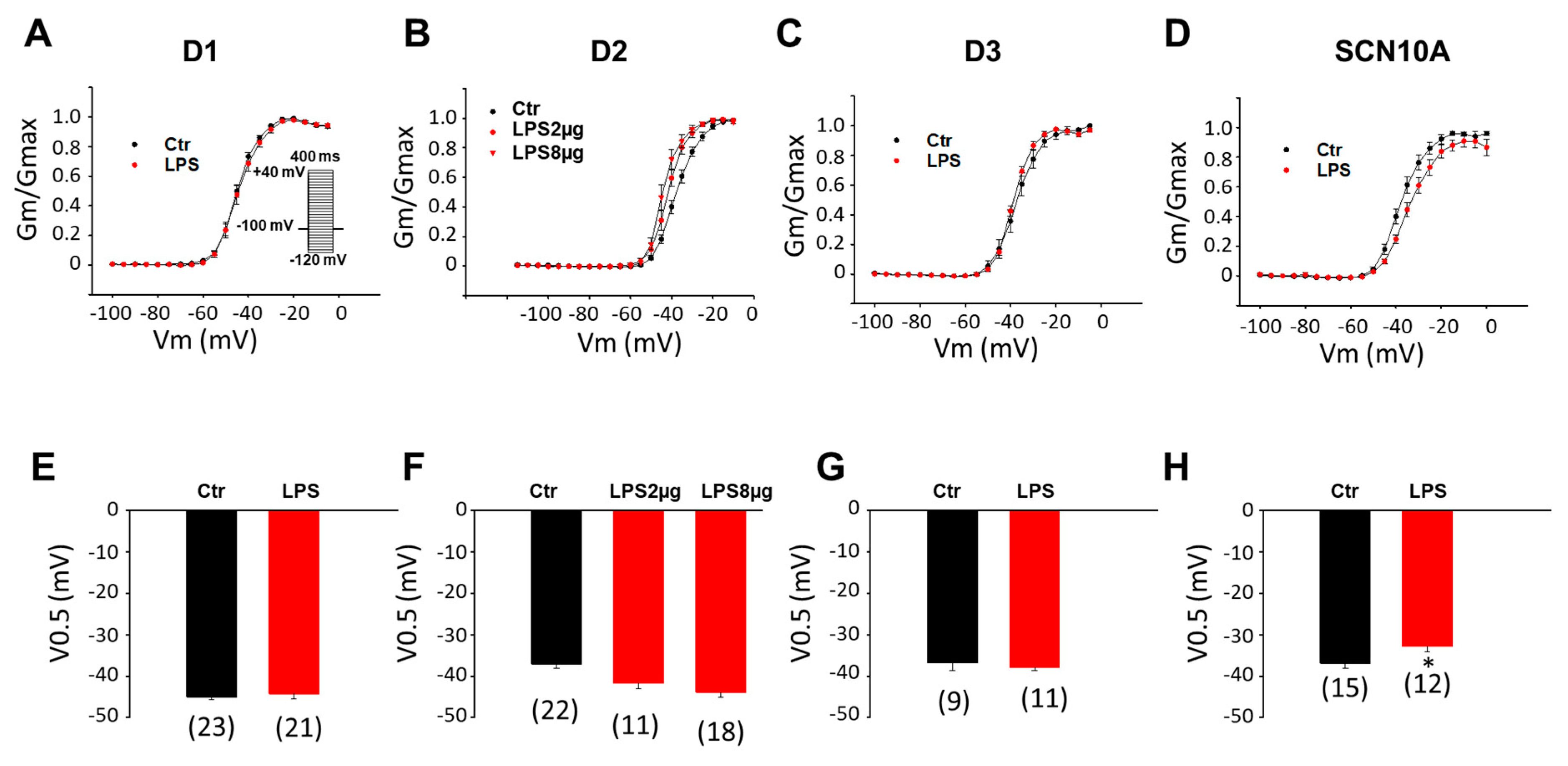
3.1. LPS Reduced Peak INa in hiPSC-CMs Derived from BrS-Patients

A recent study in our group demonstrated that hiPSC-CMs possess the molecular basis of inflammatory responses and could model inflammatory changes when they were challenged by LPS [28]. In addition, we found that the hiPSC-CMs from the patient carrying the variant (c.3749G > A) in SCN10A recapitulated the phenotypic feature (loss-of-function of cardiac sodium channel) of BrS. Therefore, we treated the SCN10A-hiPSC-CMs with LPS to mimic an inflammation to study the possible roles of inflammation on BrS phenotypic changes, focusing on changes in peak INa. First, LPS of 2 µg/mL was applied for 48 h. In the control (donor) group, LPS treatment did not change the peak INa density significantly (Figure 1A–D,F–H). In SCN10A-hiPSC-CMs, LPS treatment induced a significant reduction of the peak INa (Figure 1A,E,I). Since 2 µg/mL of LPS failed to inhibit INa in donor-hiPSC-CMs, the LPS concentration was elevated to 8 µg/mL to examine its effect in D2-hiPSC-CMs. The peak sodium current density increased slightly in the presence of 8 µg/mL of LPS (Figure 1C,G).
To investigate the mechanisms underlying the reduction of peak INa caused by LPS, the Na channel gating kinetics, including activation, inactivation, and recovery from inactivation, were analyzed. In healthy (donor) hiPSC-CMs, LPS did not significantly change the activation curves in the D1 and D3 cell lines, although it slightly shifted the curves in D2 cells to more negative potentials (Figure 2A–C,E–G). In SCN10A-hiPSC-CMs, LPS attenuated the voltage-dependent activation of the Na channel by shifting the activation curve to more positive potentials (Figure 2D,H).
To analyze the Na channel voltage-dependent inactivation, inactivation curves were obtained by plotting the relative Na currents (normalized to the maximal INa) versus voltages. In all three donor cell lines, LPS reduced the voltage-dependent inaction by shifting the inactivation curves to more positive potentials (Figure 3A–C,E–G). In D2-hiPSC-CMs, a higher dose (8 µg/mL) was needed to obtain statistically significant effects (Figure 3B,F). In SCN10A-hiPSC-CMs, LPS enhanced the voltage-dependent inactivation and shifted the inactivation curves to more negative potentials (Figure 3D,H).
To analyze the time-dependent recovery of Na channels from inactivation, the recovery curves were obtained by plotting the recovered currents against the recovery time (the interval between two pulses). In D1-hiPSC-CMs, the time-dependent recovery was accelerated (smaller tau value) by LPS of 2 µg/mL (Figure 4A,E). In D2-hiPSC-CMs, the recovery was also accelerated, but at a higher concentration (8 µg/mL) of LPS (Figure 4B,F). In D3-hiPSC-CMs, the recovery was not changed (Figure 4C,G). In SCN10A-hiPSC-CMs, the time-dependent recovery was decelerated by LPS (Figure 4D–H).

3.2. ROS and PKC Blocker Attenuated Effects of LPS on Peak INa

Studies reported that ROS contributed to the regulation of sodium currents in neurons and cardiomyocytes [29,30] and ROS signaling was involved in inflammation [31]. We suppose that ROS may be involved in the LPS-induced reduction of peak INa in the BrS-hiPSC-CMs. Hence, the SCN10A-hiPSC-CMs were treated for 48 h with LPS (2 µg/mL) or LPS plus N-acetylcysteine (NAC, 1 mM), a ROS scavenger. The result showed that peak INa was not changed by the NAC alone, but NAC prevented the inhibitory effect of LPS on peak INa (Figure 5). Likewise, NAC abolished the LPS effects on activation, inactivation, and the recovery of peak INa in SCN10A-hiPSC-CMs (Figure 6). These data suggested that ROS signaling mediated the inhibition of the Na channels by LPS. In addition to NAC, a PKC inhibitor (chelerythrine, 5 µM) also blocked the LPS effects (Figure 5 and Figure 6), indicating an involvement of PKC in the LPS effects on sodium channels.
LPS was found to cause tissue injury partially through ROS [31]. In cardiomyocytes, a low concentration of LPS (20 ng/mL) was shown to cause a significant increase in ROS [32]. Therefore, we assessed the ROS generation in donor- and SCN10A-hiPSC-CMs challenged by LPS. After hiPSC-CMs were treated with 2 μg/mL of LPS for 24 h, fluorescence imaging of 2,7-dichlorofluorescein (DCF) was applied to detect ROS generation as described previously [33]. DCF fluorescence of cells on coverslips showed that the fluorescence intensity was very weak without LPS treatment, but significantly intensified by the LPS treatment (Figure 7) in D2- and SCN10A-hiPSC-CMs, indicative of an elevation of ROS generation in donor- and BrS-hiPSC-CMs in the presence of LPS.

3.3. Peroxide Decreased the Peak INa in BrS-hiPSC-CMs

To prove the role of ROS in the change of the peak INa, hydrogen peroxide (H2O2), which is the main form of endogenous ROS in cells, was applied. In the donor cells treated with H2O2, no effects were detected (Figure 8A,C). In SCN10A-hiPSC-CMs, the peak INa decreased significantly (Figure 8B,D).
Since ROS inhibited peak INa, its effects on Na channel gating kinetics were further assessed by treating cells with 200 µM H2O2 for 2 h. In donor cells, H2O2 failed to alter the activation and inactivation curves (Figure 9A,C and Figure 10A,C) but accelerated the recovery from inactivation (Figure 11A,C). In SCN10A-hiPSC-CMs, H2O2 shifted the activation curve to a more positive potential (Figure 9B,D) and the inactivation curve to a more negative potential (Figure 10B,D) without significant influence on the recovery from inactivation (Figure 11B,D).

H2O2 Effect Was Blocked by a PKC Inhibitor

To check whether PKC was a downstream factor of ROS in the process of alteration of INa by LPS, the PKC block chelerythrine was applied. Chelerythrine (5 µM) was applied to SCN10A-hiPSC-CMs for 15 min, and then H2O2 (200 µM) was added for a further 2 h before cells were recorded. The I–V curve displayed that chelerythrine completely reversed the H2O2 induced reduction of peak INa (Figure 12A,E). In addition, chelerythrine also prevented ROS effects on the voltage-dependent activation, inactivation, and time-dependent recovery from inactivation (Figure 12B–D,F–H). These results indicate that PKC is involved in the ROS-induced peak INa reduction.

4. Discussion

Inflammation-induced arrhythmic events have been observed for quite a long time. Around 20 years ago, malignant tachy- and bradyarrhythmia in myocarditis were reported [34]. Inflammation, either acute, or chronic infection or non-infection caused, may influence the process of cardiovascular diseases. Some inflammatory factors, such as endotoxin, cytokines, and C-reactive protein (CRP), can alter the electrophysiology of the cardiomyocytes directly or indirectly [34]. Cytokines are the major modulators of the inflammatory process. Studies show that cytokines were involved in cardiac electrical and structural remodeling and the occurrence of arrhythmias [35]. For instance, TNF-α was shown to enhance arrhythmogenicity in rabbit cardiomyocytes [36].
Though increasing evidence shows that inflammation may be involved in the pathogenesis of arrhythmias, few studies assessed the connection between inflammation and BrS. There are some clinical data showing a possible relation between inflammation and BrS, but experimental improvements and mechanistic studies are lacking. This study investigated experimentally the role of the inflammatory response in the pathogenesis of Brugada Syndrome using hiPSC-CMs generated from a Brugada patient and demonstrated, for the first time, that LPS can exacerbate the loss-of-function sodium channel through ROS-PKC signaling. Data from this study may provide new insights into the arrhythmogenesis of BrS.
To study the connection between inflammation and Brs, the first question to be answered in this study is whether inflammation exacerbates the phenotypic changes of BrS. For this purpose, we used LPS to mimic inflammatory responses in healthy and diseased cells. LPS, the endotoxin of Gram-negative bacteria, was usually used to establish inflammatory models in various types of cells, including hiPSC-CMs [28]. Therefore, in this study, we used LPS to challenge hiPSC-CMs from BrS-patients to model the inflammatory responses of the cells and analyze the influences of inflammation on a key phenotypic change, loss-of-function of peak INa, in BrS.
Our recent study showed that peak INa was significantly smaller in the SCN10A-hiPSC-CMs than that in healthy donor cell lines, indicating a loss-of-function of sodium channels, which is a key phenotypic change in BrS [7]. The LPS treatment caused a further reduction of peak INa density in the SCN10A-iPSC-CMs. The reduced peak INa can reduce the depolarization speed (Vmax) of action potential and, in turn, decelerate the excitation propagation among cardiomyocytes. Since the conduction defect is a typical feature and an arrhythmogenic factor in BrS, this may help to understand how inflammation can contribute to the arrhythmogenesis of BrS. Strikingly, in all the healthy cell lines from three healthy donors, LPS failed to cause a reduction of peak INa. In one healthy donor (D2) cell line, the LPS treatment at higher concentration even increased peak INa slightly. To understand why LPS did not suppress peak INa in healthy hiPSC-CMs, we searched available data from the literature. A study in an animal model showed that peak sodium channel current in rat neurons was significantly increased after treatment of LPS [37], which was observed in our study in D2 cells with a high concentration of LPS. However, two studies assessing cardiac sodium channels of rabbits showed that LPS failed to change the peak INa [38,39], consistent with our observation in healthy hiPSC-CMs. In a rat model of sepsis, papillary muscles showed a decreased action potential (AP) magnitude and the rate of depolarization. When tetrodotoxin (TTX) was utilized to block the sodium channel, similar changes in APs were detected. It was concluded that the sepsis could suppress sodium channel current [40], which is consistent with our result in BrS-hiPSC-CMs treated by LPS. Taken all together, previous studies detected different effects of LPS on peak INa, increase, decrease, or no effect. These data imply that the LPS effects on sodium channels can be influenced by extra factors. The difference between healthy and BrS cells regarding the effects of LPS on peak INa suggests that the variant in SCN10A rendered sodium channels to be inhibited by LPS. How the variant in SCN10A renders sodium channels to be sensitive to LPS is still an open question and needs to be clarified in future studies.
The next question is how LPS inhibited peak INa in BrS cells. It is known that the amplitude of peak INa is determined by the channel open activity and channel expression level in the cell membrane. In this study, we focused on the influence of LPS on channel open activity. For this purpose, we analyzed the channel gating kinetics, including activation, inactivation, and recovery from inactivation of peak INa in cells challenged by LPS.
The reduction of peak INa in SCN10A-cells can be explained by the suppression of activation, enhancement of inactivation, and deceleration of recovery. The novel data showing that LPS induced a reduction of peak INa density in BrS cells demonstrates that the endotoxin or some inflammatory factors may be able to unmask the BrS-phenotype and exacerbate arrhythmias in some BrS-patients.
Another important point is how LPS changed sodium channel currents and gating kinetics. It is well-known that reactive oxygen species (ROS) play an important role in cell signaling. ROS is involved in the different pathology processes, including inflammation, neurodegeneration, diabetes, atherosclerosis, and aging. The phenomenon that LPS can increase ROS generation in cardiomyocytes has been reported [41,42] and is in agreement with the result of the current study. ROS may also participate in the process of ion channel regulation via direct or indirect mechanisms. In an experiment on rat neurons, Wang et al. observed that endogenous ROS increased peak sodium current in muscle afferent DRG neurons in an unknown way [29]. This is in contrast to our data in the current study. However, Liu et al. used NADH and other drugs to enhance ROS generation in HEK cells and cardiomyocytes, and they detected an increase in ROS production and a reduction of peak INa in both types of cells [30], suggesting that ROS may reduce peak INa, in agreement with our data in BrS-hiPSC-CMs. To examine the role of ROS in the LPS-induced reduction of INa in the BrS-hiPSC-CMs, we first investigated the effects of the ROS scavenger NAC (N-acetyl-l-cysteine). NAC significantly reduced the LPS effects on the sodium channels, including channel currents and gating kinetics. NAC alone showed no effect on sodium current, suggesting that ROS was involved in LPS effects. ROS generation was also observed in our hiPSC-CMs treated with LPS. DCF fluorescence assay showed that LPS induced a significant increase in ROS generation in iPSC-CMs, the second evidence for the involvement of ROS in the LPS effects. Of note, LPS induced similarly the ROS generation in both donor and BrS cells. This raised a question: why did LPS not inhibit INa in donor-hiPSC-CMs? Therefore, we further assessed the effects of H2O2, the main form of ROS in cells, on sodium channel currents and kinetics in donor- (D2) and SCN10A-hiPSC-CMs. Indeed, H2O2 mimicked the effects of LPS on sodium channel currents and gating kinetics in BrS cells, but not in D2 cells. These results confirmed that ROS contributed to the LPS-induced reduction of sodium current in the SCN10A-hiPSC-CMs and indicated that the ROS effects on sodium channels are gene variant related. Probably, the change of nucleoside leads to structural or functional changes in channel proteins and, in turn, renders the channel sensitive to ROS.
Aside from inflammation, ROS signaling can participate in the processes of many other diseases, such as stress, ischemia, diabetes, cancer, etc. Co-existence of BrS and cancer has been reported [43,44,45], but the concrete relation is unclear. The Brugada pattern has also been previously reported in the setting of fever, diabetes, stress, ischemia, and myocardial and pericardial diseases [46,47,48,49], all of which may contain pathogenic roles of ROS. ROS signaling might be involved in the pathogenesis of BrS in those diseases. In different diseases, however, the BrS phenotype can be evoked by different signaling.
Next, we tried to unveil the mechanism behind the ROS effect on sodium channels. PKC was shown to participate in LPS-induced ROS generation. A recent study reported that the genetic deletion of PKC delta in mice prevented an increase in total or mitochondrial ROS in cardiomyocytes after LPS exposure [50]. This result is in agreement with another study by Liu et al. [30]. In our current study, we used H2O2 to mimic the ROS effects on sodium channels. The application of a PKC inhibitor, chelerythrine [51], was able to completely prevent the H2O2 effects. This finding indicates that PKC is probably a downstream factor of ROS in cardiomyocytes.
PKC was shown to be activated by H2O2 [52]. It was also shown that PKC inhibitor could suppress the effect of H2O2 on late sodium current and action potential in cardiomyocytes, suggesting that PKC is an important factor in ROS signaling [53]. Qu described that the phosphorylation of S1505 in cardiac sodium channels by PKC altered the steady-state inactivation and the effect was completely abolished by the mutation S1505A. These data indicate that amino acid S1505 in the cardiac sodium channel is critical for channel regulation by PKC [54]. Our study demonstrated that H2O2 reduced peak INa and shifted the inactivation curve of INa to more negative potential in SCN10A-hiPSC-CMs and both effects were suppressed by the PKC inhibitor. This result implies that PKC mediated the effect of H2O2 on peak INa in SCN10A-hiPSC-CMs.
Taking all the data together, LPS may increase the risk of arrhythmias in BrS-patients with the SCNB10A mutation by exacerbating loss-of-function of sodium channels by enhancing ROS-PKC signaling, which can suppress peak sodium channel current via changing the channel gating kinetics.

5. Conclusions

This study demonstrated that LPS can exacerbate the loss-of-function of sodium channels by suppressing peak sodium channel currents and changing sodium channel gating kinetics. Inflammation may play an important role in the pathogenesis of some BrS-patients. Anti-inflammation treatment may be helpful in preventing the occurrence of arrhythmias in BrS-patients.

6. Study Limitations

Our study detected different LPS effects on the healthy donor iPSC-CMs and the BrS iPSC-CMs. The exact reason for the difference is unclear. We speculate that it may be related to the patient-specific gene variants or mutations. In this study, we used healthy donor iPSC-CMs as the control group, but an isogenic control using the CRISPR/Cas9-mediated genome editing was lacking.
The study exhibited that PKC was involved in LPS and ROS effects on INa in BrS-hiPSC-CMs, but which subtype of PKC exerted the effect was not addressed.
One of the main limitations of iPS-CMs as a model for arrhythmogenic diseases is the immature phenotype of hiPSC-CMs. The differences in cell properties, including electrical activities between hiPSC-CMs and native cardiomyocytes, should be also considered in interpreting the data of this study.

Author Contributions

Conceptualization, X.Z. and I.E.-B.; methodology, Z.L.; validation, Z.L., X.Z. and I.A.; formal analysis, Z.L.; investigation, Z.L., X.F., Y.L. and Z.Y.; resources, I.E.-B. and I.A.; data curation, Z.L. and Z.Y.; writing—original draft preparation, X.Z.; writing—review and editing, X.Z., I.E.-B. and I.A.; supervision, I.A.; funding acquisition, I.E.-B., X.Z. and I.A. All authors have read and agreed to the published version of the manuscript.

Funding

This work was supported by DZHK (German Center for Cardiovascular Research) and the BMBF (German Ministry of Education and Research) (No.: 81Z0500204; 81 × 2500208) and the Hector Foundation (MED 1814).

Institutional Review Board Statement

The research was approved by the Ethical Committee of the Medical Faculty Mannheim, University of Heidelberg (approval number: 2018-565N-MA).

Informed Consent Statement

Written informed consents have been obtained from the healthy donors and the patient to publish this paper.

Data Availability Statement

All the data reported in this study are contained in the paper.

Acknowledgments

We thank the Chinese Scholarship Council (CSC) for the financial support for Xuehui Fan. We thank the German Center for Cardiovascular Research (DZHK) and the Hector Foundation (Hector Stiftung) for financial support. The excellent technical support by the Stem Cell Unit, Göttingen is acknowledged.

Conflicts of Interest

The authors declare that they have no conflict of interest.

References

  1. Brugada, J.; Campuzano, O.; Arbelo, E.; Sarquella-Brugada, G.; Brugada, R. Present Status of Brugada Syndrome: JACC State-of-the-Art Review. J. Am. Coll. Cardiol. 2018, 72, 1046–1059. [Google Scholar] [CrossRef] [PubMed]
  2. Hoogendijk, M.G.; Potse, M.; Vinet, A.; de Bakker, J.M.; Coronel, R. ST segment elevation by current-to-load mismatch: An experimental and computational study. Heart Rhythm 2011, 8, 111–118. [Google Scholar] [CrossRef]
  3. Vohra, J.; Rajagopalan, S. Update on the Diagnosis and Management of Brugada Syndrome. Heart Lung Circ. 2015, 24, 1141–1148. [Google Scholar] [CrossRef] [PubMed] [Green Version]
  4. Chen, Q.; Kirsch, G.E.; Zhang, D.; Brugada, R.; Brugada, J.; Brugada, P.; Potenza, D.; Moya, A.; Borggrefe, M.; Breithardt, G. Genetic basis and molecular mechanism for idiopathic ventricular fibrillation. Nature 1998, 392, 293. [Google Scholar] [CrossRef]
  5. Campuzano, O.; Sarquella-Brugada, G.; Fernandez-Falgueras, A.; Cesar, S.; Coll, M.; Mates, J.; Arbelo, E.; Perez-Serra, A.; Del Olmo, B.; Jordá, P.; et al. Genetic interpretation and clinical translation of minor genes related to Brugada syndrome. Hum. Mutat. 2019, 40, 749–764. [Google Scholar] [CrossRef]
  6. Hosseini, S.M.; Kim, R.; Udupa, S.; Costain, G.; Jobling, R.; Liston, E.; Jamal, S.M.; Szybowska, M.; Morel, C.F.; Bowdin, S. Reappraisal of reported genes for sudden arrhythmic death: Evidence-based evaluation of gene validity for Brugada syndrome. Circulation 2018, 138, 1195–1205. [Google Scholar] [CrossRef]
  7. El-Battrawy, I.; Albers, S.; Cyganek, L.; Zhao, Z.; Lan, H.; Li, X.; Xu, Q.; Kleinsorge, M.; Huang, M.; Liao, Z.; et al. A cellular model of Brugada syndrome with SCN10A variants using human-induced pluripotent stem cell-derived cardiomyocytes. Europace 2019, 21, 1410–1421. [Google Scholar] [CrossRef]
  8. Yu, G.; Liu, Y.; Qin, J.; Wang, Z.; Hu, Y.; Wang, F.; Li, Y.; Chakrabarti, S.; Chen, Q.; Wang, Q.K. Mechanistic insights into the interaction of the MOG1 protein with the cardiac sodium channel Nav1. 5 clarify the molecular basis of Brugada syndrome. J. Biol. Chem. 2018, 293, 18207–18217. [Google Scholar] [CrossRef] [Green Version]
  9. Campuzano, O.; Berne, P.; Selga, E.; Allegue, C.; Iglesias, A.; Brugada, J.; Brugada, R. Brugada syndrome and p.E61X_RANGRF. Cardiol. J. 2014, 21, 121–127. [Google Scholar] [CrossRef] [Green Version]
  10. Huang, H.; Chen, Y.Q.; Fan, L.L.; Guo, S.; Li, J.J.; Jin, J.Y.; Xiang, R. Whole-exome sequencing identifies a novel mutation of GPD1L (R189X) associated with familial conduction disease and sudden death. J. Cell. Mol. Med. 2018, 22, 1350–1354. [Google Scholar] [CrossRef]
  11. Cerrone, M.; Lin, X.; Zhang, M.; Agullo-Pascual, E.; Pfenniger, A.; Chkourko Gusky, H.; Novelli, V.; Kim, C.; Tirasawadichai, T.; Judge, D.P.; et al. Missense mutations in plakophilin-2 cause sodium current deficit and associate with a Brugada syndrome phenotype. Circulation 2014, 129, 1092–1103. [Google Scholar] [CrossRef] [PubMed] [Green Version]
  12. Campuzano, O.; Fernandez-Falgueras, A.; Lemus, X.; Sarquella-Brugada, G.; Cesar, S.; Coll, M.; Mates, J.; Arbelo, E.; Jorda, P.; Perez-Serra, A.; et al. Short QT Syndrome: A Comprehensive Genetic Interpretation and Clinical Translation of Rare Variants. J. Clin. Med. 2019, 8, 1035. [Google Scholar] [CrossRef] [PubMed] [Green Version]
  13. Nakajima, T.; Wu, J.; Kaneko, Y.; Ashihara, T.; Ohno, S.; Irie, T.; Ding, W.G.; Matsuura, H.; Kurabayashi, M.; Horie, M. KCNE3 T4A as the Genetic Basis of Brugada-Pattern Electrocardiogram. Circ. J. 2012, 76, 2763–2772. [Google Scholar] [CrossRef] [PubMed] [Green Version]
  14. Ohno, S.; Zankov, D.P.; Ding, W.G.; Itoh, H.; Makiyama, T.; Doi, T.; Shizuta, S.; Hattori, T.; Miyamoto, A.; Naiki, N.; et al. KCNE5 (KCNE1L) Variants Are Novel Modulators of Brugada Syndrome and Idiopathic Ventricular Fibrillation. Circ. Arrhythm. Electrophysiol. 2011, 4, 352–361. [Google Scholar] [CrossRef] [Green Version]
  15. Cerrone, M.; Priori, S.G. Genetics of sudden death: Focus on inherited channelopathies. Eur. Heart J. 2011, 32, 2109–2118. [Google Scholar] [CrossRef] [Green Version]
  16. Lazzerini, P.E.; Capecchi, P.L.; Laghi-Pasini, F.; Boutjdir, M. Autoimmune channelopathies as a novel mechanism in cardiac arrhythmias. Nat. Rev. Cardiol. 2017, 14, 521. [Google Scholar] [CrossRef]
  17. Capecchi, P.L.; Laghi-Pasini, F.; El-Sherif, N.; Qu, Y.; Boutjdir, M.; Lazzerini, P.E. Autoimmune and inflammatory K(+) channelopathies in cardiac arrhythmias: Clinical evidence and molecular mechanisms. Heart Rhythm 2019, 16, 1273–1280. [Google Scholar] [CrossRef]
  18. Zhao, Y.; Sun, Q.; Zeng, Z.; Li, Q.; Zhou, S.; Zhou, M.; Xue, Y.; Cheng, X.; Xia, Y.; Wang, Q.; et al. Regulation of SCN3B/scn3b by Interleukin 2 (IL-2): IL-2 modulates SCN3B/scn3b transcript expression and increases sodium current in myocardial cells. BMC Cardiovasc. Disord. 2016, 16, 1. [Google Scholar] [CrossRef] [Green Version]
  19. Li, Q.; Qin, L.; Li, J. Enhancement by TNF-α of TTX-resistant Na(V) current in muscle sensory neurons after femoral artery occlusion. Am. J. Physiol. Regul. Integr. Comp. Physiol. 2020, 318, R772–R780. [Google Scholar] [CrossRef]
  20. Casado-Arroyo, R.; Berne, P.; Rao, J.Y.; Rodriguez-Manero, M.; Levinstein, M.; Conte, G.; Sieira, J.; Namdar, M.; Ricciardi, D.; Chierchia, G.B.; et al. Long-Term Trends in Newly Diagnosed Brugada Syndrome: Implications for Risk Stratification. J. Am. Coll. Cardiol. 2016, 68, 614–623. [Google Scholar] [CrossRef]
  21. Frustaci, A.; Priori, S.G.; Pieroni, M.; Chimenti, C.; Napolitano, C.; Rivolta, I.; Sanna, T.; Bellocci, F.; Russo, M.A. Cardiac histological substrate in patients with clinical phenotype of Brugada syndrome. Circulation 2005, 112, 3680–3687. [Google Scholar] [CrossRef] [PubMed] [Green Version]
  22. Bonny, A.; Tonet, J.; Marquez, M.F.; De Sisti, A.; Temfemo, A.; Himbert, C.; Gueffaf, F.; Larrazet, F.; Ditah, I.; Frank, R.; et al. C-reactive protein levels in the brugada syndrome. Cardiol. Res. Pract. 2011, 2011, 341521. [Google Scholar] [CrossRef] [PubMed] [Green Version]
  23. Li, A.; Tung, R.; Shivkumar, K.; Bradfield, J.S. Brugada syndrome-Malignant phenotype associated with acute cardiac inflammation? HeartRhythm Case Rep. 2017, 3, 384–388. [Google Scholar] [CrossRef] [PubMed] [Green Version]
  24. Fouda, M.A.; Ruben, P.C. Protein Kinases Mediate Anti-Inflammatory Effects of Cannabidiol and Estradiol against High Glucose in Cardiac Sodium Channels. Front. Pharmacol. 2021, 12, 668657. [Google Scholar] [CrossRef] [PubMed]
  25. El-Battrawy, I.; Lan, H.; Cyganek, L.; Zhao, Z.; Li, X.; Buljubasic, F.; Lang, S.; Yucel, G.; Sattler, K.; Zimmermann, W.H.; et al. Modeling Short QT Syndrome Using Human-Induced Pluripotent Stem Cell-Derived Cardiomyocytes. J. Am. Heart Assoc. 2018, 7, e007394. [Google Scholar] [CrossRef] [Green Version]
  26. Sattler, K.; El-Battrawy, I.; Cyganek, L.; Lang, S.; Lan, H.; Li, X.; Zhao, Z.; Utikal, J.; Wieland, T.; Borggrefe, M.; et al. TRPV1 activation and internalization is part of the LPS-induced inflammation in human iPSC-derived cardiomyocytes. Sci. Rep. 2021, 11, 14689. [Google Scholar] [CrossRef]
  27. Lian, X.; Hsiao, C.; Wilson, G.; Zhu, K.; Hazeltine, L.B.; Azarin, S.M.; Raval, K.K.; Zhang, J.; Kamp, T.J.; Palecek, S.P. Robust cardiomyocyte differentiation from human pluripotent stem cells via temporal modulation of canonical Wnt signaling. Proc. Natl. Acad. Sci. USA 2012, 109, E1848–E1857. [Google Scholar] [CrossRef] [Green Version]
  28. Yücel, G.; Zhao, Z.; El-Battrawy, I.; Lan, H.; Lang, S.; Li, X.; Buljubasic, F.; Zimmermann, W.-H.; Cyganek, L.; Utikal, J.; et al. Lipopolysaccharides induced inflammatory responses and electrophysiological dysfunctions in human-induced pluripotent stem cell derived cardiomyocytes. Sci. Rep. 2017, 7, 2935. [Google Scholar] [CrossRef]
  29. Wang, H.J.; Li, Y.L.; Zhang, L.B.; Zucker, I.H.; Gao, L.; Zimmerman, M.C.; Wang, W. Endogenous reactive oxygen species modulates voltage-gated sodium channels in dorsal root ganglia of rats. J. Appl. Physiol. 2011, 110, 1439–1447. [Google Scholar] [CrossRef]
  30. Liu, M.; Liu, H.; Dudley, S.C., Jr. Reactive oxygen species originating from mitochondria regulate the cardiac sodium channel. Circ. Res. 2010, 107, 967–974. [Google Scholar] [CrossRef] [Green Version]
  31. Hussain, T.; Tan, B.; Yin, Y.; Blachier, F.; Tossou, M.C.; Rahu, N. Oxidative Stress and Inflammation: What Polyphenols Can Do for Us? Oxidative Med. Cell. Longev. 2016, 2016, 7432797. [Google Scholar] [CrossRef] [PubMed] [Green Version]
  32. Joseph, L.C.; Kokkinaki, D.; Valenti, M.C.; Kim, G.J.; Barca, E.; Tomar, D.; Hoffman, N.E.; Subramanyam, P.; Colecraft, H.M.; Hirano, M.; et al. Inhibition of NADPH oxidase 2 (NOX2) prevents sepsis-induced cardiomyopathy by improving calcium handling and mitochondrial function. JCI Insight 2017, 2, e94248. [Google Scholar] [CrossRef]
  33. Dikalov, S.; Griendling, K.K.; Harrison, D.G. Measurement of reactive oxygen species in cardiovascular studies. Hypertension 2007, 49, 717–727. [Google Scholar] [CrossRef] [PubMed] [Green Version]
  34. Vonderlin, N.; Siebermair, J.; Kaya, E.; Köhler, M.; Rassaf, T.; Wakili, R. Critical inflammatory mechanisms underlying arrhythmias. Herz 2019, 44, 121–129. [Google Scholar] [CrossRef] [PubMed]
  35. Bartekova, M.; Radosinska, J.; Jelemensky, M.; Dhalla, N.S. Role of cytokines and inflammation in heart function during health and disease. Heart Fail. Rev. 2018, 23, 733–758. [Google Scholar] [CrossRef]
  36. Lee, S.-H.; Chen, Y.-C.; Chen, Y.-J.; Chang, S.-L.; Tai, C.-T.; Wongcharoen, W.; Yeh, H.-I.; Lin, C.-I.; Chen, S.-A. Tumor necrosis factor-α alters calcium handling and increases arrhythmogenesis of pulmonary vein cardiomyocytes. Life Sci. 2007, 80, 1806–1815. [Google Scholar] [CrossRef]
  37. Lu, Y.; Peng, F.; Dong, M.; Yang, H. Endocannabinoid 2-arachidonylglycerol protects primary cultured neurons against LPS-induced impairments in rat caudate nucleus. J. Mol. Neurosci. 2014, 54, 49–58. [Google Scholar] [CrossRef]
  38. Hwang, H.R.; Tai, B.Y.; Cheng, P.Y.; Chen, P.N.; Sung, P.J.; Wen, Z.H.; Hsu, C.H. Excavatolide B Modulates the Electrophysiological Characteristics and Calcium Homeostasis of Atrial Myocytes. Mar. Drugs 2017, 15, 25. [Google Scholar] [CrossRef] [Green Version]
  39. Tai, B.Y.; Wen, Z.H.; Cheng, P.Y.; Yang, H.Y.; Duh, C.Y.; Chen, P.N.; Hsu, C.H. Lemnalol Modulates the Electrophysiological Characteristics and Calcium Homeostasis of Atrial Myocytes. Mar. Drugs 2019, 17, 619. [Google Scholar] [CrossRef] [Green Version]
  40. Koesters, A.; Engisch, K.L.; Rich, M.M. Decreased cardiac excitability secondary to reduction of sodium current may be a significant contributor to reduced contractility in a rat model of sepsis. Crit. Care 2014, 18, R54. [Google Scholar] [CrossRef] [Green Version]
  41. Shang, F.; Zhao, L.; Zheng, Q.; Wang, J.; Xu, Z.; Liang, W.; Liu, H.; Liu, S.; Zhang, L. Simvastatin inhibits lipopolysaccharide-induced tumor necrosis factor-alpha expression in neonatal rat cardiomyocytes: The role of reactive oxygen species. Biochem. Biophys. Res. Commun. 2006, 351, 947–952. [Google Scholar] [CrossRef] [PubMed]
  42. Huang, J.; Peng, W.; Zheng, Y.; Hao, H.; Li, S.; Yao, Y.; Ding, Y.; Zhang, J.; Lyu, J.; Zeng, Q. Upregulation of UCP2 Expression Protects against LPS-Induced Oxidative Stress and Apoptosis in Cardiomyocytes. Oxidative Med. Cell. Longev. 2019, 2019, 2758262. [Google Scholar] [CrossRef] [PubMed]
  43. Sasaki, A.; Nakazato, Y. Brugada-like electrocardiogram detected after reconstructive operation for oesophageal cancer. Europace 2010, 12, 1542. [Google Scholar] [CrossRef] [PubMed]
  44. Tarín, N.; Farré, J.; Rubio, J.M.; Tuñón, J.; Castro-Dorticós, J. Brugada-like electrocardiographic pattern in a patient with a mediastinal tumor. Pacing Clin. Electrophysiol. PACE 1999, 22, 1264–1266. [Google Scholar] [CrossRef] [PubMed]
  45. Kusano, K.F. Brugada phenotype and prostate cancer. Circ. J. Off. J. Jpn. Circ. Soc. 2009, 73, 35–36. [Google Scholar] [CrossRef] [Green Version]
  46. Haseeb, S.; Kariyanna, P.T.; Jayarangaiah, A.; Thirunavukkarasu, G.; Hegde, S.; Marmur, J.D.; Neurgaonkar, S.; McFarlane, S.I. Brugada Pattern in Diabetic Ketoacidosis: A Case Report and Scoping Study. Am. J. Med. Case Rep. 2018, 6, 173–179. [Google Scholar] [CrossRef]
  47. Anselm, D.D.; Gottschalk, B.H.; Baranchuk, A. Brugada phenocopies: Consideration of morphologic criteria and early findings from an international registry. Can. J. Cardiol. 2014, 30, 1511–1515. [Google Scholar] [CrossRef]
  48. Batra, A.S.; Watson, R.; McCanta, A.C. Exercise-induced syncope and Brugada syndrome. Ann. Pediatr. Cardiol. 2019, 12, 292–294. [Google Scholar] [CrossRef]
  49. Wu, C.I.; Postema, P.G.; Arbelo, E.; Behr, E.R.; Bezzina, C.R.; Napolitano, C.; Robyns, T.; Probst, V.; Schulze-Bahr, E.; Remme, C.A.; et al. SARS-CoV-2, COVID-19, and inherited arrhythmia syndromes. Heart Rhythm 2020, 17, 1456–1462. [Google Scholar] [CrossRef]
  50. Joseph, L.C.; Reyes, M.V.; Lakkadi, K.R.; Gowen, B.H.; Hasko, G.; Drosatos, K.; Morrow, J.P. PKCδ causes sepsis-induced cardiomyopathy by inducing mitochondrial dysfunction. Am. J. Physiol. Heart Circ. Physiol. 2020, 318, H778–H786. [Google Scholar] [CrossRef]
  51. Herbert, J.; Augereau, J.; Gleye, J.; Maffrand, J. Chelerythrine is a potent and specific inhibitor of protein kinase C. Biochem. Biophys. Res. Commun. 1990, 172, 993–999. [Google Scholar] [CrossRef]
  52. Rhee, S.G.; Woo, H.A.; Kang, D. The role of peroxiredoxins in the transduction of H2O2 signals. Antioxid. Redox Signal. 2018, 28, 537–557. [Google Scholar] [CrossRef] [PubMed]
  53. Ward, C.A.; Giles, W.R. Ionic mechanism of the effects of hydrogen peroxide in rat ventricular myocytes. J. Physiol. 1997, 500 Pt 3, 631–642. [Google Scholar] [CrossRef] [PubMed] [Green Version]
  54. Qu, Y.; Rogers, J.C.; Tanada, T.N.; Catterall, W.A.; Scheuer, T. Phosphorylation of S1505 in the cardiac Na+ channel inactivation gate is required for modulation by protein kinase C. J. Gen. Physiol. 1996, 108, 375–379. [Google Scholar] [CrossRef] [PubMed]
Figure 1. LPS suppressed peak sodium channel currents in hiPSC-CMs from BrS-patients. Cells were treated with vehicle (same amount of water, Ctr) or LPS of 2 µg/mL or 8 µg/mL (in D2 cells) for 48 h and then peak sodium channel currents (INa) were recorded. (A) Representative traces of peak INa at −30 mV were recorded in hiPSC-CMs from healthy donors (D1, D2, and D3) and the BrS-patient (SCN10A) in absence (Ctr) and presence of LPS (2 µg/mL). (B) Current-Voltage (I–V) relationship curves of peak INa in hiPS-CMs from D1 donor in absence (Ctr) and presence of LPS (2 µg/mL). (C) Current-Voltage (I–V) relationship curves of peak INa in hiPS-CMs from D2 donor in absence (Ctr) and presence of LPS (2 µg/mL and 8 µg/mL). (D) Current-Voltage (I–V) relationship curves of peak INa in hiPS-CMs from D3 donor in absence (Ctr) and presence of LPS (2 µg/mL). (E) Current-Voltage (I–V) relationship curves of peak INa in hiPS-CMs from the BrS-patient (SCN10A) in absence (Ctr) and presence of LPS (2 µg/mL). (F) Mean values of peak INa at −30 mV in hiPS-CMs from D1 donor in absence (Ctr) and presence of LPS (2 µg/mL). (G) Mean values of peak INa at −30 mV in hiPS-CMs from D2 donor in absence (Ctr) and presence of LPS (2 µg/mL and 8 µg/mL). (H) Mean values of peak INa at −30 mV in hiPS-CMs from D3 donor in absence (Ctr) and presence of LPS (2 µg/mL). (I) Mean values of peak INa at −30 mV in hiPS-CMs from the BrS-patient in absence (Ctr) and presence of LPS (2 µg/mL). Numbers given in (FI) represent the number of cells of measurements also for (B–E). * p < 0.05 according to t-test.
Jcdd 09 00119 g001
Figure 2. LPS effects on activation of sodium channels in hiPSC-CMs from healthy donors and the BrS-patient. Cells were treated with vehicle (same amount of water, Ctr) of LPS of 2 µg/mL or 8 µg/mL (in D2 cells) for 48 h. (A) Activation curves of peak sodium channel currents (INa) recorded in hiPSC-CMs from D1 healthy donor. (B) Activation curves of peak INa recorded in hiPSC-CMs from D2 healthy donor. (C) Activation curves of peak INa recorded in hiPSC-CMs from D3 healthy donor. (D) Activation curves of peak sodium channel currents (INa) recorded in SCN10A-hiPSC-CMs. (E) Mean values of potential at 50% activation (V0.5) in hiPSC-CMs from D1 healthy donor. (F) Mean values of potential at 50% activation (V0.5) in hiPSC-CMs from D2 healthy donor. (G) Mean values of potential at 50% activation (V0.5) in hiPSC-CMs from D3 healthy donor. (H) Mean values of potential at 50% activation (V0.5) in SCN10A-hiPSC-CMs. Numbers given in (EH) represent the number of cells of measurements also for (AD). * p < 0.05 versus Ctr according to t-test or one-way ANOVA with Holm-Sidak post-test (F).
Jcdd 09 00119 g002
Figure 3. LPS effects on inactivation of sodium channels in hiPSC-CMs from healthy donors and the BrS-patient. Cells were treated with vehicle (Ctr) of LPS of 2 µg/mL or 8 µg/mL (in D2 cells) for 48 h. (A) Inactivation curves of peak sodium channel currents (INa) recorded in hiPSC-CMs from D1 healthy donor. (B) Inactivation curves of peak INa recorded in hiPSC-CMs from D2 healthy donor. (C) Inactivation curves of peak INa recorded in hiPSC-CMs from D3 healthy donor. (D) Inactivation curves of peak sodium channel currents (INa) recorded in SCN10A-hiPSC-CMs. (E) Mean values of potential at 50% inactivation (V0.5) in hiPSC-CMs from D1 healthy donor. (F) Mean values of potential at 50% inactivation (V0.5) in hiPSC-CMs from D2 healthy donor. (G) Mean values of potential at 50% inactivation (V0.5) in hiPSC-CMs from D3 healthy donor. (H) Mean values of potential at 50% inactivation (V0.5) in SCN10A-hiPSC-CMs. Numbers given in (EH) represent the number of cells of measurements also for (AD). * p < 0.05 versus Ctr according to t-test or one-way ANOVA with Holm-Sidak post-test (F).
Jcdd 09 00119 g003
Figure 4. LPS effects on recovery from inactivation of sodium channels in hiPSC-CMs from healthy donors and the BrS-patient. Cells were treated with vehicle (Ctr) of LPS of 2 µg/mL or 8 µg/mL (in D2 cells) for 48 h. (A) Recovery curves of peak sodium channel currents (INa) recorded in hiPSC-CMs from D1 healthy donor. (B) Recovery curves of peak INa recorded in hiPSC-CMs from D2 healthy donor. (C) Recovery curves of peak INa recorded in hiPSC-CMs from D3 healthy donor. (D) Recovery curves of peak sodium channel currents (INa) recorded in SCN10A-hiPSC-CMs. (E) Mean values of time constant (Tau) of recovery from inactivation in hiPSC-CMs from D1 healthy donor. (F) Mean values of time constant (Tau) of recovery from inactivation in hiPSC-CMs from D2 healthy donor. (G) Mean values of time constant (Tau) of recovery from inactivation in hiPSC-CMs from D3 healthy donor. (H) Mean values of time constants (Tau) in SCN10A-hiPSC-CMs. Numbers given in (EH) represent the number of cells of measurements also for (AD). * p < 0.05 versus Ctr according to t-test or one-way ANOVA with Holm–Sidak post-test (F).
Jcdd 09 00119 g004
Figure 5. A ROS and PKC blocker prevented the LPS effects on sodium channel currents in SCN10A-hiPSC-CMs. Cells were treated for 48 h with vehicle (Ctr) or LPS of 2 µg/mL (LPS) or LPS plus 1 mM N-acetylcysteine (LPS + NAC) or LPS plus 5 µM chelerythrine (LPS + Che). (A) I–V curves of peak sodium channel currents (INa) recorded in SCN10A-hiPSC-CMs. (B) Mean value of peak INa at −30 mV. Numbers given in (B) represent the number of cells of measurements also for (A). * p < 0.05 versus Ctr according to one-way ANOVA with Holm–Sidak post-test.
Jcdd 09 00119 g005
Figure 6. A ROS and PKC blocker prevented the LPS effects on sodium channel gating kinetics in SCN10A-hiPSC-CMs. Cells were treated for 48 h with vehicle (Ctr) or LPS of 2 µg/mL (LPS) or LPS plus 1 mM N-acetylcysteine (LPS + NAC) or LPS plus 5 µM chelerythrine (LPS + Che). (A) Activation curves of peak sodium channel currents (INa) recorded in SCN10A-hiPSC-CMs. (B) Inactivation curves of peak INa recorded in SCN10A-hiPSC-CMs. (C) Recovery curves of peak INa recorded in SCN10A-hiPSC-CMs. (D) Mean values of V0.5 of activation. (E) Mean values of V0.5 of inactivation. (F) Mean values of time constants (Tau) of recovery from inactivation. Numbers given in (DF) represent the number of cells of measurements also for (AC). * p < 0.05 versus Ctr according to one-way ANOVA with Holm–Sidak post-test.
Jcdd 09 00119 g006
Figure 7. LPS-induced ROS generation in the donor and BrS-hiPSC-CMs. Cells from D2 donor (Ctr) and BrS-patient (SCN10A) were incubated with LPS (2 µg/mL) for 48 h. Next, hiPSC-CMs were washed twice and incubated with 5 μM DCFH-DA solution in serum-free medium at 37 °C for 30 min in the dark. A fluorescence microscope was employed for evaluating the DCF fluorescence of cells on coverslips. (A,B) Fluorescence intensity in control cells (Ctr) without LPS treatment. (C,D) Fluorescence in the LPS treated cardiomyocytes. Magnification, ×400.
Jcdd 09 00119 g007
Figure 8. Peroxide (H2O2) decreased the peak INa in SCN10A cardiomyocytes. Cells were treated with vehicle (water, Ctr) or H2O2 (200 µM) for 2 h. (A) I–V curves of peak INa in donor-hiPSC-CMs (D2) in absence (Ctr) or presence (H2O2) of H2O2. (B) I–V curves of peak INa in SCN10A-hiPSC-CMs (SCN10A) in absence (Ctr) or presence (H2O2) of H2O2. (C) Mean values of peak INa at −30 mV in donor-hiPSC-CMs in absence (Ctr) or presence (H2O2) of H2O2. (D) Mean values of peak INa at −30 mV in SCN10A-hiPSC-CMs in absence (Ctr) or presence (H2O2) of H2O2. Values given are mean  ±  SEM. Numbers given in (C,D) represent the number of cells of measurements also for (A,B). * p < 0.05 versus Ctr according to t-test for two groups.
Jcdd 09 00119 g008
Figure 9. Effects of H2O2 on activation of sodium channels in hiPSC-CMs from BrS-patient. Cells were treated with vehicle (Ctr) or H2O2 (200 µM) for 2 h. (A) Activation curves of peak sodium channel currents (INa) recorded in hiPSC-CMs by the D2 healthy donor. (B) Activation curves of peak INa recorded in SCN10A-hiPSC-CMs. (C) Mean values of potential at 50% activation (V0.5) in hiPSC-CMs from D2 healthy donor. (D) Mean values of potential at 50% activation (V0.5) in SCN10A-hiPSC-CMs. Numbers given in (C,D) represent the number of cells of measurements also for (A,B). * p < 0.05 versus Ctr according to t-test for two groups.
Jcdd 09 00119 g009
Figure 10. Effects of H2O2 on inactivation of sodium channels in hiPSC-CMs from BrS-patient. Cells were treated with vehicle (Ctr) or H2O2 (200 µM) for 2 h. (A) Inactivation curves of peak sodium channel currents (INa) recorded in hiPSC-CMs from the D2 healthy donor. (B) Inactivation curves of peak INa recorded in SCN10A-hiPSC-CMs. (C) Mean values of potential at 50% inactivation (V0.5) in hiPSC-CMs from D2 healthy donor. (D) Mean values of potential at 50% inactivation (V0.5) in SCN10A-hiPSC-CMs. Numbers given in (C,D) represent the number of cells of measurements also for (A,B). * p < 0.05 versus Ctr according to t-test for two groups.
Jcdd 09 00119 g010
Figure 11. Effects of H2O2 on recovery of sodium channels in hiPSC-CMs from donor and BrS-patient. Cells were treated with vehicle (Ctr) or H2O2 (200 µM) for 2 h. (A) Recovery curves of peak sodium channel currents (INa) recorded in hiPSC-CMs from the D2 healthy donor. (B) Recovery curves of peak INa recorded in SCN10A-hiPSC-CMs. (C) Mean values of time constants (Tau) of recovery from inactivation in hiPSC-CMs from D2 healthy donor. (D) Mean values of time constants (Tau) of recovery from inactivation in SCN10A-hiPSC-CMs. Numbers given in (C,D) represent the number of cells of measurements also for (A,B). * p < 0.05 versus Ctr according to t-test for two groups.
Jcdd 09 00119 g011
Figure 12. A PKC inhibitor abolished the effects of H2O2 on sodium channels in hiPSC-CMs from the BrS-patient. Cells were treated with vehicle (water and DMSO, Ctr) or H2O2 (200 µM) or H2O2 plus 5 µM chelerythrine, a PKC inhibitor (H2O2 + Chel) for 2 h. (A) I–V curves of peak sodium channel currents (INa) recorded in SCN10A-hiPSC-CMs. (B) Activation curves of peak INa recorded in SCN10A-hiPSC-CMs. (C) Inactivation curves of peak INa recorded in SCN10A-hiPSC-CMs. (D) Recovery curves of peak INa recorded in SCN10A-hiPSC-CMs. (E) Mean values of peak sodium channel currents at −30 mV in SCN10A-hiPSC-CMs. (F) Mean values of V0.5 of activation in SCN10A-hiPSC-CMs. (G) Mean values of V0.5 of inactivation in SCN10A-hiPSC-CMs. (H) Mean values of time constants (Tau) of recovery from inactivation in SCN10A-hiPSC-CMs. Numbers given in H represent the number of cells of measurements for (AH). * p < 0.05 versus Ctr according to one-way ANOVA with Holm–Sidak post-test.
Jcdd 09 00119 g012
Publisher’s Note: MDPI stays neutral with regard to jurisdictional claims in published maps and institutional affiliations.

