Article

Role of the osaA Gene in Aspergillus fumigatus Development, Secondary Metabolism and Virulence

1
Department of Biological Sciences, Northern Illinois University, DeKalb, IL 60115, USA
2
Department of Biological Sciences and Eukaryotic Pathogen Innovation Center, Clemson University, Clemson, SC 29634, USA
3
Toxalim (Research Centre in Food Toxicology), Université de Toulouse, INRAE, ENVT, INP-Purpan, UPS, 31027 Toulouse, France
*
Author to whom correspondence should be addressed.
J. Fungi 2024, 10(2), 103; https://doi.org/10.3390/jof10020103
Submission received: 20 December 2023 / Revised: 17 January 2024 / Accepted: 22 January 2024 / Published: 26 January 2024
(This article belongs to the Section Fungal Genomics, Genetics and Molecular Biology)

Abstract

:
Aspergillus fumigatus is the leading cause of aspergillosis, associated with high mortality rates, particularly in immunocompromised individuals. In search of novel genetic targets against aspergillosis, we studied the WOPR transcription factor OsaA. The deletion of the osaA gene resulted in colony growth reduction. Conidiation is also influenced by osaA; both osaA deletion and overexpression resulted in a decrease in spore production. Wild-type expression levels of osaA are necessary for the expression of the conidiation regulatory genes brlA, abaA, and wetA. In addition, osaA is necessary for normal cell wall integrity. Furthermore, the deletion of osaA resulted in a reduction in the ability of A. fumigatus to adhere to surfaces, decreased thermotolerance, as well as increased sensitivity to oxidative stress. Metabolomics analysis indicated that osaA deletion or overexpression led to alterations in the production of multiple secondary metabolites, including gliotoxin. This was accompanied by changes in the expression of genes in the corresponding secondary metabolite gene clusters. These effects could be, at least in part, due to the observed reduction in the expression levels of the veA and laeA global regulators when the osaA locus was altered. Importantly, our study shows that osaA is indispensable for virulence in both neutropenic and corticosteroid-immunosuppressed mouse models.

1. Introduction

The ubiquitous fungus Aspergillus fumigatus is one of the most common fungal pathogens of humans worldwide [1], particularly affecting immunosuppressed patients, including those with hematological malignancies (such as leukemia), solid-organ and hematopoietic stem cell transplant patients, individuals with prolonged corticosteroid therapy, patients with genetic immunodeficiencies such as chronic granulomatous disease, cancer patients undergoing chemotherapy, and individuals infected with HIV [2,3,4,5,6,7]. The main reason for the increase in systemic infections lies in the constant increase in the number of immunocompromised individuals [8], such as a higher number of transplants for end-organ disease, the development of immunosuppressive and myeloablative therapies for autoimmune and neoplastic diseases, the HIV pandemic [2,8,9,10], and the COVID-19 pandemic [11,12,13,14]. Mortality rates range from 40% to 90% [8,9,15,16]. The World Health Organization recently ranked A. fumigatus third in the critical priority group of fungal pathogens [17]. The leap from being an organism on the watch list in 2019 to a globally critical fungal pathogen in three years can be traced back to unmet health problems that are associated with aspergillosis [17,18].
For most patients, the main site of entry and infection for A. fumigatus is the respiratory tract. In immunosuppressed patients, A. fumigatus infection can cause aspergilloma and invasive aspergillosis (IA). Diagnostic methods are still limited, as well as the possibilities of therapeutic treatments, leading to high mortality rates [19,20,21]. A. fumigatus causes approximately 90% of systemic Aspergillus infections [22]. Fungal asexual spores (conidia) are only 2.5–3.0 μm in diameter, which allows them to reach the lung alveoli [23,24]. In healthy individuals, conidia that are not removed via mucociliary clearance encounter epithelial cells or alveolar macrophages, which are responsible for the phagocytosis and killing of conidia, as well as for initiating a pro-inflammatory response that recruits neutrophils (PMN type). IA is primarily a consequence of a dysfunction in these defenses. The most common host immunodeficiencies responsible for an increased risk of IA are neutropenia and corticosteroid-induced immunosuppression [2,25,26,27,28]. In neutropenic patients or in a murine model of chemotherapy-induced neutropenia, IA can cause thrombosis and hemorrhage due to rapid hyphal angioinvasion and systemic dissemination to other organs [9,29]. Corticosteroid-induced immunosuppressed patients usually present tissue necrosis and excessive inflammation. Corticosteroid treatments impair the function of phagocytes [30], thus rendering them unable to kill both conidia and hyphae [31,32,33,34,35,36]. In most cases, consequent excessive inflammation leading to tissue damage is considered the cause of death.
Aspergillus fumigatus possesses a variety of traits that allow it to become an opportunistic pathogen in the case of immunocompromised patients. Examples of these factors include thermotolerance and cell wall components of conidia [37,38,39,40,41]. Metabolic requirements can influence the propagation in vivo [42,43,44,45]. Secondary metabolites are considered part of the chemical arsenal required for niche specialization [46], including host–fungus interactions. A. fumigatus produces several toxic secondary metabolites, such as fumitoxin, fumigaclavines, fumigatin, fumagillin, verruculogen, fumitremorgins, gliotoxin, trypacidin, and helvolic acid [16,47,48,49]. Some of these mycotoxins act as immunosuppressants, which may be associated with pathogenesis processes.
Common treatments with azole drugs present side effects such as skin rashes and photopsia, especially in the early stages of treatment [50]. Studies have also revealed the presence of resistant isolates of A. fumigatus to azole treatment [51]. Prolonged azole therapy could result in the development of azole-resistant A. fumigatus strains during infection [52]. The widespread application of azole fungicides in crop fields is also a driving force for azole-resistant infections worldwide [53,54]. It is paramount to find new strategies against fungal infections such as aspergillosis, including novel genetic targets for the treatment or prevention of A. fumigatus infection. In the present study, we investigated the role of OsaA, a WOPR-domain transcription factor, in A. fumigatus.
The name WOPR evolved from closely related proteins, Wor1 (in Candida albicans), Pac2 (in Schizosaccharomyce pombe), and Ryp1 (in Histoplasma capsulatum), where these proteins play a major role in morphological development and pathogenesis [55,56,57]. Wor1 regulates white-opaque switching in C. albicans [55], affecting mating frequency and tissue specificity during infection [58]. Pac2 may mediate signaling between nutritional conditions and the activation of the transcription factor gene ste11, which is necessary for sexual development in S. pombe [56]. Ryp1, from the human pathogen H. capsulatum, is a regulator of the yeast–mycelia transition and affects the transcription of hundreds of genes [57]. In Fusarium graminearum, the WOPR protein Fgp1 is a key regulator of reproduction, toxin biosynthesis, and pathogenesis [59], as well as Sge1 in Fusarium oxysporum [60]. In Aspergillus nidulans, osaA was shown to be epistatic to the veA regulatory gene in maintaining a balance between sexual and asexual development [61].
Our present work revealed that osaA is necessary for proper colony growth and conidiation in A. fumigatus. In addition, our analysis indicated that secondary metabolite production was also influenced by osaA. Furthermore, osaA is indispensable for virulence in both the neutropenic and corticosteroid-immunosuppressed mouse models. Other virulence factors, such as cell wall stability, thermotolerance, adhesion capacity, and sensitivity to oxidative stress, were also decreased in the absence of osaA in this opportunistic human pathogen.

2. Materials and Methods

2.1. Sequence Analysis

The Aspergillus fumigatus osaA nucleotide and deduced amino acid sequence were acquired from FungiDB (https://fungidb.org/fungidb/app, 11 August 2020) with the accession number AFUB_093810. A search of OsaA homologs was carried out using the BLASTP search tool (https://blast.ncbi.nlm.nih.gov/, 11 June 2023). Clustal W (https://www.ebi.ac.uk/Tools/msa/clustalo/, 11 June 2023) and ESPript (https://espript.ibcp.fr/ESPript/ESPript/, 11 June 2023) were used for multiple sequence alignment (MSA). The phylogenetic tree was constructed using MEGA v11.0 software and a maximum likelihood model with a bootstrap value of 1000 (https://www.megasoftware.net/, 11 June 2023).

2.2. Strains and Culture Conditions

The strains of A. fumigatus used in this study are listed in Table 1. All the strains were grown in glucose minimal medium (GMM) [62] plus the appropriate supplements for the corresponding auxotrophic markers [62] unless otherwise indicated. A solid medium was prepared by adding 1% agar. Strains were stored as 30% glycerol stocks at −80 °C.

2.3. Generation of the osaA Deletion Strain

To construct the osaA deletion strain (ΔosaA) in A. fumigatus, a deletion of the DNA cassette was generated to replace the osaA gene with the Aspergillus parasiticus pyrG selectable marker [63]. First, the 5′UTR and 3′UTR were PCR-amplified from A. fumigatus genomic DNA using the primers osaA_F1, osaA_R2, and osaA_F3, osaA_R4, respectively (the primers used in this study are included in Table S1). The pyrG marker gene was amplified from A. parasiticus genomic DNA using osaA_F5 and osaA_R6 primers. The three fragments were fused using the nested primers, osaA_F7, and osaA_R8, resulting in a 5157 bp PCR product. The obtained deletion fusion cassette was transformed in A. fumigatus CEA17 via polythene glycol-mediated fungal transformation. Transformants were selected on a stabilized minimal medium (SMM) [64] containing 1 M sucrose and no supplemental uracil. The ΔosaA strain was confirmed using a diagnostic PCR with primers osaA_F1 and A para pyrG_R and designated as TAD1.1.

2.4. Generation of the osaA Complementation Strain

To generate the osaA complementation strain (Com), a recombinant plasmid was constructed with pBC-hygro as the backbone vector. The osaA coding region, along with 1.5 kb of 5′ and 3′UTR were PCR-amplified using the primers osaA_com1 and osaA_com2. The forward primer contained an engineered HindIII site, whereas the reverse primer contained a NotI site. The amplified fragment was digested with the respective restriction enzymes and ligated to the pBC-hygro vector, previously digested with the same enzymes. The pBC-hygro vector includes the selection marker Hyg. The recombinant plasmid was named pAD1.1 and was transformed in the ∆osaA strain, resulting in the complementation strain (TAD2.1). The strain was confirmed using a diagnostic PCR with primers osaA_OE1 and osaA_OE2.

2.5. Construction of the osaA Overexpression Strain

A strain overexpressing osaA (OE) was conducted as follows: the coding region of osaA was PCR-amplified from A. fumigatus genomic DNA using the primers osaA_OE1 and osaA_OE2 with AscI and NotI engineered restriction sites, respectively. The 1443 bp PCR product was then digested using the corresponding enzymes and ligated into the pSSP1.1 plasmid, previously digested with the same enzymes. The pSSP1.1 vector includes the A. nidulans gpdA promoter and trpC terminator, along with A. fumigatus pyrG. The resulting recombinant plasmid (pAD3.1) was transformed into the CEA17 strain. Transformants were confirmed using a diagnostic PCR with the primers osaA_OE1 and A fum pyrG_R.

2.6. Morphological Analysis

To examine the role of osaA in colony growth, spores of wild-type, ΔosaA, Com, and OE strains were point-inoculated on GMM and incubated at 37 °C in the dark. Colony growth was estimated as the colony diameter, which was measured every two days. The experiment was carried out with four replicates.
For an assessment of the effect of osaA on conidiation, the same A. fumigatus strains were top-agar-inoculated on GMM (5 × 10⁶ spores/plate) and incubated at 37 °C in the dark. Cores were harvested from each plate at 24 h, 48 h, and 72 h after inoculation and homogenized in water. Conidia were quantified using a haemocytometer (Hausser Scientific, Horsham, PA, USA) and a Nikon Eclipse E-400 bright field microscope (Nikon, Inc., Melville, NY, USA). The experiment was performed in triplicates. Also, the spore size was measured under the Nikon E-400 microscope. Measurements of spore diameter (n = 40) were taken.
To evaluate whether osaA affects the spore germination rate in A. fumigatus, approximately 106 spores/mL of each of the strains were grown in liquid-shaken cultures containing 50 mL of GMM at 250 rpm. After 4 h post-inoculation, the cultures were observed under a bright field microscope, using a haemocytometer, every 2 h. This experiment was carried out in triplicates.

2.7. Environmental Stress Tests

To test sensitivity to a range of temperatures, wild-type, ΔosaA, Com and OE strains were point-inoculated on GMM and incubated in the dark at 25 °C, 30 °C, 37 °C, and 42 °C for 5 days when photographs and colony diameter measurements were taken. This experiment included three replicates.
To evaluate the importance of osaA in the resistance to oxidative stress, the strains were point-inoculated on solid GMM supplemented with 0, 15, 20, 25, or 30 μM menadione and incubated at 37 °C. Photographs were taken after an incubation period of 5 days. The experiment was performed with three replicates.

2.8. Cell Wall Tests

To elucidate possible alterations of cell wall integrity due to changes in the osaA locus, the wild-type, and ΔosaA, Com, and OE strains were point-inoculated in a 24-well plate on solid GMM supplemented with different concentrations (0, 0.005, 0.01, 0.015, and 0.02%) of sodium dodecyl sulfate (SDS) [65], and incubated at 37 °C for 72 h. In a separate experiment, the strains were inoculated on GMM with Congo red at concentrations of 0, 5, 10, 15, 50, or 100 μg/mL and incubated at 37 °C. Photographs were also taken at 72 h after inoculation. The experiments were carried out with three replicates.
Whether osmotic stabilization allowed the recovery of the wild-type phenotype in strains with an altered osaA locus and possible cell wall defects was assessed as follows. The conidia (106 spores/mL) of each strain were point-inoculated on GMM supplemented with either 0.6 M of KCl, 1 M of sucrose, 0.7 M of NaCl, or 1.2 M of sorbitol, and incubated at 37 °C for 72 h when photographs were taken, and colony diameters were measured. This experiment was performed in triplicate.

2.9. Adhesion Assay

To assess the role of osaA in adhesion capacity, each strain was inoculated in 50 mL of GMM (106 spores/mL). Aliquots (130 μL) from these suspensions were then added to each well of a 96-well microtiter plate. Cultures were incubated at 37 °C for a period of 24 h and 48 h. The mycelial mat and supernatant were removed from the wells. Biomass adhered to the well walls and was washed three times with water and then stained with 130 μL of 0.01% Crystal Violet (in water) for 20 min at room temperature. The stained wells were washed again three times with water, allowed to dry, and then de-stained using 130 μL of 30% acetic acid. The absorbance was measured at 560 nm on an Epoch spectrophotometer (Biotek, Winooski, VT, USA).

2.10. Metabolomics Analysis

For these analyses, the wild-type, ΔosaA, Com, and OE strains were top-agar-inoculated on GMM (5 × 10⁶ spores/plate) and cultured at 37 °C for 72 h. Three cores (16 mm diameter) were harvested from each plate and extracted with chloroform. The strain set was also grown in a liquid yeast extract–glucose–supplements (YES) medium [66] and cultured at 37 °C and 250 rpm for 7 days. The filtrate (30 mL) was collected and extracted with chloroform. Extracts were analyzed using liquid chromatography coupled to high-resolution mass spectrometry (LC-HRMS). Samples were dissolved in 500 μL of an acetonitrile/water mixture (50:50, v/v) and shaken vigorously for 30 s, and then treated with a sonicator (Bransonic 221 Ultrasonic bath, Emerson Electric, St. Louis, MO, USA) for 2 h. A volume of 250 μL of pure acetonitrile was added to each sample, and vigorously shaken for 30 s and centrifuged (pulse). LC-MS analyses were performed using Acquity HPLC (Waters, Saint-Quentin-en-Yvelines, France) linked to an LTQ Orbitrap XL high-resolution mass spectrometer (Thermo Fisher Scientific, Les Ulis, France). A reversed-phase Luna® C18 column (150 mm × 2.0 mm, 5 μm) (Phenomenex, Torrance, CA, USA) was utilized with water acidified with 0.1% formic acid and 100% acetonitrile as the mobile phases. The elution gradient was as follows: 0–30 min, B: 20–50%; 30–35 min, B: 50–90%; 35–45 min, B: 90%; 45–50 min, B: 90–20%; and 50–60 min, B: 20%. The flow rate was 0.2 mL min−1, the column temperature was 30 °C, and a volume of 10 μL was injected. HRMS acquisitions were carried out with electrospray ionization (ESI) in positive and negative modes, as previously reported [67]. MS/MS spectra were obtained with the CID mode at low resolution and a collision energy of 35%.

2.11. Gene Expression Analysis

Total RNA was obtained from lyophilized mycelial samples using TRIsureTM (Meridian Bioscience, Bioline, Cincinnati, OH, USA), following the manufacturer’s instructions. Gene expression was assessed using qRT-PCR. Ten micrograms of total RNA was treated with the Ambion Turbo DNA-free Kit (ThermoFisher Scientific, Waltham, MA, USA). cDNA was synthesized with Moloney murine leukemia virus (MMLV) reverse transcriptase (Promega, Madison, WI, USA). qRT-PCR was performed with the BioRad CFX96 Real-Time PCR System or Applied Biosystems 7000, using Q SYBR green Supermix (BioRad, Hercules, CA, USA) or SYBR green Jumpstart Taq Ready mix (Sigma, St. Louis, MO, USA) for fluorescence detection. The primer pairs used for qRT-PCR are listed in Table S1. The gene expression data for each gene were normalized to that of the A. fumigatus histone H4 unless specified otherwise. Relative expression levels were calculated using the 2−ΔΔCT method [68].

2.12. Virulence Assay

The inoculum was generated as follows: all strains were inoculated on stabilized minimal medium (GMM + 1.2 M sorbitol) and grown at 37 °C for 5 days. Conidia were collected by flooding the plates with 0.01% Tween-80 and scraping them with sterile cotton tip applicators. The conidia were filtered through Miracloth, washed twice with sterile water, and resuspended in sterile phosphate-buffered saline (PBS, ThermoFisher Scientific, Waltham, MA, USA).
Outbred female CD-1 mice (Charles River), six to eight weeks old, weighing 18–24 g, were used for the virulence experiments. For a neutropenic mouse model, immunosuppression was carried out via intraperitoneal injection with 175 mg/kg cyclophosphamide (Baxter, Deerfield, IL, USA) two days before inoculation [69], followed by a subcutaneous injection 40 mg/kg of triamcinolone acetonide (Kenalog-10 from Bristol-Myers Squibb, Lawrenceville, NJ, USA) one day before inoculation with A. fumigatus spores. Immunosuppressed mice were briefly anesthetized using a small rodent anesthesia machine and inoculated with A. fumigatus wild-type, ΔosaA, osaA Com, or osaA OE in the concentration of 2 × 10⁶ spores in 40 µL of PBS. A control group of five mice were immunosuppressed and injected with PBS. All the mice were monitored three times daily from days 1 to 7 and once daily from days 8 to 14. Injections with cyclophosphamide and Kenalog were repeated on days +3 and +6, respectively. A statistical comparison of the associated Kaplan–Meier curves was conducted with the log-rank test using GraphPad Prism (version 8.0).
For the corticosteroid-induced immune suppression mouse model, mice were immunosuppressed with a single 40 mg/kg injection of Kenalog-10 one day before infection with A. fumigatus conidia. Mice were monitored three times daily from days 1 to 7 and once daily from days 8–14. A statistical comparison of the associated Kaplan–Meier curves was also conducted with the log-rank test using GraphPad Prism (version 8.0).
This study was carried out in strict accordance with the Guide for the Care and Use of Laboratory Animals of the National Research Council. The protocol was approved by the Institutional Animal Care and Use Committee of Clemson University (Permit #AUP2022-0236). All efforts were made to minimize suffering. Humane euthanasia via CO2 inhalation was performed when mice met the criteria indicating a moribund state; these endpoints include behaviors of unresponsiveness to tactile stimuli, inactivity, lethargy, staggering, anorexia, and/or clinical signs of bleeding from the nose or mouth, labored breathing, agonal respirations, a purulent exudate from the eyes or nose, abnormally ruffled fur, or greater than 20% weight loss. The method of euthanasia via CO2 inhalation is consistent with recommendations of the Panel on Euthanasia of the American Veterinary Medical Association.

2.13. Statistical Analysis

Statistical analysis was carried out using analysis of variance (ANOVA) in conjunction with Tukey’s multiple test comparison test, unless stated otherwise.

3. Results

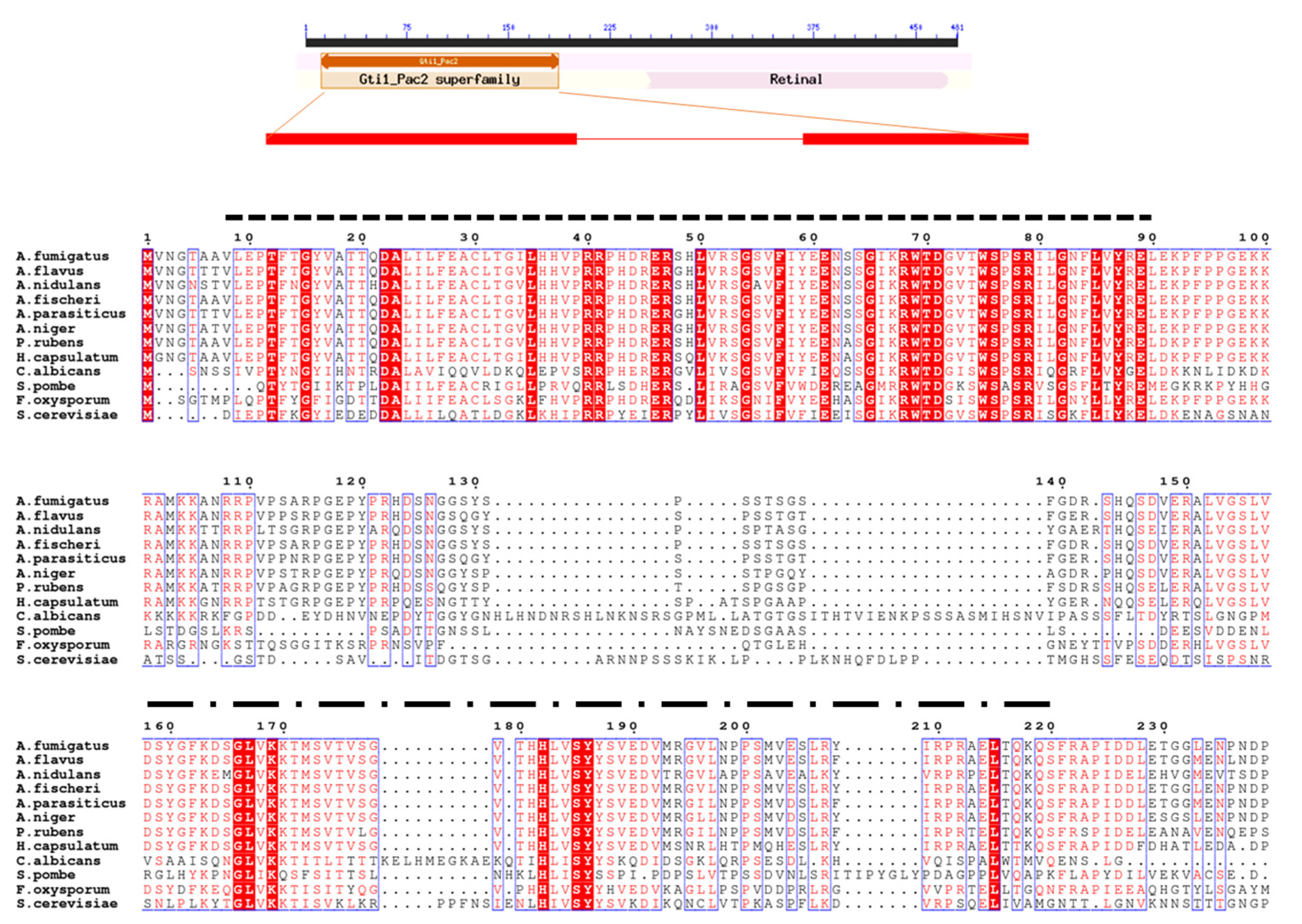
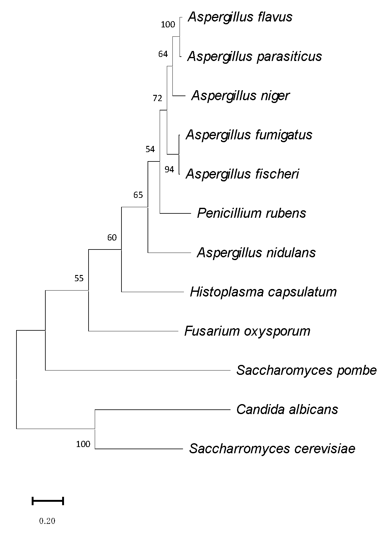
3.1. Identification of OsaA in A. fumigatus

The OsaA protein (corresponding to AFUB_093810) of A. fumigatus is conserved with respect to homologs in other species of the genus Aspergillus (Figure 1 and Figure 2, and Table 2). Our BLASTP analysis demonstrated that A. fumigatus OsaA presents the highest similarity to Aspergillus fischeri (98.96% percent identity, accession number: XP_001257654.1), followed by the homolog in Aspergillus flavus (82.32% identity, accession number: AFLA_004600). The OsaA in A. nidulans (AN6578) showed a 64.24% identity with OsaA in A. fumigatus (Table 2). Our BLASTP analysis also revealed the high conservation of OsaA with other species within Ascomycota, beyond Aspergillus. The sequence alignment of A. fumigatus OsaA with other fungal homologs showed the conservation of the WOPR domain (Figure 1). The WOPR domain in A. fumigatus OsaA was divided into two regions (9–94 and 158–222 residues), connected to each other by an interlinked sequence. Conserved domain analysis revealed the presence of a Gti1/Pac2 domain (Accession pfam09729) in the N-terminal region of the protein. Members of this family are orthologs of Saccharomyces pombe, such as the Pac2 WOPR fragment [56].

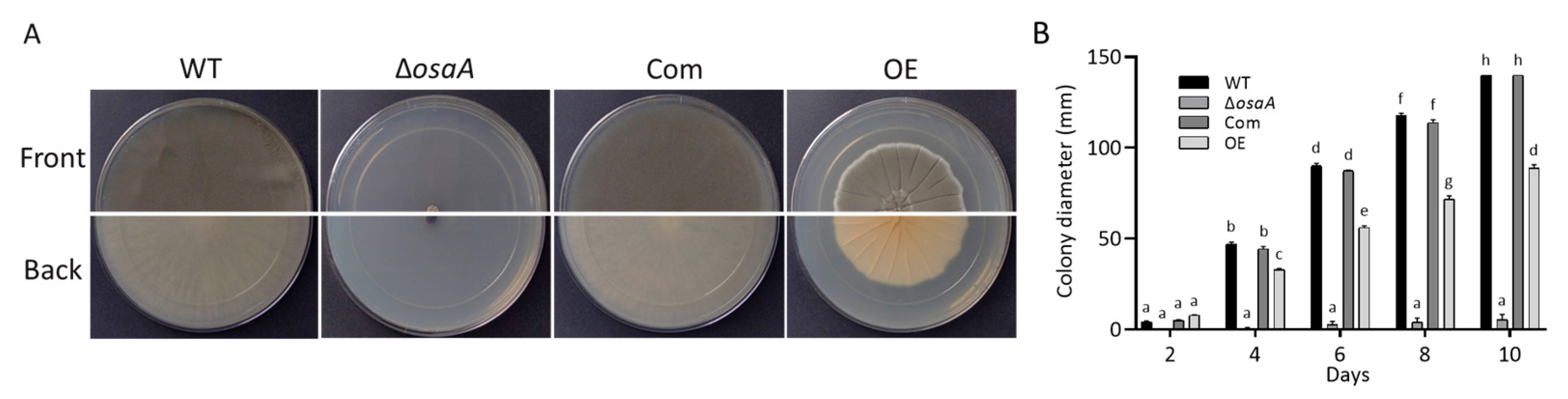
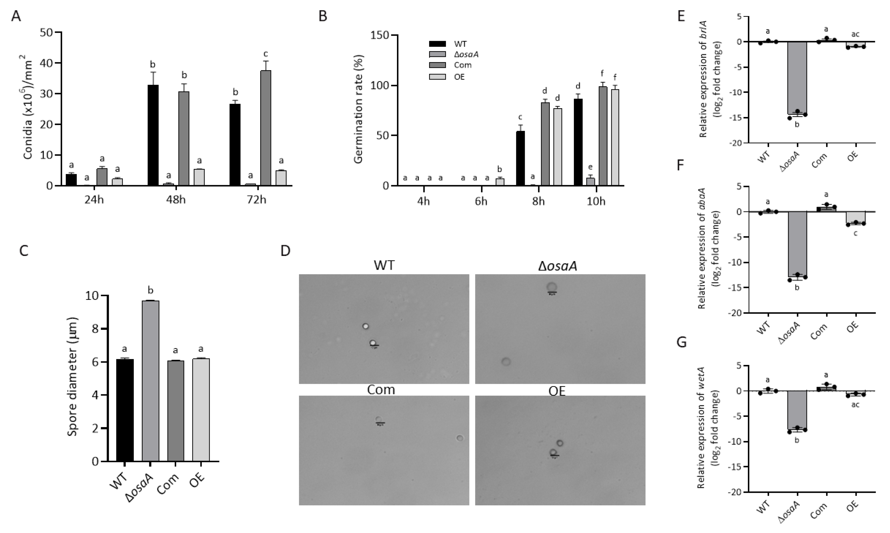
3.2. Growth, Conidiation, and Germination Rate Are Regulated by osaA in A. fumigatus

To determine the function of osaA in A. fumigatus, an osaA deletion, ΔosaA, as well as complementation and overexpression strains were generated (Figure S1). A diagnostic PCR confirmed the gene replacement of osaA with the pyrG marker and obtained the expected 2.64 kb product. PCR was also used to verify the integration of the wild-type allele in the ΔosaA strain in the Com strain, amplifying a 1.4 kb product (Figure S1). In addition, osaA overexpression strains were confirmed via a PCR amplifying the expected 1.97 kb product.
An evaluation of the colony growth revealed a significant reduction in colony diameter in the osaA deletion mutant compared to the wild-type (Figure 3). The back of the mutant colonies showed dark pigmentation, which was not present in the wild-type colonies. The complementation of ΔosaA with the osaA wild-type allele recovered the wild-type phenotype. The constitutive overexpression of osaA also reduced colony growth but to a lesser extent. The back of the osaA overexpression strain colony exhibited a yellowish pigmentation and distinct furrowing when compared to the wild-type colony.
Conidia are the inoculum for A. fumigatus infections. Previous studies in A. nidulans showed that the production of these asexual air-borne spores was affected by osaA in the model fungus [61]. In the present study, the role of osaA in conidiation was assessed in A. fumigatus. The ΔosaA and OE strains showed a significant reduction in conidial production compared to the wild-type and Com strain, particularly in the deletion strain (Figure 4). Our experiments revealed that both the deletion and overexpression of osaA resulted in alterations in expression levels of the transcription factor genes brlA, abaA, and wetA of the central regulatory pathway leading to conidiation [70], particularly in the absence of osaA, resulting in a significant reduction (Figure 4). Interestingly, micrographs of ΔosaA spores indicated a significant enlargement of conidial size (57%) compared to spores produced by the wild-type. Furthermore, a reduction in germination rate was observed in the absence of osaA, while the overexpression of osaA resulted in precocious germination (Figure 4).

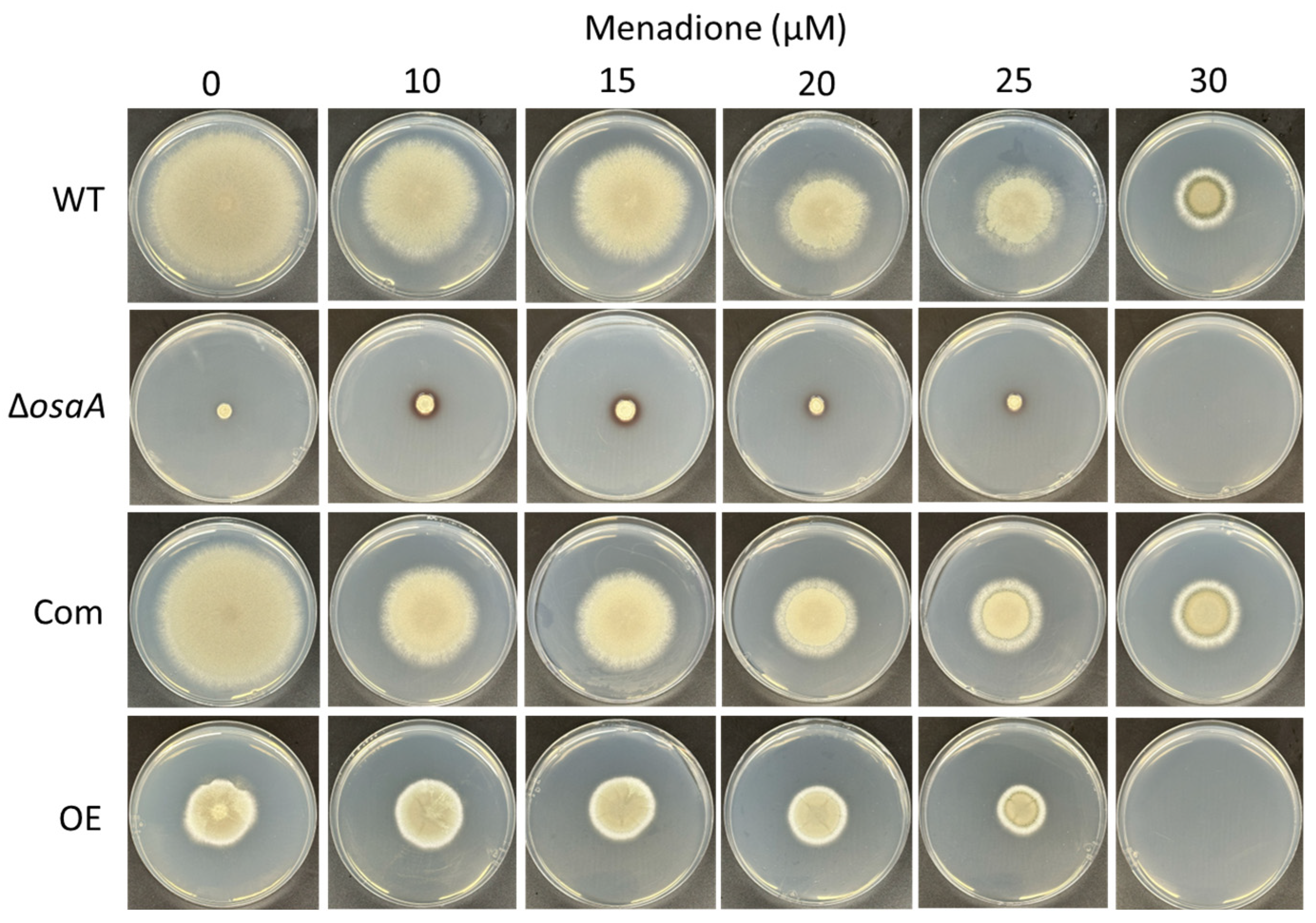
3.3. osaA Influences A. fumigatus Temperature and Oxidative Stress Resistance

Our results revealed that the absence of osaA alters A. fumigatus thermotolerance (Figure 5). The osaA gene is required for growth at 42 °C. This is relevant since the pathogenic nature of A. fumigatus is attributed to its thermotolerance, allowing this fungus to survive in a warmer environment and to combat human inflammatory responses. The colony growth defect of this mutant is slightly remediated at temperatures lower than 37 °C. We also noted that the knockout mutant showed increased pigmentation compared to the wild-type, particularly when grown at 25 °C and 30 °C.
An important component in the host’s defense against A. fumigatus infections includes the formation of an oxidative stress environment [16,71]. In our study, either the absence or constitutive overexpression of osaA both resulted in increased sensitivity to oxidative stress. Although at 10 μM of menadione, the deletion strain colony grew slightly healthier, both deletion and overexpression osaA strain colonies were unable to grow in the presence of 30 μM of menadione, a condition that still allowed for growth in wild-type and Com colonies (Figure 6).

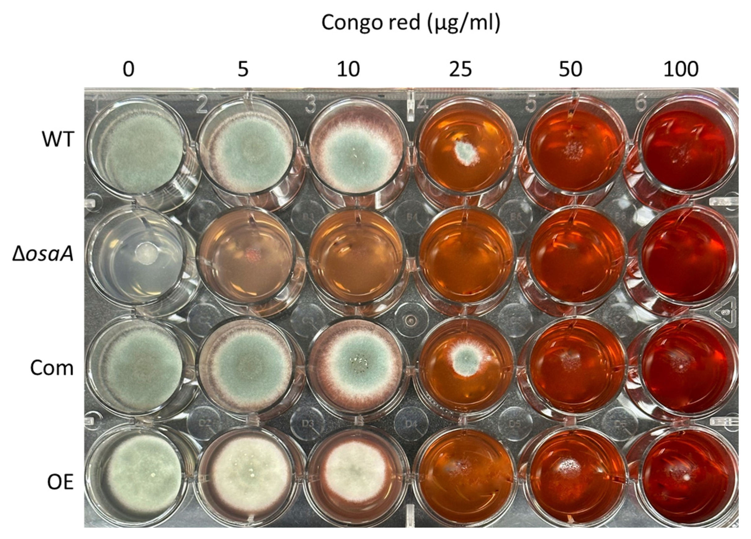
3.4. The Effect of osaA on A. fumigatus Cell Wall and Adhesion Capacity

The fungal cell wall is the first point of contact with the host. To elucidate whether osaA influences cell wall integrity, we exposed the deletion and overexpression of osaA strains, as well as their controls, to cell wall stressors. When grown on a medium supplemented with SDS, the growth of the osaA deletion mutant was compromised (Figure S2). In addition, when Congo red was supplemented to the medium, the growth of ΔosaA was severely affected at a concentration of 5 μg/mL and was unable to grow at 10 μg/mL (Figure 7). The growth of the OE strain was also dramatically affected at 25 μg/mL of Congo red.
The presence of osmotic stabilizers (0.6 M KCl, 1 M sucrose, 0.7 M NaCl, or 1.2 M sorbitol) in the medium allowed for the healthier growth of the deletion osaA mutant than in their absence, although the colony growth was still significantly smaller than that of the wild-type growing under the same experimental conditions (Figure S3).
Biofilm formation in the host is relevant in A. fumigatus resistance to anti-fungal drugs and to evade the host immune system. Importantly, adhesion to surfaces is required for the formation of biofilm [60]. Our analysis indicated a delay in the capacity to bind to abiotic surfaces in the absence of osaA at 24 h after inoculation (Figure S4). At 48 h, the adhesion capacity in ΔosaA increased over time; however, it was still significantly lower with respect to that of the wild-type. Interestingly, the overexpression of osaA resulted in a significant loss of adhesion capacity after 48 h.

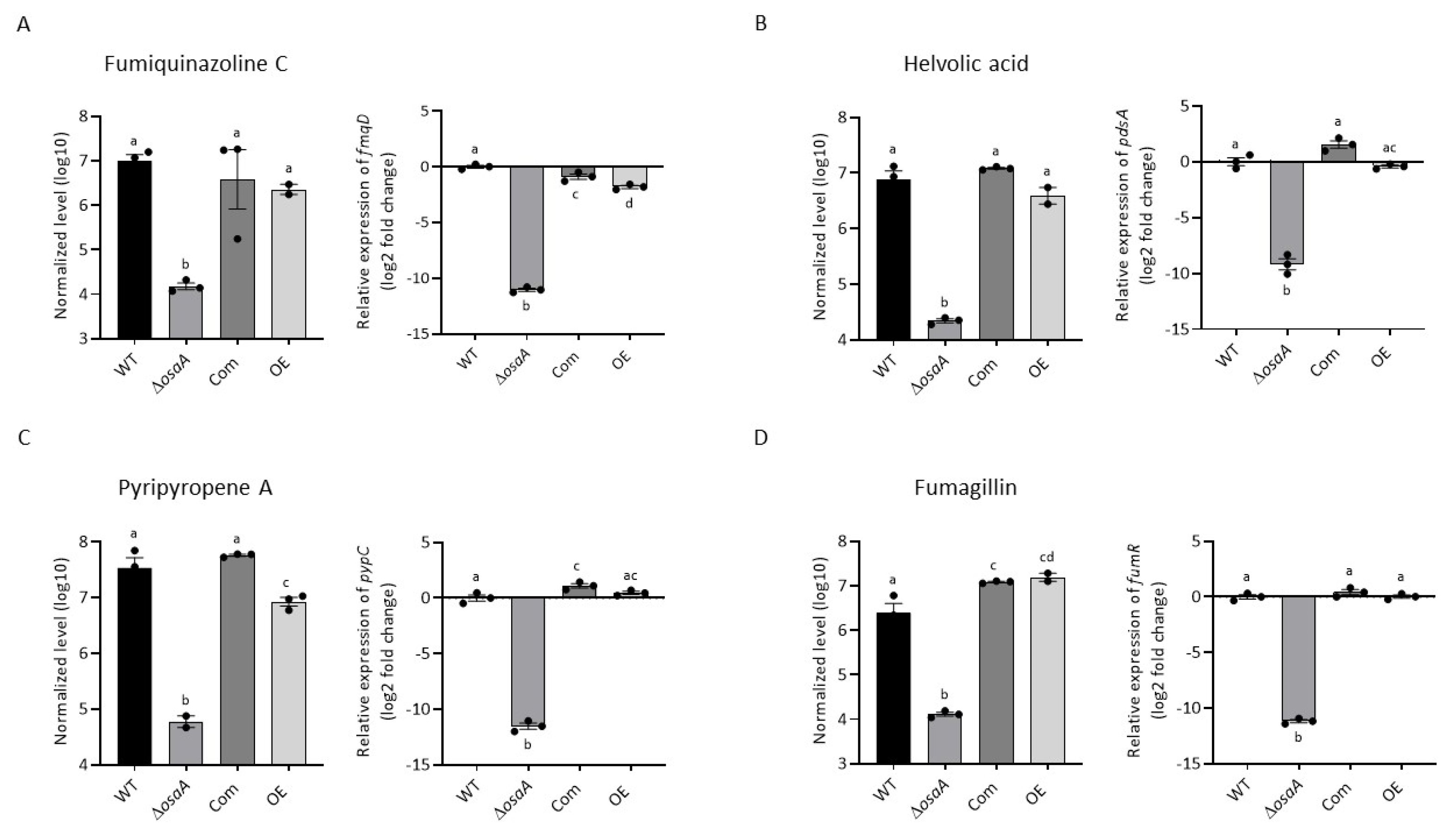
3.5. osaA Regulates Secondary Metabolism in A. fumigatus

Aspergillus fumigatus produces numerous bioactive compounds that serve multiple functions, including those involved in pathogenicity. Our metabolomics analysis revealed that osaA positively regulated secondary metabolism, including the production of fumiquinazoline C, helvolic acid, pyripyropene A, and fumagillin when growing on GMM (Figure 8). In addition, the deletion of osaA completely abolished the production of pseurotin A (Figure S5) when growing on the same medium. In the case of the OE strain, the production of pseurotin A was significantly overproduced compared to the wild-type under these culture conditions (Figure S5). When the YES medium was used (Table 3), the osaA deletion mutant presented a reduction or was unable to produce the above-mentioned metabolites, along with helvolinic acid, 1,2 dihydrohelvolic acid and gliotoxin, which were absent in the deletion mutant. The overexpression of osaA in YES medium leads to a significant decrease or lack of production of fumiquinazoline C, helvolinic acid, 1,2-dihydrohelvolic acid, fumagillin, and gliotoxin detected in the wild-type strain under the same experimental conditions.

3.6. Gene Expression Analysis of Secondary Metabolite Genes

Genes from gene clusters associated with the secondary metabolites whose synthesis was found to be osaA-dependent were selected as indicators of cluster activation. Their expression was analyzed in the wild-type, ΔosaA, Com, and OE strains using qRT-PCR. Our results indicated that osaA is necessary for the normal transcription of the fmqD gene, encoding a FAD-dependent oxidoreductase in the fumiquinazoline C biosynthetic gene cluster [72]. The transcription of the pdsA cyclase gene, involved in the synthesis of helvolic acid [73], and pypC, necessary for the synthesis of pyripyropene A [74], were significantly downregulated in the deletion strain. In addition, the transcription factor gene, fumR [75], involved in the regulation of other genes in the fumagillin gene cluster, displayed a considerable loss of expression when osaA was deleted (Figure 8). Furthermore, our study revealed that osaA was required for the normal transcription of psoA, encoding a polyketide synthase, in the pseurotin A gene cluster [65,76] (Figure S5).
Since gliotoxin production was reduced in the ΔosaA strain, we also examined whether the expression of key genes in the gliotoxin gene cluster was osaA-dependent. Our results indicated a drastic reduction in the expression of the Zn(II)2Cys6 binuclear transcription factor gliZ [77] in the absence of osaA with respect to the wild-type control (Figure S6). In addition, the expression of gliP, encoding a nonribosomal peptide synthase [78], was also downregulated in the osaA deletion strain. Interestingly, our results also showed that the expression of the global regulatory genes veA and laeA is osaA-dependent (Figure 9).

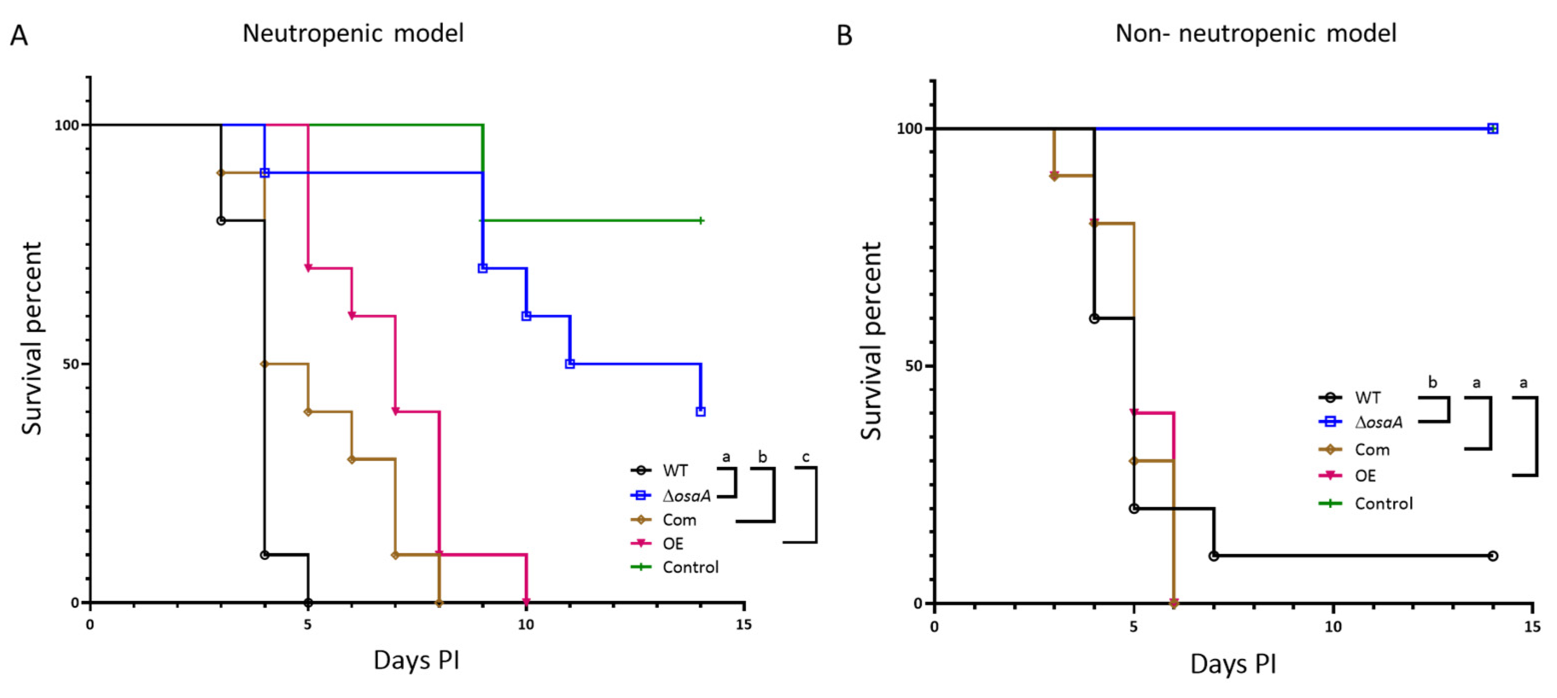
3.7. osaA Is Indispensable for Normal A. fumigatus Virulence in Both Neutropenic and Corticosteroid-Immunodepressed Murine Models

Since A. fumigatus osaA affects multiple cellular processes in this fungus, including those influencing pathogenicity, we tested whether osaA is involved in virulence (Figure 10). Importantly, the groups infected with spores from the deletion osaA mutant showed higher survival rates when compared to the groups infected with wild-type spores, indicating that osaA is indispensable for virulence in either neutropenic or corticosteroid immune-repressed environments. The complementation strain presented a similar virulence pattern to that of the wild-type strain in both experiments, indicating the recovery of the wild-type phenotype when the osaA wild-type allele was reintroduced in the deletion mutant. The overexpression of osaA resulted in a slight reduction in the mortality rate in the neutropenic model, while, in the corticosteroid model, OE presented a similar mortality rate to that of the wild-type.

4. Discussion

Transcriptional regulators with a WOPR domain are found across different fungal genera [79] and have been known to act as master regulators governing morphological development, sporogenesis, and pathogenesis [55,56,57,58,59,60]. Our work shows that OsaA in A. fumigatus includes a WOPR domain in the N-terminal region. This domain is present in two sub-regions and interconnected with a short linker region, as is the case in the same conserved domain in the C. albicans Wor1 protein [55,79,80].
In closely related dimorphic fungi, H. capsulatum and Coccidioides spp., the WOPR protein Ryp1 is involved in regulating host responses to temperature that result in a switch between two phenotypic forms necessary for the organism’s commensalism [81,82]. Specifically, in the absence of Ryp1, the mycelial form of H. capsulatum is genetically dominant at 37 °C [57], whereas, in Coccidioides, it maintains genetic development in the environmental hyphae form as well as the pathogenic spherulated form [81]. In A. nidulans, the role of osaA was identified to be a crucial factor in development. Our analysis also indicated that although A. fumigatus osaA presents commonalities regarding its function in other species, including A. nidulans, it also presents some distinct roles. For example, as in A. nidulans [61], osaA is important for proper conidiation in A. fumigatus; however, osaA also has a role in germination and vegetative growth in this opportunistic pathogen.
Importantly, in this study, we demonstrated that osaA is indispensable for virulence in both neutropenic and corticosteroid-induced murine models. The pathogenicity of A. fumigatus is associated with multiple virulent factors; some of these factors are affected by osaA. Our microscopic observation indicated that the spore size of the deletion osaA strain is larger than that of the wild-type strain. The small spore size of A. fumigatus allows them to easily enter the respiratory tract of humans and other animals. Spore enlargement in the absence of osaA could reduce the capability to reach the lung alveoli, reducing systemic infection [37,83,84]. In addition, decreased thermotolerance in a strain with an osaA-altered locus could negatively impact its ability to grow inside mammalian lungs [85,86]. Recent studies have also indicated that thermotolerance regulatory genes play a vital role in the A. fumigatus cell wall integrity pathway [86]. Our results revealed that both the deletion and overexpression of osaA (particularly the former) are more susceptible to cell wall stressors, suggesting a loss of integrity. The addition of osmotic stabilizers to the deletion of osaA cultures partially restored colony growth, further supporting the role of osaA in the cell-wall function. Importantly, osaA affects the capacity of the fungus to adhere to surfaces, a condition necessary for the formation of biofilm, which is crucial for fungal pathogenicity, protecting the fungus from external stresses, including antifungal agents [40,87,88].
During A. fumigatus infection, the host immune system elicits an elevated production of the macrophage and neutrophil-mediated killing of the conidia, one of which is through the generation of reactive oxygen species (ROS) [16]. Here, we also explored whether osaA is necessary for resistance to oxidative stress in A. fumigatus. Our results showed an increase in sensitivity to high levels of menadione in the deletion and overexpression of osaA strains compared to the wild-type. The addition of menadione in the medium simulates the production of ROS inside the cell [89,90], thus creating an oxidative stressful condition. This higher sensitivity to ROS in A. fumigatus could influence the survival of the infected host.
In addition, A. fumigatus is known to produce numerous secondary metabolites that impact host defenses at different stages [91,92]. These compounds are known to either suppress or avoid immune sentinel cells [93]. Our metabolomics analysis revealed a profound effect of osaA in A. fumigatus secondary metabolism. For example, pseurotin A and fumagillin, both associated with the same biosynthetic gene cluster [57,94], are not produced in the osaA mutant. This is relevant, as fumagillin alters neutrophil activity [95], and pseurotin A is an antioxidant [96]. It is possible that the lack of pseurotin A in the osaA mutant could contribute to the observed higher sensitivity to ROS in this strain. Other compounds not produced in the osaA deletion strain and produced at significantly lower levels when osaA is overexpressed are helvolic acid, a cilioinhibitor, and the derivatives helvolinic acid and 1,2-dihydrohelvolic acid, which presents antibacterial properties against Streptococcus aureus [97], fumiquinazoline, which presents cytotoxic activity, and pyripyropenes, an acyl-CoA/cholesterol acyltransferase inhibitor [98,99,100,101]. Furthermore, the production of gliotoxin was severely compromised when osaA was differentially expressed in A. fumigatus. Gliotoxin’s role in A. fumigatus infection has been previously studied in depth [8,16,93,102], where it was shown to affect host macrophage phagocytosis and to inhibit the activity of other immune cells [93].
The osaA gene was required for the expression of numerous genes in biosynthetic gene clusters associated with the production of secondary metabolites; for instance, gliZ and gliP [77,78], involved in the production of gliotoxin, and fmqD, pdsA, pypC, and fumR, involved in the synthesis of fumiquinazoline C, helvolic acid, pyripyropene, and fumagillin, respectively [65,72,73,75,76,103] were downregulated in the osaA deletion mutant with respect to the wild-type. Based on our results, the effect of osaA on the expression of these gene clusters could be, at least in part, mediated by osaA’s influence on the expression of the global regulatory genes veA and laeA. VeA and LaeA are components of the velvet protein complex [16,48], which governs several biological processes in numerous fungi, including secondary metabolism and development [48,74,104,105,106,107,108,109]. In A. nidulans, osaA has been shown to regulate the developmental balance between conidiation and sexual reproduction in a veA-dependent manner [61]. In A. fumigatus, we observed the downregulation of the expression of the developmental genes brlA, abaA, and wetA in the absence of osaA, which was associated with a decrease in conidiation. Based on our expression analysis results, it is likely that the effect of osaA on veA and laeA in A. fumigatus could result in multiple outputs, affecting secondary metabolism, development, and sensitivity to oxidative stress, among other biological processes [105].

5. Conclusions

In conclusion, A. fumigatus osaA is indispensable for virulence in neutropenic and corticosteroid-induced immune-repressed environments. The osaA gene influences growth, development, and sensitivity to environmental stressors, such as oxidative and temperature stresses, which could affect pathogenicity in this fungus. Furthermore, osaA was found to positively regulate the capacity of adhesion to surfaces, which is an important characteristic for the formation of biofilm. Importantly, this WOPR domain transcription factor gene is also necessary for the normal expression of secondary metabolite gene clusters and the concomitant synthesis of natural products, many of which are known to play a role during infection, including gliotoxin. In addition, osaA regulates the expression of veA and laeA genes, known global regulators governing development and secondary metabolism in A. fumigatus and other fungi. This study suggests that osaA or its product are promising targets with the potential for use in a control strategy against A. fumigatus infections.

Supplementary Materials

The following supporting information can be downloaded at: https://www.mdpi.com/article/10.3390/jof10020103/s1, Figure S1: Generation of the osaA deletion, complementation and overexpression strains; Figure S2: The deletion of osaA is more susceptible than wild-type to the presence of sodium dodecyl sulphate (SDS); Figure S3: Growth reduction in the A. fumigatus ΔosaA strain is partially remediated in the presence of osmotic stabilizers; Figure S4: osaA is necessary for normal adhesion to solid surfaces in A. fumigatus; Figure S5: osaA affects the production of pseurotin A in A. fumigatus; Figure S6: The gene expression of gliZ (A) and gliP (B) is osaA-dependent. Table S1: The primers used in this study.

Author Contributions

A.D. performed experiments, analyzed data and contributed to the manuscript writing; S.P. performed experiments; R.D. and S.D. performed experiments; S.L. and O.P. performed experiments; A.M.C. provided the concept and direction of the study, contributed to the manuscript writing and provided funding. All authors have read and agreed to the published version of the manuscript.

Funding

This work was funded by Northern Illinois University. The funders had no role in the project design, experimentation, and interpretation of results or the decision to submit the manuscript for publication.

Institutional Review Board Statement

Not applicable.

Informed Consent Statement

Not applicable.

Data Availability Statement

Data are contained within the article and Supplementary Materials.

Acknowledgments

We would like to thank Scott Grayburn, Erica Porter and Emma Whitlock for their technical support.

Conflicts of Interest

The authors declare no conflicts of interest.

References

  1. Winters, B.; Custer, J.; Galvagno, S.M.; Colantuoni, E.; Kapoor, S.G.; Lee, H.; Goode, V.; Robinson, K.; Nakhasi, A.; Pronovost, P.; et al. Diagnostic Errors in the Intensive Care Unit: A Systematic Review of Autopsy Studies. BMJ Qual. Saf. 2012, 21, 894–902. [Google Scholar] [CrossRef] [PubMed]
  2. Denning, D.W. Invasive Aspergillosis. Clin. Infect. Dis. 1998, 26, 781–803; quiz 804–805. [Google Scholar] [CrossRef] [PubMed]
  3. Kliasova, G.A.; Petrova, N.A.; Parovichnikova, E.N.; Gotman, L.N.; Isaev, V.G.; Mikhaĭlova, E.A.; Ustinova, E.N.; Khoroshko, N.D.; Vishnevskaia, E.S.; Kremenetskaia, A.M.; et al. Invasive Pulmonary Aspergillesis. Ter. Arkh. 2005, 77, 65–71. [Google Scholar]
  4. Pagano, L.; Girmenia, C.; Mele, L.; Ricci, P.; Tosti, M.E.; Nosari, A.; Buelli, M.; Picardi, M.; Allione, B.; Corvatta, L.; et al. Infections Caused by Filamentous Fungi in Patients with Hematologic Malignancies. A Report of 391 Cases by GIMEMA Infection Program. Haematologica 2001, 86, 862–870. [Google Scholar] [PubMed]
  5. Marr, K.A.; Carter, R.A.; Boeckh, M.; Martin, P.; Corey, L. Invasive Aspergillosis in Allogeneic Stem Cell Transplant Recipients: Changes in Epidemiology and Risk Factors. Blood 2002, 100, 4358–4366. [Google Scholar] [CrossRef]
  6. Wiederhold, N.P.; Lewis, R.E.; Kontoyiannis, D.P. Invasive Aspergillosis in Patients with Hematologic Malignancies. Pharmacother. J. Hum. Pharmacol. Drug Ther. 2003, 23, 1592–1610. [Google Scholar] [CrossRef]
  7. Post, M.J.; Lass-Floerl, C.; Gastl, G.; Nachbaur, D. Invasive Fungal Infections in Allogeneic and Autologous Stem Cell Transplant Recipients: A Single-Center Study of 166 Transplanted Patients. Transpl. Infect. Dis. 2007, 9, 189–195. [Google Scholar] [CrossRef] [PubMed]
  8. Latgé, J.P. Aspergillus fumigatus and Aspergillosis. Clin. Microbiol. Rev. 1999, 12, 310–350. [Google Scholar] [CrossRef]
  9. Sherif, R.; Segal, B.H. Pulmonary Aspergillosis: Clinical Presentation, Diagnostic Tests, Management and Complications. Curr. Opin. Pulm. Med. 2010, 16, 242–250. [Google Scholar] [CrossRef]
  10. Hohl, T.M.; Feldmesser, M. Aspergillus fumigatus: Principles of Pathogenesis and Host Defense. Eukaryot. Cell 2007, 6, 1953–1963. [Google Scholar] [CrossRef]
  11. Dimopoulos, G.; Almyroudi, M.-P.; Myrianthefs, P.; Rello, J. COVID-19-Associated Pulmonary Aspergillosis (CAPA). J. Intensive Med. 2021, 1, 71–80. [Google Scholar] [CrossRef]
  12. Ergün, M.; Brüggemann, R.J.M.; Alanio, A.; Dellière, S.; van Arkel, A.; Bentvelsen, R.G.; Rijpstra, T.; van der Sar-van der Brugge, S.; Lagrou, K.; Janssen, N.A.F.; et al. Aspergillus Test Profiles and Mortality in Critically Ill COVID-19 Patients. J. Clin. Microbiol. 2021, 59, e0122921. [Google Scholar] [CrossRef] [PubMed]
  13. Bartoletti, M.; Pascale, R.; Cricca, M.; Rinaldi, M.; Maccaro, A.; Bussini, L.; Fornaro, G.; Tonetti, T.; Pizzilli, G.; Francalanci, E.; et al. Epidemiology of Invasive Pulmonary Aspergillosis Among Intubated Patients with COVID-19: A Prospective Study. Clin. Infect. Dis. 2021, 73, e3606–e3614. [Google Scholar] [CrossRef]
  14. Machado, M.; Valerio, M.; Álvarez-Uría, A.; Olmedo, M.; Veintimilla, C.; Padilla, B.; De la Villa, S.; Guinea, J.; Escribano, P.; Ruiz-Serrano, M.J.; et al. Invasive Pulmonary Aspergillosis in the COVID-19 Era: An Expected New Entity. Mycoses 2021, 64, 132–143. [Google Scholar] [CrossRef] [PubMed]
  15. Lin, S.-J.; Schranz, J.; Teutsch, S.M. Aspergillosis Case-Fatality Rate: Systematic Review of the Literature. Clin. Infect. Dis. 2001, 32, 358–366. [Google Scholar] [CrossRef] [PubMed]
  16. Dagenais, T.R.T.; Keller, N.P. Pathogenesis of Aspergillus fumigatus in Invasive Aspergillosis. Clin. Microbiol. Rev. 2009, 22, 447–465. [Google Scholar] [CrossRef] [PubMed]
  17. Parums, D.V. Editorial: The World Health Organization (WHO) Fungal Priority Pathogens List in Response to Emerging Fungal Pathogens During the COVID-19 Pandemic. Med. Sci. Monit. 2022, 28, e939088. [Google Scholar] [CrossRef]
  18. CDC. Antibiotic Resistance Threats in the United States; U.S. Department of Health and Human Services, CDC: Atlanta, GA, USA, 2019. Available online: www.cdc.gov/DrugResistance/Biggest-Threats.html (accessed on 27 March 2023).
  19. Ellis, M. Therapy of Aspergillus fumigatus-Related Diseases. Contrib. Microbiol. 1999, 2, 105–129. [Google Scholar] [CrossRef]
  20. Oren, I.; Goldstein, N. Invasive Pulmonary Aspergillosis. Curr. Opin. Pulm. Med. 2002, 8, 195–200. [Google Scholar] [CrossRef]
  21. Kontoyiannis, D.P.; Bodey, G.P. Invasive Aspergillosis in 2002: An Update. Eur. J. Clin. Microbiol. Infect. Dis. 2002, 21, 161–172. [Google Scholar] [CrossRef]
  22. Schmitt, H.J.; Blevins, A.; Sobeck, K.; Armstrong, D. Aspergillus Species from Hospital Air and from Patients. Mycoses 1990, 33, 539–541. [Google Scholar] [CrossRef] [PubMed]
  23. Rüchel, R.; Reichard, U. Pathogenesis and Clinical Presentation of Aspergillosis. Contrib. Microbiol. 1999, 2, 21–43. [Google Scholar] [CrossRef] [PubMed]
  24. Samson, R.A. The Genus Aspergillus with Special Regard to the Aspergillus fumigatus Group. Contrib. Microbiol. 1999, 2, 5–20. [Google Scholar] [CrossRef] [PubMed]
  25. Berenguer, J.; Allende, M.C.; Lee, J.W.; Garrett, K.; Lyman, C.; Ali, N.M.; Bacher, J.; Pizzo, P.A.; Walsh, T.J. Pathogenesis of Pulmonary Aspergillosis. Granulocytopenia versus Cyclosporine and Methylprednisolone-Induced Immunosuppression. Am. J. Respir. Crit. Care Med. 1995, 152, 1079–1086. [Google Scholar] [CrossRef]
  26. Denning, D.W. Aspergillosis: Diagnosis and Treatment. Int. J. Antimicrob. Agents 1996, 6, 161–168. [Google Scholar] [CrossRef] [PubMed]
  27. Balloy, V.; Huerre, M.; Latgé, J.-P.; Chignard, M. Differences in Patterns of Infection and Inflammation for Corticosteroid Treatment and Chemotherapy in Experimental Invasive Pulmonary Aspergillosis. Infect. Immun. 2005, 73, 494–503. [Google Scholar] [CrossRef] [PubMed]
  28. Stergiopoulou, T.; Meletiadis, J.; Roilides, E.; Kleiner, D.E.; Schaufele, R.; Roden, M.; Harrington, S.; Dad, L.; Segal, B.; Walsh, T.J. Host-Dependent Patterns of Tissue Injury in Invasive Pulmonary Aspergillosis. Am. J. Clin. Pathol. 2007, 127, 349–355. [Google Scholar] [CrossRef]
  29. Chiang, L.Y.; Sheppard, D.C.; Gravelat, F.N.; Patterson, T.F.; Filler, S.G. Aspergillus fumigatus Stimulates Leukocyte Adhesion Molecules and Cytokine Production by Endothelial Cells in Vitro and during Invasive Pulmonary Disease. Infect. Immun. 2008, 76, 3429–3438. [Google Scholar] [CrossRef]
  30. Lionakis, M.S.; Kontoyiannis, D.P. Glucocorticoids and Invasive Fungal Infections. Lancet 2003, 362, 1828–1838. [Google Scholar] [CrossRef]
  31. Brummer, E.; Maqbool, A.; Stevens, D.A. In Vivo GM-CSF Prevents Dexamethasone Suppression of Killing of Aspergillus fumigatus Conidia by Bronchoalveolar Macrophages. J. Leukoc. Biol. 2001, 70, 868–872. [Google Scholar] [CrossRef]
  32. Kamberi, M.; Brummer, E.; Stevens, D.A. Regulation of Bronchoalveolar Macrophage Proinflammatory Cytokine Production by Dexamethasone and Granulocyte-Macrophage Colony-Stimulating Factor after Stimulation by Aspergillus Conidia or Lipopolysaccharide. Cytokine 2002, 19, 14–20. [Google Scholar] [CrossRef]
  33. Meier-Osusky, I.; Schoedon, G.; Bläuer, F.; Schneemann, M.; Schaffner, A. Comparison of the Antimicrobial Activity of Deactivated Human Macrophages Challenged with Aspergillus fumigatus and Listeria monocytogenes. J. Infect. Dis. 1996, 174, 651–654. [Google Scholar] [CrossRef]
  34. Roilides, E.; Uhlig, K.; Venzon, D.; Pizzo, P.A.; Walsh, T.J. Prevention of Corticosteroid-Induced Suppression of Human Polymorphonuclear Leukocyte-Induced Damage of Aspergillus fumigatus Hyphae by Granulocyte Colony-Stimulating Factor and Gamma Interferon. Infect. Immun. 1993, 61, 4870–4877. [Google Scholar] [CrossRef] [PubMed]
  35. Roilides, E.; Holmes, A.; Blake, C.; Pizzo, P.A.; Walsh, T.J. Impairment of Neutrophil Antifungal Activity against Hyphae of Aspergillus fumigatus in Children Infected with Human Immunodeficiency Virus. J. Infect. Dis. 1993, 167, 905–911. [Google Scholar] [CrossRef] [PubMed]
  36. Bhatia, S.; Fei, M.; Yarlagadda, M.; Qi, Z.; Akira, S.; Saijo, S.; Iwakura, Y.; van Rooijen, N.; Gibson, G.A.; St., Croix, C.M.; et al. Rapid Host Defense against Aspergillus fumigatus Involves Alveolar Macrophages with a Predominance of Alternatively Activated Phenotype. PLoS ONE 2011, 6, e15943. [Google Scholar] [CrossRef] [PubMed]
  37. Brakhage, A.A.; Langfelder, K.; Wanner, G.; Schmidt, A.; Jahn, B. Pigment Biosynthesis and Virulence. Contrib. Microbiol. 1999, 2, 205–215. [Google Scholar] [CrossRef] [PubMed]
  38. Tsai, H.F.; Wheeler, M.H.; Chang, Y.C.; Kwon-Chung, K.J. A Developmentally Regulated Gene Cluster Involved in Conidial Pigment Biosynthesis in Aspergillus fumigatus. J. Bacteriol. 1999, 181, 6469–6477. [Google Scholar] [CrossRef] [PubMed]
  39. Jahn, B.; Boukhallouk, F.; Lotz, J.; Langfelder, K.; Wanner, G.; Brakhage, A.A. Interaction of Human Phagocytes with Pigmentless Aspergillus Conidia. Infect. Immun. 2000, 68, 3736–3739. [Google Scholar] [CrossRef]
  40. Langfelder, K.; Jahn, B.; Gehringer, H.; Schmidt, A.; Wanner, G.; Brakhage, A.A. Identification of a Polyketide Synthase Gene (PksP) of Aspergillus fumigatus Involved in Conidial Pigment Biosynthesis and Virulence. Med. Microbiol. Immunol. 1998, 187, 79–89. [Google Scholar] [CrossRef]
  41. D’Enfert, C.; Diaquin, M.; Delit, A.; Wuscher, N.; Debeaupuis, J.P.; Huerre, M.; Latge, J.P. Attenuated Virulence of Uridine-Uracil Auxotrophs of Aspergillus fumigatus. Infect. Immun. 1996, 64, 4401–4405. [Google Scholar] [CrossRef]
  42. Sandhu, D.K.; Sandhu, R.S.; Khan, Z.U.; Damodaran, V.N. Conditional Virulence of a P-Aminobenzoic Acid-Requiring Mutant of Aspergillus fumigatus. Infect. Immun. 1976, 13, 527–532. [Google Scholar] [CrossRef]
  43. Brown, J.S.; Aufauvre-Brown, A.; Brown, J.; Jennings, J.M.; Arst, H.; Holden, D.W. Signature-Tagged and Directed Mutagenesis Identify PABA Synthetase as Essential for Aspergillus fumigatus Pathogenicity. Mol. Microbiol. 2000, 36, 1371–1380. [Google Scholar] [CrossRef] [PubMed]
  44. Krappmann, S.; Bignell, E.M.; Reichard, U.; Rogers, T.; Haynes, K.; Braus, G.H. The Aspergillus fumigatus Transcriptional Activator CpcA Contributes Significantly to the Virulence of This Fungal Pathogen. Mol. Microbiol. 2004, 52, 785–799. [Google Scholar] [CrossRef] [PubMed]
  45. Calvo, A.M.; Wilson, R.A.; Bok, J.W.; Keller, N.P. Relationship between Secondary Metabolism and Fungal Development. Microbiol. Mol. Biol. Rev. 2002, 66, 447–459. [Google Scholar] [CrossRef] [PubMed]
  46. Tsang, C.-C.; Tang, J.Y.M.; Lau, S.K.P.; Woo, P.C.Y. Taxonomy and Evolution of Aspergillus, Penicillium and Talaromyces in the Omics Era—Past, Present and Future. Comput. Struct. Biotechnol. J. 2018, 16, 197–210. [Google Scholar] [CrossRef] [PubMed]
  47. Khoufache, K.; Puel, O.; Loiseau, N.; Delaforge, M.; Rivollet, D.; Coste, A.; Cordonnier, C.; Escudier, E.; Botterel, F.; Bretagne, S. Verruculogen Associated with Aspergillus fumigatus Hyphae and Conidia Modifies the Electrophysiological Properties of Human Nasal Epithelial Cells. BMC Microbiol. 2007, 7, 5. [Google Scholar] [CrossRef] [PubMed]
  48. Dhingra, S.; Andes, D.; Calvo, A.M. VeA Regulates Conidiation, Gliotoxin Production, and Protease Activity in the Opportunistic Human Pathogen Aspergillus fumigatus. Eukaryot. Cell 2012, 11, 1531–1543. [Google Scholar] [CrossRef] [PubMed]
  49. Gauthier, T.; Wang, X.; Sifuentes Dos Santos, J.; Fysikopoulos, A.; Tadrist, S.; Canlet, C.; Artigot, M.P.; Loiseau, N.; Oswald, I.P.; Puel, O. Trypacidin, a spore-borne toxin from Aspergillus fumigatus, is cytotoxic to lung cells. PLoS ONE 2012, 7, e29906. [Google Scholar] [CrossRef]
  50. Kauffman, C.A. Fungal Infections. Proc. Am. Thorac. Soc. 2006, 3, 35–40. [Google Scholar] [CrossRef]
  51. Cowen, L.E.; Sanglard, D.; Howard, S.J.; Rogers, P.D.; Perlin, D.S. Mechanisms of Antifungal Drug Resistance. Cold Spring Harb. Perspect. Med. 2014, 5, a019752. [Google Scholar] [CrossRef]
  52. Hagiwara, D.; Watanabe, A.; Kamei, K.; Goldman, G.H. Epidemiological and Genomic Landscape of Azole Resistance Mechanisms in Aspergillus Fungi. Front. Microbiol. 2016, 7, 1382. [Google Scholar] [CrossRef]
  53. Jeanvoine, A.; Rocchi, S.; Bellanger, A.P.; Reboux, G.; Millon, L. Azole-Resistant Aspergillus fumigatus: A Global Phenomenon Originating in the Environment? Med. Mal. Infect. 2020, 50, 389–395. [Google Scholar] [CrossRef]
  54. Lelièvre, L.; Groh, M.; Angebault, C.; Maherault, A.-C.; Didier, E.; Bougnoux, M.-E. Azole Resistant Aspergillus fumigatus: An Emerging Problem. Med. Mal. Infect. 2013, 43, 139–145. [Google Scholar] [CrossRef] [PubMed]
  55. Crystal Structure of the WOPR-DNA Complex and Implications for Wor1 Function in White-Opaque Switching of Candida albicans—PMC. Available online: https://www.ncbi.nlm.nih.gov/pmc/articles/PMC4152732/ (accessed on 18 August 2023).
  56. Kunitomo, H.; Sugimoto, A.; Wilkinson, C.R.; Yamamoto, M. Schizosaccharomyces pombe Pac2+ Controls the Onset of Sexual Development via a Pathway Independent of the CAMP Cascade. Curr. Genet. 1995, 28, 32–38. [Google Scholar] [CrossRef] [PubMed]
  57. Nguyen, V.Q.; Sil, A. Temperature-Induced Switch to the Pathogenic Yeast Form of Histoplasma capsulatum Requires Ryp1, a Conserved Transcriptional Regulator. Proc. Natl. Acad. Sci. USA 2008, 105, 4880–4885. [Google Scholar] [CrossRef] [PubMed]
  58. Zordan, R.E.; Galgoczy, D.J.; Johnson, A.D. Epigenetic Properties of White-Opaque Switching in Candida albicans Are Based on a Self-Sustaining Transcriptional Feedback Loop. Proc. Natl. Acad. Sci. USA 2006, 103, 12807–12812. [Google Scholar] [CrossRef] [PubMed]
  59. Jonkers, W.; Dong, Y.; Broz, K.; Kistler, H.C. The Wor1-like Protein Fgp1 Regulates Pathogenicity, Toxin Synthesis and Reproduction in the Phytopathogenic Fungus Fusarium graminearum. PLoS Pathog. 2012, 8, e1002724. [Google Scholar] [CrossRef] [PubMed]
  60. Michielse, C.B.; van Wijk, R.; Reijnen, L.; Manders, E.M.M.; Boas, S.; Olivain, C.; Alabouvette, C.; Rep, M. The Nuclear Protein Sge1 of Fusarium oxysporum Is Required for Parasitic Growth. PLoS Pathog. 2009, 5, e1000637. [Google Scholar] [CrossRef]
  61. Alkahyyat, F.; Ni, M.; Kim, S.C.; Yu, J.-H. The WOPR Domain Protein OsaA Orchestrates Development in Aspergillus nidulans. PLoS ONE 2015, 10, e0137554. [Google Scholar] [CrossRef]
  62. Käfer, E. Meiotic and Mitotic Recombination in Aspergillus and Its Chromosomal Aberrations. In Advances in Genetics; Caspari, E.W., Ed.; Academic Press: Cambridge, MA, USA, 1977; Volume 19, pp. 33–131. [Google Scholar]
  63. Szewczyk, E.; Nayak, T.; Oakley, C.E.; Edgerton, H.; Xiong, Y.; Taheri-Talesh, N.; Osmani, S.A.; Oakley, B.R. Fusion PCR and Gene Targeting in Aspergillus nidulans. Nat. Protoc. 2006, 1, 3111–3120. [Google Scholar] [CrossRef]
  64. Thammahong, A.; Caffrey-Card, A.K.; Dhingra, S.; Obar, J.J.; Cramer, R.A. Aspergillus fumigatus Trehalose-Regulatory Subunit Homolog Moonlights to Mediate Cell Wall Homeostasis through Modulation of Chitin Synthase Activity. mBio 2017, 8, e00056-17. [Google Scholar] [CrossRef] [PubMed]
  65. Myers, R.R.; Smith, T.D.; Elsawa, S.F.; Puel, O.; Tadrist, S.; Calvo, A.M. RtfA Controls Development, Secondary Metabolism, and Virulence in Aspergillus fumigatus. PLoS ONE 2017, 12, e0176702. [Google Scholar] [CrossRef]
  66. Satterlee, T.; Nepal, B.; Lorber, S.; Puel, O.; Calvo, A.M. The Transcriptional Regulator HbxA Governs Development, Secondary Metabolism, and Virulence in Aspergillus fumigatus. Appl. Environ. Microbiol. 2020, 86, e01779-19. [Google Scholar] [CrossRef]
  67. Pandit, S.S.; Zheng, J.; Yin, Y.; Lorber, S.; Puel, O.; Dhingra, S.; Espeso, E.A.; Calvo, A.M. Homeobox transcription factor HbxA Influences Expression of Over One Thousand Genes in the Model Fungus Aspergillus nidulans. PLoS ONE 2023, 18, e0286271. [Google Scholar] [CrossRef]
  68. Livak, K.J.; Schmittgen, T.D. Analysis of Relative Gene Expression Data Using Real-Time Quantitative PCR and the 2−ΔΔCT Method. Methods 2001, 25, 402–408. [Google Scholar] [CrossRef] [PubMed]
  69. Dhingra, S.; Buckey, J.C.; Cramer, R.A. Hyperbaric Oxygen Reduces Aspergillus fumigatus Proliferation In Vitro and Influences In Vivo Disease Outcomes. Antimicrob. Agents Chemother. 2018, 62, e01953-17. [Google Scholar] [CrossRef] [PubMed]
  70. Yu, J.-H. Regulation of Development in Aspergillus nidulans and Aspergillus fumigatus. Mycobiology 2010, 38, 229–237. [Google Scholar] [CrossRef]
  71. Bultman, K.M.; Kowalski, C.H.; Cramer, R.A. Aspergillus fumigatus Virulence through the Lens of Transcription Factors. Med. Mycol. 2017, 55, myw120. [Google Scholar] [CrossRef]
  72. Lim, F.Y.; Ames, B.; Walsh, C.T.; Keller, N.P. Co-Ordination between BrlA Regulation and Secretion of the Oxidoreductase FmqD Directs Selective Accumulation of Fumiquinazoline C to Conidial Tissues in Aspergillus fumigatus. Cell. Microbiol. 2014, 16, 1267–1283. [Google Scholar] [CrossRef]
  73. Lodeiro, S.; Xiong, Q.; Wilson, W.K.; Ivanova, Y.; Smith, M.L.; May, G.S.; Matsuda, S.P.T. Protostadienol Biosynthesis and Metabolism in the Pathogenic Fungus Aspergillus fumigatus. Org. Lett. 2009, 11, 1241–1244. [Google Scholar] [CrossRef]
  74. Lind, A.L.; Lim, F.Y.; Soukup, A.A.; Keller, N.P.; Rokas, A. An LaeA- and BrlA-Dependent Cellular Network Governs Tissue-Specific Secondary Metabolism in the Human Pathogen Aspergillus fumigatus. mSphere 2018, 3, e00050-18. [Google Scholar] [CrossRef]
  75. Lin, H.-C.; Chooi, Y.-H.; Dhingra, S.; Xu, W.; Calvo, A.M.; Tang, Y. The Fumagillin Biosynthetic Gene Cluster in Aspergillus fumigatus Encodes a Cryptic Terpene Cyclase Involved in the Formation of β-Trans-Bergamotene. J. Am. Chem. Soc. 2013, 135, 4616–4619. [Google Scholar] [CrossRef] [PubMed]
  76. Maiya, S.; Grundmann, A.; Li, X.; Li, S.-M.; Turner, G. Identification of a Hybrid PKS/NRPS Required for Pseurotin A Biosynthesis in the Human Pathogen Aspergillus fumigatus. Chembiochem 2007, 8, 1736–1743. [Google Scholar] [CrossRef] [PubMed]
  77. Bok, J.W.; Chung, D.; Balajee, S.A.; Marr, K.A.; Andes, D.; Nielsen, K.F.; Frisvad, J.C.; Kirby, K.A.; Keller, N.P. GliZ, a Transcriptional Regulator of Gliotoxin Biosynthesis, Contributes to Aspergillus fumigatus Virulence. Infect. Immun. 2006, 74, 6761–6768. [Google Scholar] [CrossRef] [PubMed]
  78. Kupfahl, C.; Heinekamp, T.; Geginat, G.; Ruppert, T.; Härtl, A.; Hof, H.; Brakhage, A.A. Deletion of the GliP Gene of Aspergillus fumigatus Results in Loss of Gliotoxin Production but Has No Effect on Virulence of the Fungus in a Low-Dose Mouse Infection Model. Mol. Microbiol. 2006, 62, 292–302. [Google Scholar] [CrossRef] [PubMed]
  79. Lohse, M.B.; Rosenberg, O.S.; Cox, J.S.; Stroud, R.M.; Finer-Moore, J.S.; Johnson, A.D. Structure of a New DNA-Binding Domain Which Regulates Pathogenesis in a Wide Variety of Fungi. Proc. Natl. Acad. Sci. USA 2014, 111, 10404–10410. [Google Scholar] [CrossRef] [PubMed]
  80. Lohse, M.B.; Zordan, R.E.; Cain, C.W.; Johnson, A.D. Distinct Class of DNA-Binding Domains Is Exemplified by a Master Regulator of Phenotypic Switching in Candida albicans. Proc. Natl. Acad. Sci. USA 2010, 107, 14105–14110. [Google Scholar] [CrossRef]
  81. Mandel, M.A.; Beyhan, S.; Voorhies, M.; Shubitz, L.F.; Galgiani, J.N.; Orbach, M.J.; Sil, A. The WOPR Family Protein Ryp1 Is a Key Regulator of Gene Expression, Development, and Virulence in the Thermally Dimorphic Fungal Pathogen Coccidioides posadasii. PLoS Pathog. 2022, 18, e1009832. [Google Scholar] [CrossRef]
  82. Maresca, B.; Carratù, L.; Kobayashi, G.S. Morphological Transition in the Human Fungal Pathogen Histoplasma capsulatum. Trends Microbiol. 1994, 2, 110–114. [Google Scholar] [CrossRef]
  83. Kwon-Chung, K.J.; Sugui, J.A. Aspergillus fumigatus—What Makes the Species a Ubiquitous Human Fungal Pathogen? PLoS Pathog. 2013, 9, e1003743. [Google Scholar] [CrossRef]
  84. Croft, C.A.; Culibrk, L.; Moore, M.M.; Tebbutt, S.J. Interactions of Aspergillus fumigatus Conidia with Airway Epithelial Cells: A Critical Review. Front. Microbiol. 2016, 7, 472. [Google Scholar] [CrossRef] [PubMed]
  85. Bhabhra, R.; Askew, D.S. Thermotolerance and Virulence of Aspergillus fumigatus: Role of the Fungal Nucleolus. Med. Mycol. 2005, 43, S87–S93. [Google Scholar] [CrossRef] [PubMed]
  86. Fabri, J.H.T.M.; Rocha, M.C.; Fernandes, C.M.; Persinoti, G.F.; Ries, L.N.A.; da Cunha, A.F.; Goldman, G.H.; Del Poeta, M.; Malavazi, I. The Heat Shock Transcription Factor HsfA Is Essential for Thermotolerance and Regulates Cell Wall Integrity in Aspergillus fumigatus. Front. Microbiol. 2021, 12, 656548. [Google Scholar] [CrossRef] [PubMed]
  87. Sheppard, D.C. Molecular Mechanism of Aspergillus fumigatus Adherence to Host Constituents. Curr. Opin. Microbiol. 2011, 14, 375–379. [Google Scholar] [CrossRef] [PubMed]
  88. Gupta, L.; Sen, P.; Bhattacharya, A.K.; Vijayaraghavan, P. Isoeugenol Affects Expression Pattern of Conidial Hydrophobin Gene RodA and Transcriptional Regulators MedA and SomA Responsible for Adherence and Biofilm Formation in Aspergillus fumigatus. Arch. Microbiol. 2022, 204, 214. [Google Scholar] [CrossRef] [PubMed]
  89. Loor, G.; Kondapalli, J.; Schriewer, J.M.; Chandel, N.S.; Vanden Hoek, T.L.; Schumacker, P.T. Menadione Triggers Cell Death through ROS-Dependent Mechanisms Involving PARP Activation without Requiring Apoptosis. Free Radic. Biol. Med. 2010, 49, 1925–1936. [Google Scholar] [CrossRef] [PubMed]
  90. Li, J.; Zuo, X.; Cheng, P.; Ren, X.; Sun, S.; Xu, J.; Holmgren, A.; Lu, J. The Production of Reactive Oxygen Species Enhanced with the Reduction of Menadione by Active Thioredoxin Reductase. Metallomics 2019, 11, 1490–1497. [Google Scholar] [CrossRef]
  91. Cui, C.B.; Kakeya, H.; Osada, H. Novel Mammalian Cell Cycle Inhibitors, Tryprostatins A, B and Other Diketopiperazines Produced by Aspergillus fumigatus. II. Physico-Chemical Properties and Structures. J. Antibiot. 1996, 49, 534–540. [Google Scholar] [CrossRef]
  92. Keller, N.P. Fungal Secondary Metabolism: Regulation, Function and Drug Discovery. Nat. Rev. Microbiol. 2019, 17, 167–180. [Google Scholar] [CrossRef]
  93. Raffa, N.; Keller, N.P. A Call to Arms: Mustering Secondary Metabolites for Success and Survival of an Opportunistic Pathogen. PLoS Pathog. 2019, 15, e1007606. [Google Scholar] [CrossRef]
  94. Seo, H.; Kang, S.; Lee, D.; Yun, C.-W. Regulation of Pseurotin A Biosynthesis by GliZ and Zinc in Aspergillus fumigatus. Sci. Rep. 2023, 13, 2431. [Google Scholar] [CrossRef]
  95. Fallon, J.P.; Reeves, E.P.; Kavanagh, K. Inhibition of Neutrophil Function Following Exposure to the Aspergillus fumigatus Toxin Fumagillin. J. Med. Microbiol. 2010, 59, 625–633. [Google Scholar] [CrossRef]
  96. Chen, K.; Qiu, P.; Yuan, Y.; Zheng, L.; He, J.; Wang, C.; Guo, Q.; Kenny, J.; Liu, Q.; Zhao, J.; et al. Pseurotin A Inhibits Osteoclastogenesis and Prevents Ovariectomized-Induced Bone Loss by Suppressing Reactive Oxygen Species. Theranostics 2019, 9, 1634–1650. [Google Scholar] [CrossRef]
  97. Zhang, Z.-B.; Du, S.-Y.; Ji, B.; Ji, C.-J.; Xiao, Y.-W.; Yan, R.-M.; Zhu, D. New Helvolic Acid Derivatives with Antibacterial Activities from Sarocladium oryzae DX-THL3, an Endophytic Fungus from Dongxiang Wild Rice (Oryza rufipogon Griff.). Molecules 2021, 26, 1828. [Google Scholar] [CrossRef]
  98. Coyle, C.M.; Kenaley, S.C.; Rittenour, W.R.; Panaccione, D.G. Association of Ergot Alkaloids with Conidiation in Aspergillus fumigatus. Mycologia 2007, 99, 804–811. [Google Scholar] [CrossRef]
  99. Li, Y.-X.; Himaya, S.W.A.; Dewapriya, P.; Zhang, C.; Kim, S.-K. Fumigaclavine C from a Marine-Derived Fungus Aspergillus fumigatus Induces Apoptosis in MCF-7 Breast Cancer Cells. Mar. Drugs 2013, 11, 5063–5086. [Google Scholar] [CrossRef] [PubMed]
  100. Guruceaga, X.; Perez-Cuesta, U.; Abad-Diaz de Cerio, A.; Gonzalez, O.; Alonso, R.M.; Hernando, F.L.; Ramirez-Garcia, A.; Rementeria, A. Fumagillin, a Mycotoxin of Aspergillus fumigatus: Biosynthesis, Biological Activities, Detection, and Applications. Toxins 2019, 12, 7. [Google Scholar] [CrossRef] [PubMed]
  101. Maiya, S.; Grundmann, A.; Li, S.-M.; Turner, G. The Fumitremorgin Gene Cluster of Aspergillus fumigatus: Identification of a Gene Encoding Brevianamide F Synthetase. Chembiochem 2006, 7, 1062–1069. [Google Scholar] [CrossRef] [PubMed]
  102. Kwon-Chung, K.J.; Sugui, J.A. What Do We Know about the Role of Gliotoxin in the Pathobiology of Aspergillus fumigatus? Med. Mycol. 2009, 47, S97–S103. [Google Scholar] [CrossRef] [PubMed]
  103. Boysen, J.M.; Saeed, N.; Hillmann, F. Natural Products in the Predatory Defence of the Filamentous Fungal Pathogen Aspergillus fumigatus. Beilstein J. Org. Chem. 2021, 17, 1814–1827. [Google Scholar] [CrossRef] [PubMed]
  104. Bok, J.W.; Balajee, S.A.; Marr, K.A.; Andes, D.; Nielsen, K.F.; Frisvad, J.C.; Keller, N.P. LaeA, a Regulator of Morphogenetic Fungal Virulence Factors. Eukaryot. Cell 2005, 4, 1574–1582. [Google Scholar] [CrossRef] [PubMed]
  105. Calvo, A.M.; Lohmar, J.M.; Ibarra, B.; Satterlee, T. 18 Velvet Regulation of Fungal Development. In Growth, Differentiation and Sexuality; Wendland, J., Ed.; The Mycota; Springer International Publishing: Cham, Switzerland, 2016; pp. 475–497. ISBN 978-3-319-25844-7. [Google Scholar]
  106. Lind, A.L.; Smith, T.D.; Satterlee, T.; Calvo, A.M.; Rokas, A. Regulation of Secondary Metabolism by the Velvet Complex Is Temperature-Responsive in Aspergillus. G3 2016, 6, 4023–4033. [Google Scholar] [CrossRef]
  107. Bok, J.W.; Keller, N.P. LaeA, a Regulator of Secondary Metabolism in Aspergillus spp. Eukaryot. Cell 2004, 3, 527–535. [Google Scholar] [CrossRef]
  108. Moon, H.; Lee, M.-K.; Bok, I.; Bok, J.W.; Keller, N.P.; Yu, J.-H. Unraveling the Gene Regulatory Networks of the Global Regulators VeA and LaeA in Aspergillus nidulans. Microbiol. Spectr. 2023, 11, e0016623. [Google Scholar] [CrossRef] [PubMed]
  109. Perrin, R.M.; Fedorova, N.D.; Bok, J.W.; Cramer, R.A.; Wortman, J.R.; Kim, H.S.; Nierman, W.C.; Keller, N.P. Transcriptional regulation of chemical diversity in Aspergillus fumigatus by LaeA. PLoS Pathog. 2007, 3, e50. [Google Scholar] [CrossRef] [PubMed]
Figure 1. Alignment of OsaA homologs. Alignment of OsaA of A. fumigatus (AFUB_093810) with homologs of A. flavus (AFLA_004600), A. nidulans (AN6578.2), A. fischeri (XP_001257654.1), A. parasiticus (KAB8200295.1), A. niger (XM_001396613.1), P. rubens (XM_002563660.1), H. capsulatum Ryp1 (ABX74945.1), C. albicans Wor1(Q5AP80), S. pombe Pac2 (BAC54908.1), S. cerevisiae Mit1 (NC_001137), and F. oxysporum Sge1 (XM_018389881). Sequences were obtained from NCBI and aligned using Clustal W Multiple Sequence Alignment tool and ESPript. Two conserved regions of WOPR domain were identified as WOPRa (dash-lined) and WOPRb (dash-dot-lined). Red box with white characters indicates strict identical residues, whereas red characters indicate similarity in a group. Blue frame depicts similarity across groups.
Jof 10 00103 g001
Figure 2. Phylogenetic tree of OsaA homologs from different fungal species. Construction of a phylogenetic tree was carried out using MEGA v11.0. The tree was generated with the maximum likelihood model with a bootstrap value of 1000 (http://megasoftware.net/, 11 June 2023).
Jof 10 00103 g002
Figure 3. osaA influences growth and development in A. fumigatus. (A) The wild-type (WT), ΔosaA, complementation strain (Com), and overexpression strain (OE) were point-inoculated on GMM and grown in the dark at 37 °C. Photographs were taken after 10 days. (B) Colony diameter was measured every 2 days. The error bars represent standard errors. Different letters on the columns indicate values that are statistically different (p < 0.05), as determined using one-way ANOVA with Tukey’s test comparison.
Jof 10 00103 g003
Figure 4. osaA regulates conidial production and spore size. (A) Conidial quantification of top-agar-inoculated plates grown at 37 °C for 24 h, 48 h, and 72 h. Conidia were observed under a bright field microscope and counted with a hemocytometer. (B) Germination rate assessment. Liquid GMM cultures were grown at 37 °C under shaking conditions. Germination was evaluated every 2 h under the microscope using a hemocytometer. (C) Measurements of spore diameter (n = 40 for each strain). Error bars represent the standard error. Different letters on the columns indicate values that are statistically different (p < 0.05). (D) Micrographs of A. fumigatus wild-type (WT), ΔosaA, osaA complementation (Com), and osaA overexpression (OE) conidia. The scale bar represents 10 µm. (EG) Gene expression analysis of developmental genes brlA (E), abaA (F), and wetA (G). WT, ΔosaA, Com, and OE strains were grown in GMM liquid stationary cultures at 37 °C for 48 h. Expression was calculated using the 2−ΔΔCT method [68]. Error bars represent the standard error, and different letters on the bar represent significantly different values (p < 0.05). Color key in (B) also applies to (AC) and (EG).
Jof 10 00103 g004
Figure 5. Thermotolerance is affected by osaA in A. fumigatus. A. fumigatus wild-type (WT), ΔosaA, complementation (Com), and overexpression (OE) strains were point-inoculated on GMM and incubated in the dark at 25 °C, 30 °C, 37 °C, or 42 °C. Photographs were taken 5 days after inoculation. The experiment was carried out with three replicates.
Jof 10 00103 g005
Figure 6. osaA is necessary for resistance to oxidative stress. Plates of solid GMM and GMM supplemented with increasing concentrations of menadione (from 0 to 30 µM) were point-inoculated with A. fumigatus wild-type (WT), ΔosaA, complementation (Com) and overexpression (OE) strains and incubated at 37 °C for 5 days. The experiment was conducted with three replicates.
Jof 10 00103 g006
Figure 7. Effect of osaA in response to cell wall stress. A. fumigatus wild-type (WT), ΔosaA, complementation (Com), and overexpression (OE) strains were point-inoculated on GMM and GMM supplemented with different concentrations of Congo red. Plates were incubated at 37 °C for 72 h. The experiment was carried out in triplicates.
Jof 10 00103 g007
Figure 8. osaA regulates the production of (A) fumiquinazoline C, (B) helvolic acid, (C) pyripyropene A and (D) fumagillin in A. fumigatus. The A. fumigatus wild-type (WT), ΔosaA, complementation (Com), and overexpression (OE) strains were top-agar-inoculated on GMM and incubated at 37 °C for 72 h. Extracts were analyzed using liquid chromatography coupled to high-resolution mass spectrometry (LC-HRMS). On the right in each panel, the expression of key genes in the corresponding secondary metabolite genes clusters, fmqD, pdsA, pypC, and fumR, respectively, were selected as indicators of cluster activation. Samples were obtained from GMM liquid stationary cultures incubated for 48 h. Expression was calculated following the 2−ΔΔCT method [68]. Error bars represent the standard error. Different letters on the columns indicate values that are statistically different (p < 0.05), as determined using one-way ANOVA with Tukey’s test comparison. All the experiments were performed in triplicates.
Jof 10 00103 g008
Figure 9. osaA affects the gene expression of the global regulators veA and laeA in A. fumigatus. A. fumigatus wild-type (WT), ΔosaA, complementation (Com) and overexpression (OE) strains were grown on GMM stationary cultures for 48 h. The relative gene expression of (A) veA and (B) laeA was calculated using the 2−ΔΔCT method [68]. Error bars represent the standard error. Different letters on the columns indicate values that are statistically different (p < 0.05).
Jof 10 00103 g009
Figure 10. osaA is indispensable for A. fumigatus virulence in both neutropenic and corticosteroid-induced immune-repressed murine models. The A. fumigatus wild-type (WT), ΔosaA, complementation (Com), and overexpression (OE) strains (2 × 10⁶ spores/40 μL PBS) were used to infect female outbred CD-1 mice. (A) For the neutropenic model, mice received an intraperitoneal injection of cyclophosphamide and Kenalog, as detailed in the Section 2. Uninfected controls were treated with cyclophosphamide and Kenalog but not fungal spores. Post-infection, mice were observed three times daily for the first 7 days and once daily from days 8 to 14. Each group included 10 mice. Statistical analysis was performed utilizing GraphPad PRISM and the Mantel–Cox test. Different letters indicate that values are statistically different. Survival was significantly different between ΔosaA and WT (p < 0.0001) and OE and WT (p < 0.0001). (B) For the non-neutropenic model, mice received only Kenalog one day prior to the infection. Uninfected controls were treated with Kenalog but not fungal spores. Mice were monitored for survival for 14 days. Statistical analysis was performed using GraphPad PRISM statistics. This was significantly different between ΔosaA and WT (p < 0.0001) and between OE and WT (p ≤ 0.001).
Jof 10 00103 g010
Table 1. Strains used in this study.
Strain NameGenotypeSource
CEA10Wild-typeGift from Robert Cramer
CEA17pyrG1Gift from Robert Cramer
TAD1.1pyrG1ΔosaA::pyrGAparasiticusThis study
TAD2.1pyrG1 ΔosaA::pyrG osaA::hygThis study
TSSP41.1gpdA::osaA::trpC::pyrGThis study
Table 2. Percentage of identity and similarity of A. fumigatus OsaA with homologous proteins in species within the Ascomycota phylum.
Accession NumberOrganism Name% Identity% Similarity
RAQ53329.1Aspergillus flavus82.3289.5
AN6578.2Aspergillus nidulans64.2473.1
XP_001257654.1Aspergillus fischeri98.9699.4
KAB8200295.1Aspergillus parasiticus82.3289.7
XM_001396613.1Aspergillus niger82.4288.4
XM_002563660.1Penicillium rubens72.5780.4
ABX74945.1Histoplasma capsulatum58.1364.9
Q5AP80Candida albicans30.0224.1
BAC54908.1Saccharomyces pombe35.4525.5
XM_018389881Fusarium oxysporum44.1638.9
NC_001137Saccharomyces cerevisiae30.0033.5
Table 3. LC-HRMS analysis data from YES cultures (average of normalized values ± standard error).
CompoundWTΔosaAComOE
Fumiquinazoline C2.58 × 107 ± 2.80 × 1061.43 × 104 ± 1.06 × 1032.99 × 107 ± 2.99 × 1062.92 × 106 ± 1.38 × 10⁶
Pyripyropene A1.20 × 108 ± 1.86 × 1071.02 × 106 ± 5.52 × 1051.15 × 108 ± 3.14 × 1071.88 × 107 ± 1.16 × 10⁷
Helvolic acid 9.55 × 10⁶ ± 2.70 × 10⁶Not detected1.50 × 107 ± 1.34 × 1062.96 × 106 ± 1.50 × 10⁶
Helvolinic acid 3.59 × 10⁶ ± 3.96 × 10⁵Not detected5.03 × 106 ± 6.99 × 1058.91 × 105 ± 4.01 × 10⁵
1,2-dihydrohelvolic acid 1.88 × 10⁶ ± 4.36 × 10⁵Not detected3.11 × 106 ± 5.14 × 1055.34 × 105 ± 2.14 × 10⁵
Fumagillin 2.27 × 10⁷ ± 1.74 × 10⁶Not detected2.76 × 107 ± 4.85 × 1068.59 × 106 ± 2.83 × 10⁶
Gliotoxin 7.50 × 10⁴ ± 1.55 × 10⁴Not detected7.01 × 104 ± 1.47 × 10⁴Not detected
Disclaimer/Publisher’s Note: The statements, opinions and data contained in all publications are solely those of the individual author(s) and contributor(s) and not of MDPI and/or the editor(s). MDPI and/or the editor(s) disclaim responsibility for any injury to people or property resulting from any ideas, methods, instructions or products referred to in the content.

